Development of a multi-axis X-ray CT for highly accurate inspection of electronic devices
|
|
|
- Dale Boone
- 5 years ago
- Views:
Transcription


1 Development of a multi-axis X-ray CT for highly accurate inspection of electronic devices Toru Kano 1, Michihiko Koseki 2 More info about this article: 1 Tokyo University of Technology, Katakura-cho, Hachioji, Tokyo, Japan, kanohtr@stf.teu.ac.jp 2 Shinshu University, Tokida, Ueda, Nagano, Japan, koseki@shinshu-u.ac.jp Abstract X-ray CT is a non-destructive inspection device that can generate cross-sectional images of an object. This technology has been primarily used as a diagnostic tool and an inspection technique. However, significant problems with metal artifacts remain. A metal artifact is a strong radial noise, which makes it difficult to diagnose patients and inspect products that contains metal implants. Although many studies for metal artifact reduction were conducted, it has never reached a drastic solution. In particular, it's very tough to inspect electronic devices containing a lot of metals. In this paper, we propose new concepts of X-ray CT for inspecting electronic devices that is based on multi-axis rotation mechanism. The feasibility of the multi-axis X-ray CT was verified by a preliminary experiment and simulation. Keywords: X-ray CT, metal artifact, non-destructive inspection, reconstruction algorithm 1 Background Computed tomography (CT) is a method for generating cross-sectional images of an object. This technology is commonly used as a diagnostic tool in medicine such as in the early detection of cancer, as well as for non-destructive inspection in industrial engineering applications. Despite its widespread use, technical challenges remain. A pervasive problem is an imaging artifact caused by radial noise resultant of objects containing high-z materials; typically metals (Figure 1). Metal artifacts make it difficult to diagnose patients and inspect products containing metal implants, and this continues to be problematic in CT imaging. E. Meyer et al. proposed normalize metal artifact reduction (NMAR) [1], which is composed of a segmentation and a normalizing operation. This method partially removed metal artifacts, but other streaking artifacts were generated. Also, it is necessary to extract accurate metal regions by thresholding for applying this algorithm. Zhao et al. proposed a method using a wavelet-based weighting function in the field of view [2], Zhang et al. modified metal regions on cross-sectional images by using projection data from two directions [3], and Abdoli et al. corrected projection data using weighted virtual sinograms [4]. There are also several methods based on reconstructed images. M. Bal et al. conducted adaptive filtering algorithm [5], and segmented reconstructed images into different material classes. Y. Chen et al. enhanced metal artifacts with a large-scale non-local mean filter [6], and segmented images into metal parts and artifact parts. T. Kano et al. focused on X-ray energy and proposed an iterative reconstruction algorithm from a deteriorated image [7]. Although many other methods of metal artifact reduction have proposed, most of them include the process of interpolation of projection data, and it s really difficult to apply them to electronic devices that contains a lot of metals, because they have many regions that have to be interpolated, and make strong metal artifacts due to saturation of X-ray transmission intensities. In this paper, we propose a new X-ray CT system based on multi-axis rotation mechanism, in order to establish highly accurate inspection technology of electronic devices. Figure 1: CT images showing metal artifacts on an electronic circuit and a cell phone. 1


2 2 Metal artifacts Causes of metal artifacts can be divided into two main factors shown in the following: The X-ray energy spectrum is not considered properly during image reconstruction. The X-ray transmission intensities are saturated -- X-rays don't reach the detector. The above two causes are often treated as the same, and the differences are rarely mentioned even in articles related to metal artifact reduction. However, the amount of available information is quite different between them. 2.1 Cases where X-rays reach the detector In cases where enough X-rays reach the detector, metal artifacts are due to improper consideration of the X-ray energy spectrum. We have examined about the fact, and obtained some positive results [7]. Modern CT imaging systems define the projection data for reconstruction calculation as the following equation. =, where denotes the material-specific X-ray attenuation coefficient, and is the path length. This equation indicates that is linear with respect to an object thickness when the object is homogeneous, but this is only true for monochromatic X-ray fields. Because attenuation depends on X-ray energies, the actual projection data becomes the integration of energy as follows: = ln in exp. in In order to solve the discrepancy due to the nonlinearity of projection data, we assumed the distribution of X-ray attenuation coefficients, and embedded the information in iterative calculation. The equation for the calculation is: + = + [ + ln { in exp( ) } ], in where is a coefficient that decides the feedback ratio. By using this calculation, we confirmed that metal artifacts were theoretically reduced on simulation. That is to say, when X-rays transmitted through metals reach the detector, metal artifacts can be technically removed. Figure 2: Metal artifact reduction using iterative reconstruction based on X-ray energy. Figure 3: The simulation result of metal artifact reduction. is the stomach phantom containing metals, is the application result. 2.2 Cases where X-rays don t reach the detector On the other hand, in cases that the X-ray transmission intensities are saturated by metals, the saturation regions on the projection data aren't available in any correction processing. Although some metal artifacts would be reduced by interpolating them, it's impossible to restore accurate information of non-metallic regions that were lost by metals, and the non-metallic regions adjoining metals can't be reconstructed. The new X-ray imaging mechanism, which is proposed in this paper, targets at such cases. 2
.")


.")
3 3 Multi-axis X-ray CT Consider a projection to an electronic device from a certain direction. Even if X-ray transmission intensities are saturated at the direction by superimposed metals, there will be a lot of three-dimensional directions that X-ray intensities aren't saturated (Figure 4). Therefore, if projection data of the object in various postures can be acquired by using a multi-axis mechanism, lost nonmetallic regions will be interpolated, and accurate reconstruction can be conducted. In this study, we propose two concepts of multi-axis X-ray CT below. Both mechanisms are assumed to place in a single-axis X-ray CT for industrial fields that the X-ray source is fixed and the stage rotates. Single-axis projection Multi-axis projection Figure 4: A basic idea of multi-axis X-ray CT. 3.1 Dome-type mechanism The dome-type mechanism is composed of: the rotation of the stage, the rotation to the tilt direction of the dome, and the rotation of the upper side of the dome (Figure 5 ). By placing an object on the top of the dome, X-ray attenuation due to the dome itself can be suppressed. However, since the rotation range of the tilt direction of the dome is limited, it cannot give the object arbitrary posture angle. 3.2 Gimbal-type mechanism The gimbal-type mechanism is composed of: the rotation of the stage, and the rotations of two frames (Figure 5 ). An object is fixed using the central holder, and this mechanism can give the object arbitrary posture angle. However, it's necessary to control the posture and perform reconstruction considering attenuation due to the stepping motors and the frames. Dome-type Gimbal-type Figure 5: Concepts of multi-axis mechanisms. 3.3 Reconstruction algorithm As long as projection data are recorded with angles of each axis of the mechanism, we can perform back-projection calculations to three-dimensional space using the Feldkamp method. In this calculation, if regions that transmission distance of metals is short are preferentially back-projected, metal artifacts will be reduced. Also, the more projection data you collect, the more quality the reconstructed image has. However, a large amount of projection directions means a waste of a huge memory space and a significant increase in calculation time. In order to develop a practical inspection technology for electronic devices, we should avoid unnecessary projections as much as possible for saving memory space and calculation time. As a new projection flow, we propose following steps (Figure 6): 1. Perform a conventional X-ray projection with a single-axis rotation. 2. Extract saturation regions and directions based on the projection data. 3. Collect additional projection data around the saturation directions by rotating other two-axes. 4. Perform the Feldkamp algorithm using data that aren t saturated. If enough data for performing back-projection are collected, metal artifacts will be reduced. Furthermore, by fusing this method with iterative reconstruction algorithm which is considering X-ray energy, the effectiveness will be more improved. Figure 6: The process flow of the multi-axis X-ray CT. 3
,")
,")
.")

 projection for")
.")


4 4 Preliminary experiment As a fundamental experiment for validation of our proposed method, we conducted CT imaging of an experimental sample (Figure 7), which is composed of a cross-shaped metal column and a resin. One projection was performed while placing the sample horizontally (Figure 8 ), and the other projection was performed while tilting the stage with 30 degrees (Figure 8 ). You can see that more information about the resin was collected and saturation regions were reduced in the latter projection result. Also, by replacing the non-metal regions of the former (horizontal) projection with the corresponding regions of the latter (tilting 30 degrees) projection for these reconstruction results, the metal artifacts in the former projection were partially reduced (Figure 9). This result indicates that image quality could be improved and metal artifacts could be reduced by using projection data collected from various directions three-dimensionally. Figure 7: An experimental sample. Figure 8: The preliminary experiment. is the projection result with horizontal placing and is the projection result with 30 degrees tilting. ` (c) Figure 9: The preliminary reconstruction results. and are the FBP results with horizontal placing and 30 degrees tilting, respectively. (c) is the synthesis result. 4


 database [8].")
![Energy X-ray Air, dry Resin Iron [KeV] Intensity [/mm] [/mm] [/mm] 10 0.000 6.170E-04 0.39948 134.3 20 1.604 9.374E-05 0.06800 20.22 30 26.93 4.263E-05 0.03608 6.438 40 49.12 2.994E-05 0.02797 2.](/docs-images/90/103050361/images/5-3.jpg "857 50 46.77 2.506E-05 0.02468 1.542 60 42.78 2.259E-05 0.02290 0.9488 Figure 10: A numerical three-dimensional phantom composed of a resin embedded with a cross-shaped iron.")
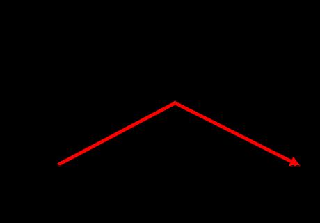
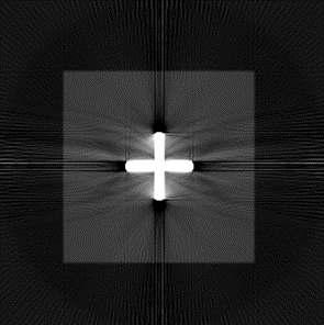
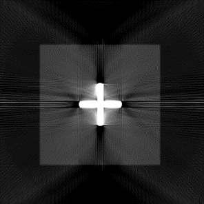
5 5 Simulation experiments In this section, the simulation experiments performed to confirm the application potency of multi-axis back projection algorithm are described. 5.1 Simulation conditions A numerical three-dimensional phantom that imitates the experimental sample shown in Figure 10 was prepared. This phantom was composed of two materials -- a resin and a cross-shaped iron. The image size was, the source-to-image distance was 1946 mm, and the number of projection was 256. The X-ray source was a cone beam, and the tube voltage was 100 KeV. Also, a discrete X-ray energy distribution and X-ray attenuation coefficients to each energy were prepared for generating metal artifacts on simulation (Table 1). The X-ray attenuation coefficients for each element were calculated from mass attenuation coefficients found in the National Institutes of Standards and Technology (NIST) database [8]. Energy X-ray Air, dry Resin Iron [KeV] Intensity [/mm] [/mm] [/mm] E E E E E E Figure 10: A numerical three-dimensional phantom composed of a resin embedded with a cross-shaped iron. is a crosssectional image at the height of the center, and is the volume rendering result E E Table 1: X-ray intensity and attenuation coefficients for materials used in forward projection. 5.2 Multi-axis projection In this simulation, we added a rotation angle that tilts three-dimensional space on the stage in addition to a rotation angle of the stage (Figure 11). By changing continuously with the change of from to, a simple multi-axis projection is performed. The multi-axis back projection can also be performed by Feldkamp algorithm based on recorded values of and. The projection result at the initial state ( =, = ) is shown in Figure 12. Figure 11: Projection image for illustration purposes and definition of two angles. Figure 12: The projection result of the phantom shown in Figure 10 at the initial state ( =, = ). As experiments of multi-axis projection, three kinds of rotation patterns were performed -- =, =, =. The relationship between two rotate angles are shown in Figure 11. The condition = indicates an ordinary projection, and other two conditions indicate multi-axis projection. 5
6 5.3 Multi-axis back projection Back projection results for each condition are shown in Figure 13. As cross-sectional images for presentation, three different heights ( =, 8, ) were selected. At the height of =, there is only the resin region, and at the heights of = 8,, there is the cross-shaped iron embedded in the resin. In the result at =, while metal artifacts didn't generate at the height of =, distinctive metal artifacts were generated from the iron at the heights of = 8 and. On the other hand, in the results of =, 8 4 at the height of = 8, we can see that metal artifacts were reduced and the cross-shape would be observed. However, we also see that the results at other heights were changed. Although there were no metal artifacts at =, =, some artifacts were generated for multi-axis projection results at the height of =. This is because metal artifacts that were generated only two-dimensionally came to spread to three-dimensional space by multi-axis projection. Here, remember that the metal artifacts were reduced at the height of = 8. These results indicate that multiaxis projection could distribute and weaken metal artifacts three-dimensionally. Furthermore, if metal artifacts were weakened and saturation regions of projection data reduced, you can apply our previous iterative algorithm that is considering X-ray energy. 6 Conclusion We proposed new systems of multi-axis X-ray CT, and conducted preliminary experiments both on actual CT scan and simulation. As the result, it was indicated that metal artifact would be reduced and electronic devices could be inspected with a high degree of accuracy. By using the two proposed mechanisms selectively depending on the situation, and combining them with our previous algorithm based on X-ray energy, effective metal artifact reduction will be realized. As a next step, we will first implement a function selecting rotation angles of multi-axis projection automatically. Acknowledgements We wish to acknowledge valuable discussions with Mr. Kazuo Kikuchi and Mr. Takashi Tomitsuka of Comscantecno Co., Ltd. References [1] E. Meyer, R. Raupach, M. Lell, B. Schmidt and M. Kachelrieß, Normalized metal artifact reduction (NMAR) in computed tomography, Medical Physics, (2010), [2] S. Zhao, K.T. Bae, B. Whiting and G. Wang, A wavelet method for metal artifact reduction with multiple metallic objects in the field of view, Journal of X-ray Science and Technology, (2002), [3] Y. Zhang, L. Zhang, X.R. Zhu, A.K. Lee, M. Chambers and L. Dong, Reducing metal artifacts in cone-beam CT images by preprocessing projection data, International Journal of Radiation Oncology Biology Physics, 67-3 (2007), [4] M. Abdoli, M. R. Ay, A. Ahmadian, R. A. Dierckx and H. Zaidi, Reduction of dental filling metallic artifacts in CTbased attenuation correction of PET data using weighted virtual sinograms optimized by a genetic algorithm, Medical Physics (2010), [5] M. Bal and L. Spies, Metal artifact reduction in CT using tissue-class modeling and adaptive prefiltering, Medical Physics 33-8 (2006), [6] Y. Chen, Y. Li, H. Guo, Y. Hu, L. Luo, X. Yin, J. Gu and C. Toumoulin, CT Metal Artifact Reduction Method Based on Improved Image Segmentation and Sinogram In-Painting, Mathematical Problems in Engineering 2012 (2012), [7] T. Kano and M. Koseki, A new metal artifact reduction algorithm based on a deteriorated CT image, Journal of X- Ray Science and Technology 24-6 (2016), [8] J. H. Hubbell and S. M. Seltzer, "Tables of X-Ray Mass Attenuation Coefficients and Mass Energy-Absorption Coefficients from 1 kev to 20 MeV for Elements Z = 1 to 92 and 48 Additional Substances of Dosimetric Interest, NIST, (2015) 6


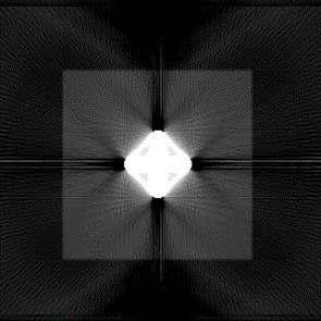
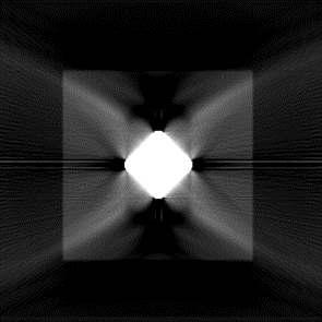
7 = = 8 = Line charts of relationship 7th Conference on Industrial Computed Tomography, Leuven, Belgium (ict 2017) = = = Figure 13: The simulation results of multi-axis projection and back projection. 7
A Curvelet based Sinogram Correction Method for Metal Artifact Reduction
 A based Sinogram Correction Method for Metal Artifact Reduction Kiwan Jeon 1 and Hyoung Suk Park 1 More info about this article: http://www.ndt.net/?id=3715 1 National Institute for Mathematical Sciences,
Optimization of CT Simulation Imaging. Ingrid Reiser Dept. of Radiology The University of Chicago
 Optimization of CT Simulation Imaging Ingrid Reiser Dept. of Radiology The University of Chicago Optimization of CT imaging Goal: Achieve image quality that allows to perform the task at hand (diagnostic
Digital Image Processing
 Digital Image Processing SPECIAL TOPICS CT IMAGES Hamid R. Rabiee Fall 2015 What is an image? 2 Are images only about visual concepts? We ve already seen that there are other kinds of image. In this lecture
Moving Metal Artifact Reduction for Cone-Beam CT (CBCT) Scans of the Thorax Region
 Scans of the Thorax Region") Moving Metal Artifact Reduction for Cone-Beam CT (CBCT) Scans of the Thorax Region Andreas Hahn 1,2, Sebastian Sauppe 1,2, Michael Knaup 1, and Marc Kachelrieß 1,2 1 German Cancer Research Center (DKFZ),
Moving Metal Artifact Reduction for Cone-Beam CT (CBCT) Scans of the Thorax Region Andreas Hahn 1,2, Sebastian Sauppe 1,2, Michael Knaup 1, and Marc Kachelrieß 1,2 1 German Cancer Research Center (DKFZ),
An Acquisition Geometry-Independent Calibration Tool for Industrial Computed Tomography
 4th International Symposium on NDT in Aerospace 2012 - Tu.3.A.3 An Acquisition Geometry-Independent Calibration Tool for Industrial Computed Tomography Jonathan HESS *, Patrick KUEHNLEIN *, Steven OECKL
Recognition and Measurement of Small Defects in ICT Testing
 19 th World Conference on Non-Destructive Testing 2016 Recognition and Measurement of Small Defects in ICT Testing Guo ZHIMIN, Ni PEIJUN, Zhang WEIGUO, Qi ZICHENG Inner Mongolia Metallic Materials Research
Fast iterative beam hardening correction based on frequency splitting in computed tomography
 Fast iterative beam hardening correction based on frequency splitting in computed tomography Qiao Yang a,b, Matthias Elter b, Ingo Schasiepen b, Nicole Maass b and Joachim Hornegger a,c a Pattern Recognition
Projection and Reconstruction-Based Noise Filtering Methods in Cone Beam CT
 Projection and Reconstruction-Based Noise Filtering Methods in Cone Beam CT Benedikt Lorch 1, Martin Berger 1,2, Joachim Hornegger 1,2, Andreas Maier 1,2 1 Pattern Recognition Lab, FAU Erlangen-Nürnberg
Frequency split metal artifact reduction (FSMAR) in computed tomography
 in computed tomography") The Johns Hopkins University Advanced Computer Integrated Surgery Group 4 Metal Artifact Removal in C-arm Cone-Beam CT Paper Seminar Critical Review of Frequency split metal artifact reduction (FSMAR)
The Johns Hopkins University Advanced Computer Integrated Surgery Group 4 Metal Artifact Removal in C-arm Cone-Beam CT Paper Seminar Critical Review of Frequency split metal artifact reduction (FSMAR)
Metal Artifact Reduction CT Techniques. Tobias Dietrich University Hospital Balgrist University of Zurich Switzerland
 Metal Artifact Reduction CT Techniques R S S S Tobias Dietrich University Hospital Balgrist University of Zurich Switzerland N. 1 v o 4 1 0 2. Postoperative CT Metal Implants CT is accurate for assessment
Novel evaluation method of low contrast resolution performance of dimensional X-ray CT
 More Info at Open Access Database www.ndt.net/?id=18754 Novel evaluation method of low contrast resolution performance of dimensional X-ray CT Makoto Abe 1, Hiroyuki Fujimoto 1, Osamu Sato 1, Kazuya Matsuzaki
Quality control phantoms and protocol for a tomography system
 Quality control phantoms and protocol for a tomography system Lucía Franco 1 1 CT AIMEN, C/Relva 27A O Porriño Pontevedra, Spain, lfranco@aimen.es Abstract Tomography systems for non-destructive testing
Computed tomography (Item No.: P )
") Computed tomography (Item No.: P2550100) Curricular Relevance Area of Expertise: Biology Education Level: University Topic: Modern Imaging Methods Subtopic: X-ray Imaging Experiment: Computed tomography
Computed tomography (Item No.: P2550100) Curricular Relevance Area of Expertise: Biology Education Level: University Topic: Modern Imaging Methods Subtopic: X-ray Imaging Experiment: Computed tomography
Thickness Measurement of Metal Plate Using CT Projection Images and Nominal Shape
 Thickness Measurement of Metal Plate Using CT Projection Images and Nominal Shape Tasuku Ito 1, Yutaka Ohtake 1, Yukie Nagai 2, Hiromasa Suzuki 1 More info about this article: http://www.ndt.net/?id=23662
DUE to beam polychromacity in CT and the energy dependence
 1 Empirical Water Precorrection for Cone-Beam Computed Tomography Katia Sourbelle, Marc Kachelrieß, Member, IEEE, and Willi A. Kalender Abstract We propose an algorithm to correct for the cupping artifact
Ch. 4 Physical Principles of CT
 Ch. 4 Physical Principles of CT CLRS 408: Intro to CT Department of Radiation Sciences Review: Why CT? Solution for radiography/tomography limitations Superimposition of structures Distinguishing between
An Iterative Approach to the Beam Hardening Correction in Cone Beam CT (Proceedings)
") Marquette University e-publications@marquette Biomedical Engineering Faculty Research and Publications Engineering, College of 1-1-1999 An Iterative Approach to the Beam Hardening Correction in Cone Beam
Marquette University e-publications@marquette Biomedical Engineering Faculty Research and Publications Engineering, College of 1-1-1999 An Iterative Approach to the Beam Hardening Correction in Cone Beam
Improvement of Efficiency and Flexibility in Multi-slice Helical CT
 J. Shanghai Jiaotong Univ. (Sci.), 2008, 13(4): 408 412 DOI: 10.1007/s12204-008-0408-x Improvement of Efficiency and Flexibility in Multi-slice Helical CT SUN Wen-wu 1 ( ), CHEN Si-ping 2 ( ), ZHUANG Tian-ge
An Automated Image-based Method for Multi-Leaf Collimator Positioning Verification in Intensity Modulated Radiation Therapy
 An Automated Image-based Method for Multi-Leaf Collimator Positioning Verification in Intensity Modulated Radiation Therapy Chenyang Xu 1, Siemens Corporate Research, Inc., Princeton, NJ, USA Xiaolei Huang,
Moscow-Bavarian Joint Advanced Student School 2006 / Medical Imaging Principles of Computerized Tomographic Imaging and Cone-Beam Reconstruction
 Line Integrals Line integrals represent the integral of some parameter of the object along the line (e.g. attenuation of x-rays) Object: f(x,y) Line: x cosθ + y sinθ = t Line integral / Radon transform:
Computed tomography of simple objects. Related topics. Principle. Equipment TEP Beam hardening, artefacts, and algorithms
 Related topics Beam hardening, artefacts, and algorithms Principle The CT principle is demonstrated with the aid of simple objects. In the case of very simple targets, only a few images need to be taken
A new calibration-free beam hardening reduction method for industrial CT
 A new calibration-free beam hardening reduction method for industrial CT ECC 2 for industrial CT Tobias Würfl 1, Nicole Maaß 2, Frank Dennerlein 2, Andreas K. Maier 1 1 Pattern Recognition Lab, FAU Erlangen-Nürnberg;
3D Computed Tomography (CT) Its Application to Aerospace Industry
 Its Application to Aerospace Industry") 3D Computed Tomography (CT) Its Application to Aerospace Industry C. Muralidhar, M. P. Subramanian, V. Ravi Shankar and G. Chandrasekhar Directorate of Non Destructive Evaluation, Defence Research & Development
3D Computed Tomography (CT) Its Application to Aerospace Industry C. Muralidhar, M. P. Subramanian, V. Ravi Shankar and G. Chandrasekhar Directorate of Non Destructive Evaluation, Defence Research & Development
Application of 450 kv Computed Tomography to Engine Blocks with Steel Liners
 Application of 450 kv Computed Tomography to Engine Blocks with Steel Liners Charles R. Smith, Kevin Holt BIR, Inc. Uwe Bischoff, Bernd Georgi, Ferdinand Hansen, Frank Jeltsch Volkswagen Commercial Vehicles
Computed Tomography. Principles, Design, Artifacts, and Recent Advances. Jiang Hsieh THIRD EDITION. SPIE PRESS Bellingham, Washington USA
 Computed Tomography Principles, Design, Artifacts, and Recent Advances THIRD EDITION Jiang Hsieh SPIE PRESS Bellingham, Washington USA Table of Contents Preface Nomenclature and Abbreviations xi xv 1 Introduction
Low-Dose Dual-Energy CT for PET Attenuation Correction with Statistical Sinogram Restoration
 Low-Dose Dual-Energy CT for PET Attenuation Correction with Statistical Sinogram Restoration Joonki Noh, Jeffrey A. Fessler EECS Department, The University of Michigan Paul E. Kinahan Radiology Department,
3D X-ray Laminography with CMOS Image Sensor Using a Projection Method for Reconstruction of Arbitrary Cross-sectional Images
 Ke Engineering Materials Vols. 270-273 (2004) pp. 192-197 online at http://www.scientific.net (2004) Trans Tech Publications, Switzerland Online available since 2004/08/15 Citation & Copright (to be inserted
Physical bases of X-ray diagnostics
 Physical bases of X-ray diagnostics Dr. István Voszka Possibilities of X-ray production (X-ray is produced, when charged particles of high velocity are stopped) X-ray tube: Relatively low accelerating
Spiral ASSR Std p = 1.0. Spiral EPBP Std. 256 slices (0/300) Kachelrieß et al., Med. Phys. 31(6): , 2004
 Kachelrieß et al., Med. Phys. 31(6): , 2004") Spiral ASSR Std p = 1.0 Spiral EPBP Std p = 1.0 Kachelrieß et al., Med. Phys. 31(6): 1623-1641, 2004 256 slices (0/300) Advantages of Cone-Beam Spiral CT Image quality nearly independent of pitch Increase
Spiral ASSR Std p = 1.0 Spiral EPBP Std p = 1.0 Kachelrieß et al., Med. Phys. 31(6): 1623-1641, 2004 256 slices (0/300) Advantages of Cone-Beam Spiral CT Image quality nearly independent of pitch Increase
MEDICAL IMAGING 2nd Part Computed Tomography
 MEDICAL IMAGING 2nd Part Computed Tomography Introduction 2 In the last 30 years X-ray Computed Tomography development produced a great change in the role of diagnostic imaging in medicine. In convetional
Tomographic Reconstruction
 Tomographic Reconstruction 3D Image Processing Torsten Möller Reading Gonzales + Woods, Chapter 5.11 2 Overview Physics History Reconstruction basic idea Radon transform Fourier-Slice theorem (Parallel-beam)
First CT Scanner. How it Works. Contemporary CT. Before and After CT. Computer Tomography: How It Works. Medical Imaging and Pattern Recognition
 Computer Tomography: How t Works Medical maging and Pattern Recognition Lecture 7 Computed Tomography Oleh Tretiak Only one plane is illuminated. Source-subject motion provides added information. 2 How
Multi-slice CT Image Reconstruction Jiang Hsieh, Ph.D.
 Multi-slice CT Image Reconstruction Jiang Hsieh, Ph.D. Applied Science Laboratory, GE Healthcare Technologies 1 Image Generation Reconstruction of images from projections. textbook reconstruction advanced
ML reconstruction for CT
 ML reconstruction for CT derivation of MLTR rigid motion correction resolution modeling polychromatic ML model dual energy ML model Bruno De Man, Katrien Van Slambrouck, Maarten Depypere, Frederik Maes,
MEDICAL EQUIPMENT: COMPUTED TOMOGRAPHY. Prof. Yasser Mostafa Kadah
 MEDICAL EQUIPMENT: COMPUTED TOMOGRAPHY Prof. Yasser Mostafa Kadah www.k-space.org Recommended Textbook X-Ray Computed Tomography in Biomedical Engineering, by Robert Cierniak, Springer, 211 Computed Tomography
Radiology. Marta Anguiano Millán. Departamento de Física Atómica, Molecular y Nuclear Facultad de Ciencias. Universidad de Granada
 Departamento de Física Atómica, Molecular y Nuclear Facultad de Ciencias. Universidad de Granada Overview Introduction Overview Introduction Tecniques of imaging in Overview Introduction Tecniques of imaging
Non-Stationary CT Image Noise Spectrum Analysis
 Non-Stationary CT Image Noise Spectrum Analysis Michael Balda, Björn J. Heismann,, Joachim Hornegger Pattern Recognition Lab, Friedrich-Alexander-Universität Erlangen Siemens Healthcare, Erlangen michael.balda@informatik.uni-erlangen.de
A method and algorithm for Tomographic Imaging of highly porous specimen using Low Frequency Acoustic/Ultrasonic signals
 More Info at Open Access Database www.ndt.net/?id=15210 A method and algorithm for Tomographic Imaging of highly porous specimen using Low Frequency Acoustic/Ultrasonic signals Subodh P S 1,a, Reghunathan
NON-COLLIMATED SCATTERED RADIATION TOMOGRAPHY
 NON-COLLIMATED SCATTERED RADIATION TOMOGRAPHY Gorshkov V.A., Space Research Institute, Moscow, Russia Yumashev V.M., State corporation "Rosatom", Centre "Atom-innovation", Moscow, Russia Kirilenko K.V.,
Enhanced material contrast by dual-energy microct imaging
 Enhanced material contrast by dual-energy microct imaging Method note Page 1 of 12 2 Method note: Dual-energy microct analysis 1. Introduction 1.1. The basis for dual energy imaging Micro-computed tomography
DEVELOPMENT OF CONE BEAM TOMOGRAPHIC RECONSTRUCTION SOFTWARE MODULE
 Rajesh et al. : Proceedings of the National Seminar & Exhibition on Non-Destructive Evaluation DEVELOPMENT OF CONE BEAM TOMOGRAPHIC RECONSTRUCTION SOFTWARE MODULE Rajesh V Acharya, Umesh Kumar, Gursharan
Radon Transform and Filtered Backprojection
 Radon Transform and Filtered Backprojection Jørgen Arendt Jensen October 13, 2016 Center for Fast Ultrasound Imaging, Build 349 Department of Electrical Engineering Center for Fast Ultrasound Imaging Department
Introduction to Biomedical Imaging
 Alejandro Frangi, PhD Computational Imaging Lab Department of Information & Communication Technology Pompeu Fabra University www.cilab.upf.edu X-ray Projection Imaging Computed Tomography Digital X-ray
Efficient 3D Crease Point Extraction from 2D Crease Pixels of Sinogram
 Efficient 3D Crease Point Extraction from 2D Crease Pixels of Sinogram Ryo Jinnouchi 1, Yutaka Ohtake 1, Hiromasa Suzuki 1, Yukie Nagai 1 1 The University of Tokyo, Japan, e-mail: {jinnouchi, yu-ohtake,
Digital Laminography and Computed Tomography with 600 kv for Aerospace Applications
 4th International Symposium on NDT in Aerospace 2012 - Tu.3.A.1 Digital Laminography and Computed Tomography with 600 kv for Aerospace Applications Malte KURFISS 1, Gerd STRECKENBACH 2 1 YXLON International
Empirical cupping correction: A first-order raw data precorrection for cone-beam computed tomography
 Empirical cupping correction: A first-order raw data precorrection for cone-beam computed tomography Marc Kachelrieß, a Katia Sourbelle, and Willi A. Kalender Institute of Medical Physics, University of
Central Slice Theorem
 Central Slice Theorem Incident X-rays y f(x,y) R x r x Detected p(, x ) The thick line is described by xcos +ysin =R Properties of Fourier Transform F [ f ( x a)] F [ f ( x)] e j 2 a Spatial Domain Spatial
Image Acquisition Systems
 Image Acquisition Systems Goals and Terminology Conventional Radiography Axial Tomography Computer Axial Tomography (CAT) Magnetic Resonance Imaging (MRI) PET, SPECT Ultrasound Microscopy Imaging ITCS
BME I5000: Biomedical Imaging
 1 Lucas Parra, CCNY BME I5000: Biomedical Imaging Lecture 4 Computed Tomography Lucas C. Parra, parra@ccny.cuny.edu some slides inspired by lecture notes of Andreas H. Hilscher at Columbia University.
2D Fan Beam Reconstruction 3D Cone Beam Reconstruction
 2D Fan Beam Reconstruction 3D Cone Beam Reconstruction Mario Koerner March 17, 2006 1 2D Fan Beam Reconstruction Two-dimensional objects can be reconstructed from projections that were acquired using parallel
Shadow casting. What is the problem? Cone Beam Computed Tomography THE OBJECTIVES OF DIAGNOSTIC IMAGING IDEAL DIAGNOSTIC IMAGING STUDY LIMITATIONS
 Cone Beam Computed Tomography THE OBJECTIVES OF DIAGNOSTIC IMAGING Reveal pathology Reveal the anatomic truth Steven R. Singer, DDS srs2@columbia.edu IDEAL DIAGNOSTIC IMAGING STUDY Provides desired diagnostic
Evaluation of Spectrum Mismatching using Spectrum Binning Approach for Statistical Polychromatic Reconstruction in CT
 Evaluation of Spectrum Mismatching using Spectrum Binning Approach for Statistical Polychromatic Reconstruction in CT Qiao Yang 1,4, Meng Wu 2, Andreas Maier 1,3,4, Joachim Hornegger 1,3,4, Rebecca Fahrig
Metal Streak Artifacts in X-ray Computed Tomography: A Simulation Study
 Metal Streak Artifacts in X-ray Computed Tomography: A Simulation Study B. De Man, J. Nuyts, P. Dupont, G. Marchal and P. Suetens Medical Image Computing, ESAT-PSI, K.U.Leuven, B-3000 Leuven, Belgium Department
Design and performance characteristics of a Cone Beam CT system for Leksell Gamma Knife Icon
 Design and performance characteristics of a Cone Beam CT system for Leksell Gamma Knife Icon WHITE PAPER Introduction Introducing an image guidance system based on Cone Beam CT (CBCT) and a mask immobilization
Index. aliasing artifacts and noise in CT images, 200 measurement of projection data, nondiffracting
 Index Algebraic equations solution by Kaczmarz method, 278 Algebraic reconstruction techniques, 283-84 sequential, 289, 293 simultaneous, 285-92 Algebraic techniques reconstruction algorithms, 275-96 Algorithms
Phase problem and the Radon transform
 Phase problem and the Radon transform Andrei V. Bronnikov Bronnikov Algorithms The Netherlands The Radon transform and applications Inverse problem of phase-contrast CT Fundamental theorem Image reconstruction
Scatter Correction for Dual source Cone beam CT Using the Pre patient Grid. Yingxuan Chen. Graduate Program in Medical Physics Duke University
 Scatter Correction for Dual source Cone beam CT Using the Pre patient Grid by Yingxuan Chen Graduate Program in Medical Physics Duke University Date: Approved: Lei Ren, Supervisor Fang Fang Yin, Chair
Registration concepts for the just-in-time artefact correction by means of virtual computed tomography
 DIR 2007 - International Symposium on Digital industrial Radiology and Computed Tomography, June 25-27, 2007, Lyon, France Registration concepts for the just-in-time artefact correction by means of virtual
Beam Attenuation Grid Based Scatter Correction Algorithm for. Cone Beam Volume CT
 11th European Conference on Non-Destructive Testing (ECNDT 2014), October 6-10, 2014, Prague, Czech Republic Beam Attenuation Grid Based Scatter Correction Algorithm for More Info at Open Access Database
Combining Analytical and Monte Carlo Modelling for Industrial Radiology
 19 th World Conference on Non-Destructive Testing 2016 Combining Analytical and Monte Carlo Modelling for Industrial Radiology Carsten BELLON, Gerd-Rüdiger JAENISCH, Andreas DERESCH BAM Bundesanstalt für
Multi-spectral (W, Mo, Cu, and Ag) XCT measurements
 XCT measurements") Multi-spectral (W, Mo, Cu, and Ag) XCT measurements Marko Katić 1, Damir Markučič 1, Gorana Baršić 1 Faculty of mechanical engineering and naval architecture, Ivana Lucica 5, Zagreb, Croatia, marko.katic@fsb.hr
Multi-spectral (W, Mo, Cu, and Ag) XCT measurements Marko Katić 1, Damir Markučič 1, Gorana Baršić 1 Faculty of mechanical engineering and naval architecture, Ivana Lucica 5, Zagreb, Croatia, marko.katic@fsb.hr
A Novel Image Super-resolution Reconstruction Algorithm based on Modified Sparse Representation
 , pp.162-167 http://dx.doi.org/10.14257/astl.2016.138.33 A Novel Image Super-resolution Reconstruction Algorithm based on Modified Sparse Representation Liqiang Hu, Chaofeng He Shijiazhuang Tiedao University,
Reconstruction of CT Images from Sparse-View Polyenergetic Data Using Total Variation Minimization
 1 Reconstruction of CT Images from Sparse-View Polyenergetic Data Using Total Variation Minimization T. Humphries and A. Faridani Abstract Recent work in CT image reconstruction has seen increasing interest
Corso di laurea in Fisica A.A Fisica Medica 4 TC
 Corso di laurea in Fisica A.A. 2007-2008 Fisica Medica 4 TC Computed Tomography Principles 1. Projection measurement 2. Scanner systems 3. Scanning modes Basic Tomographic Principle The internal structure
A Comparative Study of Two Methods for the Correction of Beam Hardening Artifacts in X-ray Computed Tomography
 A Comparative Study of Two Methods for the Correction of Beam Hardening Artifacts in X-ray Computed Tomography by Maryam Khalid Alarfaj submitted to Oregon State University in partial fulfillment of the
Chapter6 Image Reconstruction
 Chapter6 Image Reconstruction Preview 61I 6.1 Introduction 6.2 Reconstruction by Fourier Inversion 6.3 Reconstruction by convolution and backprojection 6.4 Finite series-expansion 1 Preview Reconstruction
Metal Artifact Reduction for Multi-Material Objects
 Metal Artifact Reduction for Multi-Material Objects Bärbel Kratz 1,#, Frank Herold 1, Malte Kurfiß 1, Thorsten M. Buzug 2 1 YXLON International GmbH, Essener Bogen 15, 22419 Hamburg, Germany, e-mail: baerbel.kratz@hbg.yxlon.com,
Translational Computed Tomography: A New Data Acquisition Scheme
 2nd International Symposium on NDT in Aerospace 2010 - We.1.A.3 Translational Computed Tomography: A New Data Acquisition Scheme Theobald FUCHS 1, Tobias SCHÖN 2, Randolf HANKE 3 1 Fraunhofer Development
An approximate cone beam reconstruction algorithm for gantry-tilted CT
 An approximate cone beam reconstruction algorithm for gantry-tilted CT Ming Yan a, Cishen Zhang ab, Hongzhu Liang a a School of Electrical & Electronic Engineering, Nanyang Technological University, Singapore;
CT Reconstruction with Good-Orientation and Layer Separation for Multilayer Objects
 17th World Conference on Nondestructive Testing, 25-28 Oct 2008, Shanghai, China CT Reconstruction with Good-Orientation and Layer Separation for Multilayer Objects Tong LIU 1, Brian Stephan WONG 2, Tai
2D Fan Beam Reconstruction 3D Cone Beam Reconstruction. Mario Koerner
 2D Fan Beam Reconstruction 3D Cone Beam Reconstruction Mario Koerner Moscow-Bavarian Joint Advanced Student School 2006 March 19 2006 to March 29 2006 Overview 2D Fan Beam Reconstruction Shortscan Reconstruction
Ultrasonic Multi-Skip Tomography for Pipe Inspection
 18 th World Conference on Non destructive Testing, 16-2 April 212, Durban, South Africa Ultrasonic Multi-Skip Tomography for Pipe Inspection Arno VOLKER 1, Rik VOS 1 Alan HUNTER 1 1 TNO, Stieltjesweg 1,
Computed tomography - outline
 Computed tomography - outline Computed Tomography Systems Jørgen Arendt Jensen and Mikael Jensen (DTU Nutech) October 6, 216 Center for Fast Ultrasound Imaging, Build 349 Department of Electrical Engineering
Applying Hounsfield unit density calibration in SkyScan CT-analyser
 1 Bruker-microCT Method note Applying Hounsfield unit density calibration in SkyScan CT-analyser Hounsfield units (HU) are a standard unit of x-ray CT density, in which air and water are ascribed values
Non-Destructive Failure Analysis and Measurement for Molded Devices and Complex Assemblies with X-ray CT and 3D Image Processing Techniques
 SINCE2013 Singapore International NDT Conference & Exhibition 2013, 19-20 July 2013 Non-Destructive Failure Analysis and Measurement for Molded Devices and Complex Assemblies with X-ray CT and 3D Image
Feldkamp-type image reconstruction from equiangular data
 Journal of X-Ray Science and Technology 9 (2001) 113 120 113 IOS Press Feldkamp-type image reconstruction from equiangular data Ben Wang a, Hong Liu b, Shiying Zhao c and Ge Wang d a Department of Elec.
RADIOMICS: potential role in the clinics and challenges
 27 giugno 2018 Dipartimento di Fisica Università degli Studi di Milano RADIOMICS: potential role in the clinics and challenges Dr. Francesca Botta Medical Physicist Istituto Europeo di Oncologia (Milano)
REAL-TIME ADAPTIVITY IN HEAD-AND-NECK AND LUNG CANCER RADIOTHERAPY IN A GPU ENVIRONMENT
 REAL-TIME ADAPTIVITY IN HEAD-AND-NECK AND LUNG CANCER RADIOTHERAPY IN A GPU ENVIRONMENT Anand P Santhanam Assistant Professor, Department of Radiation Oncology OUTLINE Adaptive radiotherapy for head and
TEP Hounsfield units. Related topics Attenuation coefficient, Hounsfield units
 Hounsfield units TEP Related topics Attenuation coefficient, Hounsfield units Principle Depending on the type of CT scanner and the settings, the result of a CT scan of the same material can be different
New Features of SIMTech s Unique CT Reconstruction and Visualization Software
 New Features of SIMTech s Unique CT Reconstruction and Visualization Software Liu Tong and Jiang Ting Ying More info about this article: http://www.ndt.net/?id=22215 Abstract Singapore Institute of Manufacturing
DEVELOPMENT OF POSITION MEASUREMENT SYSTEM FOR CONSTRUCTION PILE USING LASER RANGE FINDER
 S17- DEVELOPMENT OF POSITION MEASUREMENT SYSTEM FOR CONSTRUCTION PILE USING LASER RANGE FINDER Fumihiro Inoue 1 *, Takeshi Sasaki, Xiangqi Huang 3, and Hideki Hashimoto 4 1 Technica Research Institute,
PURE. ViSION Edition PET/CT. Patient Comfort Put First.
 PURE ViSION Edition PET/CT Patient Comfort Put First. 2 System features that put patient comfort and safety first. Oncology patients deserve the highest levels of safety and comfort during scans. Our Celesteion
Adaptive region of interest method for analytical micro-ct reconstruction
 Journal of X-Ray Science and Technology 19 (2011) 23 33 23 DOI 10.3233/XST-2010-0274 IOS Press Adaptive region of interest method for analytical micro-ct reconstruction Wanneng Yang, Xiaochun Xu, Kun Bi,
Reduction of Metal Artifacts in Computed Tomographies for the Planning and Simulation of Radiation Therapy
 Reduction of Metal Artifacts in Computed Tomographies for the Planning and Simulation of Radiation Therapy T. Rohlfing a, D. Zerfowski b, J. Beier a, P. Wust a, N. Hosten a, R. Felix a a Department of
CIVA Computed Tomography Modeling
 CIVA Computed Tomography Modeling R. FERNANDEZ, EXTENDE, France S. LEGOUPIL, M. COSTIN, D. TISSEUR, A. LEVEQUE, CEA-LIST, France page 1 Summary Context From CIVA RT to CIVA CT Reconstruction Methods Applications
Metal artifacts reduction in x-ray CT based on segmentation and forward-projection
 Bio-Medical Materials and Engineering 24 (2014) 3287 3293 DOI 10.3233/BME-141151 IOS Press 3287 Metal artifacts reduction in x-ray CT based on segmentation and forward-projection Shoukat Nawaz, Jian Fu
MULTI-PURPOSE 3D COMPUTED TOMOGRAPHY SYSTEM
 MULTI-PURPOSE 3D COMPUTED TOMOGRAPHY SYSTEM M. Simon, C. Sauerwein, I. Tiseanu, S. Burdairon Hans Wälischmiller GmbH Klingleweg 8, D-88709 Meersburg, Germany e-mail: ms@hwm.com ABSTRACT A new flexible
Accelerated quantitative multi-material beam hardening correction(bhc) in cone-beam CT
 in cone-beam CT") Accelerated quantitative multi-material beam hardening correction(bhc) in cone-beam CT Award: Poster No.: C-2161 Certificate of Merit Congress: ECR 2012 Type: Authors: Scientific Exhibit Q. Yang, M. Elter,
Accelerated quantitative multi-material beam hardening correction(bhc) in cone-beam CT Award: Poster No.: C-2161 Certificate of Merit Congress: ECR 2012 Type: Authors: Scientific Exhibit Q. Yang, M. Elter,
Biomedical Imaging. Computed Tomography. Patrícia Figueiredo IST
 Biomedical Imaging Computed Tomography Patrícia Figueiredo IST 2013-2014 Overview Basic principles X ray attenuation projection Slice selection and line projections Projection reconstruction Instrumentation
Performance Evaluation of 3-Axis Scanner Automated For Industrial Gamma- Ray Computed Tomography
 National Seminar & Exhibition on Non-Destructive Evaluation, NDE 2014, Pune, December 4-6, 2014 (NDE-India 2014) Vol.20 No.6 (June 2015) - The e-journal of Nondestructive Testing - ISSN 1435-4934 www.ndt.net/?id=17822
Enhancement Image Quality of CT Using Single Slice Spiral Technique
 Enhancement Image Quality of CT Using Single Slice Spiral Technique Doaa. N. Al Sheack 1 and Dr.Mohammed H. Ali Al Hayani 2 1 2 Electronic and Communications Engineering Department College of Engineering,
Fresnel Reflection. angle of transmission. Snell s law relates these according to the
 Fresnel Reflection 1. Reflectivity of polarized light The reflection of a polarized beam of light from a dielectric material such as air/glass was described by Augustin Jean Fresnel in 1823. While his
DUAL energy CT (DECT) is a modality where one and. Empirical Dual Energy Calibration (EDEC) for Cone-Beam Computed Tomography
 is a modality where one and. Empirical Dual Energy Calibration (EDEC) for Cone-Beam Computed Tomography") Empirical Dual Energy Calibration (EDEC) for Cone-Beam Computed Tomography Marc Kachelrieß, Member, IEEE, Timo Berkus, Philip Stenner, Willi A. Kalender Abstract Material selective imaging using dual energy
Empirical Dual Energy Calibration (EDEC) for Cone-Beam Computed Tomography Marc Kachelrieß, Member, IEEE, Timo Berkus, Philip Stenner, Willi A. Kalender Abstract Material selective imaging using dual energy
Diagnostic imaging techniques. Krasznai Zoltán. University of Debrecen Medical and Health Science Centre Department of Biophysics and Cell Biology
 Diagnostic imaging techniques Krasznai Zoltán University of Debrecen Medical and Health Science Centre Department of Biophysics and Cell Biology 1. Computer tomography (CT) 2. Gamma camera 3. Single Photon
SYSTEM LINEARITY LAB MANUAL: 2 Modifications for P551 Fall 2013 Medical Physics Laboratory
 SYSTEM LINEARITY LAB MANUAL: 2 Modifications for P551 Fall 2013 Medical Physics Laboratory Introduction In this lab exercise, you will investigate the linearity of the DeskCAT scanner by making measurements
REMOVAL OF THE EFFECT OF COMPTON SCATTERING IN 3-D WHOLE BODY POSITRON EMISSION TOMOGRAPHY BY MONTE CARLO
 REMOVAL OF THE EFFECT OF COMPTON SCATTERING IN 3-D WHOLE BODY POSITRON EMISSION TOMOGRAPHY BY MONTE CARLO Abstract C.S. Levin, Y-C Tai, E.J. Hoffman, M. Dahlbom, T.H. Farquhar UCLA School of Medicine Division
CT NOISE POWER SPECTRUM FOR FILTERED BACKPROJECTION AND ITERATIVE RECONSTRUCTION
 CT NOISE POWER SPECTRUM FOR FILTERED BACKPROJECTION AND ITERATIVE RECONSTRUCTION Frank Dong, PhD, DABR Diagnostic Physicist, Imaging Institute Cleveland Clinic Foundation and Associate Professor of Radiology
Some reference material
 Some reference material Physics reference book on medical imaging: A good one is The Essential Physics of Medical Imaging, 3 rd Ed. by Bushberg et al. ($170! new). However, there are several similar books
Financial disclosure. Onboard imaging modality for IGRT
 Tetrahedron Beam Computed Tomography Based On Multi-Pixel X- Ray Source and Its Application in Image Guided Radiotherapy Tiezhi Zhang, Ph.D. Advanced X-ray imaging Lab Financial disclosure Patent royalty
Simulation of Mammograms & Tomosynthesis imaging with Cone Beam Breast CT images
 Simulation of Mammograms & Tomosynthesis imaging with Cone Beam Breast CT images Tao Han, Chris C. Shaw, Lingyun Chen, Chao-jen Lai, Xinming Liu, Tianpeng Wang Digital Imaging Research Laboratory (DIRL),