Detection of Sub-resolution Dots in Microscopy Images
|
|
|
- Erin Price
- 5 years ago
- Views:
Transcription
1 Detection of Sub-resolution Dots in Microscopy Images Karel Štěpka, 2012 Centre for Biomedical Image Analysis, FI MU supervisor: prof. RNDr. Michal Kozubek, Ph.D.
2 Outline Introduction Existing approaches Method evaluation Future work

3 Introduction In biology and medicine, parts of living cells can be observed using fluorescence microscopy Fluorescence in-situ hybridization (FISH) Allows us to visualize individual parts of genetic material: chromosomes or their parts Probes appear as small dots in the result Image courtesy of Wikimedia Commons
4 Observable Parts of a Cell Cytoplasm, cytoskeleton, nucleus Whole chromosomes Conditions related to the number of chromosomes (e.g. Down syndrome) Telomeres, kinetochores, centromeres Individual genes Translocations (e.g. BCR/ABL genes and their relation to certain kinds of leukemia)
5 Observable Parts of a Cell Dots Cytoplasm, cytoskeleton, nucleus Whole chromosomes Conditions related to the number of chromosomes (e.g. Down syndrome) Telomeres, kinetochores, centromeres Individual genes Translocations (e.g. BCR/ABL genes and their relation to certain kinds of leukemia)

6 Fluorescence Dots Real size as small as 10 nm In the resulting image, often 1 pixel > 60 nm Because of the diffraction limit of visible light, the magnification cannot be easily improved Due to image degradations, the sensor detects a blurred image of the dot Image of a dot has a few pixels across
 Degradation by point spread function (PSF) Present even in an ideal optical system PSF of a system can be")
7 Image Degradation Noise Multiple types, with different causes and statistical distributions: Photon shot noise (Poisson) Readout noise (Gaussian) Laser speckle noise Different methods for suppression Gaussian blurring Non-linear filters (median, non-linear diffusion) Degradation by point spread function (PSF) Present even in an ideal optical system PSF of a system can be experimentally measured
8 Existing Approaches to Dot Detection
9 Classical Detection Foundations Thresholding Otsu, unimodal, adaptive Mathematical morphology Opening, top-hat Pattern matching
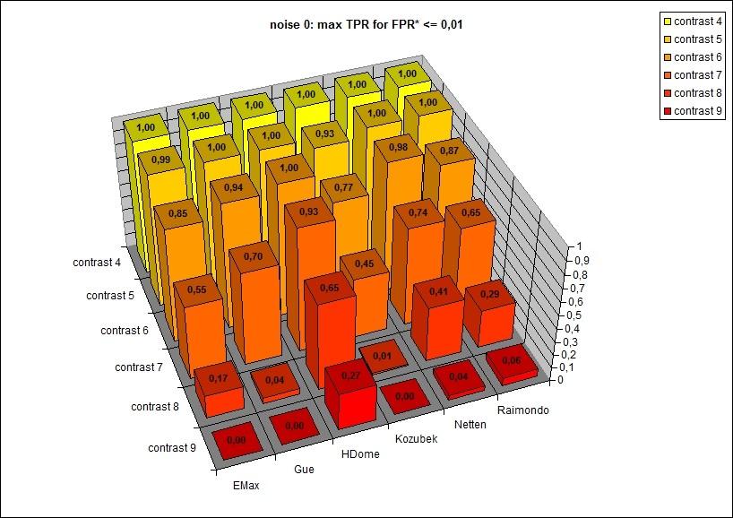
10 Recent Classical-Based Methods EMax Extended maxima transform, size-based filtering Gué Top-hat, thresholding, region growing HDome HDome transformation, mean shift clustering Kozubek Gradual thresholding, size-based filtering Netten Top-hat, dot label ( sweep through all intensity levels) Raimondo Top-hat, modified unimodal thresholding, pattern matching
11 Machine Learning Approach Examine potential dot locations, classify as positive/negative Usually using a sliding sub-window May lead to excessive time consumption in 3D Training required, overtraining undesirable Training set with image patches from which the classifier learns Test set necessary to determine the quality of the classifier Neural networks AdaBoost Combination of multiple weak classifiers
12 Measuring Success Rate
13 Measuring Success Rate Comparison of the results with the ground truth (GT) We can obtain GT by manually annotating real images We can generate synthetic images together with their GT Real testing data, manual GT Different people, or the same person over multiple attempts, generally annotate images differently Time consuming, expensive Synthetic testing data, generated GT GT is accurate and undebatable (created before the images) The synthetic data must correspond to the real images
14 Measuring Success Rate Detection precision = recall = TP TP + FP TP TP + FN present not present found TP FP not found FN TN F 1 score = 2 precision recall precision + recall Distinguishing between large dots and double-dots To identify chromosomal conditions such as polysomy
15 Recent Survey by I. Smal et al. Compared performance of several methods 2D data Real images Simplified synthetic images Dots represented by Gaussian profiles Did not evaluate the influence of method parameters Good starting point Ihor Smal et al.: Quantitative Comparison of Spot Detection Methods in Fluorescence Microscopy. IEEE Transactions on Medical Imaging 29(2): (2010)
16 Parametrization No Size Fits All No method can be used on all types of images without any adjustments On the pixel level, images can be very different, even when displaying the same class of objects Noise level Base intensity Dynamic range Contrast Background (non-)uniformity Illumination artifacts Amount of objects of interest
17 Parametrization Usability Usability of a method depends on: Number of its parameters Sensitivity to parameter changes Intuitiveness of its parameters for the end user A thorough parametric study is required Curse of dimensionality Some of the methods have 4 6 free parameters
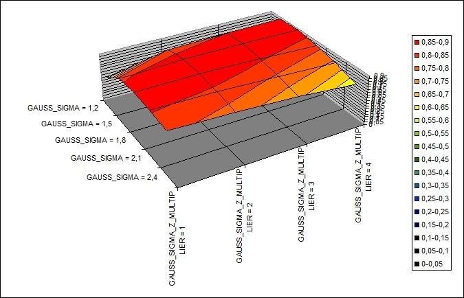
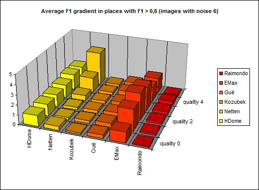
18 Parametrization Usability Proposed new method for evaluating sensitivity to parameter settings: Mean squared magnitude of the F 1 gradient, Higher value = higher sensitivity
19 Intermediate results
20 Further Work Publish the evaluation of existing methods Methods were tested on various 3D images Real, manually annotated data Simulated data with known GT Parametric study, sensitivity evaluation Prepare a set of benchmark data Cover testing of all important measurements Detection, localization, double-dots Possibly make the set publicly available through CBIA web-site
21 Further Work Investigate the conceptual difference between 2D and 3D fluorescence images Dots do not lie in the same focal plane Microscopy images exhibit strong anisotropy PSF does not correspond to simple Gaussian Per-slice processing or direct extension to 3D do not take any of this into account Design a method natively working with 3D images Most of the existing methods are natively 2D (or nd), and use no special approach for 3D data Include the new method in the comparison
Bioimage Informatics. Lecture 8, Spring Bioimage Data Analysis (II): Point Feature Detection
: Point Feature Detection") Bioimage Informatics Lecture 8, Spring 2012 Bioimage Data Analysis (II): Point Feature Detection Lecture 8 February 13, 2012 1 Outline Project assignment 02 Comments on reading assignment 01 Review: pixel
Bioimage Informatics Lecture 8, Spring 2012 Bioimage Data Analysis (II): Point Feature Detection Lecture 8 February 13, 2012 1 Outline Project assignment 02 Comments on reading assignment 01 Review: pixel
Topic 4 Image Segmentation
 Topic 4 Image Segmentation What is Segmentation? Why? Segmentation important contributing factor to the success of an automated image analysis process What is Image Analysis: Processing images to derive
Linear Operations Using Masks
 Linear Operations Using Masks Masks are patterns used to define the weights used in averaging the neighbors of a pixel to compute some result at that pixel Expressing linear operations on neighborhoods
Introduction. Loading Images
 Introduction CellProfiler is a free Open Source software for automated image analysis. Versions for Mac, Windows and Linux are available and can be downloaded at: http://www.cellprofiler.org/. CellProfiler
Tracking of Virus Particles in Time-Lapse Fluorescence Microscopy Image Sequences
 Tracking of Virus Particles in Time-Lapse Fluorescence Microscopy Image Sequences W. J. Godinez 1, M. Lampe 2, S. Wörz 1, B. Müller 2, R. Eils 1 and K. Rohr 1 1 University of Heidelberg, IPMB, and DKFZ
DT-Binarize: A Hybrid Binarization Method using Decision Tree for Protein Crystallization Images
 DT-Binarize: A Hybrid Binarization Method using Decision Tree for Protein Crystallization Images İmren Dinç 1, Semih Dinç 1, Madhav Sigdel 1, Madhu S. Sigdel 1, Marc L. Pusey 2, Ramazan S. Aygün 1 1 DataMedia
Outline 7/2/201011/6/
 Outline Pattern recognition in computer vision Background on the development of SIFT SIFT algorithm and some of its variations Computational considerations (SURF) Potential improvement Summary 01 2 Pattern
Engineering Problem and Goal
 Engineering Problem and Goal Engineering Problem: Traditional active contour models can not detect edges or convex regions in noisy images. Engineering Goal: The goal of this project is to design an algorithm
Edge detection. Winter in Kraków photographed by Marcin Ryczek
 Edge detection Winter in Kraków photographed by Marcin Ryczek Edge detection Goal: Identify sudden changes (discontinuities) in an image Intuitively, most semantic and shape information from the image
Robust Detection for Red Blood Cells in Thin Blood Smear Microscopy Using Deep Learning
 Robust Detection for Red Blood Cells in Thin Blood Smear Microscopy Using Deep Learning By Yasmin Kassim PhD Candidate in University of Missouri-Columbia Supervised by Dr. Kannappan Palaniappan Mentored
Learning and Inferring Depth from Monocular Images. Jiyan Pan April 1, 2009
 Learning and Inferring Depth from Monocular Images Jiyan Pan April 1, 2009 Traditional ways of inferring depth Binocular disparity Structure from motion Defocus Given a single monocular image, how to infer
Edge detection. Winter in Kraków photographed by Marcin Ryczek
 Edge detection Winter in Kraków photographed by Marcin Ryczek Edge detection Goal: Identify sudden changes (discontinuities) in an image Intuitively, edges carry most of the semantic and shape information
Subpixel accurate refinement of disparity maps using stereo correspondences
 Subpixel accurate refinement of disparity maps using stereo correspondences Matthias Demant Lehrstuhl für Mustererkennung, Universität Freiburg Outline 1 Introduction and Overview 2 Refining the Cost Volume
Optimal Separation, Detection, and Analysis of FISH Images
 Optimal Separation, Detection, and Analysis of FISH Images Aparna Rajpurkar Department of Genetics arrajpur@stanford.edu Maggie Engler Department of Electrical Engineering mengler@stanford.edu Abstract
Edge Detection (with a sidelight introduction to linear, associative operators). Images
. Images") Images (we will, eventually, come back to imaging geometry. But, now that we know how images come from the world, we will examine operations on images). Edge Detection (with a sidelight introduction to
Images (we will, eventually, come back to imaging geometry. But, now that we know how images come from the world, we will examine operations on images). Edge Detection (with a sidelight introduction to
A Model Based Neuron Detection Approach Using Sparse Location Priors
 A Model Based Neuron Detection Approach Using Sparse Location Priors Electronic Imaging, Burlingame, CA 30 th January 2017 Soumendu Majee 1 Dong Hye Ye 1 Gregery T. Buzzard 2 Charles A. Bouman 1 1 Department
doi: /
 Yiting Xie ; Anthony P. Reeves; Single 3D cell segmentation from optical CT microscope images. Proc. SPIE 934, Medical Imaging 214: Image Processing, 9343B (March 21, 214); doi:1.1117/12.243852. (214)
Edge Detection CSC 767
 Edge Detection CSC 767 Edge detection Goal: Identify sudden changes (discontinuities) in an image Most semantic and shape information from the image can be encoded in the edges More compact than pixels
Restoring the Invisible Details in Differential Interference Contrast Microscopy Images
 Restoring the Invisible Details in Differential Interference Contrast Microscopy Images Wenchao Jiang and Zhaozheng Yin Department of Computer Science, Missouri University of Science and Technology, wjm84@mst.edu,
Norbert Schuff VA Medical Center and UCSF
 Norbert Schuff Medical Center and UCSF Norbert.schuff@ucsf.edu Medical Imaging Informatics N.Schuff Course # 170.03 Slide 1/67 Objective Learn the principle segmentation techniques Understand the role
Single-Frame Image Processing Techniques for Low-SNR Infrared Imagery
 Single-Frame Image Processing Techniques for Low-SNR Infrared Imagery Richard Edmondson, Michael Rodgers, Michele Banish, Michelle Johnson Sensor Technologies Huntsville, AL Heggere Ranganath University
Back Illuminated Scientific CMOS
 Prime 95B Scientific CMOS Camera Datasheet CMOS, EMCCD AND CCD CAMERAS FOR LIFE SCIENCES Back Illuminated Scientific CMOS Discovery depends on every photon Primary applications: Super-Resolution Microscopy
COSC160: Detection and Classification. Jeremy Bolton, PhD Assistant Teaching Professor
 COSC160: Detection and Classification Jeremy Bolton, PhD Assistant Teaching Professor Outline I. Problem I. Strategies II. Features for training III. Using spatial information? IV. Reducing dimensionality
Edge detection. Goal: Identify sudden. an image. Ideal: artist s line drawing. object-level knowledge)
") Edge detection Goal: Identify sudden changes (discontinuities) in an image Intuitively, most semantic and shape information from the image can be encoded in the edges More compact than pixels Ideal: artist
Edge detection Goal: Identify sudden changes (discontinuities) in an image Intuitively, most semantic and shape information from the image can be encoded in the edges More compact than pixels Ideal: artist
Bioimage Informatics
 Bioimage Informatics Lecture 10, Spring 2012 Bioimage Data Analysis (II): Applications of Point Feature Detection Techniques: Super Resolution Fluorescence Microscopy Bioimage Data Analysis (III): Edge
Medical images, segmentation and analysis
 Medical images, segmentation and analysis ImageLab group http://imagelab.ing.unimo.it Università degli Studi di Modena e Reggio Emilia Medical Images Macroscopic Dermoscopic ELM enhance the features of
RESTORATION OF DEGRADED DOCUMENTS USING IMAGE BINARIZATION TECHNIQUE
 RESTORATION OF DEGRADED DOCUMENTS USING IMAGE BINARIZATION TECHNIQUE K. Kaviya Selvi 1 and R. S. Sabeenian 2 1 Department of Electronics and Communication Engineering, Communication Systems, Sona College
Removing Atmospheric Turbulence
 Removing Atmospheric Turbulence Xiang Zhu, Peyman Milanfar EE Department University of California, Santa Cruz SIAM Imaging Science, May 20 th, 2012 1 What is the Problem? time 2 Atmospheric Turbulence
Optical Sectioning. Bo Huang. Pharmaceutical Chemistry
 Optical Sectioning Bo Huang Pharmaceutical Chemistry Approaches to 3D imaging Physical cutting Technical difficulty Highest resolution Highest sensitivity Optical sectioning Simple sample prep. No physical
Edge and corner detection
 Edge and corner detection Prof. Stricker Doz. G. Bleser Computer Vision: Object and People Tracking Goals Where is the information in an image? How is an object characterized? How can I find measurements
Exploiting scene constraints to improve object detection algorithms for industrial applications
 Exploiting scene constraints to improve object detection algorithms for industrial applications PhD Public Defense Steven Puttemans Promotor: Toon Goedemé 2 A general introduction Object detection? Help
Image Segmentation and Registration
 Image Segmentation and Registration Dr. Christine Tanner (tanner@vision.ee.ethz.ch) Computer Vision Laboratory, ETH Zürich Dr. Verena Kaynig, Machine Learning Laboratory, ETH Zürich Outline Segmentation
Biometrics Technology: Image Processing & Pattern Recognition (by Dr. Dickson Tong)
") Biometrics Technology: Image Processing & Pattern Recognition (by Dr. Dickson Tong) References: [1] http://homepages.inf.ed.ac.uk/rbf/hipr2/index.htm [2] http://www.cs.wisc.edu/~dyer/cs540/notes/vision.html
Biometrics Technology: Image Processing & Pattern Recognition (by Dr. Dickson Tong) References: [1] http://homepages.inf.ed.ac.uk/rbf/hipr2/index.htm [2] http://www.cs.wisc.edu/~dyer/cs540/notes/vision.html
MICROARRAY IMAGE SEGMENTATION USING CLUSTERING METHODS
 Mathematical and Computational Applications, Vol. 5, No. 2, pp. 240-247, 200. Association for Scientific Research MICROARRAY IMAGE SEGMENTATION USING CLUSTERING METHODS Volkan Uslan and Đhsan Ömür Bucak
BioImaging facility update: from multi-photon in vivo imaging to highcontent high-throughput image-based screening. Alex Laude The BioImaging Unit
 BioImaging facility update: from multi-photon in vivo imaging to highcontent high-throughput image-based screening Alex Laude The BioImaging Unit Multi-dimensional, multi-modal imaging at the sub-cellular
An ICA based Approach for Complex Color Scene Text Binarization
 An ICA based Approach for Complex Color Scene Text Binarization Siddharth Kherada IIIT-Hyderabad, India siddharth.kherada@research.iiit.ac.in Anoop M. Namboodiri IIIT-Hyderabad, India anoop@iiit.ac.in
Rapidly Adaptive Cell Detection using Transfer Learning with a Global Parameter
 Rapidly Adaptive Cell Detection using Transfer Learning with a Global Parameter Nhat H. Nguyen 1, Eric Norris 2, Mark G. Clemens 2, and Min C. Shin 1 1 Department of Computer Science, University of North
Probabilistic Tracking of Virus Particles in Fluorescence Microscopy Image Sequences
 Probabilistic Tracking of Virus Particles in Fluorescence Microscopy Image Sequences W. J. Godinez 1,2, M. Lampe 3, S. Wörz 1,2, B. Müller 3, R. Eils 1,2, K. Rohr 1,2 1 BIOQUANT, IPMB, University of Heidelberg,
Epithelial rosette detection in microscopic images
 Epithelial rosette detection in microscopic images Kun Liu,3, Sandra Ernst 2,3, Virginie Lecaudey 2,3 and Olaf Ronneberger,3 Department of Computer Science 2 Department of Developmental Biology 3 BIOSS
Distributed Ray Tracing
 CT5510: Computer Graphics Distributed Ray Tracing BOCHANG MOON Distributed Ray Tracing Motivation The classical ray tracing produces very clean images (look fake) Perfect focus Perfect reflections Sharp
Segmentation and Tracking of Partial Planar Templates
 Segmentation and Tracking of Partial Planar Templates Abdelsalam Masoud William Hoff Colorado School of Mines Colorado School of Mines Golden, CO 800 Golden, CO 800 amasoud@mines.edu whoff@mines.edu Abstract
Other Linear Filters CS 211A
 Other Linear Filters CS 211A Slides from Cornelia Fermüller and Marc Pollefeys Edge detection Convert a 2D image into a set of curves Extracts salient features of the scene More compact than pixels Origin
Experiments with Edge Detection using One-dimensional Surface Fitting
 Experiments with Edge Detection using One-dimensional Surface Fitting Gabor Terei, Jorge Luis Nunes e Silva Brito The Ohio State University, Department of Geodetic Science and Surveying 1958 Neil Avenue,
Line, edge, blob and corner detection
 Line, edge, blob and corner detection Dmitri Melnikov MTAT.03.260 Pattern Recognition and Image Analysis April 5, 2011 1 / 33 Outline 1 Introduction 2 Line detection 3 Edge detection 4 Blob detection 5
[ ] Review. Edges and Binary Images. Edge detection. Derivative of Gaussian filter. Image gradient. Tuesday, Sept 16
![[ ] Review. Edges and Binary Images. Edge detection. Derivative of Gaussian filter. Image gradient. Tuesday, Sept 16](/thumbs/89/98496315.jpg "[ ] Review. Edges and Binary Images. Edge detection. Derivative of Gaussian filter. Image gradient. Tuesday, Sept 16") Review Edges and Binary Images Tuesday, Sept 6 Thought question: how could we compute a temporal gradient from video data? What filter is likely to have produced this image output? original filtered output
Review Edges and Binary Images Tuesday, Sept 6 Thought question: how could we compute a temporal gradient from video data? What filter is likely to have produced this image output? original filtered output
Segmentation and Grouping
 Segmentation and Grouping How and what do we see? Fundamental Problems ' Focus of attention, or grouping ' What subsets of pixels do we consider as possible objects? ' All connected subsets? ' Representation
Digital Image Processing COSC 6380/4393
 Digital Image Processing COSC 6380/4393 Lecture 21 Nov 16 th, 2017 Pranav Mantini Ack: Shah. M Image Processing Geometric Transformation Point Operations Filtering (spatial, Frequency) Input Restoration/
Keywords: Thresholding, Morphological operations, Image filtering, Adaptive histogram equalization, Ceramic tile.
 Volume 3, Issue 7, July 2013 ISSN: 2277 128X International Journal of Advanced Research in Computer Science and Software Engineering Research Paper Available online at: www.ijarcsse.com Blobs and Cracks
Digital Image Processing. Image Enhancement in the Spatial Domain (Chapter 4)
") Digital Image Processing Image Enhancement in the Spatial Domain (Chapter 4) Objective The principal objective o enhancement is to process an images so that the result is more suitable than the original
Digital Image Processing Image Enhancement in the Spatial Domain (Chapter 4) Objective The principal objective o enhancement is to process an images so that the result is more suitable than the original
Lecture: Edge Detection
 CMPUT 299 Winter 2007 Lecture: Edge Detection Irene Cheng Overview. What is a pixel in an image? 2. How does Photoshop, + human assistance, detect an edge in a picture/photograph? 3. Behind Photoshop -
SIFT: SCALE INVARIANT FEATURE TRANSFORM SURF: SPEEDED UP ROBUST FEATURES BASHAR ALSADIK EOS DEPT. TOPMAP M13 3D GEOINFORMATION FROM IMAGES 2014
 SIFT: SCALE INVARIANT FEATURE TRANSFORM SURF: SPEEDED UP ROBUST FEATURES BASHAR ALSADIK EOS DEPT. TOPMAP M13 3D GEOINFORMATION FROM IMAGES 2014 SIFT SIFT: Scale Invariant Feature Transform; transform image
Adaptative Elimination of False Edges for First Order Detectors
 Adaptative Elimination of False Edges for First Order Detectors Djemel ZIOU and Salvatore TABBONE D~partement de math~matiques et d'informatique, universit~ de Sherbrooke, Qc, Canada, J1K 2R1 Crin/Cnrs
Digital Volume Correlation for Materials Characterization
 19 th World Conference on Non-Destructive Testing 2016 Digital Volume Correlation for Materials Characterization Enrico QUINTANA, Phillip REU, Edward JIMENEZ, Kyle THOMPSON, Sharlotte KRAMER Sandia National
Computer Vision I. Announcement. Corners. Edges. Numerical Derivatives f(x) Edge and Corner Detection. CSE252A Lecture 11
 Edge and Corner Detection. CSE252A Lecture 11") Announcement Edge and Corner Detection Slides are posted HW due Friday CSE5A Lecture 11 Edges Corners Edge is Where Change Occurs: 1-D Change is measured by derivative in 1D Numerical Derivatives f(x)
Announcement Edge and Corner Detection Slides are posted HW due Friday CSE5A Lecture 11 Edges Corners Edge is Where Change Occurs: 1-D Change is measured by derivative in 1D Numerical Derivatives f(x)
The latest trend of hybrid instrumentation
 Multivariate Data Processing of Spectral Images: The Ugly, the Bad, and the True The results of various multivariate data-processing methods of Raman maps recorded with a dispersive Raman microscope are
Image Processing
 Image Processing 159.731 Canny Edge Detection Report Syed Irfanullah, Azeezullah 00297844 Danh Anh Huynh 02136047 1 Canny Edge Detection INTRODUCTION Edges Edges characterize boundaries and are therefore
convolution shift invariant linear system Fourier Transform Aliasing and sampling scale representation edge detection corner detection
 COS 429: COMPUTER VISON Linear Filters and Edge Detection convolution shift invariant linear system Fourier Transform Aliasing and sampling scale representation edge detection corner detection Reading:
Deep Tracking: Biologically Inspired Tracking with Deep Convolutional Networks
 Deep Tracking: Biologically Inspired Tracking with Deep Convolutional Networks Si Chen The George Washington University sichen@gwmail.gwu.edu Meera Hahn Emory University mhahn7@emory.edu Mentor: Afshin
High Resolution BSI Scientific CMOS
 CMOS, EMCCD AND CCD CAMERAS FOR LIFE SCIENCES High Resolution BSI Scientific CMOS Prime BSI delivers the perfect balance between high resolution imaging and sensitivity with an optimized pixel design and
COMPUTER AND ROBOT VISION
 VOLUME COMPUTER AND ROBOT VISION Robert M. Haralick University of Washington Linda G. Shapiro University of Washington A^ ADDISON-WESLEY PUBLISHING COMPANY Reading, Massachusetts Menlo Park, California
SURVEY ON IMAGE PROCESSING IN THE FIELD OF DE-NOISING TECHNIQUES AND EDGE DETECTION TECHNIQUES ON RADIOGRAPHIC IMAGES
 SURVEY ON IMAGE PROCESSING IN THE FIELD OF DE-NOISING TECHNIQUES AND EDGE DETECTION TECHNIQUES ON RADIOGRAPHIC IMAGES 1 B.THAMOTHARAN, 2 M.MENAKA, 3 SANDHYA VAIDYANATHAN, 3 SOWMYA RAVIKUMAR 1 Asst. Prof.,
Classification. Vladimir Curic. Centre for Image Analysis Swedish University of Agricultural Sciences Uppsala University
 Classification Vladimir Curic Centre for Image Analysis Swedish University of Agricultural Sciences Uppsala University Outline An overview on classification Basics of classification How to choose appropriate
Local Features: Detection, Description & Matching
 Local Features: Detection, Description & Matching Lecture 08 Computer Vision Material Citations Dr George Stockman Professor Emeritus, Michigan State University Dr David Lowe Professor, University of British
Edge and Texture. CS 554 Computer Vision Pinar Duygulu Bilkent University
 Edge and Texture CS 554 Computer Vision Pinar Duygulu Bilkent University Filters for features Previously, thinking of filtering as a way to remove or reduce noise Now, consider how filters will allow us
Classification of Subject Motion for Improved Reconstruction of Dynamic Magnetic Resonance Imaging
 1 CS 9 Final Project Classification of Subject Motion for Improved Reconstruction of Dynamic Magnetic Resonance Imaging Feiyu Chen Department of Electrical Engineering ABSTRACT Subject motion is a significant
Today. Anti-aliasing Surface Parametrization Soft Shadows Global Illumination. Exercise 2. Path Tracing Radiosity
 Today Anti-aliasing Surface Parametrization Soft Shadows Global Illumination Path Tracing Radiosity Exercise 2 Sampling Ray Casting is a form of discrete sampling. Rendered Image: Sampling of the ground
Edges and Binary Images
 CS 699: Intro to Computer Vision Edges and Binary Images Prof. Adriana Kovashka University of Pittsburgh September 5, 205 Plan for today Edge detection Binary image analysis Homework Due on 9/22, :59pm
SCENE TEXT BINARIZATION AND RECOGNITION
 Chapter 5 SCENE TEXT BINARIZATION AND RECOGNITION 5.1 BACKGROUND In the previous chapter, detection of text lines from scene images using run length based method and also elimination of false positives
Model-Based Segmentation and Colocalization Quantification in 3D Microscopy Images
 Model-Based Segmentation and Colocalization Quantification in 3D Microscopy Images Stefan Wörz 1, Petra Sander 2, Martin Pfannmöller 1, Ralf J. Rieker 3,4, Stefan Joos 2, Gunhild Mechtersheimer 3, Petra
Biomedical Image Analysis. Point, Edge and Line Detection
 Biomedical Image Analysis Point, Edge and Line Detection Contents: Point and line detection Advanced edge detection: Canny Local/regional edge processing Global processing: Hough transform BMIA 15 V. Roth
Filtering Images. Contents
 Image Processing and Data Visualization with MATLAB Filtering Images Hansrudi Noser June 8-9, 010 UZH, Multimedia and Robotics Summer School Noise Smoothing Filters Sigmoid Filters Gradient Filters Contents
Feature descriptors and matching
 Feature descriptors and matching Detections at multiple scales Invariance of MOPS Intensity Scale Rotation Color and Lighting Out-of-plane rotation Out-of-plane rotation Better representation than color:
Weka ( )
") Weka ( http://www.cs.waikato.ac.nz/ml/weka/ ) The phases in which classifier s design can be divided are reflected in WEKA s Explorer structure: Data pre-processing (filtering) and representation Supervised
Weka ( http://www.cs.waikato.ac.nz/ml/weka/ ) The phases in which classifier s design can be divided are reflected in WEKA s Explorer structure: Data pre-processing (filtering) and representation Supervised
Vision Based Metal Spectral Analysis using
 1/27 Vision Based Metal Spectral Analysis using Eranga Ukwatta Department of Electrical and Computer Engineering The University of Western Ontario May 25, 2009 2/27 Outline 1 Overview of Element Spectroscopy
Digital Image Processing (CS/ECE 545) Lecture 5: Edge Detection (Part 2) & Corner Detection
 Lecture 5: Edge Detection (Part 2) & Corner Detection") Digital Image Processing (CS/ECE 545) Lecture 5: Edge Detection (Part 2) & Corner Detection Prof Emmanuel Agu Computer Science Dept. Worcester Polytechnic Institute (WPI) Recall: Edge Detection Image processing
Digital Image Processing (CS/ECE 545) Lecture 5: Edge Detection (Part 2) & Corner Detection Prof Emmanuel Agu Computer Science Dept. Worcester Polytechnic Institute (WPI) Recall: Edge Detection Image processing
COMP 465: Data Mining Still More on Clustering
 3/4/015 Exercise COMP 465: Data Mining Still More on Clustering Slides Adapted From : Jiawei Han, Micheline Kamber & Jian Pei Data Mining: Concepts and Techniques, 3 rd ed. Describe each of the following
Advanced phase retrieval: maximum likelihood technique with sparse regularization of phase and amplitude
 Advanced phase retrieval: maximum likelihood technique with sparse regularization of phase and amplitude A. Migukin *, V. atkovnik and J. Astola Department of Signal Processing, Tampere University of Technology,
Hybrid Approach for MRI Human Head Scans Classification using HTT based SFTA Texture Feature Extraction Technique
 Volume 118 No. 17 2018, 691-701 ISSN: 1311-8080 (printed version); ISSN: 1314-3395 (on-line version) url: http://www.ijpam.eu ijpam.eu Hybrid Approach for MRI Human Head Scans Classification using HTT
3D Convolutional Neural Networks for Landing Zone Detection from LiDAR
 3D Convolutional Neural Networks for Landing Zone Detection from LiDAR Daniel Mataruna and Sebastian Scherer Presented by: Sabin Kafle Outline Introduction Preliminaries Approach Volumetric Density Mapping
How and what do we see? Segmentation and Grouping. Fundamental Problems. Polyhedral objects. Reducing the combinatorics of pose estimation
 Segmentation and Grouping Fundamental Problems ' Focus of attention, or grouping ' What subsets of piels do we consider as possible objects? ' All connected subsets? ' Representation ' How do we model
Lecture 19: Depth Cameras. Visual Computing Systems CMU , Fall 2013
 Lecture 19: Depth Cameras Visual Computing Systems Continuing theme: computational photography Cameras capture light, then extensive processing produces the desired image Today: - Capturing scene depth
Improving Positron Emission Tomography Imaging with Machine Learning David Fan-Chung Hsu CS 229 Fall
 Improving Positron Emission Tomography Imaging with Machine Learning David Fan-Chung Hsu (fcdh@stanford.edu), CS 229 Fall 2014-15 1. Introduction and Motivation High- resolution Positron Emission Tomography
Blood vessel tracking in retinal images
 Y. Jiang, A. Bainbridge-Smith, A. B. Morris, Blood Vessel Tracking in Retinal Images, Proceedings of Image and Vision Computing New Zealand 2007, pp. 126 131, Hamilton, New Zealand, December 2007. Blood
Quality Guided Image Denoising for Low-Cost Fundus Imaging
 Quality Guided Image Denoising for Low-Cost Fundus Imaging Thomas Köhler1,2, Joachim Hornegger1,2, Markus Mayer1,2, Georg Michelson2,3 20.03.2012 1 Pattern Recognition Lab, Ophthalmic Imaging Group 2 Erlangen
Neighborhood operations
 Neighborhood operations Generate an output pixel on the basis of the pixel and its neighbors Often involve the convolution of an image with a filter kernel or mask g ( i, j) = f h = f ( i m, j n) h( m,
Extracting Rankings for Spatial Keyword Queries from GPS Data
 Extracting Rankings for Spatial Keyword Queries from GPS Data Ilkcan Keles Christian S. Jensen Simonas Saltenis Aalborg University Outline Introduction Motivation Problem Definition Proposed Method Overview
Low-level Vision Processing Algorithms Speaker: Ito, Dang Supporter: Ishii, Toyama and Y. Murakami
 Low-level Vision Processing Algorithms Speaker: Ito, Dang Supporter: Ishii, Toyama and Y. Murakami Adaptive Systems Lab The University of Aizu Overview Introduction What is Vision Processing? Basic Knowledge
Texture. Frequency Descriptors. Frequency Descriptors. Frequency Descriptors. Frequency Descriptors. Frequency Descriptors
 Texture The most fundamental question is: How can we measure texture, i.e., how can we quantitatively distinguish between different textures? Of course it is not enough to look at the intensity of individual
Cover Page. The handle holds various files of this Leiden University dissertation
 Cover Page The handle http://hdl.handle.net/1887/48877 holds various files of this Leiden University dissertation Author: Li, Y. Title: A new method to reconstruct the structure from crystal images Issue
Background Motion Video Tracking of the Memory Watershed Disc Gradient Expansion Template
 , pp.26-31 http://dx.doi.org/10.14257/astl.2016.137.05 Background Motion Video Tracking of the Memory Watershed Disc Gradient Expansion Template Yao Nan 1, Shen Haiping 2 1 Department of Jiangsu Electric
BME I5000: Biomedical Imaging
 BME I5000: Biomedical Imaging Lecture 1 Introduction Lucas C. Parra, parra@ccny.cuny.edu 1 Content Topics: Physics of medial imaging modalities (blue) Digital Image Processing (black) Schedule: 1. Introduction,
CS4670: Computer Vision
 CS4670: Computer Vision Noah Snavely Lecture 6: Feature matching and alignment Szeliski: Chapter 6.1 Reading Last time: Corners and blobs Scale-space blob detector: Example Feature descriptors We know
Filtering and Enhancing Images
 KECE471 Computer Vision Filtering and Enhancing Images Chang-Su Kim Chapter 5, Computer Vision by Shapiro and Stockman Note: Some figures and contents in the lecture notes of Dr. Stockman are used partly.
Computer Vision I - Filtering and Feature detection
 Computer Vision I - Filtering and Feature detection Carsten Rother 30/10/2015 Computer Vision I: Basics of Image Processing Roadmap: Basics of Digital Image Processing Computer Vision I: Basics of Image
Contents. Supplementary Information. Detection and Segmentation of Cell Nuclei in Virtual Microscopy Images: A Minimum-Model Approach
 Supplementary Information Detection and Segmentation of Cell Nuclei in Virtual Microscopy Images: A Minimum-Model Approach Stephan Wienert 1,2, Daniel Heim 2, Kai Saeger 2, Albrecht Stenzinger 3, Michael
Algorithm User Guide:
 Algorithm User Guide: Membrane Quantification Use the Aperio algorithms to adjust (tune) the parameters until the quantitative results are sufficiently accurate for the purpose for which you intend to
Dynamic Reconstruction for Coded Aperture Imaging Draft Unpublished work please do not cite or distribute.
 Dynamic Reconstruction for Coded Aperture Imaging Draft 1.0.1 Berthold K.P. Horn 2007 September 30. Unpublished work please do not cite or distribute. The dynamic reconstruction technique makes it possible
A SUPER-RESOLUTION MICROSCOPY WITH STANDING EVANESCENT LIGHT AND IMAGE RECONSTRUCTION METHOD
 A SUPER-RESOLUTION MICROSCOPY WITH STANDING EVANESCENT LIGHT AND IMAGE RECONSTRUCTION METHOD Hiroaki Nishioka, Satoru Takahashi Kiyoshi Takamasu Department of Precision Engineering, The University of Tokyo,
Accurate extraction of reciprocal space information from transmission electron microscopy images. Edward Rosten and Susan Cox LA-UR
 Accurate extraction of reciprocal space information from transmission electron microscopy images Edward Rosten and Susan Cox LA-UR-06-8287 Diffraction patterns Diffraction pattern is the Power Spectral
CMPUT 206. Introduction to Digital Image Processing
 CMPUT 206 Introduction to Digital Image Processing Overview. What is a pixel in an image? 2. How does Photoshop, + human assistance, detect an edge in a picture/photograph? 3. Behind Photoshop - How does