RADIOMICS: potential role in the clinics and challenges
|
|
|
- Leon Garrett
- 5 years ago
- Views:
Transcription


1 27 giugno 2018 Dipartimento di Fisica Università degli Studi di Milano RADIOMICS: potential role in the clinics and challenges Dr. Francesca Botta Medical Physicist Istituto Europeo di Oncologia (Milano)
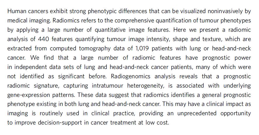
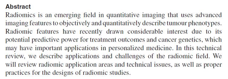
2 RADIOMICS: definition Radiomics is a field of medical study that aims to extract large amount of quantitative features from medical images using mathematical algorithms. These features, termed radiomic features, have the potential to uncover disease characteristics that fail to be appreciated by the naked eye. The hypothesis of radiomics is that the distinctive imaging features between disease forms may be useful for predicting prognosis and therapeutic response for various conditions, thus providing valuable information for personalized therapy. Radiomics emerged from the medical field of oncology and is the most advanced in applications within that field. However, the technique can be applied to any medical study where a disease or a condition can be tomographically imaged.


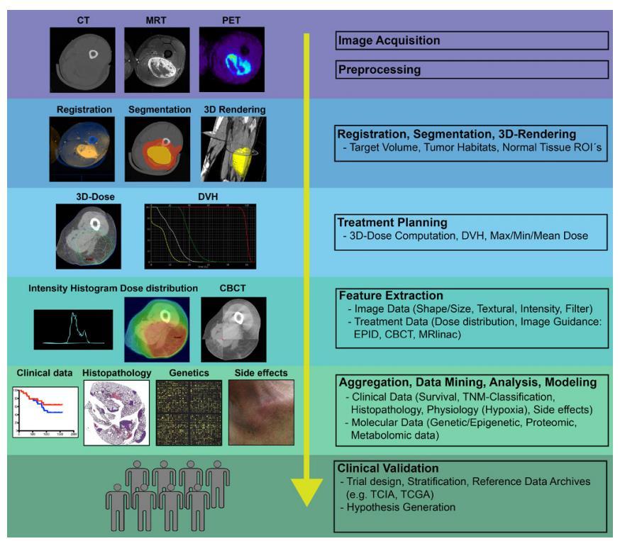
3 RADIOMICS: definition & workflow Computed Tomography CT Positron Emission Tomography PET Magnetic Resonance - MR Predictive / Prognostic models
4 RADIOMICS: history


5 RADIOMICS: history Texture analysis Extraction of quantitative parameters from images Big data analysis extraction of LARGE amount of quantitative features Computational power Haralick, 1973: - omics Experience from other fields (Molecular biology, genetics, ) Clinical data availability Store & retrieval of large amount of clinical data and images Digital Imaging
6 RADIOMICS: history
7 RADIOMICS: history

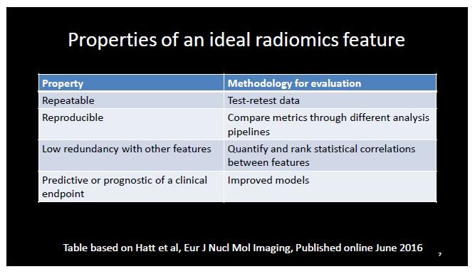
8 RADIOMICS: potential role in the clinics Considering that: 1. Imaging is routinely performed for oncologic patients: - diagnosis - treatment planning - follow up 2. Imaging is not invasive and minimally detrimental invasive alternatives: biopsy, blood sampling plenty of retrospective data database continuously updated no additional cost no additional patient discomfort 3. Radiomics quantifies the properties of the whole volume reduced risk of under-sampling as compared to e.g. biospy more complete information
9 RADIOMICS: history Pubmed search Radiomics
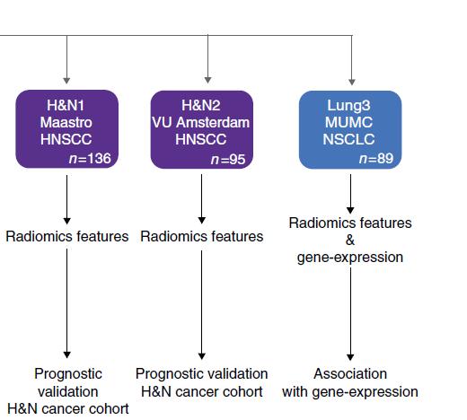
10 RADIOMICS: potential role in the clinics - Differentiate malignant / benign tissue - Tumour staging: differentiate between early and advanced stage disease - Prognostic models: correlation with survival - Predictive models: predict treatment response (chemotherapy, radiation therapy) - Assessment of the metastatic potential of tumours - Assessment of cancer genetics / biological or histopathological properties (biological basis of clinical application of radiomics) - Improve predictivity of models based on clinical,biological, genetic data
11 RADIOMICS: potential role in the clinics
12 RADIOMICS: potential role in the clinics


13 Models generalizability

14
15 Computed Tomography CT Positron Emission Tomography PET Magnetic Resonance - MR Data quality: «imaging biomarkers» are needed
16 Computed Tomography CT Positron Emission Tomography PET Magnetic Resonance - MR Imaging biomarker: Which requirements?
17 Interpretation?
18 RADIOMICS: workflow Computed Tomography CT Positron Emission Tomography PET Magnetic Resonance - MR
19 RADIOMICS: workflow 1. Image acquisition CT images: the voxel intensity describes the composition and the density of the tissue PET images: the voxel intensity is a measure of the concentration of the radiotracer MR images: according to the sequence applied, the voxel intensity can be representative of different properties of the tissue (relaxation times T1, T2, proton density), diffusion, perfusion, Discrete sampling
20 RADIOMICS: workflow Computed Tomography CT Positron Emission Tomography PET Magnetic Resonance - MR


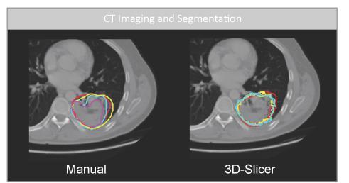
21 RADIOMICS: workflow 2. Region segmentation The Volume Of Interest is a 3D array of numbers, from which many different parameters can be calculated VOI: Volume Of Interest Manual segmentation Semi-automatic / Automatic segmentation algorithms Machine learning
22 RADIOMICS: workflow Computed Tomography CT Positron Emission Tomography PET Magnetic Resonance - MR


23 RADIOMICS: features extraction Shape features: describe the shape of the Region Of Interest in 3D Geometric properties, like the Volume, the maximum diameter or the 3 diameters along the 3 orthogonal directions, the maximum surface
24 RADIOMICS: features categories Histogram-based (First order statistics) features: Describe the distribution of values of individual voxels without concern for spatial relationships Different spatial arrangement BUT Same Histogram!
")


 and")
25 RADIOMICS: features categories Histogram-based (First order statistics) features: Describe the distribution of values of individual voxels without concern for spatial relationships (histogram-based methods as mean, median, maximum, minimum, uniformity or randomness (entropy) of the intensities, skewness (asymmetry) and kurtosis (flatness) of the histogram of values.
26 RADIOMICS: features categories Textural (Second order statistics) features: Textural features describing statistical interrelationships between voxels with similar (or dissimilar) values and take into account the spatial arrangement of the values. «Haralick features»
27 RADIOMICS: features categories Image GLCM: Gray Level Cooccurrence Matrix.
28 RADIOMICS: features categories v Image GLRLM: Gray Level Run Length Matrix.
 Fractal dimension wavelet transform; fractal analyses, wherein patterns are imposed on the image and the number of grid")

29 RADIOMICS: features categories Higher order statistics features: Higher-order statistical methods applies filter grids or mathematical transforms to the image (for example, to extract repetitive or nonrepetitive patterns) Fractal dimension wavelet transform; fractal analyses, wherein patterns are imposed on the image and the number of grid elements containing voxels of a specified value is computed; Laplacian transforms of Gaussian bandpass filters that can extract areas with increasingly coarse texture patterns from the image; Radiomic features calculation is performed on the filtered or decomposed images in order to extract multiple or more informative parameters from a single image.
30 Computed Tomography CT Positron Emission Tomography PET Magnetic Resonance - MR The voxel intensity is the starting point for features calculation
31 Computed Tomography CT Positron Emission Tomography PET Magnetic Resonance - MR A. What affects the voxel intensity? Does it also affect the radiomic feature value?

32 1. Image acquisition affects the informative content of the image Discrete sampling Partial Volume Effect

33 1. Image acquisition affects the informative content of the image Voxel size: Pixel size Slice Thickness Discrete sampling


34 1. Image acquisition affects the informative content of the image Scanner properties Acquisition protocol Reconstruction algorithm Spatial resolution Image from Soret et al., Partial Volume Effect in PET tumour imaging, Journal of Nuclear Medicine (2007), 48(6):


35 1. Image acquisition affects the informative content of the image Scanner properties Acquisition protocol Reconstruction algorithm Noise

36 1. Image acquisition affects the informative content of the image Bit depth
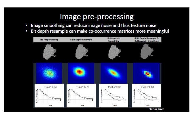
37 2. Image post-processing Post-reconstruction image filtering Useful for physicians, visual inspection, clinical reporting impact on quantification? Discretization N possible values in the image Size of GLCM: NxN


38 A. what affects the voxel intensity? Pixel size Slice Thickness Scanner properties Acquisition protocol Reconstruction algorithm Bit depth Post-reconstruction image filtering Discretization Does it also affect the radiomic feature value? Reproducibility: different modalities are comparable? Repeatability: one modality more stable than others?


39 A. what affects the voxel intensity? Pixel size Slice Thickness Scanner properties Acquisition protocol Reconstruction algorithm Bit depth Post-reconstruction image filtering Discretization Does it also affect the radiomic feature value? Reproducibility: different modalities are comparable? Repeatability: one modality more stable than others?


40 A. what affects the voxel intensity? Pixel size Slice Thickness Scanner properties Acquisition protocol Reconstruction algorithm Bit depth Post-reconstruction image filtering Discretization Does it also affect the radiomic feature value? Reproducibility: different modalities are comparable? Repeatability: one modality more stable than others?


41 Repeatability - CT Concordance Correlation Coefficient > 0.9

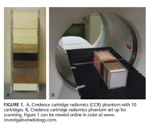
42 Repeatability - CT Example: Phantom experiment, CT Acquisition/ Reconstruction setting % Coefficient of Variation among 10 consecutive acquisitions Feature Different acquisition/reconstruction settings have different repeatability
43 Repeatability - PET
44 about Repeatability: It should be assessed according to the Dynamic Range observed in-vivo, and according to the difference between the groups Distribution of values assessed after repeated acquisitions Feature value
45 about Repeatability: It should be assessed according to the Dynamic Range observed in-vivo, and according to the difference between the groups Distribution among many subjects of a group Feature value
46 about Repeatability: It should be assessed according to the Dynamic Range observed in-vivo, and according to the difference between the groups Group 1 Group 2 Feature value
47 Reproducibility - CT

48 Reproducibility - CT Example: Phantom experiment, CT Acquisition/ Reconstruction setting Feature Values obtained for 40 different acquisition and reconstruction settings Some features are highly dependent on acquisition/reconstruction settings Feature
49 Image processing
50 Computed Tomography CT Positron Emission Tomography PET Magnetic Resonance - MR A. What affects the voxel intensity? Does it also affect the radiomic feature value? B. Other factors affecting the radiomic feature value?

51 3. Volume: segmentation


52 3. Volume: size Small volumes, not only partial volume effect:

53 Rigorous methodology Standardization
54 Rigorous methodology Standardization Ask yourself: is the feature really quantifying a biological property? Or am I «measuring» noise? am I «measuring» volume?

55 Rigorous methodology Standardization Ask yourself: A. is the feature really quantifying a biological property? Or am I «measuring» noise? am I «measuring» volume? B. can I explain the meaning of the features extracted from images?


56 RADIOMICS: statistical analysis Redundancy: Hierarchical Clustering High correlation among features High intra-cluster correlation Low inter-cluster correlation


57 RADIOMICS: the future Data analysis - radiomic features - clinical data Feature calculation Machine Learning - image + Region Of Interest - clinical data Segmentation - image - clinical data
Chapter 3 Set Redundancy in Magnetic Resonance Brain Images
 16 Chapter 3 Set Redundancy in Magnetic Resonance Brain Images 3.1 MRI (magnetic resonance imaging) MRI is a technique of measuring physical structure within the human anatomy. Our proposed research focuses
A dedicated tool for PET scanner simulations using FLUKA
 A dedicated tool for PET scanner simulations using FLUKA P. G. Ortega FLUKA meeting June 2013 1 Need for in-vivo treatment monitoring Particles: The good thing is that they stop... Tumour Normal tissue/organ
Automated segmentation methods for liver analysis in oncology applications
 University of Szeged Department of Image Processing and Computer Graphics Automated segmentation methods for liver analysis in oncology applications Ph. D. Thesis László Ruskó Thesis Advisor Dr. Antal
UNIVERSITY OF SOUTHAMPTON
 UNIVERSITY OF SOUTHAMPTON PHYS2007W1 SEMESTER 2 EXAMINATION 2014-2015 MEDICAL PHYSICS Duration: 120 MINS (2 hours) This paper contains 10 questions. Answer all questions in Section A and only two questions
Available Online through
 Available Online through www.ijptonline.com ISSN: 0975-766X CODEN: IJPTFI Research Article ANALYSIS OF CT LIVER IMAGES FOR TUMOUR DIAGNOSIS BASED ON CLUSTERING TECHNIQUE AND TEXTURE FEATURES M.Krithika
COMPREHENSIVE QUALITY CONTROL OF NMR TOMOGRAPHY USING 3D PRINTED PHANTOM
 COMPREHENSIVE QUALITY CONTROL OF NMR TOMOGRAPHY USING 3D PRINTED PHANTOM Mažena MACIUSOVIČ *, Marius BURKANAS *, Jonas VENIUS *, ** * Medical Physics Department, National Cancer Institute, Vilnius, Lithuania
Computer Aided Diagnosis Based on Medical Image Processing and Artificial Intelligence Methods
 International Journal of Information and Computation Technology. ISSN 0974-2239 Volume 3, Number 9 (2013), pp. 887-892 International Research Publications House http://www. irphouse.com /ijict.htm Computer
MEDICAL IMAGE ANALYSIS
 SECOND EDITION MEDICAL IMAGE ANALYSIS ATAM P. DHAWAN g, A B IEEE Engineering in Medicine and Biology Society, Sponsor IEEE Press Series in Biomedical Engineering Metin Akay, Series Editor +IEEE IEEE PRESS
Computer-Aided Diagnosis in Abdominal and Cardiac Radiology Using Neural Networks
 Computer-Aided Diagnosis in Abdominal and Cardiac Radiology Using Neural Networks Du-Yih Tsai, Masaru Sekiya and Yongbum Lee Department of Radiological Technology, School of Health Sciences, Faculty of
Virtual Phantoms for IGRT QA
 TM Virtual Phantoms for IGRT QA Why ImSimQA? ImSimQA was developed to overcome the limitations of physical phantoms for testing modern medical imaging and radiation therapy software systems, when there
White Paper. EQ PET: Achieving NEMAreferenced. Technologies. Matthew Kelly, PhD, Siemens Healthcare
 White Paper EQ PET: Achieving NEMAreferenced SUV Across Technologies Matthew Kelly, PhD, Siemens Healthcare Table of Contents Introduction 1 Case Study 1 Cross-Scanner Response Assessment 2 Clinical Example
Automatic Lesion Detection for Measuring Response using Dynamic FDG-PET
 Automatic Lesion Detection for Measuring Response using Dynamic FDG-PET Xiujuan Zheng a,b, Guangjian Tian a, Shaoli Song b, Gang Huang b, David Dagan Feng a,c a Department of Electronic and Information
GPU implementation for rapid iterative image reconstruction algorithm
 GPU implementation for rapid iterative image reconstruction algorithm and its applications in nuclear medicine Jakub Pietrzak Krzysztof Kacperski Department of Medical Physics, Maria Skłodowska-Curie Memorial
Medical Image Analysis
 Computer assisted Image Analysis VT04 29 april 2004 Medical Image Analysis Lecture 10 (part 1) Xavier Tizon Medical Image Processing Medical imaging modalities XRay,, CT Ultrasound MRI PET, SPECT Generic
Association between pathology and texture features of multi parametric MRI of the prostate
 Association between pathology and texture features of multi parametric MRI of the prostate 1,2 Peter Kuess, 3 D. Nilsson, 1,2 P. Andrzejewski, 2,4 P. Georg, 1 J. Knoth, 5 M. Susani, 3 J. Trygg, 2,6 T.
Lecture 6: Medical imaging and image-guided interventions
 ME 328: Medical Robotics Winter 2019 Lecture 6: Medical imaging and image-guided interventions Allison Okamura Stanford University Updates Assignment 3 Due this Thursday, Jan. 31 Note that this assignment
Image Registration. Prof. Dr. Lucas Ferrari de Oliveira UFPR Informatics Department
 Image Registration Prof. Dr. Lucas Ferrari de Oliveira UFPR Informatics Department Introduction Visualize objects inside the human body Advances in CS methods to diagnosis, treatment planning and medical
Machine Learning for Medical Image Analysis. A. Criminisi
 Machine Learning for Medical Image Analysis A. Criminisi Overview Introduction to machine learning Decision forests Applications in medical image analysis Anatomy localization in CT Scans Spine Detection
Early Stage Oral Cavity Cancer Detection: Anisotropic Pre-Processing and Fuzzy C-Means Segmentation
 Early Stage Oral Cavity Cancer Detection: Anisotropic Pre-Processing and Fuzzy C-Means Segmentation Zhalong Hu 1 *, Abeer Alsadoon 1 *, Paul Manoranjan 2*, P.W.C. Prasad 1*, Salih Ali 3 * 1 School of Computing
CHAPTER 4 FEATURE EXTRACTION AND SELECTION TECHNIQUES
 69 CHAPTER 4 FEATURE EXTRACTION AND SELECTION TECHNIQUES 4.1 INTRODUCTION Texture is an important characteristic for analyzing the many types of images. It can be seen in all images, from multi spectral
Medical Image Fusion using Rayleigh Contrast Limited Adaptive Histogram Equalization and Ant Colony Edge Method
 Medical Image Fusion using Rayleigh Contrast Limited Adaptive Histogram Equalization and Ant Colony Edge Method Ramandeep 1, Rajiv Kamboj 2 1 Student, M. Tech (ECE), Doon Valley Institute of Engineering
Abbie M. Diak, PhD Loyola University Medical Center Dept. of Radiation Oncology
 Abbie M. Diak, PhD Loyola University Medical Center Dept. of Radiation Oncology Outline High Spectral and Spatial Resolution MR Imaging (HiSS) What it is How to do it Ways to use it HiSS for Radiation
Introduction to Medical Image Processing
 Introduction to Medical Image Processing Δ Essential environments of a medical imaging system Subject Image Analysis Energy Imaging System Images Image Processing Feature Images Image processing may be
TUMOR DETECTION IN MRI IMAGES
 TUMOR DETECTION IN MRI IMAGES Prof. Pravin P. Adivarekar, 2 Priyanka P. Khatate, 3 Punam N. Pawar Prof. Pravin P. Adivarekar, 2 Priyanka P. Khatate, 3 Punam N. Pawar Asst. Professor, 2,3 BE Student,,2,3
8/3/2017. Contour Assessment for Quality Assurance and Data Mining. Objective. Outline. Tom Purdie, PhD, MCCPM
 Contour Assessment for Quality Assurance and Data Mining Tom Purdie, PhD, MCCPM Objective Understand the state-of-the-art in contour assessment for quality assurance including data mining-based techniques
INDUSTRIAL SYSTEM DEVELOPMENT FOR VOLUMETRIC INTEGRITY
 INDUSTRIAL SYSTEM DEVELOPMENT FOR VOLUMETRIC INTEGRITY VERIFICATION AND ANALYSIS M. L. Hsiao and J. W. Eberhard CR&D General Electric Company Schenectady, NY 12301 J. B. Ross Aircraft Engine - QTC General
An Automatic Algorithm for Quality Assurance of MRI Scanners using a DWI Phantom
 An Automatic Algorithm for Quality Assurance of MRI Scanners using a DWI Phantom Aalborg University Biomedical Engineering and informatics School of Medicine and Health Master s Thesis By Andreas Ormstrup
Computational Medical Imaging Analysis
 Computational Medical Imaging Analysis Chapter 1: Introduction to Imaging Science Jun Zhang Laboratory for Computational Medical Imaging & Data Analysis Department of Computer Science University of Kentucky
Brain tumour detection using discrete wavelet transform based medical image fusion.
 Biomedical Research 017; 8 (): 684-688 ISSN 0970-938X www.biomedres.info Brain tumour detection using discrete wavelet transform based medical image fusion. Udhaya Suriya TS 1*, Rangarajan P 1 Department
Ch. 4 Physical Principles of CT
 Ch. 4 Physical Principles of CT CLRS 408: Intro to CT Department of Radiation Sciences Review: Why CT? Solution for radiography/tomography limitations Superimposition of structures Distinguishing between
C a t p h a n / T h e P h a n t o m L a b o r a t o r y
 C a t p h a n 5 0 0 / 6 0 0 T h e P h a n t o m L a b o r a t o r y C a t p h a n 5 0 0 / 6 0 0 Internationally recognized for measuring the maximum obtainable performance of axial, spiral and multi-slice
Reconstruction in CT and relation to other imaging modalities
 Reconstruction in CT and relation to other imaging modalities Jørgen Arendt Jensen November 1, 2017 Center for Fast Ultrasound Imaging, Build 349 Department of Electrical Engineering Center for Fast Ultrasound
Tumor Detection and classification of Medical MRI UsingAdvance ROIPropANN Algorithm
 International Journal of Engineering Research and Advanced Technology (IJERAT) DOI:http://dx.doi.org/10.31695/IJERAT.2018.3273 E-ISSN : 2454-6135 Volume.4, Issue 6 June -2018 Tumor Detection and classification
Semi-Quantitative Metrics in Positron Emission Tomography. Michael Adams. Department of Biomedical Engineering Duke University.
 Semi-Quantitative Metrics in Positron Emission Tomography by Michael Adams Department of Biomedical Engineering Duke University Date: Approved: Timothy G. Turkington, Supervisor Adam P. Wax Terence Z.
Dynamic digital phantoms
 Dynamic digital phantoms In radiation research the term phantom is used to describe an inanimate object or system used to tune the performance of radiation imaging or radiotherapeutic devices. A wide range
CHAPTER-1 INTRODUCTION
 CHAPTER-1 INTRODUCTION 1.1 Fuzzy concept, digital image processing and application in medicine With the advancement of digital computers, it has become easy to store large amount of data and carry out
MULTIMODAL MEDICAL IMAGE FUSION BASED ON HYBRID FUSION METHOD
 MULTIMODAL MEDICAL IMAGE FUSION BASED ON HYBRID FUSION METHOD Sinija.T.S MTECH, Department of computer science Mohandas College of Engineering Karthik.M Assistant professor in CSE Mohandas College of Engineering
Implementation and evaluation of a fully 3D OS-MLEM reconstruction algorithm accounting for the PSF of the PET imaging system
 Implementation and evaluation of a fully 3D OS-MLEM reconstruction algorithm accounting for the PSF of the PET imaging system 3 rd October 2008 11 th Topical Seminar on Innovative Particle and Radiation
Medical Image Processing: Image Reconstruction and 3D Renderings
 Medical Image Processing: Image Reconstruction and 3D Renderings 김보형 서울대학교컴퓨터공학부 Computer Graphics and Image Processing Lab. 2011. 3. 23 1 Computer Graphics & Image Processing Computer Graphics : Create,
A Generation Methodology for Numerical Phantoms with Statistically Relevant Variability of Geometric and Physical Properties
 A Generation Methodology for Numerical Phantoms with Statistically Relevant Variability of Geometric and Physical Properties Steven Dolly 1, Eric Ehler 1, Yang Lou 2, Mark Anastasio 2, Hua Li 2 (1) University
maximum likelihood estimates. The performance of
 International Journal of Computer Science and Telecommunications [Volume 2, Issue 6, September 2] 8 ISSN 247-3338 An Efficient Approach for Medical Image Segmentation Based on Truncated Skew Gaussian Mixture
NON OB ULTRASOUND MORPHOMETRICS AS BIOMARKERS
 NON OB ULTRASOUND MORPHOMETRICS AS BIOMARKERS Brian S Garra Washington DC VA Medical Center and Division of Imaging & Applied Mathematics, OSEL, CDRH, FDA GOALS Review Major Types of Clinical Ultrasonic
BME I5000: Biomedical Imaging
 1 Lucas Parra, CCNY BME I5000: Biomedical Imaging Lecture 4 Computed Tomography Lucas C. Parra, parra@ccny.cuny.edu some slides inspired by lecture notes of Andreas H. Hilscher at Columbia University.
The Effects of PET Reconstruction Parameters on Radiotherapy Response Assessment. and an Investigation of SUV peak Sampling Parameters.
 The Effects of PET Reconstruction Parameters on Radiotherapy Response Assessment and an Investigation of SUV peak Sampling Parameters by Leith Rankine Graduate Program in Medical Physics Duke University
Volume 2, Issue 9, September 2014 ISSN
 Fingerprint Verification of the Digital Images by Using the Discrete Cosine Transformation, Run length Encoding, Fourier transformation and Correlation. Palvee Sharma 1, Dr. Rajeev Mahajan 2 1M.Tech Student
Computer Assisted Image Analysis TF 3p and MN1 5p Lecture 1, (GW 1, )
") Centre for Image Analysis Computer Assisted Image Analysis TF p and MN 5p Lecture, 422 (GW, 2.-2.4) 2.4) 2 Why put the image into a computer? A digital image of a rat. A magnification of the rat s nose.
Centre for Image Analysis Computer Assisted Image Analysis TF p and MN 5p Lecture, 422 (GW, 2.-2.4) 2.4) 2 Why put the image into a computer? A digital image of a rat. A magnification of the rat s nose.
DENOISING OF COMPUTER TOMOGRAPHY IMAGES USING CURVELET TRANSFORM
 VOL. 2, NO. 1, FEBRUARY 7 ISSN 1819-6608 6-7 Asian Research Publishing Network (ARPN). All rights reserved. DENOISING OF COMPUTER TOMOGRAPHY IMAGES USING CURVELET TRANSFORM R. Sivakumar Department of Electronics
Improvement of contrast using reconstruction of 3D Image by PET /CT combination system
 Available online at www.pelagiaresearchlibrary.com Advances in Applied Science Research, 2013, 4(1):285-290 ISSN: 0976-8610 CODEN (USA): AASRFC Improvement of contrast using reconstruction of 3D Image
Mutual Information Based Methods to Localize Image Registration
 Mutual Information Based Methods to Localize Image Registration by Kathleen P. Wilkie A thesis presented to the University of Waterloo in fulfilment of the thesis requirement for the degree of Master of
Deviceless respiratory motion correction in PET imaging exploring the potential of novel data driven strategies
 g Deviceless respiratory motion correction in PET imaging exploring the potential of novel data driven strategies Presented by Adam Kesner, Ph.D., DABR Assistant Professor, Division of Radiological Sciences,
CLASS HOURS: 4 CREDIT HOURS: 4 LABORATORY HOURS: 0
 Revised 10/10 COURSE SYLLABUS TM 220 COMPUTED TOMOGRAPHY PHYSICS CLASS HOURS: 4 CREDIT HOURS: 4 LABORATORY HOURS: 0 CATALOG COURSE DESCRIPTION: This course is one of a three course set in whole body Computed
Block Diagram. Physical World. Image Acquisition. Enhancement and Restoration. Segmentation. Feature Selection/Extraction.
 Block Diagram Physical World Image Acquisition Imaging Image Sampling, Quantization, Compression Image Processing Enhancement and Restoration Segmentation Image Analysis Feature Selection/Extraction Image
Generalized Hough transform for segmentation of X-ray Computed Tomography images
 Generalized Hough transform for segmentation of X-ray Computed Tomography images Cristina Campi CNR - SPIN, Genova, Italy cristina.campi@spin.cnr.it Dipartimento di Matematica, Università Di Roma Sapienza
Texture Classification Using Curvelet Transform
 International Journal of Advancements in Research & Technology, Volume, Issue4, April-3 49 ISSN 78-7763 Texture Classification Using Curvelet Transform S. Prabha, Dr. M. Sasikala Department of Electronics
ANALYSIS OF PULMONARY FIBROSIS IN MRI, USING AN ELASTIC REGISTRATION TECHNIQUE IN A MODEL OF FIBROSIS: Scleroderma
 ANALYSIS OF PULMONARY FIBROSIS IN MRI, USING AN ELASTIC REGISTRATION TECHNIQUE IN A MODEL OF FIBROSIS: Scleroderma ORAL DEFENSE 8 th of September 2017 Charlotte MARTIN Supervisor: Pr. MP REVEL M2 Bio Medical
Global Journal of Engineering Science and Research Management
 ADVANCED K-MEANS ALGORITHM FOR BRAIN TUMOR DETECTION USING NAIVE BAYES CLASSIFIER Veena Bai K*, Dr. Niharika Kumar * MTech CSE, Department of Computer Science and Engineering, B.N.M. Institute of Technology,
BRAIN CANCER CLASSIFICATION USING BACK PROPAGATION NEURAL NETWORK AND PRINCIPLE COMPONENT ANALYSIS Ganesh Ram Nayak 1, Mr.
 International Journal of Technical Research and Applications e-issn:2320-8163, www.ijtra.com Volume 2, Issue 4 (July-Aug 2014), PP. 26-31 BRAIN CANCER CLASSIFICATION USING BACK PROPAGATION NEURAL NETWORK
Sphere Extraction in MR Images with Application to Whole-Body MRI
 Sphere Extraction in MR Images with Application to Whole-Body MRI Christian Wachinger a, Simon Baumann a, Jochen Zeltner b, Ben Glocker a, and Nassir Navab a a Computer Aided Medical Procedures (CAMP),
FOREWORD TO THE SPECIAL ISSUE ON MOTION DETECTION AND COMPENSATION
 Philips J. Res. 51 (1998) 197-201 FOREWORD TO THE SPECIAL ISSUE ON MOTION DETECTION AND COMPENSATION This special issue of Philips Journalof Research includes a number of papers presented at a Philips
Classification of Subject Motion for Improved Reconstruction of Dynamic Magnetic Resonance Imaging
 1 CS 9 Final Project Classification of Subject Motion for Improved Reconstruction of Dynamic Magnetic Resonance Imaging Feiyu Chen Department of Electrical Engineering ABSTRACT Subject motion is a significant
The Institute of Telecommunications and Computer Sciences, UTP University of Science and Technology, Bydgoszcz , Poland
 Computer Technology and Application 6 (2015) 64-69 doi: 10.17265/1934-7332/2015.02.002 D DAVID PUBLISHIN An Image Analysis of Breast Thermograms Ryszard S. Choras The Institute of Telecommunications and
Intraoperative Prostate Tracking with Slice-to-Volume Registration in MR
 Intraoperative Prostate Tracking with Slice-to-Volume Registration in MR Sean Gill a, Purang Abolmaesumi a,b, Siddharth Vikal a, Parvin Mousavi a and Gabor Fichtinger a,b,* (a) School of Computing, Queen
New Technology Allows Multiple Image Contrasts in a Single Scan
 These images were acquired with an investigational device. PD T2 T2 FLAIR T1 MAP T1 FLAIR PSIR T1 New Technology Allows Multiple Image Contrasts in a Single Scan MR exams can be time consuming. A typical
Radiology. Marta Anguiano Millán. Departamento de Física Atómica, Molecular y Nuclear Facultad de Ciencias. Universidad de Granada
 Departamento de Física Atómica, Molecular y Nuclear Facultad de Ciencias. Universidad de Granada Overview Introduction Overview Introduction Tecniques of imaging in Overview Introduction Tecniques of imaging
Technical aspects of SPECT and SPECT-CT. John Buscombe
 Technical aspects of SPECT and SPECT-CT John Buscombe What does the clinician need to know? For SPECT What factors affect SPECT How those factors should be sought Looking for artefacts For SPECT-CT Issues
Biomedical Image Analysis based on Computational Registration Methods. João Manuel R. S. Tavares
 Biomedical Image Analysis based on Computational Registration Methods João Manuel R. S. Tavares tavares@fe.up.pt, www.fe.up.pt/~tavares Outline 1. Introduction 2. Methods a) Spatial Registration of (2D
Utilizing Salient Region Features for 3D Multi-Modality Medical Image Registration
 Utilizing Salient Region Features for 3D Multi-Modality Medical Image Registration Dieter Hahn 1, Gabriele Wolz 2, Yiyong Sun 3, Frank Sauer 3, Joachim Hornegger 1, Torsten Kuwert 2 and Chenyang Xu 3 1
A WAVELET BASED BIOMEDICAL IMAGE COMPRESSION WITH ROI CODING
 Available Online at www.ijcsmc.com International Journal of Computer Science and Mobile Computing A Monthly Journal of Computer Science and Information Technology IJCSMC, Vol. 4, Issue. 5, May 2015, pg.407
MEDICAL IMAGE NOISE REDUCTION AND REGION CONTRAST ENHANCEMENT USING PARTIAL DIFFERENTIAL EQUATIONS
 MEDICAL IMAGE NOISE REDUCTION AND REGION CONTRAST ENHANCEMENT USING PARTIAL DIFFERENTIAL EQUATIONS Miguel Alemán-Flores, Luis Álvarez-León Departamento de Informática y Sistemas, Universidad de Las Palmas
Introduction to Neuroimaging Janaina Mourao-Miranda
 Introduction to Neuroimaging Janaina Mourao-Miranda Neuroimaging techniques have changed the way neuroscientists address questions about functional anatomy, especially in relation to behavior and clinical
Breast Cancer Detection and Classification Using Ultrasound and Ultrasound Elastography Images
 Breast Cancer Detection and Classification Using Ultrasound and Ultrasound Elastography Images Ramya S. 1, Nanda S. 2 1IV Sem, M.Tech, Biomedical Signal Processing & Instrumentation, SJCE, Mysuru, Karnataka,
Tumor Detection in Breast Ultrasound images
 I J C T A, 8(5), 2015, pp. 1881-1885 International Science Press Tumor Detection in Breast Ultrasound images R. Vanithamani* and R. Dhivya** Abstract: Breast ultrasound is becoming a popular screening
Automated segmentation methods for liver analysis in oncology applications
 University of Szeged Department of Image Processing and Computer Graphics Automated segmentation methods for liver analysis in oncology applications Ph. D. Thesis László Ruskó Thesis Advisor Dr. Antal
Weakly Supervised Fully Convolutional Network for PET Lesion Segmentation
 Weakly Supervised Fully Convolutional Network for PET Lesion Segmentation S. Afshari a, A. BenTaieb a, Z. Mirikharaji a, and G. Hamarneh a a Medical Image Analysis Lab, School of Computing Science, Simon
A Study of Medical Image Analysis System
 Indian Journal of Science and Technology, Vol 8(25), DOI: 10.17485/ijst/2015/v8i25/80492, October 2015 ISSN (Print) : 0974-6846 ISSN (Online) : 0974-5645 A Study of Medical Image Analysis System Kim Tae-Eun
We are IntechOpen, the world s leading publisher of Open Access books Built by scientists, for scientists. International authors and editors
 We are IntechOpen, the world s leading publisher of Open Access books Built by scientists, for scientists 4,000 116,000 120M Open access books available International authors and editors Downloads Our
Detection & Classification of Lung Nodules Using multi resolution MTANN in Chest Radiography Images
 The International Journal Of Engineering And Science (IJES) ISSN (e): 2319 1813 ISSN (p): 2319 1805 Pages 98-104 March - 2015 Detection & Classification of Lung Nodules Using multi resolution MTANN in
An Integrated Visual Analysis System for Fusing MR Spectroscopy and Multi-Modal Radiology Imaging
 An Integrated Visual Analysis System for Fusing MR Spectroscopy and Multi-Modal Radiology Imaging Miguel Nunes, Benjamin Rowland, Matthias Schlachter, Soléakhéna Ken, Kresimir Matkovic, Anne Laprie and
Prostate Detection Using Principal Component Analysis
 Prostate Detection Using Principal Component Analysis Aamir Virani (avirani@stanford.edu) CS 229 Machine Learning Stanford University 16 December 2005 Introduction During the past two decades, computed
Medical Image Feature, Extraction, Selection And Classification
 Medical Image Feature, Extraction, Selection And Classification 1 M.VASANTHA *, 2 DR.V.SUBBIAH BHARATHI, 3 R.DHAMODHARAN 1. Research Scholar, Mother Teresa Women s University, KodaiKanal, and Asst.Professor,
CS 5630/6630 Scientific Visualization. Volume Rendering I: Overview
 CS 5630/6630 Scientific Visualization Volume Rendering I: Overview Motivation Isosurfacing is limited It is binary A hard, distinct boundary is not always appropriate Slice Isosurface Volume Rendering
Medical Images Analysis and Processing
 Medical Images Analysis and Processing - 25642 Emad Course Introduction Course Information: Type: Graduated Credits: 3 Prerequisites: Digital Image Processing Course Introduction Reference(s): Insight
SPECT QA and QC. Bruce McBride St. Vincent s Hospital Sydney.
 SPECT QA and QC Bruce McBride St. Vincent s Hospital Sydney. SPECT QA and QC What is needed? Why? How often? Who says? QA and QC in Nuclear Medicine QA - collective term for all the efforts made to produce
PET AND MRI BRAIN IMAGE FUSION USING REDUNDANT WAVELET TRANSFORM
 International Journal of Latest Engineering and Management Research (IJLEMR) ISSN: 2455-4847 Volume 1 Issue 4 ǁ May 2016 ǁ PP.21-26 PET AND MRI BRAIN IMAGE FUSION USING REDUNDANT WAVELET TRANSFORM Gayathri
Whole Body MRI Intensity Standardization
 Whole Body MRI Intensity Standardization Florian Jäger 1, László Nyúl 1, Bernd Frericks 2, Frank Wacker 2 and Joachim Hornegger 1 1 Institute of Pattern Recognition, University of Erlangen, {jaeger,nyul,hornegger}@informatik.uni-erlangen.de
Skull Segmentation of MR images based on texture features for attenuation correction in PET/MR
 Skull Segmentation of MR images based on texture features for attenuation correction in PET/MR CHAIBI HASSEN, NOURINE RACHID ITIO Laboratory, Oran University Algeriachaibih@yahoo.fr, nourine@yahoo.com
Advanced Visual Medicine: Techniques for Visual Exploration & Analysis
 Advanced Visual Medicine: Techniques for Visual Exploration & Analysis Interactive Visualization of Multimodal Volume Data for Neurosurgical Planning Felix Ritter, MeVis Research Bremen Multimodal Neurosurgical
Tumor Lesion Segmentation from 3D PET using a Machine Learning driven Active Surface
 Tumor Lesion Segmentation from 3D PET using a Machine Learning driven Active Surface Payam Ahmadvand 1, Nóirín Duggan 1, François Bénard 2,3, and Ghassan Hamarneh 1 1 Medical Image Analysis Lab, Simon
A simple method to test geometrical reliability of digital reconstructed radiograph (DRR)
") JOURNAL OF APPLIED CLINICAL MEDICAL PHYSICS, VOLUME 11, NUMBER 1, WINTER 2010 A simple method to test geometrical reliability of digital reconstructed radiograph (DRR) Stefania Pallotta, a Marta Bucciolini
JOURNAL OF APPLIED CLINICAL MEDICAL PHYSICS, VOLUME 11, NUMBER 1, WINTER 2010 A simple method to test geometrical reliability of digital reconstructed radiograph (DRR) Stefania Pallotta, a Marta Bucciolini
Hybrid Approach for MRI Human Head Scans Classification using HTT based SFTA Texture Feature Extraction Technique
 Volume 118 No. 17 2018, 691-701 ISSN: 1311-8080 (printed version); ISSN: 1314-3395 (on-line version) url: http://www.ijpam.eu ijpam.eu Hybrid Approach for MRI Human Head Scans Classification using HTT
Lucy Phantom MR Grid Evaluation
 Lucy Phantom MR Grid Evaluation Anil Sethi, PhD Loyola University Medical Center, Maywood, IL 60153 November 2015 I. Introduction: The MR distortion grid, used as an insert with Lucy 3D QA phantom, is
Textural Feature Extraction and Analysis for Brain Tumors using MRI
 Textural Feature Extraction and Analysis for Brain Tumors using MRI 185 Anantha Padmanabha A G*, S Y Pattar** * Student, M.Tech, ML, BMSCE, Bangalore, INDIA. ** Associate. Professor, ML, BMSCE, Bangalore,
Medical Imaging Introduction
 Medical Imaging Introduction Jan Kybic February 16, 2010 Medical imaging: a collaborative paradigm picture from Atam P. Dhawan: Medical Imaging From physiology to information processing (what we should
Extraction and Features of Tumour from MR brain images
 IOSR Journal of Electronics and Communication Engineering (IOSR-JECE) e-issn: 2278-2834,p- ISSN: 2278-8735.Volume 13, Issue 2, Ver. I (Mar. - Apr. 2018), PP 67-71 www.iosrjournals.org Sai Prasanna M 1,
Automated Image Analysis Software for Quality Assurance of a Radiotherapy CT Simulator
 Automated Image Analysis Software for Quality Assurance of a Radiotherapy CT Simulator Andrew J Reilly Imaging Physicist Oncology Physics Edinburgh Cancer Centre Western General Hospital EDINBURGH EH4
CHAPTER 4 TEXTURE FEATURE EXTRACTION
 83 CHAPTER 4 TEXTURE FEATURE EXTRACTION This chapter deals with various feature extraction technique based on spatial, transform, edge and boundary, color, shape and texture features. A brief introduction
A Comparative Study between Two Hybrid Medical Image Compression Methods
 A Comparative Study between Two Hybrid Medical Image Compression Methods Clarissa Philana Shopia Azaria 1, and Irwan Prasetya Gunawan 2 Abstract This paper aims to compare two hybrid medical image compression
Tumor Detection and Classification using Decision Tree in Brain MRI
 Tumor Detection and Classification using Decision Tree in Brain MRI Janki Naik 1, Prof. Sagar Patel 2 Gujarat Technological University, Ahmedabad, Gujarat, India. janki.naik88@gmail.com 1, sagar_in_2006@yahoo.com
Texture Analysis in Quantitative Osteoporosis Assessment
 Texture Analysis in Quantitative Osteoporosis Assessment Characterizing Micro-architecture in High Resolution Peripheral Quantitative Computed Tomography Alexander Valentinitsch 1, Janina Patsch 1, Dirk
Refraction Corrected Transmission Ultrasound Computed Tomography for Application in Breast Imaging
 Refraction Corrected Transmission Ultrasound Computed Tomography for Application in Breast Imaging Joint Research With Trond Varslot Marcel Jackowski Shengying Li and Klaus Mueller Ultrasound Detection