The 3D Accuitomo 80 offers a minute voxel. size of just 80 μm (micrometer). This super-fine. voxel combined with the unit s 13 bit grayscale
|
|
|
- Blaise Weaver
- 5 years ago
- Views:
Transcription
1 2

2 Thinking ahead.

3 Focused on life.

4 REALIZED: Groundbreaking Resolution of 80 µm Voxel


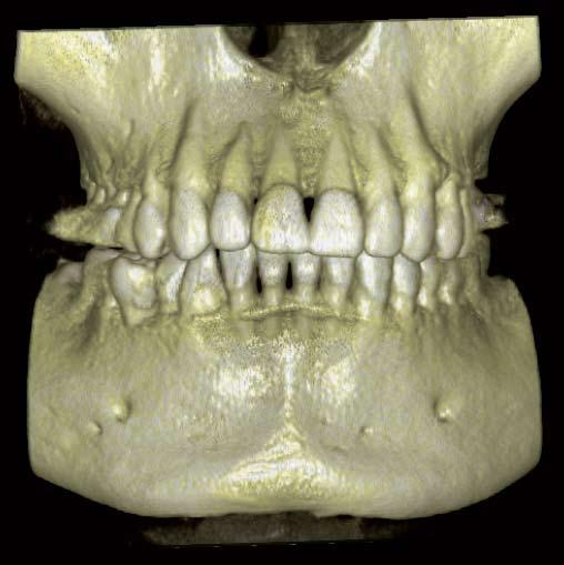
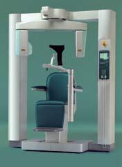
5 05 The Crystallization of cutting-edge mechatronics and High Definition display algorithm The 3D Accuitomo 80 offers a minute voxel size of just 80 μm (micrometer). This super-fine voxel combined with the unit s 13 bit grayscale capability displays an amazing level of clarity never before seen in the world of 3D imaging. Newly developed Zoom Reconstruction function Three image sizes available ø 40 H 40 mm, ø 60 H 60 mm, ø 80 H 80 mm Offers High definition 3D images with low patient dose Displays both hard and soft tissue A wide dynamic range and precise grayscale differentiation capability Enables comprehensive examination for diagnosing apical lesions, TMJ problems, caries, and treatment planning for implants, restorative, surgery, etc. One Data Viewer Software Volume rendering function Intra-clinic network compatibility Compact Floor space: 1,620 mm 1,200 mm Basis technology provided by NUBIC (Nihon University Business, Research and Intellectural Property Center). Developed in collaboration with Nihon University School of Dentistry and J.Morita Mfg. Corp.
6


 to a narrow image area (ø 40 H 40 mm), high resolution is maintained with no")

7 X-ray Flat Panel Detector (FPD) Automatic Positioning Function For ø 40 x H 40 mm and ø 60 x H 60 mm images, increasing the distance between the center of the object being X-rayed and the FPD, the beams are almost parallel, delivering high resolution images with no distortion. Collimation of the beams also makes it possible to minimize patient radiation dose and reduce scatter. ø 40 H 40 mm image is suitable for 90% of all cases while keeping patient dose to a minimum. High Resolution is Maintained Even When the Image Area is Increased From a wide image area (ø 80 H 80 mm) to a narrow image area (ø 40 H 40 mm), high resolution is maintained with no distortion even as the image is expanded from ø 40 H 40 mm to ø 80 H 80 mm. ø 60 H 60 mm image provides a wide image area and simultaneous examination of maxillary and mandibular teeth. ø 80 H 80 mm ø 60 H 60 mm ø 40 H 40 mm ø 80 H 80 mm image allows for a full mouth examination.


8 Zoom Reconstruction The zoom reconstruction function provides high resolution, 80 µm voxel size, 3D images of any region of interest. This allows for a comprehensive examination and diagnosis. There is no need to retake images since all data has already been acquired. Choice of Three Resolution Options Voxel sizes of 80 µm, 125 µm or 160 µm can be selected as the resolution for each image area. Default setting ø 40 H 40 mm: 80 μm ø 60 H 60 mm: 125 μm ø 80 H 80 mm: 160 μm ø 80 H 80 mm, voxel size: 160 µm resliced image Ø 40 H 40 mm, voxel size: 80 µm In viewing an ø 80 H 80 mm image, the clinician can zoom in on any region of interest using the Zoom Reconstruction function.


9 09 80 mm 80 mm Radiographic Procedure A small cone-shaped X-ray beam irradiates the FPD for approximately 18 seconds while the C-arm makes one 360 rotation around the region of interest. The raw images are digitized in the FPD and are transferred to the host computer. After post-processing, a reconstruction algorithm generates super-high resolution, 3-dimensional images displayed on the computer monitor. Low Effective Dose The exposure time is approximately 18 sec. and the effective dose is only about 1.6 times* as high as that of the panoramic X-ray with film exposure and 1/7** the CTDIw value of a conventional CT. Anisotropic voxel Voxel size 0.16 mm 0.08 mm 0.08 mm 0.16 mm 0.16 mm 0.08 mm 0.7 mm mm mm 0.7 mm mm 1.0 mm Isotropic voxel Isotropic Voxel The voxel is an isotropic cube that provides a highly detailed image without artifacts that are usually caused by slice pitch and helical pitch that are common with medical CT scans. Voxel: The minimum unit of 3D data. voxel size: 160 µm voxel size: 80 µm Wide yet smooth and distortion-free 80 µm high resolution images * Effective dose is calculated in accordance with the ICRP 2007 Draft for exposure of the Mandibular Molar Region with Morita s recommended loading factor (80 kv, 3 ma, ø 40 mm H 40 mm). Comparison is to the Veraviewepocs film (75 kv, 8 ma, 16 sec). ** CTDIw value measurement is made according to IEC with Morita s recommended loading factor. The comparison is to the reference CTDIw value for the maxillofacial and parallel sinus indicated in ICRP Pub. 87 Appendix A (ø 40 mm H 40 mm).

![Spatial Resolution* MTF: Modulation Transfer Function MTF [%] 100 90 80 70 60 50 40 30 20 10 0 0 Tube Voltage: 60 kv Tube Current: 1.0 ma MTF at 2 Lp/mm = 13.](/docs-images/94/122369287/images/10-2.jpg "2 % 1 2 3 4 This function is based on data from a typical product.")
10 High Definition / Low Dose The ultra-sensitive, high resolution Flat Panel Detector (FPD) provides high definition 3D images for a more thorough examination while diagnosing apical lesions, root fractures, accessory canals, TMJ problems, and treatment planning for implants, endodontics, surgery, restrative procedures, etc. Spatial Resolution* MTF: Modulation Transfer Function MTF [%] Tube Voltage: 60 kv Tube Current: 1.0 ma MTF at 2 Lp/mm = 13.2 % This function is based on data from a typical product. Spatial Frequency [Lp/mm] Flat Panel Detector (FPD) The FPD provides a high quality image by converting X-ray intensity into digital signals. It has superior sensitivity and a wide dynamic range with precise grayscale differentiation capability and no distortion. X-rays are converted into visible light by the directly deposited CSI scintillator. Then, a photo diode converts the light into an electrical signal. The FPD is quite thin and has a long lifespan. Super-High Resolution The resolution of the 3D Accuitomo 80 is greater than 2 line pairs per mm (MTF 10%). The highly detailed images have a voxel size of 0.08 mm. The slice width and pitch can be set between and 2 mm.


.")
11 Amplifier and horizontal shift register Directly deposited CSI scintillator 11 X-ray Vertical shift register Pixel Digital Video Signal No image Distortion The image receptor is flat so the x-ray projection occurs without any distortion. The less accurate compensation process that is needed for analog systems is eliminated with the FPD. Wide Dynamic Range ** A wide dynamic range produces a precise grayscale differentiation capability. This makes it possible to view both hard and soft tissue. The FPD provides a higher reproducibility of the grayscale using 13 bit technology. Creates very high quality radiographs of soft-tissue areas (such as the mucous membrane of the maxillary sinus and skin, etc.) as well as hard tissue areas (such as enamel, dentin, and alveolar bone). * Spatial resolution refers to how distinct an image appears the smaller it becomes; it measures the fineness of an image. Spatial frequency is the unit of measurement of line pairs per distance (mm). As the map scale decreases, the patterns of contrast become harder to see. This is known as MTF (Modulation Transfer Function). It represents the number of line pairs per 1 mm that can be distinguished based on contrast. It is said that humans can only differentiate about 10%. ** Dynamic range: Numerical values express the reproducibility of the signal and the ratio of the largest and smallest input values in dbs. The dynamic range of the digital signal is also sometimes expressed in bits. The highest signal level is taken to be the level remaining after subtracting the noise level. The value of the dynamic range indicates how weak of a signal can be reproduced or, in other words, how high the contrast resolution will actually turn out to be.

12


13 Easy Image Processing 13 Multiple image processing functions allow the i-dixel volume-rendering software to display a variety of helpful information, including both 3D and 2D data. The software is seamlessly integrated into your network environment. Volume Rendering The volume-rendering capability of i-dixel generates a three dimensional image of anatomical structure on the display monitor. High definition 3D images of both hard and soft tissue can be obtained by adjusting the histogram and threshold level depending on the region of interest. Real-Time Reslice Volume-rendering images and slice images are linked. Observation angles and/or slicing angles can be viewed simultaneously and easily manipulated in real time by simply dragging anywhere on the volume or slice images. Curved MPR Orthogonal cross-sectional images along dental arch can be observed. Other Key Features n XYZ view windows n Reslicing n Zoom n Rotate n Histogram n Edge enhancement n Distance and angle measurement n Negative image n Mirror image n Slice distance measurement n DICOM 3.0-compatible n Density control n Spatial frequency filtering n Patient orientation display n Density measurement


14 3D Accuitomo 80 i-dixel software i-dixel software i-dixel software Intra-clinic network Out of network computer In external clinic networks without 3D Accuitomo 80, 3D images can be viewed on a PC with both of the following methods: i-dixel Viewer Software One Data Viewer

15 SHARING IMAGE DATA 15 Installing i-dixel software on all intra-clinic computers enables sharing of image data on each linked computer. Observation of images on non-network computers can be achieved with One Data Viewer without installing i-dixel. One Data Viewer Software One Data Viewer is a unique Morita software feature which is a stand-alone, executable application. It allows 3D images to be viewed on computers without i-dixel. Data files can be taken anywhere and viewed on any computer without i-dixel. One Data Viewer EX 3D images and patient information can be observed. One Data Viewer Plus (Optional) In addition to the One Data Viewer EX functions, the Plus version provides distance and angle measurement, zooming, image reversal, and additional adjustments for brightness, contrast, and gamma curve. i-dixel Viewer Software By installing i-dixel Viewer software on any computer, all i-dixel features can be utilized. DICOM Support (Optional) i-dixel can be seamlessly integrated into a DICOM compatible clinic network environment. DICOM, or Digital Imaging and Communications in Medicine, creates and maintains an international standard for image data communication and storage, to achieve compatibility between various information systems in health care environments. DICOM enables the exchange of diagnostic images, patient examination data and other relevant information regardless of the manufacturer or model of the medical imaging systems involved. It also supports the system architecture link to HIS/RIS. i-dixel Supports the Following DICOM Service Classes: 1. Modality worklist management service class 2. Storage service class 3. Modality performed procedure step service class 4. Print management service class

16 X cursor Z cursor Y cursor


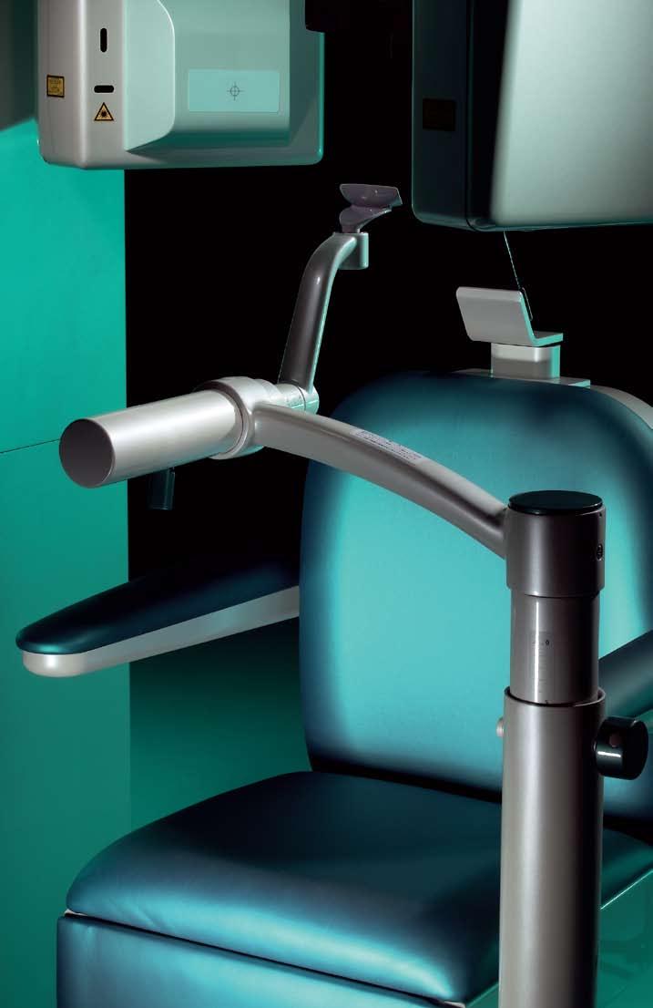
17 Simple, Accurate Positioning 17 The three positioning laser beams and an LCD make patient positioning easy. The chinrest stabilizes patient s head to avoid movement. Scout images enable even more accurate po si tion ing. Easy as One, Two, Three First, the patient s initial position is set and recorded using the three positioning laser beams. Then, the region of interest is aligned in the LCD. The chair automatically moves into the optimal position. During the X-ray exposure, the patient is stabilized by the chinrest and the headrest. 2-Directional Scout For even more accurate positioning, scout images* can be created. After positioning, take two still X-ray images of coronal and sagittal views to confirm that the position is accurate. If adjustment is necessary, positioning can be changed by dragging the cursor on the monitor and moving it to the center of the region of interest. Hitting the ready key will then automatically move the chair, and thus the region of interest, to the desired position. 3D image with the region of interest located at the center. *Taking scout images (80 kv, 2 ma) increases X-ray dosage by approximately 2.4%.

18 Volume rendering images


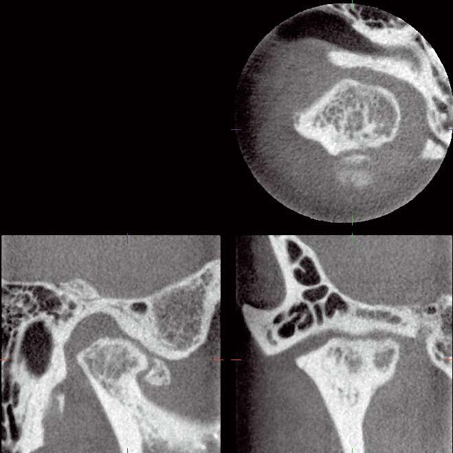
19 Zoom reconstruction of areas of interest Resolution Power of 80 µm! Resorption of alveolar bone was seen in the periapical region of the maxillary first and second molars. By specifying the area of interest and performing zoom reconstruction (80 µm voxels), a more detailed examination was completed. Lesion present on the mesio-buccal root of the maxillary right first molar and its proximity with the maxillary sinus. Lesion of the mesio-buccal root of the maxillary right second molar and its proximity to the maxillary sinus

,")

20 20 A cavity on the lingual side was detected, and by specifying the area of interest and performing zoom reconstruction (80 µm voxels), more detailed examination was completed. Cavity on lingual side (tip of arrow)

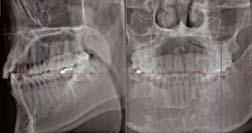
21 Clinical Cases 21 high quality 3D imaging allows for a comprehensive radiographic examination. The ø 80 mm x H 80 mm imaging area covers the entire mandibular, maxillary, and TMJ areas. This includes soft tissue, membranes, and skin as well as hard tissues such as enamel, dentin, and bone. Periodontitis and Apical Lesions Resorption of alveolar bone can be seen in the maxillary right posterior periapical region. ø 80 H 80, 160 µm, 90 kv, 6 ma


22 ø 40 H 40, 80 μm 90 kv, 5 ma


 was applied to assist in diagnosis.")
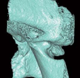
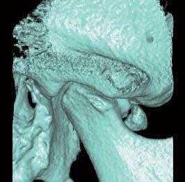
23 Clinical Cases 23 The ø 40 H 40 mm imaging area is suitable for 90% of cases and has a low patient dose. TMJ Osteoarthritis It can be seen that the condylar head is interfering with an articular tubercle and causing deterioration. Moreover, a bone fragment can be seen at the anterior edge of the condylar head. Implant Follow-up Follow up radiographic examination of an implant reveals a gross lesion apical to the fixture. Zoom reconstruction (80 µm voxels) was applied to assist in diagnosis. After ø 80 H 80, 160 µm imaging (90 kv, 4 ma), ø 40 H 40, 80 µm zoom reconstruction was performed.
24
25 Technical Specifications 25 Dimensions Control Box unit: mm X-ray room Internal size: W x D x H 2,000 mm x 1,800 mm x 2,400 mm Dedicated PCs Specifications Trade Name Model Type Input Voltage Power Consumption X-ray Head Tube Voltage Tube Current Focal Spot Size Exposure Time Size of Imaging Area 3D Accuitomo XYZ Slice View Tomograph MCT-1 EX F8 (EX-1F8/EX-2F8) 100/110/120VAC (EX-1F8) 220/230/240VAC (EX-2F8) max. 2.0 kva kv 1-10 ma (8 ma Max at kv) 0.5 mm 18 seconds or less Diameter 40 Height 40 mm Diameter 60 Height 60 mm Diameter 80 Height 80 mm Voxel Size 80 µm, 125 µm, 160 µm Outer Dimensions Main Unit 1,620 mm 1,200 mm 2,080 mm (W x D x H) Weight Control Box (W x D x H) 96 mm 40 mm 115 mm Approx. 400 kg Outlet of computer cable and operation cable Outlet of power supply Outlet of operation cable Outlet of computer cable * Clinical images are provided by Kitasenju Radist Dental Clinic, i-view Imaging Center, Japan * X-ray protection should be provided for the patient when X-rays are emitted. * Design and specifications are subject to change without notification. Developed and Manufactured by J. MORITA Mfg. Corp. 680 Higashihama Minami-cho, Fushimi-ku, Kyoto, Japan Tel: , Fax: , Distributed by J. MORITA CORPORATION 33-18, 3-Chome, Tarumi-cho Suita City, Osaka, Japan Tel: , Fax: , J. MORITA USA, Inc. 9 Mason lrvine, CA U.S.A. Tel: , Fax: , J. MORITA EUROPE GMBH Justus-von-Liebig-Strasse 27A, D Dietzenbach, Germany Tel: , Fax: , Siamdent Co., Ltd. 71/10 Bangpakong Industrial Park 1, Bangna-Trad KM 52, Bangpakong, Chachuengsao 24130, Thailand Tel: , Fax: ,
26
Profound understanding of anatomy
 ENGLISH Profound understanding of anatomy The unique Planmeca ProMax 3D product family offers equipment for all maxillofacial imaging. All volume sizes from the smallest special cases to whole head images
One Machine... Mac OS Compatible! Multiple Modalities. ProMax 3D & 3D s
 One Machine... Multiple Modalities Mac OS Compatible! 3D Bitewing Panoramic Cephalometric Features Availability of multiple imaging modalities in one machine (3D, bitewing, panoramic, and cephalometric)
Shadow casting. What is the problem? Cone Beam Computed Tomography THE OBJECTIVES OF DIAGNOSTIC IMAGING IDEAL DIAGNOSTIC IMAGING STUDY LIMITATIONS
 Cone Beam Computed Tomography THE OBJECTIVES OF DIAGNOSTIC IMAGING Reveal pathology Reveal the anatomic truth Steven R. Singer, DDS srs2@columbia.edu IDEAL DIAGNOSTIC IMAGING STUDY Provides desired diagnostic
Z-MOTION. Universal Digital Radiographic System Z-MOTION. Control-X Medical CONTROL-X MEDICAL
 Control-X Medical Z-MOTION Compact design, low ceiling height requirement Motorized and manual movement capability Wide motion / SID range Best-in-class image quality Flexible connectivity to PACS systems
icatvision Quick Reference
 icatvision Quick Reference Navigating the i-cat Interface This guide shows how to: View reconstructed images Use main features and tools to optimize an image. REMINDER Images are displayed as if you are
PLANMECA PROMAX 3D MID CBCT UNIT
 1(5) PLANMECA PROMAX 3D MID CBCT UNIT Introduction The Planmeca ProMax 3D Mid X-ray unit uses cone beam computerized tomography (CBCT) to produce three-dimensional X-ray images. Panoramic and cephalometric
Planmeca ProMax 3D Max CBCT unit
 D00107623 1(6) Planmeca ProMax 3D Max CBCT unit Introduction The Planmeca ProMax 3D Max X-ray unit uses cone beam computerized tomography (CBCT) to produce threedimensional X-ray images. Panoramic and
Planmeca ProMax 3D s CBCT unit
 D0010759 1(5) Planmeca ProMax 3D s CBCT unit Introduction The Planmeca ProMax 3D s X-ray unit uses cone beam computerized tomography (CBCT) to produce threedimensional X-ray images. Panoramic and cephalometric
icatvision Software Manual
 University of Minnesota School of Dentistry icatvision Software Manual April 19, 2013 Mansur Ahmad, BDS, PhD Associate Professor, University of Minnesota School of Dentistry Director, American Board of
(restyle 2011) Anatomic programs
 Anatomic programs") Product Data Panoramic X-Ray System with CCD sensor (restyle 2011) Examination programs Panoramic - adult - child - child with deceleration ramp for spine compensation TMJ - open/close mouth (4 views on
Product Data Panoramic X-Ray System with CCD sensor (restyle 2011) Examination programs Panoramic - adult - child - child with deceleration ramp for spine compensation TMJ - open/close mouth (4 views on
Fujifilm DR Solution. FDR AcSelerate. The new pinnacle in diagnostic imaging from Fujifilm ISS. CsI. Dynamic Visualization. Technology.
 Fujifilm DR Solution FDR AcSelerate The new pinnacle in diagnostic imaging from Fujifilm CsI Scintillator ISS Technology Dynamic Visualization Welcome to the X-ray room of the future! A streamlined solution
Digital Image Processing
 Digital Image Processing SPECIAL TOPICS CT IMAGES Hamid R. Rabiee Fall 2015 What is an image? 2 Are images only about visual concepts? We ve already seen that there are other kinds of image. In this lecture
DX-D 100 with WITH ITS EXCELLENT IMAGE QUALITY AND
 M O B I L E D R S O L U T I O N DX-D 100 with wireless detector Patients who most need imaging exams may lack the mobility necessary to move to the X-ray room or to position themselves properly for optimum
DX-D WITH DX-D 40/45 DETECTOR FAMILY MOBILE DR SOLUTION
 DX-D 100 + WITH DX-D 40/45 DETECTOR FAMILY MOBILE DR SOLUTION WITH ITS EXCELLENT IMAGE QUALITY AND FLEXIBLE HANDLING, THE MOBILE DX-D 100 + WITH WIRELESS DETECTOR OFFERS FAST IMAGING THAT CAN BE VALIDATED
Dolphin 3D Imaging 11.7 beta
 Dolphin 3D Imaging 11.7 beta The Dolphin 3D software module is a powerful tool that makes processing 3D data extremely simple, enabling dental specialists from a wide variety of disciplines to diagnose,
product data Raybow dr mobile digital radiographic system with wireless detector product data
 Raybow dr mobile digital radiographic system with wireless detector Integrating all the advanced technologies, the ergonomic and high quality system Raybow dr lets you perform digital radiographies anywhere:
Ch. 4 Physical Principles of CT
 Ch. 4 Physical Principles of CT CLRS 408: Intro to CT Department of Radiation Sciences Review: Why CT? Solution for radiography/tomography limitations Superimposition of structures Distinguishing between
tr dent 2D-3D-PAN-CEPH
 tr dent 2D-3D-PAN-CEPH X-VIEW evolves and develops in response to your needs 2D PAN 2D-PAN-CEPH Single Shot 3D-PAN Multilayer 3D-PAN Multilayer CEPH Single Shot Cone Beam 3D Multilayer 2D Panoramic 24
Operation Instructions (Ver )
") Operation Instructions (Ver. 2.0.0) 1 i-dixel One Volume Viewer: Conditions of Use 1. Data Export Conditions This application can be used for any CT data which is compatible with i-dixel software. However,
Operation Instructions (Ver. 2.0.0) 1 i-dixel One Volume Viewer: Conditions of Use 1. Data Export Conditions This application can be used for any CT data which is compatible with i-dixel software. However,
DX-D 100 with wireless detector
 M O B I L E D R S O L U T I O N DX-D 100 with wireless detector Patients who most need imaging exams may lack the mobility necessary to move to the X-ray room or to position themselves properly for optimum
Image Acquisition Systems
 Image Acquisition Systems Goals and Terminology Conventional Radiography Axial Tomography Computer Axial Tomography (CAT) Magnetic Resonance Imaging (MRI) PET, SPECT Ultrasound Microscopy Imaging ITCS
DX-D 100 WITH WIRELESS DETECTOR
 DX-D 100 WITH WIRELESS DETECTOR MOBILE DR SOLUTION WITH ITS EXCELLENT IMAGE QUALITY AND FLEXIBLE HANDLING, THE MOBILE DX-D 100 WITH WIRELESS DETECTOR OFFERS FAST IMAGING THAT CAN BE VALIDATED IMMEDIATELY.
As flexible and individual as your practice.
 Intraoral imaging instruments treatment centers Imaging systems cad/cam systems XIOS PLUS - INTRAORAL SENSOR HELIODENT Plus - INTRAORAL X-RAY SOURCE As flexible and individual as your practice. XIOS PLUS
Spiral CT. Protocol Optimization & Quality Assurance. Ge Wang, Ph.D. Department of Radiology University of Iowa Iowa City, Iowa 52242, USA
 Spiral CT Protocol Optimization & Quality Assurance Ge Wang, Ph.D. Department of Radiology University of Iowa Iowa City, Iowa 52242, USA Spiral CT Protocol Optimization & Quality Assurance Protocol optimization
INTRODUCTION TO MEDICAL IMAGING- 3D LOCALIZATION LAB MANUAL 1. Modifications for P551 Fall 2013 Medical Physics Laboratory
 INTRODUCTION TO MEDICAL IMAGING- 3D LOCALIZATION LAB MANUAL 1 Modifications for P551 Fall 2013 Medical Physics Laboratory Introduction Following the introductory lab 0, this lab exercise the student through
LAB DEMONSTRATION COMPUTED TOMOGRAPHY USING DESKCAT Lab Manual: 0
 LAB DEMONSTRATION COMPUTED TOMOGRAPHY USING DESKCAT Lab Manual: 0 Introduction This lab demonstration explores the physics and technology of Computed Tomography (CT) and guides the student and instructor
Mobile X-Ray System C504-E025
 Mobile X-Ray System C504-E025 The Advent of an Advanced Mobile System Featuring Superb Image Quality and Easy Handling Modern medical facilities require rapid examinations under a variety of situations.
Radiology. Marta Anguiano Millán. Departamento de Física Atómica, Molecular y Nuclear Facultad de Ciencias. Universidad de Granada
 Departamento de Física Atómica, Molecular y Nuclear Facultad de Ciencias. Universidad de Granada Overview Introduction Overview Introduction Tecniques of imaging in Overview Introduction Tecniques of imaging
OnDemand3D Application
 OnDemand3D Application Operating Manual Build 1.0.9.2225 Version 1.0 Copyright 2012 Cybermed Inc. - 0 - Index Index - 1-1. Introduction - 5-1.1 Main Functions... - 5-2. Installation - 6-2.1 System Requirements...
Design and performance characteristics of a Cone Beam CT system for Leksell Gamma Knife Icon
 Design and performance characteristics of a Cone Beam CT system for Leksell Gamma Knife Icon WHITE PAPER Introduction Introducing an image guidance system based on Cone Beam CT (CBCT) and a mask immobilization
DX-D 300 FLEXIBLE DIRECT RADIOGRAPHY SYSTEM DX-D 300
 DX-D 300 FLEXIBLE DIRECT RADIOGRAPHY SYSTEM MUSICA processing provides excellent contrast detail and exam-independent, consistent image quality Cesium Iodide DR detector technology offers potential for
The digital EVOlution in cassette size format
 http://cr-pacs.com/ Platzhalter Wireless Mobile Fast Retrofit Light Low Dose Highest DQE Highest MTF CsI GOS The digital EVOlution in cassette size format Your wireless entry to the world of Digital Radiography
Medical Image Processing: Image Reconstruction and 3D Renderings
 Medical Image Processing: Image Reconstruction and 3D Renderings 김보형 서울대학교컴퓨터공학부 Computer Graphics and Image Processing Lab. 2011. 3. 23 1 Computer Graphics & Image Processing Computer Graphics : Create,
MEDICAL IMAGING 2nd Part Computed Tomography
 MEDICAL IMAGING 2nd Part Computed Tomography Introduction 2 In the last 30 years X-ray Computed Tomography development produced a great change in the role of diagnostic imaging in medicine. In convetional
Medical X-ray Imaging
 Medical X-ray Imaging HYBRID PORTABLE X-RAY 20BT lite / 20BT / 40BT The world s only hybrid powered portable X-ray Light weight, compact size and durable cover Light weight portable X-ray with high power
Medical X-ray Imaging
 Medical X-ray Imaging HYBRID PORTABLE X-RAY 20BT lite / 20BT / 40BT The world s only hybrid powered portable X-ray Light weight, compact size and durable cover Light weight portable X-ray with high power
Imaging protocols for navigated procedures
 9732379 G02 Rev. 1 2015-11 Imaging protocols for navigated procedures How to use this document This document contains imaging protocols for navigated cranial, DBS and stereotactic, ENT, and spine procedures
CT Basics Principles of Spiral CT Dose. Always Thinking Ahead.
 1 CT Basics Principles of Spiral CT Dose 2 Who invented CT? 1963 - Alan Cormack developed a mathematical method of reconstructing images from x-ray projections Sir Godfrey Hounsfield worked for the Central
Product Overview. A Smart and Complete DR Solution. Key Highlights. Premium detector options. Single Console Workflow and Generator Integration
 DIGITAL RADIOGRAPHY Product Overview A Smart and Complete DR Solution Key Highlights The DELWORKS E-Series DR Upgrade delivers exceptional diagnostic imaging using a powerful image acquisition and processing
PURE. ViSION Edition PET/CT. Patient Comfort Put First.
 PURE ViSION Edition PET/CT Patient Comfort Put First. 2 System features that put patient comfort and safety first. Oncology patients deserve the highest levels of safety and comfort during scans. Our Celesteion
XRADIA microxct Manual
 XRADIA microxct Manual Multiscale CT Lab Table of Contents 1. Introduction and Basics 1.1 Instrument Parts 1.2 Powering up the system 1.3 Preparing your sample 2. TXM Controller 2.1 Starting up 2.2 Finding
HIGH RESOLUTION COMPUTED TOMOGRAPHY FOR METROLOGY
 HIGH RESOLUTION COMPUTED TOMOGRAPHY FOR METROLOGY David K. Lehmann 1, Kathleen Brockdorf 1 and Dirk Neuber 2 1 phoenix x-ray Systems + Services Inc. St. Petersburg, FL, USA 2 phoenix x-ray Systems + Services
Quality control phantoms and protocol for a tomography system
 Quality control phantoms and protocol for a tomography system Lucía Franco 1 1 CT AIMEN, C/Relva 27A O Porriño Pontevedra, Spain, lfranco@aimen.es Abstract Tomography systems for non-destructive testing
Introducing the new. KODAK RVG 6500 System
 Introducing the new KODAK RVG 6500 System Innovation New features Wireless image transmission First Wi-Fi enabled sensor delivering the same film quality images as our best wired sensor Intelligent Positioning
Industrial Computed Tomography Innovations
 GE Inspection Technologies Industrial Computed Tomography Innovations Premium performance for premium quality and speed. gemeasurement.com/ct Premium CT technologies. Faster than ever before. Every industrial
Digital Laminography and Computed Tomography with 600 kv for Aerospace Applications
 4th International Symposium on NDT in Aerospace 2012 - Tu.3.A.1 Digital Laminography and Computed Tomography with 600 kv for Aerospace Applications Malte KURFISS 1, Gerd STRECKENBACH 2 1 YXLON International
Developments in Dimensional Metrology in X-ray Computed Tomography at NPL
 Developments in Dimensional Metrology in X-ray Computed Tomography at NPL Wenjuan Sun and Stephen Brown 10 th May 2016 1 Possible factors influencing XCT measurements Components Influencing variables Possible
Hybrid. powered. 20BT lite / 20BT / 40BT. 20 lite / 60. Model mex+20bt lite mex+20bt mex+40bt. Model mex+20 lite mex+20 mex+40 mex+60 mex+100
 20BT lite / 20BT / 40BT 20 lite 20 Hybrid powered 40 / 60 Model mex+20bt lite mex+20bt mex+40bt Output Tube Vol. / Current 90 kv / 20 ma 100 kv / 20 ma 100 kv / 35 ma Voltage range 50-90 kv in 1 kv steps
Position accuracy analysis of the stereotactic reference defined by the CBCT on Leksell Gamma Knife Icon
 Position accuracy analysis of the stereotactic reference defined by the CBCT on Leksell Gamma Knife Icon WHITE PAPER Introduction An image guidance system based on Cone Beam CT (CBCT) is included in Leksell
FLEXIBLE DIRECT RADIOGRAPHY SYSTEM DX-D 300
 FLEXIBLE DIRECT RADIOGRAPHY SYSTEM DX-D 300 The DX-D 300 DR system unites excellent image quality with complete convenience. It offers top-of-the-line technology, a single detector and a fully-motorized
Solid Capabilities Are Built Into the Supria Plus. Putting You On The Path of High Quality, Cost-Effective CT Scanning
 Specification Data Putting You On The Path of High Quality, Cost-Effective CT Scanning Solid Capabilities Are Built Into the Supria Plus Addressing the challenges of controlling healthcare organization
CLASS HOURS: 4 CREDIT HOURS: 4 LABORATORY HOURS: 0
 Revised 10/10 COURSE SYLLABUS TM 220 COMPUTED TOMOGRAPHY PHYSICS CLASS HOURS: 4 CREDIT HOURS: 4 LABORATORY HOURS: 0 CATALOG COURSE DESCRIPTION: This course is one of a three course set in whole body Computed
Data Fusion Virtual Surgery Medical Virtual Reality Team. Endo-Robot. Database Functional. Database
 2017 29 6 16 GITI 3D From 3D to 4D imaging Data Fusion Virtual Surgery Medical Virtual Reality Team Morphological Database Functional Database Endo-Robot High Dimensional Database Team Tele-surgery Robotic
Advanced Reconstruction Techniques Applied to an On-Site CT System
 2nd International Symposium on NDT in Aerospace 2010 - We.1.A.4 Advanced Reconstruction Techniques Applied to an On-Site CT System Jonathan HESS, Markus EBERHORN, Markus HOFMANN, Maik LUXA Fraunhofer Development
Whole Body Submillimeter Scan
 128 Whole Body Submillimeter Scan The real value of 64ch/128slice CT systems does not come from the ability to perfom cardiac scans but the capability to scan all parts of the body in high definition submillimeter
The Vision To Advance Your Quality Of Care
 The Vision To Advance Your Quality Of Care Cone Beam 3D Imaging Systems Panoramic X-ray Systems Intraoral X-ray Systems P Digital Intraoral Sensors Digital X-ray Phosphor Plates Intraoral Cameras Imaging
PRODUCT DATA. Advanced 128
 PRODUCT DATA Advanced 128 Supria 16 Slice CT Puts You On The Path of High Quality, Cost-Effective CT Scanning Solid Capabilities Are Built Into the Supria Addressing the challenges of controlling healthcare
Introduction to Biomedical Imaging
 Alejandro Frangi, PhD Computational Imaging Lab Department of Information & Communication Technology Pompeu Fabra University www.cilab.upf.edu X-ray Projection Imaging Computed Tomography Digital X-ray
DX-D 300. Flexible Direct Radiography System
 DX-D 300 Flexible Direct Radiography System MUSICA 2 processing provides superior contrast detail and exam-independent, consistent image quality Cesium Iodide DR detector technology offers potential for
Equipment Specification
 MULTISLICE CT SCANNER ( 16 Slices ) Merk : Hitachi Japan Model : SUPRIA 5 MHU Price : Rp 6.512.360.215,27 No Equipment Specification 1 2 3 Scanner Gantry Object for scanning : Whole body including head
Corso di laurea in Fisica A.A Fisica Medica 4 TC
 Corso di laurea in Fisica A.A. 2007-2008 Fisica Medica 4 TC Computed Tomography Principles 1. Projection measurement 2. Scanner systems 3. Scanning modes Basic Tomographic Principle The internal structure
Planmeca ProMax 3D family
 3D Plus ENGLISH Planmeca ProMax 3D family True all-in-one units for all your imaging needs Planmeca ProMax 3D is a product family consisting of exceptional all-in-one units. With three different types
ImPACT. Information Leaflet No. 1: CT Scanner Acceptance Testing
 ImPACT Information Leaflet No. 1: CT Scanner Acceptance Testing Version 1.02, 18/05/01 CONTENTS: 1. SCOPE OF LEAFLET 2. GENERAL PRINCIPLES OF ACCEPTANCE AND COMMISSIONING 2.1 PHANTOMS 2.2 EXPOSURE AND
Digital Innovation for Medical Equipment
 Digital Innovation for Medical Equipment 01 CONTENTS Established in 1992, Sitec Medical Co., Ltd. has been in the conventional diagnostic radiology field for more than thirty years with equipments of its
EXAMION DR EasyGo ... A SYSTEM OF CHOICES. EXAMION DR Mobile solution. EXAMION Teleradiology EXAMION AQS EXAMION PACS.
 EXAMION DR Mobile solution EasyGo Soft Pack Import your external X-rays in standard formats from external media (CD, USB stick, memory cards, etc.) Simple operation of the work List Easy application to
PH-03: Clinical Photography Production Checklist With The Sony Cybershot T-9 Digital Camera
 PH-03: Clinical Photography Production Checklist With The Sony Cybershot T-9 Digital Camera Date: Name: EXTRAORAL SERIOUS 1. Prepare the camera Program the camera to the recommended settings Set camera
C504-E013A. Mobile X-Ray System A-NS
 C504-E013A Mobile X-Ray System 6295-11902-30A-NS The Advent of an Advanced Mobile System Featuring Superb Image Quality and Easy Handling Modern medical facilities require rapid examinations under a variety
Computed Tomography & 3D Metrology Application of the VDI/VDE Directive 2630 and Optimization of the CT system
 Computed Tomography & 3D Metrology Application of the VDI/VDE Directive 2630 and Optimization of the CT system ECNDT 2014 Prague October 6-10, 2014 Dr. Eberhard Neuser Dr. Alexander Suppes Imagination
SCENARIA 64-Slice CT Putting Advanced CT Within Reach.
 SCENARIA 64-Slice CT Putting Advanced CT Within Reach. Advancing the technology of Computed Tomography for over 30 years, Hitachi is a recognized innovator of lower dose*, high diagnostic value CT solutions.
Modifications for P551 Fall 2014
 LAB DEMONSTRATION COMPUTED TOMOGRAPHY USING DESKCAT 1 Modifications for P551 Fall 2014 Introduction This lab demonstration explores the physics and technology of Computed Tomography (CT) and guides the
A study of densitometry comparison among three radiographic processing solutions
 Iran. J. Radiat. Res., 2006; 4 (2): 81-86 A study of densitometry comparison among three radiographic processing solutions V. Changizi 1*, E. Jazayeri 1,A.Talaeepour 2 1 Department of Radiology Technology,
3/27/2012 WHY SPECT / CT? SPECT / CT Basic Principles. Advantages of SPECT. Advantages of CT. Dr John C. Dickson, Principal Physicist UCLH
 3/27/212 Advantages of SPECT SPECT / CT Basic Principles Dr John C. Dickson, Principal Physicist UCLH Institute of Nuclear Medicine, University College London Hospitals and University College London john.dickson@uclh.nhs.uk
HIGH-SPEED THEE-DIMENSIONAL TOMOGRAPHIC IMAGING OF FRAGMENTS AND PRECISE STATISTICS FROM AN AUTOMATED ANALYSIS
 23 RD INTERNATIONAL SYMPOSIUM ON BALLISTICS TARRAGONA, SPAIN 16-20 APRIL 2007 HIGH-SPEED THEE-DIMENSIONAL TOMOGRAPHIC IMAGING OF FRAGMENTS AND PRECISE STATISTICS FROM AN AUTOMATED ANALYSIS P. Helberg 1,
Empirical cupping correction: A first-order raw data precorrection for cone-beam computed tomography
 Empirical cupping correction: A first-order raw data precorrection for cone-beam computed tomography Marc Kachelrieß, a Katia Sourbelle, and Willi A. Kalender Institute of Medical Physics, University of
MEDICAL EQUIPMENT: COMPUTED TOMOGRAPHY. Prof. Yasser Mostafa Kadah
 MEDICAL EQUIPMENT: COMPUTED TOMOGRAPHY Prof. Yasser Mostafa Kadah www.k-space.org Recommended Textbook X-Ray Computed Tomography in Biomedical Engineering, by Robert Cierniak, Springer, 211 Computed Tomography
Designing a new workflow
 Designing a new workflow How often do you encounter the problem with mobile X-ray in a daily medical situation? FUJIFILM Provides a New Solution with Compact Digital X-ray Cart system for Critical Moments
Acknowledgements. Atlas-based automatic measurements of the morphology of the tibiofemoral joint
 Atlas-based automatic measurements of the morphology of the tibiofemoral joint M Brehler 1, G Thawait 2, W Shyr 1, J Ramsay 3, JH Siewerdsen 1,2, W Zbijewski 1 1 Dept. of Biomedical Engineering, Johns
DOM 3000 Series Dental Microscopes For Dentistry, For you
 See more,treat more Version: DOM3000s MS en 2016/A. Semorr Medical Tech 2016. Printed in China. Pics, Specifications and Technical data subject to change without advance notifications. DOM 3000 Series
RADIOLOGY AND DIAGNOSTIC IMAGING
 Day 2 part 2 RADIOLOGY AND DIAGNOSTIC IMAGING Dr hab. Zbigniew Serafin, MD, PhD serafin@cm.umk.pl 2 3 4 5 CT technique CT technique 6 CT system Kanal K: RSNA/AAPM web module: CT Systems & CT Image Quality
8/2/2016. Measures the degradation/distortion of the acquired image (relative to an ideal image) using a quantitative figure-of-merit
 using a quantitative figure-of-merit") Ke Li Assistant Professor Department of Medical Physics and Department of Radiology School of Medicine and Public Health, University of Wisconsin-Madison This work is partially supported by an NIH Grant
Ke Li Assistant Professor Department of Medical Physics and Department of Radiology School of Medicine and Public Health, University of Wisconsin-Madison This work is partially supported by an NIH Grant
Digital Innovation for Medical Equipment.
 Digital Innovation for Medical Equipment www.sitec-med.com CONTENTS Established in 1992, Sitec Medical Co., Ltd. has been in the conventional diagnostic radiology field for more than thirty years with
PREMIER DIAGNOSTIC ULTRASOUND. Diagnostic ultrasound. that delivers unparalleled. image resolution.
 PREMIER DIAGNOSTIC ULTRASOUND Diagnostic ultrasound that delivers unparalleled image resolution. High Resolution Goes Ultra THE NEW GENERATION EYE CUBED TM M A K E S I T P O S S I B L E T O EVALUATE OCULAR
 & Superior Image Quality Seamlessly Compatible with EVA-Vet Digital Dental Sensors Thousands of installations and growing Customer Care Program www.imageworkscorporation.com ImageWorks Generations of Imaging
Experience Boundless Performance
 Experience Boundless Performance About Samsung Samsung Electronics Co., Ltd. inspires the world and shapes the future with transformative ideas and technologies, redefining the worlds of TVs, smartphones,
Tomographic Reconstruction
 Tomographic Reconstruction 3D Image Processing Torsten Möller Reading Gonzales + Woods, Chapter 5.11 2 Overview Physics History Reconstruction basic idea Radon transform Fourier-Slice theorem (Parallel-beam)
Fundamentals of CT imaging
 SECTION 1 Fundamentals of CT imaging I History In the early 1970s Sir Godfrey Hounsfield s research produced the first clinically useful CT scans. Original scanners took approximately 6 minutes to perform
BME I5000: Biomedical Imaging
 1 Lucas Parra, CCNY BME I5000: Biomedical Imaging Lecture 4 Computed Tomography Lucas C. Parra, parra@ccny.cuny.edu some slides inspired by lecture notes of Andreas H. Hilscher at Columbia University.
Competition benchmark How do we win against...?
 Competition Vatech Sirona Gendex Kodak - Carestream Soredex Planmeca Morita Instrumentarium Competition benchmark How do we win against...? All data in this document is for information purpose only and
VistaScan Mini View - compact, fast, and effi cient
 VistaScan Mini View - compact, fast, and effi cient The image plate scanner with a touchscreen for intraoral formats COMPRESSED AIR SUCTION IMAGING DENTAL CARE HYGIENE A new standard for digital X-rays
REXTAR Exo Ultimate Portable X-ray System
 REXTAR Exo 1414 Ultimate Portable X-ray System Contents Company Introduction Special Features Overview Specifications How To Use Package Posdion Co., Ltd. About US Posdion Co., Ltd. is specialized in handheld
A Study of Medical Image Analysis System
 Indian Journal of Science and Technology, Vol 8(25), DOI: 10.17485/ijst/2015/v8i25/80492, October 2015 ISSN (Print) : 0974-6846 ISSN (Online) : 0974-5645 A Study of Medical Image Analysis System Kim Tae-Eun
Construction of Voxel-type Phantom Based on Computed Tomographic Data of RANDO Phantom for the Monte Carlo Simulations
 Construction of Voxel-type Phantom Based on Computed Tomographic Data of RANDO Phantom for the Monte Carlo Simulations K. Minami 1, K. Ejiri 1, M. Shimo 1, M. Kato, Y. Takeuchi, K. Yonemochi, H. Toyama
DX-D 300. Flexible Direct Radiography System
 DX-D 300 Flexible Direct Radiography System MUSICA² processing provides outstanding contrast detail and consistently excellent image quality Universal, flexible and affordable modality combines a single
OUR FOCUS IS ON YOUR VISION
 OUR FOCUS IS ON YOUR VISION Karl Kaps Dental Microscopes Perfect Balance for your Dental Surgery Kaps Dental-Microscopes High-performance and user-friendly Powerful and user-friendly, Kaps dental microscopes
DX-D 300 FLEXIBLE DIRECT RADIOGRAPHY SYSTEM DX-D 300 DR SYSTEM
 DX-D 300 FLEXIBLE DIRECT RADIOGRAPHY SYSTEM The DX-D 300 DR system unites excellent image quality with the ultimate convenience. It offers top-of-the-line technology: a fullymotorized positioner enabling
Automated Image Analysis Software for Quality Assurance of a Radiotherapy CT Simulator
 Automated Image Analysis Software for Quality Assurance of a Radiotherapy CT Simulator Andrew J Reilly Imaging Physicist Oncology Physics Edinburgh Cancer Centre Western General Hospital EDINBURGH EH4
Computer-Tomography II: Image reconstruction and applications
 Computer-Tomography II: Image reconstruction and applications Prof. Dr. U. Oelfke DKFZ Heidelberg Department of Medical Physics (E040) Im Neuenheimer Feld 280 69120 Heidelberg, Germany u.oelfke@dkfz.de
VXvue User Manual (For Human Use)
") VXvue User Manual (For Human Use) Page 2 of 90 Revision History Version Date Description 1.0 2012-03-20 Initial Release Page 3 of 90 Contents Safety and Regulatory... 8 Safety Notice... 8 1. Introduction...
VXvue User Manual (For Human Use) Page 2 of 90 Revision History Version Date Description 1.0 2012-03-20 Initial Release Page 3 of 90 Contents Safety and Regulatory... 8 Safety Notice... 8 1. Introduction...
Diagnostic imaging techniques. Krasznai Zoltán. University of Debrecen Medical and Health Science Centre Department of Biophysics and Cell Biology
 Diagnostic imaging techniques Krasznai Zoltán University of Debrecen Medical and Health Science Centre Department of Biophysics and Cell Biology 1. Computer tomography (CT) 2. Gamma camera 3. Single Photon
Digital Imaging and Communications in Medicine (DICOM) Supplement 167: X-Ray 3D Angiographic IOD Informative Annex
 Supplement 167: X-Ray 3D Angiographic IOD Informative Annex") 5 Digital Imaging and Communications in Medicine (DICOM) Supplement 167: X-Ray 3D Angiographic IOD Informative Annex 10 15 Prepared by: DICOM Standards Committee, Working Group 2, Projection Radiography
5 Digital Imaging and Communications in Medicine (DICOM) Supplement 167: X-Ray 3D Angiographic IOD Informative Annex 10 15 Prepared by: DICOM Standards Committee, Working Group 2, Projection Radiography