One Machine... Mac OS Compatible! Multiple Modalities. ProMax 3D & 3D s
|
|
|
- Adele Pierce
- 5 years ago
- Views:
Transcription


 Space saving -")


1 One Machine... Multiple Modalities Mac OS Compatible! 3D Bitewing Panoramic Cephalometric Features Availability of multiple imaging modalities in one machine (3D, bitewing, panoramic, and cephalometric) Versatile volume sizes (large ø8 x 11 cm or ø8 x 14 cm with vertical stitching, medium ø8 x 8 cm, small ø4 x 5 cm) for a single impaction to full dentition, and beyond Over 30+ imaging programs High resolution flat panel technology The ONLY upgradeable all-in-one technology on the market (upgrade your pan to 3D or cephalometric without buying a new unit) Space saving - small footprint and compact design Full view, open patient positioning for standing, sitting, and wheelchair accessibility No annual service fee Unique Optional 2D SmartPan Programs Panoramic images are taken with the same flat panel sensor as the 3D images Algorithm calculates nine different panoramic layers with a 1 mm shift The user can browse between the layers in Romexis Imaging Software ProMax 3D & 3D s USA INC. 100 N. Gary Ave., Suite A, Roselle, IL Phone: (630) Fax: (630)


 4 panoramic programs including a")


2 FEATURES and s Availability of multiple imaging modalities in one system; panoramic, bitewing, cephalometric, 3D, and more Versatile volume sizes for a single impaction to full dentition, and beyond Over 30+ imaging programs High resolution flat panel technology Upgradeable all-in-one technology Patented SCARA technology, allowing limitless imaging possibilities Unique optional SmartPan 2D programs, and 3D imaging taken with the same sensor Optional DIMAX 2D sensor Full view, open patient positioning for standing, sitting, and wheelchair accessibility DICOM compatibility Now compatible with Mac OS environment Large View image Three volumes stitched together for a full view. Multiple Modalities Superior 2D imaging modalities from the ProMax Although there is great interest in CBVT technology, 80% of diagnosis is done through traditional 2D imaging. For this, the ProMax platform offers the largest range of 2D imaging programs. 2D Imaging Programs (with Dimax 2D sensor) 4 panoramic programs including a true bitewing program 7 TMJ programs 6 sinus programs Optional cephalometric programs Standard Panoramic The unique / 3D s product family offers equipment for all maxillofacial imaging. Volume sizes ranging from the smallest special cases to large field of views are available. The intelligent and multi-purpose X-ray units are designed to obtain complete information on patient anatomy in the minutest detail. Cephalometric Imaging Horizontal and Vertical Lateral Views The 3D ø8 x 8 cm imaging sensor covers the whole dentition area, giving a clear view of the mandible and maxilla. It is the optimal imaging solution for orthodontic, TMJ, and sinus studies. The 3D s ø5 x 8 cm volume is perfect for periodontal work, implant cases, or small areas of interest. Both units provide 3D imaging, an optional 2D SmartPan, and optional cephalometric imaging, as well as advanced imaging software tools to comply with every possible need in dental radiology. 2


3 All of Your 2D / 3D Imaging Needs in ONE Flat Panel Sensor Optional SmartPan SmartPan Programs 4 panoramic programs including a true bitewing program 6 sinus programs Unique Optional SmartPan (9 selectable layers from 1 panoramic exposure) Now take all of your 2D and 3D imaging needs with the added convenience of using only one sensor. The optional SmartPan feature can be added to or the 3D s. This exclusive feature creates 9 selectable layers with a 1 mm shift from 1 panoramic exposure. The user can browse through these 9 layers and select one or several images they prefer. This technology reduces the risk of patient positioning errors during panoramic exposures. Panoramic images are taken with the same flat panel sensor as the 3D images Algorithm calculates nine different panoramic layers with a 1 mm shift The user can browse between the layers in Romexis Imaging Software 4


.")


4 SCARA (Selectively Compliant Articulated Robotic Arm) No mechanical constraints, resulting in optimized imaging geometry for specific diagnostic needs Image layer fully formed by software Unlimited future upgrade possibilities Limits patient exposure to excess radiation Extraoral Bitewing Unlimited Upgradeability / 3D s extraoral bitewings are ideal for pedo, elderly patients, claustrophobic patients, patients that gag, or a patient in pain. Extraoral bitewings enhance clinic efficiency and take less time and effort than a conventional intraoral bitewing. The SCARA robotic arm technology reduces patient radiation and allows for better interproximal separation between contacts. Apices and 3rd molars can also be captured, improving the diagnostic value of the bitewing. Upgradeability... Past, present, or future, ProMax s available upgradeability is due to the software driven SCARA (Selectively Compliant Articulated Robot Arm). This patented technology, enables free geometry based on image formation and can produce any movement pattern required. This allows accurate and reliable volume positioning, volume diameter adjustment, and a reduction in radiation exposure to the patient. SCARA technology further separates from comparable X-ray units by allowing existing ProMax users to have the added option of upgrading an already existing unit. Whether it be replacing a film with a 3D unit, or the addition of a cephalometric arm, offers solutions for every upgrade need. This single piece of technology makes ProMax the most versatile all-in-one X-ray unit available on the market. 6


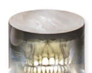
5 Easier positioning of the volume location with the ProMax... Patient positioning needs to be simple and efficient. The and 3D s interface allows the choice of pre-programmed areas; the SCARA arm will go to the appropriate region of interest. Select from any of the following pre-programmed areas: Incisors Canines Premolars Molars Teeth TMJ and Ramus Precise Positioning ø8 x 8 cm Reducing radiation to the patient and 3D s meet the needs of modern surgical dentistry and supply clear dependable imaging in a three-dimensional format with limited patient dose. The precise free-flowing arm movements allow for a wide variety of imaging programs not possible with other X-ray units with fixed rotations. During the scan, each image is generated using a short X-ray pulse instead of continuous radiation. The total rotation time is 18 seconds for one volume, but the actual exposure time is only 2.8 seconds with the smallest size volume. The flat panel sensor produces accurate, distortion-free images for 3D reconstruction. Unlike image intensifier sensors that use old vacuum tube technology and multi-step focusing, flat panels use single step image readouts with no geometric distortion or loss of sensitivity, and therefore no need for frequent calibration. The volumes are processed by computer software into one cylindrical image for viewing. The reconstructed image volume consists of more than 100 million voxels, producing a very detailed high-resolution image. These voxels are isotropic, enabling accurate 1:1 measurements and ensuring geometric proportion and consistency throughout the image. 8


6 3D s KEY ø diameter x height ø5 cm s ø5 cm Volume Sizes ø5 x 8cm 8 cm ø5 x 5cm 5 cm Standard 3D s volume sizes without stitching ø5 x 5 cm ø5 x 8 cm 3D s Focusing on your clinical needs s complies with a multitude of diagnostic requirements: those of endodontics, periodontics, orthodontics, implantology, prosthodontics, as well as dental and maxillofacial surgery. It is also an excellent tool for diagnosing ear and maxillary sinus problems. Pre-surgical planning has reached a new level of precision, as the prospective site becomes visible in all three imaging planes: sagittal, axial, and coronal. This makes it possible to locate and trace the mandibular nerve canal and correctly position implants. s is also perfect for molar area studies, or planning 3rd molar extractions by providing high-resolution studies for true and accurate evaluations. With its advanced reconstruction technology, s establishes the new standard for 3D dental radiology. 10


7 3D KEY ø diameter x height ø8 cm ø4 cm 5 cm 5 cm ø4 x 5cm ø8 x 5cm ø8 cm ø8 cm Volume Sizes 8 cm 8 cm ø8 x 8cm Standard 3D volume sizes without stitching ø4 x 5 cm ø4 x 8 cm ø8 x 5 cm ø8 x 8 cm 3D Focusing on your clinical needs The 3D ø8 x 8 cm image size is optimal for most diagnostic applications that require the whole dentition, mandible, and maxilla in the same volume. also provides TMJ studies for true and accurate evaluations of the joint arthritides, condylar morphology, and the condyle-fossa relationship. The ø8 x 5 cm volume can be used for single views of the mandible or maxilla, lowering the radiation by almost 40%. The small ø4 x 5 cm volume is intended for molar area studies or planning 3rd molar extractions. The volumes can also be stitched together to generate an image up to 14.4 cm x cm x 8 cm horizontally and 8 cm x 8 cm x 13 cm vertically. 8 cm ø4 cm ø4 x 8cm 12


8 Mac OS Compatible! iphone ipad Impacted canine An impacted mandibular left canine. Wisdom tooth extraction The extraction of the tooth would be difficult. The mandibular canal is located lingually to the roots. Axial (Z) Romexis Coronal (Y) Software Lesion in right mandible A radiolucent lesion is clearly visible in the right mandible. The probable cause is an unsuccessful root treatment. Romexis browser View all of your images: 2D, 3D, intracam, and more. Sagittal (X) 3D Romexis software is a complete-solution software package for acquisition, viewing, and rendering in multiple dimensions, multi-views, and multi-planes such as coronal, axial, and sagittal. Romexis also encompasses printing, sharing, and interactive diagnostic aspects of 2D and 3D imaging. Romexis Software is now compatible with Apple computers. All X-ray models feature full Mac OS support, which allows 2D and 3D images to be sent to an iphone and ipad for diagnosis. 2D and 3D imaging Romexis Software Romexis is a complete dental imaging software with a wide variety of tools for image viewing, enhancement, measurements, and annotations. Romexis improves the diagnostic value of radiographs. Printing, image import and export, and DICOM functionalities are also included. The system provides direct image capture from s X-ray equipment via Romexis Software, or Twain for 3rd party imaging solutions. DICOM compatibility Media Storage saving images into removable DICOM media Print printing images on film or paper with a DICOM medical printer Storage saving images into DICOM image archive Query/ Retrieve importing digital images from DICOM image archive Worklist importing a patient list from DICOM patient management Storage Commitment confirmation of a successful image storage 14


9 3D Implant Planning Module Sinus Study A cyst and inflammation can be found in the left maxillary sinus. 3D Implant Planning Module The optional Romexis Implant Planning Module offers libraries from other implant manufacturers with accurate and realistic implant models. Romexis 3D Panoramic Module. Romexis Software 3D TMJ Module The 3D TMJ Module provides easy and accurate diagnosis. Left and right TMJ are available in one view for easy comparison. Finding in canine area A suspicious mass found behind the lower right canine. Finding in sinus area Swelling in sinus mucosa. Cross-Sections Module Produces cross-sectional images of anatomy along with a defined panoramic curve, and also includes the reconstructed panoramic view for clear marking of the nerve canal. The Romexis 3D Panoramic Module creates a panoramic image from the acquired data without the redundant shadowing, commonly found in normal panoramic images. As the image is reconstructed through software, the user can determine the location and thickness of the focal trough. The Romexis 3D Cross-Sections Module produces cross-sectional images of anatomy, along with a defined panoramic curve and reconstructed panoramic view. The optional Romexis 3D Implant Planning Module offers tools for placing implants and marking the mandibular nerve canal. The implant placements are determined with the help of an accurately sized implant model. Romexis software has DICOM functionality, which allows 3D studies to be transferred to other implant planning software, such as SimPlant, Nobel Biocare Procera, CyberMed, or any other software that receives images in DICOM format. Studies can also be transferred to PACS or to a high quality DICOM printer in the network. Each patient study can be stored on a CD with Romexis 3D Viewer for others to see. 16
10 s X-ray beam Cone X-ray beam Cone Focal spot 0.5 mm, fixed anode Focal spot 0.5 mm, fixed anode Image detector Flat panel sensor Image detector Flat panel sensor Gray scale 15 bit Gray scale 15 bit Detector resolution 630 x 1024 pixels, pixel size 127 x 127 µm Detector resolution 1024 x 1024 pixels, pixel size 127 x 127 µm Voxel size 100 x 100 x 100 µm, isotropic 200 x 200 x 200 µm, isotropic Voxel size 100 x 100 x 100 µm, isotropic 200 x 200 x 200 µm, isotropic 400 x 400 x 400 µm, isotropic Image acquisition Single 200 degree rotation Image acquisition Single 200 degree rotation Total scan time 18 seconds, pulsed X-ray Total scan time 18 seconds, pulsed X-ray Reconstruction time Typically under 1.5 min. artifact removal dependent Reconstruction time Typically under 1.5 min. artifact removal dependent Standard volume sizes (diameter x height) ø5 x 8 cm ø5 x 5 cm Standard volume sizes (diameter x height) ø8 x 8 cm ø8 x 5 cm ø4 x 8 cm ø4 x 5 cm Effective Exposure Time seconds Effective Exposure Time seconds 3D reconstruction server Proprietary Feldkamp type back projection reconstruction algorithm 3D reconstruction server Proprietary Feldkamp type back projection reconstruction algorithm Improved Artifact Removal (IAR) High Contrast Object Compensation (HCOC) Improved Artifact Removal (IAR) High Contrast Object Compensation (HCOC) 17 18
11 digital X-ray imaging hardware diagrams and system requirements Dedicated Reconstruction PC (Provided by ) The reconstruction processing combines X-ray slices (raw data taken during the patient scan) through digital geometry conversion to produce voxels. This data is transferred to Romexis Software on Image Acquisition Workstation and archived on server. NOTE: A Power Conditioner is recommended for the X-ray and Reco. NIC CPU Specifications Image Acquisition Workstation (2D/3D) 2 Ghz or more Duo Core Processor 60 GB or better HD Free Space 3 GB or more RAM WINDOWS 7, WINDOWS XP, or Vista Business / Ultimate (32 bit OS only) Monitor 1600 x 1200 res. min. 32 bit graphic card, 256MB 2-1 GBit Net Interface Cards (NICs) High-speed Internet, DSL or better 8 x DVD/RW Acquisition Workstation Romexis Software acquires the image from the Reconstruction PC After the case images are reviewed in Romexis, they can be transferred to 3rd party imaging software Romexis Software is also used to enhance, filter, measure, diagnose, archive and export the images (2D / 3D) NIC Standard 10/100/1000 Switch (Provided by ) alternate connection for 2nd NIC Network Server Specifications 2 Ghz or more Duo Core Processor 2 Hard Drives, 500 GB each min. 3 GB or more RAM WINDOWS XP / Server 2003 OS (32 bit OS-Only) NET 2.0 and IIS must be installed Ethernet 1 GB 8 x DVD/RW Network Server Stores and archives all data to MS SQL Express Romexis stores data to XML The data server should have large hard drive disk space, hardware redundancy and logical backup system Each Standard or High Resolution 3D data set is approx. 250 MB Each Low Dose 3D Large Volume data is approx. 30 MB Local Area Connection Required for sharing printer or adding additional workstations. Optional Printer CPU Specifications for Additional Workstations For 3D/2D images. (refer to Image Acquisition Workstation (2D/3D)) Physical Space Requirements Minimum Operational Space Requirements Dimensions and space requirements Width Depth Height* Width Depth Height* Weight / 3D s 113 cm (44.5 in.) 126 cm (49.5 in.) cm ( in.) 150 cm (59 in.) 163 cm (64 in.) 244 cm (96 in.) 113 kg (248 lbs) / 3D s w / Cephalostat 198 cm (78 in.) 76 cm (30 in) cm ( in.) 216 cm (85 in.) 163 cm (64 in.) 244 cm (96 in.) 128 kg (282 lbs) * The maximum height of the unit can be adjusted for offices with limited ceiling clearance Romexis Software The disk space requirements are determined by digital images. Thus, the space requirements vary, but a rough estimate is as follows: 1 MB per intraoral image, 7 9 MB per extraoral image, depending on a variety of image specific factors, and up to 250 MB per 3D image. It is NOT recommended to use the same computer for your application server as your database server. If Romexis server computer is also used for client activities, the hardware should meet both client and server specifications

12 37 94 cm cm cm cm cm cm 21 22
Profound understanding of anatomy
 ENGLISH Profound understanding of anatomy The unique Planmeca ProMax 3D product family offers equipment for all maxillofacial imaging. All volume sizes from the smallest special cases to whole head images
Planmeca ProMax 3D s CBCT unit
 D0010759 1(5) Planmeca ProMax 3D s CBCT unit Introduction The Planmeca ProMax 3D s X-ray unit uses cone beam computerized tomography (CBCT) to produce threedimensional X-ray images. Panoramic and cephalometric
PLANMECA PROMAX 3D MID CBCT UNIT
 1(5) PLANMECA PROMAX 3D MID CBCT UNIT Introduction The Planmeca ProMax 3D Mid X-ray unit uses cone beam computerized tomography (CBCT) to produce three-dimensional X-ray images. Panoramic and cephalometric
Planmeca ProMax 3D Max CBCT unit
 D00107623 1(6) Planmeca ProMax 3D Max CBCT unit Introduction The Planmeca ProMax 3D Max X-ray unit uses cone beam computerized tomography (CBCT) to produce threedimensional X-ray images. Panoramic and
Maximum usability. Electronic capture request. Digitally signed approval. X-ray exposure. Image evaluation. 2D imaging. Digitally signed approval
 ENGLISH Software refined Planmeca Romexis is an advanced, easy to use software suite providing a rich set of tools to meet the imaging and clinic management requirements of any dental facility from a small
Planmeca ProMax 3D family
 3D Plus ENGLISH Planmeca ProMax 3D family True all-in-one units for all your imaging needs Planmeca ProMax 3D is a product family consisting of exceptional all-in-one units. With three different types
icatvision Software Manual
 University of Minnesota School of Dentistry icatvision Software Manual April 19, 2013 Mansur Ahmad, BDS, PhD Associate Professor, University of Minnesota School of Dentistry Director, American Board of
tr dent 2D-3D-PAN-CEPH
 tr dent 2D-3D-PAN-CEPH X-VIEW evolves and develops in response to your needs 2D PAN 2D-PAN-CEPH Single Shot 3D-PAN Multilayer 3D-PAN Multilayer CEPH Single Shot Cone Beam 3D Multilayer 2D Panoramic 24
icatvision Quick Reference
 icatvision Quick Reference Navigating the i-cat Interface This guide shows how to: View reconstructed images Use main features and tools to optimize an image. REMINDER Images are displayed as if you are
Shadow casting. What is the problem? Cone Beam Computed Tomography THE OBJECTIVES OF DIAGNOSTIC IMAGING IDEAL DIAGNOSTIC IMAGING STUDY LIMITATIONS
 Cone Beam Computed Tomography THE OBJECTIVES OF DIAGNOSTIC IMAGING Reveal pathology Reveal the anatomic truth Steven R. Singer, DDS srs2@columbia.edu IDEAL DIAGNOSTIC IMAGING STUDY Provides desired diagnostic
(restyle 2011) Anatomic programs
 Anatomic programs") Product Data Panoramic X-Ray System with CCD sensor (restyle 2011) Examination programs Panoramic - adult - child - child with deceleration ramp for spine compensation TMJ - open/close mouth (4 views on
Product Data Panoramic X-Ray System with CCD sensor (restyle 2011) Examination programs Panoramic - adult - child - child with deceleration ramp for spine compensation TMJ - open/close mouth (4 views on
The 3D Accuitomo 80 offers a minute voxel. size of just 80 μm (micrometer). This super-fine. voxel combined with the unit s 13 bit grayscale
. This super-fine. voxel combined with the unit s 13 bit grayscale") 2 Thinking ahead. Focused on life. REALIZED: Groundbreaking Resolution of 80 µm Voxel 05 The Crystallization of cutting-edge mechatronics and High Definition display algorithm The 3D Accuitomo 80 offers
2 Thinking ahead. Focused on life. REALIZED: Groundbreaking Resolution of 80 µm Voxel 05 The Crystallization of cutting-edge mechatronics and High Definition display algorithm The 3D Accuitomo 80 offers
xorantech.com Suite of DR Products
 xorantech.com Suite of DR Products 2 / xorantech.com Xoran provides unsurpassed, white-glove customer service and high quality, reliable products that are user- and patient-friendly Xoran is the pioneer
Z-MOTION. Universal Digital Radiographic System Z-MOTION. Control-X Medical CONTROL-X MEDICAL
 Control-X Medical Z-MOTION Compact design, low ceiling height requirement Motorized and manual movement capability Wide motion / SID range Best-in-class image quality Flexible connectivity to PACS systems
Dolphin 3D Imaging 11.7 beta
 Dolphin 3D Imaging 11.7 beta The Dolphin 3D software module is a powerful tool that makes processing 3D data extremely simple, enabling dental specialists from a wide variety of disciplines to diagnose,
The Vision To Advance Your Quality Of Care
 The Vision To Advance Your Quality Of Care Cone Beam 3D Imaging Systems Panoramic X-ray Systems Intraoral X-ray Systems P Digital Intraoral Sensors Digital X-ray Phosphor Plates Intraoral Cameras Imaging
As flexible and individual as your practice.
 Intraoral imaging instruments treatment centers Imaging systems cad/cam systems XIOS PLUS - INTRAORAL SENSOR HELIODENT Plus - INTRAORAL X-RAY SOURCE As flexible and individual as your practice. XIOS PLUS
Fast. Affordable. Complete. Desktop Scanning System from Great Lakes
 Desktop Scanning System from Great Lakes Fast Affordable Complete Create Virtual Bases Occlusal Mapping Complex Measurements Accurate Digital Study Models Desktop Scanning System Now you can easily convert
D B S W I N DBSWIN. The Digital Imaging Center for The Modern Operatory.
 D B S W I N DBSWIN The Digital Imaging Center for The Modern Operatory. Digital X-Ray Diagnostics With ScanX And ProVecta S-Pan/Ceph The powerful DBSWIN imaging software works with all digital X-Ray systems
digital imaging A world of dedicated to animal healthcare Fast, affordable, easy-to-use solutions that offer high-quality digital imaging
 Digital radiography small animals A world of digital imaging dedicated to animal healthcare Fast, affordable, easy-to-use solutions that offer high-quality digital imaging 2 Digital radiography small animals
Medical Image Processing: Image Reconstruction and 3D Renderings
 Medical Image Processing: Image Reconstruction and 3D Renderings 김보형 서울대학교컴퓨터공학부 Computer Graphics and Image Processing Lab. 2011. 3. 23 1 Computer Graphics & Image Processing Computer Graphics : Create,
Digital Age Diagnostics With The Power Of Platinum
 VixWin Platinum Digital Age Diagnostics With The Power Of Platinum P Cone Beam 3D Imaging Systems Panoramic X-ray Systems Intraoral X-ray Systems Digital Intraoral Sensors Digital X-ray Phosphor Plates
Simplant. Simplant Editor 2.2. Instructions for Use
 Simplant Simplant Editor 2.2 Instructions for Use 0120 Contents Introduction 3 Product information 3 Caution 3 Indications for use 3 Contraindications 3 Warnings 4 Precautions 4 Adverse reactions 4 Step-by-Step
DIRECT RADIOGRAPHY. flexible. Delivering. imaging excellence. DX-D Direct Radiography from Agfa HealthCare
 DIRECT RADIOGRAPHY flexible Delivering imaging excellence DX-D 300 - Direct Radiography from Agfa HealthCare versatile a and compact solution for all General Radiographic exams Maximum flexibility. The
Introducing the new. KODAK RVG 6500 System
 Introducing the new KODAK RVG 6500 System Innovation New features Wireless image transmission First Wi-Fi enabled sensor delivering the same film quality images as our best wired sensor Intelligent Positioning
SIRONA.COM MY ORTHOPHOS SL
 SIRONA.COM MY ORTHOPHOS SL 02 I 03 AS DIVERSE AS YOUR PRACTICE. ORTHOPHOS SL is the new complete X-ray solution from Sirona. PROVEN SOLUTION 2D 3D The device stands for exceptional image quality and perfect
Product Overview. A Smart and Complete DR Solution. Key Highlights. Premium detector options. Single Console Workflow and Generator Integration
 DIGITAL RADIOGRAPHY Product Overview A Smart and Complete DR Solution Key Highlights The DELWORKS E-Series DR Upgrade delivers exceptional diagnostic imaging using a powerful image acquisition and processing
OnDemand3D Application
 OnDemand3D Application Operating Manual Build 1.0.9.2225 Version 1.0 Copyright 2012 Cybermed Inc. - 0 - Index Index - 1-1. Introduction - 5-1.1 Main Functions... - 5-2. Installation - 6-2.1 System Requirements...
DX-D 300. Flexible Direct Radiography System
 DX-D 300 Flexible Direct Radiography System MUSICA 2 processing provides superior contrast detail and exam-independent, consistent image quality Cesium Iodide DR detector technology offers potential for
Digital Innovation for Medical Equipment
 Digital Innovation for Medical Equipment 01 CONTENTS Established in 1992, Sitec Medical Co., Ltd. has been in the conventional diagnostic radiology field for more than thirty years with equipments of its
Imaging protocols for navigated procedures
 9732379 G02 Rev. 1 2015-11 Imaging protocols for navigated procedures How to use this document This document contains imaging protocols for navigated cranial, DBS and stereotactic, ENT, and spine procedures
Digital Laminography and Computed Tomography with 600 kv for Aerospace Applications
 4th International Symposium on NDT in Aerospace 2012 - Tu.3.A.1 Digital Laminography and Computed Tomography with 600 kv for Aerospace Applications Malte KURFISS 1, Gerd STRECKENBACH 2 1 YXLON International
DX-D 300 FLEXIBLE DIRECT RADIOGRAPHY SYSTEM DX-D 300
 DX-D 300 FLEXIBLE DIRECT RADIOGRAPHY SYSTEM MUSICA processing provides excellent contrast detail and exam-independent, consistent image quality Cesium Iodide DR detector technology offers potential for
DX-D 300. Flexible Direct Radiography System
 DX-D 300 Flexible Direct Radiography System MUSICA² processing provides outstanding contrast detail and consistently excellent image quality Universal, flexible and affordable modality combines a single
imaging management aquarium What s New in Imaging 11.5 anywheredolphin
 imaging management aquarium What s New in Imaging 11.5 3d domo What s New in Dolphin Imaging 11.5 Welcome to Dolphin Imaging 11.5 Premium! This document provides an overview of many of the new features
Competition benchmark How do we win against...?
 Competition Vatech Sirona Gendex Kodak - Carestream Soredex Planmeca Morita Instrumentarium Competition benchmark How do we win against...? All data in this document is for information purpose only and
DX-D 300 FLEXIBLE DIRECT RADIOGRAPHY SYSTEM DX-D 300 DR SYSTEM
 DX-D 300 FLEXIBLE DIRECT RADIOGRAPHY SYSTEM The DX-D 300 DR system unites excellent image quality with the ultimate convenience. It offers top-of-the-line technology: a fullymotorized positioner enabling
Ch. 4 Physical Principles of CT
 Ch. 4 Physical Principles of CT CLRS 408: Intro to CT Department of Radiation Sciences Review: Why CT? Solution for radiography/tomography limitations Superimposition of structures Distinguishing between
Simplant. Simplant Editor 3.0. Instructions for Use
 Simplant Simplant Editor 3.0 Instructions for Use Table of Contents Introduction... 3 Product information... 3 Caution... 3 Indications for use... 3 Contraindications... 3 Warnings... 4 Precautions...
Industrial Computed Tomography Innovations
 GE Inspection Technologies Industrial Computed Tomography Innovations Premium performance for premium quality and speed. gemeasurement.com/ct Premium CT technologies. Faster than ever before. Every industrial
JAZZ HARMONY User Manual
 JAZZ HARMONY User Manual Copyright 2017 Imaging. All rights reserved. This manual and the software described herein are protected by copyright laws and international copyright treaties, as well as other
 & Superior Image Quality Seamlessly Compatible with EVA-Vet Digital Dental Sensors Thousands of installations and growing Customer Care Program www.imageworkscorporation.com ImageWorks Generations of Imaging
MapCHECK 2 & 3DVH. The Gold Standard for 2D Arrays
 MapCHECK 2 & 3DVH The Gold Standard for 2D Arrays Your Most Valuable QA and Dosimetry Tools THE GOLD STANDARD FOR 2D ARRAYS The MapCHECK 2 is the world s most selected independent 2D measurement array.
Mediso AnyScan. Single-Head and Dual-Head SPECT
 Mediso AnyScan S Single-Head and Dual-Head SPECT ANYSCAN S (SINGLE-HEAD & DUAL-HEAD SPECT) Low Cost of Ownership Associated Imaging Services is the exclusive U.S. source in the Midwest for Mediso Medical
This survey was completed by Date Phone. Please leave a copy of this site survey with the Dentist/Business owner!
 Version 8.2 April 2011 3D IMAGING SITE SURVEY FORM To make sure that your planned installation of the Sirona 3D system progresses as smoothly as possible, we ask you to fill out the following pages thoroughly.
Digital Innovation for Medical Equipment.
 Digital Innovation for Medical Equipment www.sitec-med.com CONTENTS Established in 1992, Sitec Medical Co., Ltd. has been in the conventional diagnostic radiology field for more than thirty years with
MapCHECK 2 & 3DVH The Gold Standard for 2D Arrays
 MapCHECK 2 & 3DVH The Gold Standard for 2D Arrays Your Most Valuable QA and Dosimetry Tools THE GOLD STANDARD FOR 2D ARRAYS The MapCHECK 2 is the world s most selected independent 2D measurement array.
Image Acquisition Systems
 Image Acquisition Systems Goals and Terminology Conventional Radiography Axial Tomography Computer Axial Tomography (CAT) Magnetic Resonance Imaging (MRI) PET, SPECT Ultrasound Microscopy Imaging ITCS
Using New Features in CDR DICOM 3.5 Software
 Using New Features in CDR DICOM 3.5 Software Schick Technologies, Inc. 30-00 47 th Avenue Long Island City, NY 11101 (718) 937-5765 (718) 937-5962 (fax) PART NUMBER B1051053 REV. Copyright 2004 by Schick
DICOM Conformance Statement
 DICOM Conformance Statement SCENARIA Tokyo, Japan E1E-HC0002-01 Hitachi, Ltd. 2016. All rights reserved. History Revision Description Date Rev. 1.0 Initial. 2016/04/01 ii E1E-HC0002 Contents History...
Annexure XII SPECIFICATIONS FOR A NEW STATE OF ART 16 SLICE ALL PURPOSE C. T. SCANNER
 Annexure XII SPECIFICATIONS FOR A NEW STATE OF ART 16 SLICE ALL PURPOSE C. T. SCANNER A) Scanner Design X-Ray generator and tube: 1. Scanner: Whole body spiral CT scanner (16 slices) of latest technology.
REXTAR Exo Ultimate Portable X-ray System
 REXTAR Exo 1414 Ultimate Portable X-ray System Contents Company Introduction Special Features Overview Specifications How To Use Package Posdion Co., Ltd. About US Posdion Co., Ltd. is specialized in handheld
Mediso AnyScan S Single-Head and Dual-Head SPECT
 Mediso AnyScan S Single-Head and Dual-Head SPECT ANYSCAN S (SINGLE-HEAD & DUAL-HEAD SPECT) Low Cost of Ownership Small Footprint Absolute Imaging Solutions tm is the exclusive U.S. source for Mediso Medical
Compact Delta II with
 W E R E A L L A B O U T P E O P L E. The Dornier Compact Delta II with Integrated Transportable Lithotripter The Dornier The Optimal Solution: The Dornier Compact Delta II Optimal Solution Compact DeltaII
OUR FOCUS IS ON YOUR VISION
 OUR FOCUS IS ON YOUR VISION Karl Kaps Dental Microscopes Perfect Balance for your Dental Surgery Kaps Dental-Microscopes High-performance and user-friendly Powerful and user-friendly, Kaps dental microscopes
VIVIX Software.
 VIVIX Software 2 software SOFTWARE Vieworks pursues an all-in-house design that offers both hardware and software solutions. Digital Radiography Acquisition viewer for VIVIX-S Series Easy Workflow User
SCENARIA 64-Slice CT Putting Advanced CT Within Reach.
 SCENARIA 64-Slice CT Putting Advanced CT Within Reach. Advancing the technology of Computed Tomography for over 30 years, Hitachi is a recognized innovator of lower dose*, high diagnostic value CT solutions.
The DR system in a suitcase for mobile veterinary X-ray
 The DR system in a suitcase for mobile veterinary X-ray total weight only ca. 22kg The portable DR system for mobile X-ray O and R Technology Digital X-ray and Imaging Solutions OR Technology has been
pfabufp=mäìöáå=ñçê=uflp=ud
 kéï=~ë=çñw== MPKOMNP pfabufp=mäìöáå=ñçê=uflp=ud lééê~íçêdë=ã~åì~ä båöäáëü Operator's Manual SIDEXIS Plugin for XIOS XG = Sirona Dental Systems GmbH Table of contents 1 Description of plugin... 4 1.1 SIDEXIS
Computational Medical Imaging Analysis Chapter 4: Image Visualization
 Computational Medical Imaging Analysis Chapter 4: Image Visualization Jun Zhang Laboratory for Computational Medical Imaging & Data Analysis Department of Computer Science University of Kentucky Lexington,
Software Installation and Configuration Guide
 Digital Imaging Systems Classic Intraoral Duo Swift PN F3700 PN F3600 PN D1000 PN G8000 Software Installation and Configuration Guide FOREWORD General The instructions provided are limited to the integration
Whole Body X-ray CT System Supria. DICOM Conformance Statement
 Whole Body X-ray CT System Supria DICOM Conformance Statement Tokyo, Japan (SN-C633E) Copyright Hitachi Medical Corporation. 2013, 2014. All rights reserved. History Revision Description Date Rev. 1.0
product data Raybow dr mobile digital radiographic system with wireless detector product data
 Raybow dr mobile digital radiographic system with wireless detector Integrating all the advanced technologies, the ergonomic and high quality system Raybow dr lets you perform digital radiographies anywhere:
Affordable image quality. CR 30-x. your versatile table-top CR system
 C o m p u t e d R a d i o g r a p h y Affordable image quality CR 30-x your versatile table-top CR system compact Versatile and budget-friendly combined with in-room integration, enable radiographers Combining
PRODUCT DATA. Advanced 128
 PRODUCT DATA Advanced 128 Supria 16 Slice CT Puts You On The Path of High Quality, Cost-Effective CT Scanning Solid Capabilities Are Built Into the Supria Addressing the challenges of controlling healthcare
Practice-oriented robust solution for mobile X-ray. The portable DR system for mobile X-ray. Leonardo DR 121OP with dicom PACS DX-R Software
 Leonardo DR 121OP The portable DR system for mobile X-ray total weight only ca. 26kg The portable DR system for mobile X-ray Practice-oriented robust solution for mobile X-ray O and R Technology Digital
FLEXIBLE DIRECT RADIOGRAPHY SYSTEM DX-D 300
 FLEXIBLE DIRECT RADIOGRAPHY SYSTEM DX-D 300 The DX-D 300 DR system unites excellent image quality with complete convenience. It offers top-of-the-line technology, a single detector and a fully-motorized
Solid Capabilities Are Built Into the Supria Plus. Putting You On The Path of High Quality, Cost-Effective CT Scanning
 Specification Data Putting You On The Path of High Quality, Cost-Effective CT Scanning Solid Capabilities Are Built Into the Supria Plus Addressing the challenges of controlling healthcare organization
DX-D 100 with wireless detector
 M O B I L E D R S O L U T I O N DX-D 100 with wireless detector Patients who most need imaging exams may lack the mobility necessary to move to the X-ray room or to position themselves properly for optimum
EXAMION DR EasyGo ... A SYSTEM OF CHOICES. EXAMION DR Mobile solution. EXAMION Teleradiology EXAMION AQS EXAMION PACS.
 EXAMION DR Mobile solution EasyGo Soft Pack Import your external X-rays in standard formats from external media (CD, USB stick, memory cards, etc.) Simple operation of the work List Easy application to
CT Protocol Review: Practical Tips for the Imaging Physicist Physicist
 CT Protocol Review: Practical Tips for the Imaging Physicist Physicist Dianna Cody, Ph.D., DABR, FAAPM U.T.M.D. Anderson Cancer Center August 8, 2013 AAPM Annual Meeting Goals Understand purpose and importance
Superior Imaging on the Go
 Superior Imaging on the Go About Samsung Samsung Electronics Co., Ltd. is a global leader in technology, opening new possibilities for people everywhere. Through relentless innovation and discovery, we
Optiv CT160 Accurate measurement for every detail. Computed Tomography System
 Optiv CT160 Accurate measurement for every detail Computed Tomography System Computed Tomography CT Technology. New prospects for metrology. Computed tomography (CT), a technology with unique advantages,
ImplaStation. ProDigiDent. Instruction. 1. Hardware requirements. 2. Installation and update
 ProDigiDent ImplaStation Instruction ImplaStation is a medical software intended for the plan the placement of one or more implants build on an imported and aligned Computed Tomography or Cone Beam Computed
Audit4 Installation Requirements
 S4S Pty Ltd ABN: 26 104 845 909 Phone: 1300 133 308 Web: http://www.s4s.com.au Audit4 Installation Requirements Audit4 version 14+ Copyright 2012 S4S Pty Ltd The following table provides details on the
VistaScan Mini View - compact, fast, and effi cient
 VistaScan Mini View - compact, fast, and effi cient The image plate scanner with a touchscreen for intraoral formats COMPRESSED AIR SUCTION IMAGING DENTAL CARE HYGIENE A new standard for digital X-rays
5020S CR READER. Software Installation Guide & Calibration Process. Ver.: Document Part Number: CR /170208
 5020S CR READER Software Installation Guide & Calibration Process Ver.: 040608 Document Part Number: CR 11072220/170208 - 2 - CR-Tech is marketing the 5020s, an innovation in the field of compact desktop
Micro-CT Methodology Hasan Alsaid, PhD
 Micro-CT Methodology Hasan Alsaid, PhD Preclinical & Translational Imaging LAS, PTS, GlaxoSmithKline 20 April 2015 Provide basic understanding of technical aspects of the micro-ct Statement: All procedures
MEDICAL IMAGING 2nd Part Computed Tomography
 MEDICAL IMAGING 2nd Part Computed Tomography Introduction 2 In the last 30 years X-ray Computed Tomography development produced a great change in the role of diagnostic imaging in medicine. In convetional
Comparison of Probing Error in Dimensional Measurement by Means of 3D Computed Tomography with Circular and Helical Sampling
 nd International Symposium on NDT in Aerospace - We..A. Comparison of Probing Error in Dimensional Measurement by Means of D Computed Tomography with Circular and Helical Sampling Jochen HILLER, Stefan
TOP dog in veterinary PACS
 WEBX WEB-based veterinary pacs TOP dog in veterinary PACS VERSiON 6.1.3 VET-WEBX VET-WEBX THE PERFECT TOOL FOR VETERINARY FREEDOM VET-WEBX is a PACS especially designed for veterinarians, which conveniently
PREINSTALL CHECKLIST. 1. General Information. 2. Radiation Environment. 3. Electrical Specifications. 4. Network Requirements. 5. Server Requirements
 PREINSTALL CHECKLIST Thank you for your investment in the i-cat FLX Cone Beam 3D Imaging System. To ensure a smooth integration of this technology into your office, please complete this checklist to confirm
Digital Image Processing
 Digital Image Processing SPECIAL TOPICS CT IMAGES Hamid R. Rabiee Fall 2015 What is an image? 2 Are images only about visual concepts? We ve already seen that there are other kinds of image. In this lecture
GALILEOS SITE SURVEY FORM
 Version 6.1 GALILEOS SITE SURVEY FORM To make sure that your planned installation of the Sirona GALILEOS system progresses as smoothly as possible with no delays, we ask you to fill out the following pages
4/19/2016. Deborah Thames R.T. (R)(M)(QM) Theory & Technology and advancement in 3D imaging DBT
(M)(QM) Theory & Technology and advancement in 3D imaging DBT") Deborah Thames R.T. (R)(M)(QM) Theory & Technology and advancement in 3D imaging DBT 1 Three manufacturers approved for Tomo Hologic and GE, and Siemens Why 2D Digital Mammography 2D FFDM it appears to
Deborah Thames R.T. (R)(M)(QM) Theory & Technology and advancement in 3D imaging DBT 1 Three manufacturers approved for Tomo Hologic and GE, and Siemens Why 2D Digital Mammography 2D FFDM it appears to
meddix WEB DICOM-SErvEr for StOragE, teleradiology and IMagE DIStrIButIOn
 meddix WEB DICOM-Server for Storage, Teleradiology and image distribution meddix-webx combines a fast DICOM archive with a web-based viewing and a managing interface in one system meddix-webx is a complete,
1. Scanning the Patient (as provided by NewTom)
") Using NewTom VGi, with NNT2.17 software or above, and Materialise SimPlant for implant planning.. Note: Users of NewTom VG (not VGi), produced in 2007 and 2008, should contact the manufacturer s Technical
Using NewTom VGi, with NNT2.17 software or above, and Materialise SimPlant for implant planning.. Note: Users of NewTom VG (not VGi), produced in 2007 and 2008, should contact the manufacturer s Technical
PH-03: Clinical Photography Production Checklist With The Sony Cybershot T-9 Digital Camera
 PH-03: Clinical Photography Production Checklist With The Sony Cybershot T-9 Digital Camera Date: Name: EXTRAORAL SERIOUS 1. Prepare the camera Program the camera to the recommended settings Set camera
PREINSTALL CHECKLIST. 1. General Information. 2. Cone Beam Shielding Plan. 3. Electrical Specifications. 4. Network Requirements
 PREINSTALL CHECKLIST Thank you for your investment in the i-cat FLX V-Series Cone Beam Imaging Solution. To ensure a smooth integration of this technology into your office, please complete the following
WEBX. One For all. Specialists in Medical. Digital Imaging Solutions
 WEBX the DICOM server for storage, teleradiology and image distribution One For all VERSiON 6.2 Specialists in Medical Digital Imaging Solutions iq-webx iq-webx - a fast DICOM archive and a web-based viewing
This guide provides instructions in the installation and configuration of XrayVision DCV.
 Apteryx Inc. 313 S. High St. Suite 200 Akron, OH 44308 330-376-0889 voice 330-376-0788 fax sales@apteryx.com www.apteryx.com XrayVision DICOM Capture View (DCV) Installation Guide Abstract Abstract Abstract
The power of centralized computing at your fingertips
 Pinnacle 3 Professional The power of centralized computing at your fingertips Philips Pinnacle 3 Professional specifications The power of centralized computing in a scalable offering for mid-size clinics
Please Read This First
 Please Read This First Getting Started with CDR DICOM For complete details about your CDR DICOM software, please refer to the User Guide on the DVD or our website at www. schicktech.com Getting Started
Software Installation and Configuration Guide
 High Definition Intraoral Video Camera System Part Number G1000 Intraoral Video Camera System Part Number G5000 Fluorescence Caries Detection Aid System Part Number G4000 Software Installation and Configuration
O-ARM IMAGING SYSTEM TECHNICAL SPECIFICATION GUIDE
 IMAGING SYSTEM TECHNICAL SPECIFICATION GUIDE SYSTEM FEATURES CATEGORY PHYSICAL DIMENSIONS IMAGING MODALITY PERFORMANCE SPECIFICATION Length Width Height Weight Gantry Opening Bore Diameter Single Plane
Whole Body X-ray CT System Supria. DICOM Conformance Statement
 Whole Body X-ray CT System Supria DICOM Conformance Statement Tokyo, Japan E1E-HC0001-02 Hitachi, Ltd. 2016. All rights reserved. ii E1E-HC0001 History Revision Description Date Rev. 1.0 Initial. 2016/4/1
Overview. Digital X-ray imaging
 Overview Digital X-ray imaging OR Technology Headquarters The OR Technology competence group: Your competent partner for digital X-ray imaging Producing digital X-ray technology and developing image management
The digital EVOlution in cassette size format
 http://cr-pacs.com/ Platzhalter Wireless Mobile Fast Retrofit Light Low Dose Highest DQE Highest MTF CsI GOS The digital EVOlution in cassette size format Your wireless entry to the world of Digital Radiography
Ultrasound To Go. MySono U5
 Ultrasound To Go MySono U5 Ultrasound To Go With the introduction of the MySono U5, Samsung Medison brings you a fully featured ultrasound imaging system to go. Delivering exceptional image quality and
A Study of Medical Image Analysis System
 Indian Journal of Science and Technology, Vol 8(25), DOI: 10.17485/ijst/2015/v8i25/80492, October 2015 ISSN (Print) : 0974-6846 ISSN (Online) : 0974-5645 A Study of Medical Image Analysis System Kim Tae-Eun