Biomedical Imaging. Computed Tomography. Patrícia Figueiredo IST
|
|
|
- Eustace Howard
- 5 years ago
- Views:
Transcription
1 Biomedical Imaging Computed Tomography Patrícia Figueiredo IST
2 Overview Basic principles X ray attenuation projection Slice selection and line projections Projection reconstruction Instrumentation Beam collimation Gas ionization chambers From the 1 st to the 4 th generation Spiral / Helical CT Multi-slice CT Image reconstruction The Radon transform: filtered backprojection Iterative reconstruction methods

3 Basic principles Image orientation: z x y

4 Basic principles X ray attenuation projection: Coronal image z x y
axial image x z y x z")
5 Basic principles X ray attenuation projection: Coronal image Computed tomography: (Trans)axial image x z y x z y
6 Basic principles X ray attenuation projection: { } ( z) I ( z) exp ( x z) I =, dx 0 µ { } ( y, z) I0 ( y, z) exp µ ( x, y z) I =, dx -The object consists of a distribution of attenuation coefficients. -The intensity of the detected X ray beam reflects the projection of the attenuation coefficients across the beam direction z µ 11 µ 12 µ 13 µ 14 µ 15 µ 16 I 0 (y,z) y µ 21 µ 22 µ 23 µ 24 µ 25 µ 26 µ 31 µ 32 µ 33 µ 34 µ 35 µ 36 µ 41 µ 42 µ 43 µ 44 µ 45 µ 46 µ 51 µ 52 µ 53 µ 54 µ 55 µ 56 µ 61 µ 62 µ 63 µ 64 µ 65 µ 66 I(y,z) x
7 Basic principles Line projections L 1 L{ θ 1 } L 2 L{ θ 2 } y p L { f ( x, y) } f x( l), y( l) L ( )dl z x

8 Basic principles Radon transform: Object space Projection space
9 Basic principles { } π Projection reconstruction: fˆ 1 ( r, θ ) = p ( xʹ ) h( xʹ ) dθ = R p ( xʹ ) 0 φ φ Object Image 1 ang. 2 ang. 4 ang. 8 ang. 16 ang. 32 ang.
 = p ( xʹ ) φ + 2π φ f(x,y) R φ p φ (x ) φ φ 4 φ 3 θ φ 2 φ 0 φ 1 -r r x xʹ = rcos ( φ θ)")
10 Basic principles Radon transform:, p ( xʹ ) { f ( x, y) } R { f ( x y) } f ( x( l), y( l) )dl L φ R φ The sinogram Symmetry at π: p ( xʹ ) = p ( xʹ ) φ ± π φ Object space Projection space Periodicity at 2π: p ( xʹ ) = p ( xʹ ) φ + 2π φ f(x,y) R φ p φ (x ) φ φ 4 φ 3 θ φ 2 φ 0 φ 1 -r r x xʹ = rcos ( φ θ)
11 Image reconstruction Projection reconstruction 2D 3D Filtered backprojection (FB) Backprojection filtering (BF) True Three-Dimensional Reconstruction (TTR) Generalized TTR (GTTR) Parallel beam mode Fan beam mode Parallel beam mode Fan beam mode Parallel beam mode Cone beam mode Planar-Integral Projection Reconstruction (PPR) Iterative reconstruction Fourier reconstruction Algebraic Reconstruction Technique (ART) Maximum Likelihood (ML) or Expectation Maximization (EM) Direct Fourier Reconstruction (DFR) Direct Fourier Imaging (in MRI)
12 Image reconstruction Backprojection: effect of a finite number of projections Object Image 1 ang. 2 ang. 4 ang. 8 ang. 16 ang. 32 ang.
13 Image reconstruction Backprojection: effect of a finite number of projections streak artifacts
14 Image reconstruction Backprojection: coverage π ( r, φ) p ( xʹ ) h( x ) dφ fˆ ʹ = 0 φ y y x Symmetry at π: p ( xʹ ) p ( xʹ φ ± π = φ ) p ( xʹ ) = p ( xʹ ) Periodicity at 2π: y φ + 2π φ p θ (x ) L φ x r φ θ q x x f(x,y) Sampling requirements for φ: Coverage of a total scan angle of 180º, usually 360º to reduce partial volume effects.
 ( r, φ) pφ ( x ) dφ =")
15 Filtered back-projection π fˆ ʹ Filtering: simple backprojection (no filter) ( r, φ) pφ ( x ) dφ = 0
 p ( x ) h( x ) dφ")
16 Filtered back-projection π fˆ ʹ ʹ Filtering: filtered backprojection ( r, φ) p ( x ) h( x ) dφ = 0 φ

 dφ fˆ ( r, φ) p ( xʹ ) h( xʹ")
17 Filtered back-projection Filtering: simple vs filtered backprojection π π fˆ ( r, φ) p ( xʹ ) dφ fˆ ( r, φ) p ( xʹ ) h( xʹ ) dφ = 0 φ = 0 φ

18 Filtered back-projection Filtering: effect of noise


19 Filtered back-projection CONTINUAR AQUI - MEBiom Filtering: effect of different filter functions
20 Image reconstruction Iterative reconstruction The ray-by-ray method: p m N = n = 1 W mn f n To estimate the value of the image cell f n from the projection data p m = to solve the inverse problem of M linear equations with N unknowns Iterative method: Mean square error (MSE) Expectation maximization (EM) Maximum likelihood (ML)

21 Image reconstruction Iterative reconstruction Jean-Baptiste Thibault et al., GE Medical Systems
22 Instrumentation

23 Instrumentation Beam collimators: 1 st collimator: width ~45º 2 nd collimator, perpendicular to 1 st : thickness ~1-5 mm Slice profile: -Δz/2 +Δz/2

24 Instrumentation 1 st generation systems Parallel beam 1 pencil beam source 1 detector translating together rotating together scanning time ~4-5 min

25 Instrumentation 2 nd generation systems Fan beam: equilinear geometry 1 thin fan beam source multiple detectors translating together rotating together But fewer rotations required: scanning time ~20 s
26 Instrumentation 3 rd generation systems Fan beam: equiangular geometry 1 wide (~30-45 ) fan beam source ~ detectors covering object no translations rotating together scanning time ~1-3 s Use pulsed X ray sources to take advantage of significant dead time

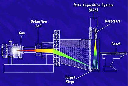
27 Instrumentation 4 th generation systems Fan beam: equiangular geometry 1 wide fan beam source complete ring of detectors source rotating detector ring stationary ~ scanning time No cumulative detector drift, But very expensive (BGO-PMT detectors)

28 Instrumentation 3 rd generation CT T = X ray tube D = Detectors X = X ray fan beam R = Rotation direction Typical parameters: kvp 140 kv E eff ma Pulse f kev ma 2-4 ms mm Thickness 1-5 mm Matrix (Resolution~0.35 mm) Nb detectors~1000
29 Instrumentation X ray detectors: X rays must be converted into radiation accessible to human vision Type of X ray detectors: - itensifying screen + photographic emulsion - cassette of photostimulable phosphor + laser scanner - scintillation detectors - crystals: NaI (Tl), CsI(Tl), BGO coupled to a photo-multiplier tube (PMT) or a photodiode array (e.g. TFT) - gas ionizing detectors: - ionizing chamber, proportional counter, Geiger-Muller counter Main characteristics of X ray detectors : - Sensitivity - Efficiency - Linearity - Energy resolution - Dead time
30 Instrumentation Ionization chamber: X rays gas ionization electron-ion pairs electron/ions attracted to cathode/anode electric current amplifier Array of interlinked Xenon-filled (~1000) ionization chambers (Z Xe = 66, P = 20 atm) ~1mm X X - + Xe+ e- 10cm - spatial resolution ADC - simplicity - more compact X - efficiency - Role of antiscatter grid X
31 Instrumentation Scintillation detectors: X rays crystal excitation electron-hole pairs electron-hole pairs collected at p-n junctions electric current pre-amplifier X rays crystal excitation optical photons photocathode ionization photoelectrons electron multiplication electric current
32 Instrumentation Conventional CT configurations One slice at a time: time inefficient susceptible to artifacts due to motion between slices

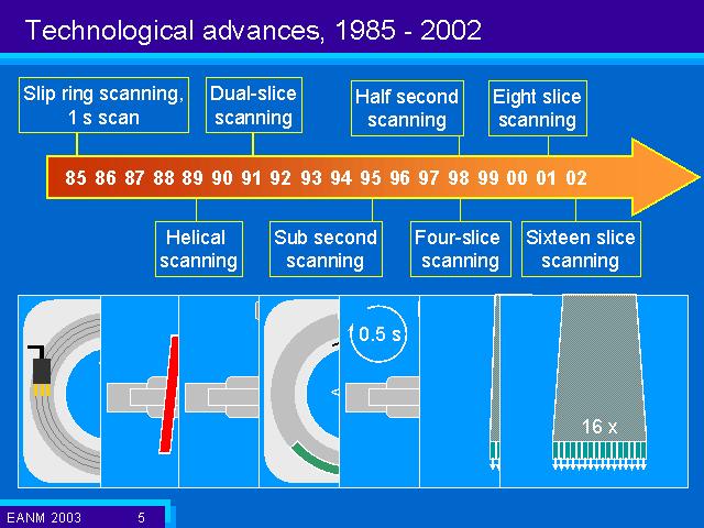
33 Instrumentation Spiral / Helical CT Data are acquired as the patient table moves continuously along z, simultaneously with the source/detectors rotation, tracing out a spiral/helix for the X ray trajectory. Continuous scanning requirements: - source: high heat capacity and efficient cooling - detectors: high efficiency Only one projection is acquired exactly in the image plane. All other projections have to be interpolated.

34 Instrumentation Spiral / Helical CT Spiral pitch: p = d / S p d = table feed per rotation S = collimated slice thickness A. p<1: slice overlap higher dose B. p>2: slice gaps lower resolution blurring 1<p<2: typical values

35 Instrumentation Multislice CT An array of detectors is incorporated along z: Spiral pitch: p ms = d / S single d = table feed per rotation S single = single slice collimated beam width 4-slice: p ms <8 8-slice: p ms <16 16-slice : p ms <32

36 Instrumentation Multislice CT - Multislice helical scans produce a set of interleaved helices interpolation is (even) more difficult to visualize - Images are reconstructed at optimized oblique planes and are then filtered to produce axial images.

37 Instrumentation Multislice CT - Multislice helical scans produce a set of interleaved helices interpolation is (even) more difficult to visualize - Images are reconstructed at optimized oblique planes and are then filtered to produce axial images.
38 Instrumentation Multislice vs single-slice CT Advantages: - Same acquisition in shorter time - Larger volumes in same time - Thinner slices: better spatial resolution - Can get isotropic volumes Disadvantages: -Larger beam width (relative to slice) -Higher dose for same quality -Cone beam artefacts
39 Instrumentation
: CT ij µ ij µ H = µ H 2 O")
40 Image characteristics Estimated object function CT numbers (Hounsfield units, HU): CT ij µ ij µ H = µ H 2 O 2 O 1000

41 Image characteristics Dosimetric quantities CT Dose Index: D z is absorbed dose at position z T is slice thickness CTDI = 1 T + 7T 7T D z dz Effective dose:
42 Image characteristics Spatial resolution: X ray tube effective focal spotsize f (~ mm) Scanner (translation and) rotation steps (~512x x1024: ~0.35x0.35mm 2 ) Collimated single-slice thickness (~0.5-5mm) and table feed Signal to noise ratio (SNR): - X ray tube voltage (~140kV): kvp SNR - X ray tube current and exposure time (~2-4ms): ma s SNR - X ray filtration (effective energy ~70-80 kev): filtration SNR Contrast to noise ratio (CNR): - X ray energy: E I scatt /I primary CNR - Object size (thickness): thickness I scatt /I primary CNR - Field-of-view: FOV I scatt CNR - Artefacts!
43 Image characteristics Artefacts: - Streak artefacts: undersampling due to finite number of projections (interaction owith motion, beam hardening or scatter) increase nb rotation steps / decrease rotation step. - Ring artefacts: imbalances in detector sensitivity calibration is performed using spatially uniform test objects. - Beam hardening: results in more attenuation in the center of the object than around the edge, but algorithms assume monochromatic X ray beams and hence uniform attenuation coefficient X ray beam filtering; calibration; reconstruction corrections. - Partial volume effects: thick slices can include, and mix up, different tissue types decrease slice thickness (using multislice systems).

44 Image characteristics Streak artefacts: Barrett J F, and Keat N Radiographics 2004;24:

45 Image characteristics Ring artefacts: when a detector is out of calibration: Barrett J F, and Keat N Radiographics 2004;24:
46 Image characteristics Beam hardening effects: Barrett J F, and Keat N Radiographics 2004;24:
47 Image characteristics Beam hardening effects Monoenergetic X rays: Polichromatic X rays: Beam hardening: I i = I i 0 exp µ ij j I i0 = Ii0 i i0 µ ij 0 j Ema x ( E) I = I ( E) exp de µ ij = µ ij i 0i exp µ ij 0 j Ema x ( E) I = I ( E) ( E) de Non-linear relation between p and µ artefacts p i = ln I I i i0 I i = ln 0 E max I 0i ( E) exp µ ( E) 0 E max I 0i j ( ) E de ij de


48 Image characteristics Beam hardening effects - minimized by: Filtration: a flat piece of attenuating material is used to pre-harden the beam before it passes through the patient (so that it becomes closer to monochromatic). Monochromatic X ray Polychromatic X ray Barrett J F, and Keat N Radiographics 2004;24:


49 Image characteristics Beam hardening effects - minimized by: Calibration correction: using phantoms in a range of sizes. Uncalibrated Calibrated Barrett J F, and Keat N Radiographics 2004;24:


50 Image characteristics Beam hardening effects - minimized by: Reconstruction: an iterative correction algorithm may be applied when images of bony regions are being reconstructed. Uncorrected Corrected Uncorrected Corrected Barrett J F, and Keat N Radiographics 2004;24:


51 Image characteristics Partial volume effects Thick slice Thin slice Barrett J F, and Keat N Radiographics 2004;24:

52 Image characteristics Partial volume effects: multi-slice vs single-slice CT
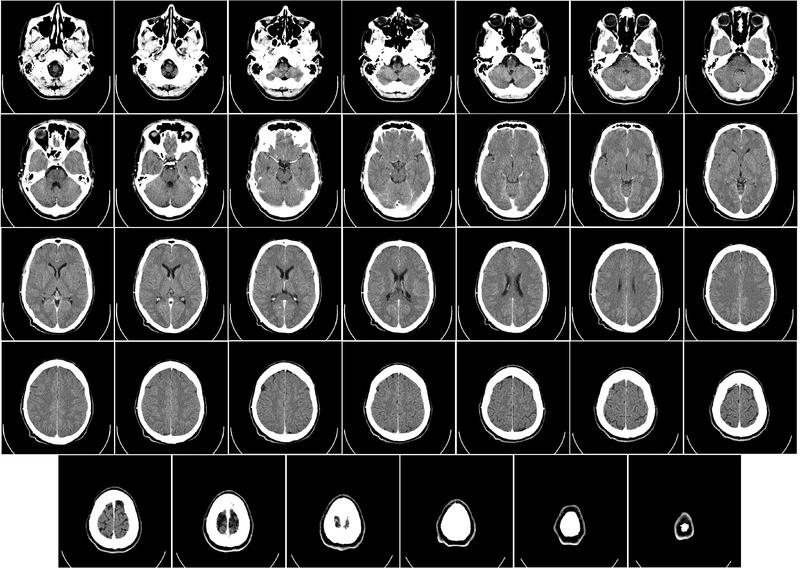

53 Image characteristics Scout image for CT Head CT
54 References Webb, Introduction to Biomedical Imaging, Wiley Cho, Foundations of Medical Imaging, Wiley Hendee, Medical Imaging Physics, Wiley 2002.
Introduction to Biomedical Imaging
 Alejandro Frangi, PhD Computational Imaging Lab Department of Information & Communication Technology Pompeu Fabra University www.cilab.upf.edu X-ray Projection Imaging Computed Tomography Digital X-ray
BME I5000: Biomedical Imaging
 1 Lucas Parra, CCNY BME I5000: Biomedical Imaging Lecture 4 Computed Tomography Lucas C. Parra, parra@ccny.cuny.edu some slides inspired by lecture notes of Andreas H. Hilscher at Columbia University.
MEDICAL IMAGING 2nd Part Computed Tomography
 MEDICAL IMAGING 2nd Part Computed Tomography Introduction 2 In the last 30 years X-ray Computed Tomography development produced a great change in the role of diagnostic imaging in medicine. In convetional
MEDICAL IMAGING 2nd Part Computed Tomography
 MEDICAL IMAGING 2nd Part Computed Tomography Introduction 2 In the last 30 years X-ray Computed Tomography development produced a great change in the role of diagnostic imaging in medicine. In convetional
Joint ICTP-TWAS Workshop on Portable X-ray Analytical Instruments for Cultural Heritage. 29 April - 3 May, 2013
 2455-5 Joint ICTP-TWAS Workshop on Portable X-ray Analytical Instruments for Cultural Heritage 29 April - 3 May, 2013 Lecture NoteBasic principles of X-ray Computed Tomography Diego Dreossi Elettra, Trieste
Moscow-Bavarian Joint Advanced Student School 2006 / Medical Imaging Principles of Computerized Tomographic Imaging and Cone-Beam Reconstruction
 Line Integrals Line integrals represent the integral of some parameter of the object along the line (e.g. attenuation of x-rays) Object: f(x,y) Line: x cosθ + y sinθ = t Line integral / Radon transform:
Digital Image Processing
 Digital Image Processing SPECIAL TOPICS CT IMAGES Hamid R. Rabiee Fall 2015 What is an image? 2 Are images only about visual concepts? We ve already seen that there are other kinds of image. In this lecture
Multi-slice CT Image Reconstruction Jiang Hsieh, Ph.D.
 Multi-slice CT Image Reconstruction Jiang Hsieh, Ph.D. Applied Science Laboratory, GE Healthcare Technologies 1 Image Generation Reconstruction of images from projections. textbook reconstruction advanced
Computed Tomography. Principles, Design, Artifacts, and Recent Advances. Jiang Hsieh THIRD EDITION. SPIE PRESS Bellingham, Washington USA
 Computed Tomography Principles, Design, Artifacts, and Recent Advances THIRD EDITION Jiang Hsieh SPIE PRESS Bellingham, Washington USA Table of Contents Preface Nomenclature and Abbreviations xi xv 1 Introduction
Spiral CT. Protocol Optimization & Quality Assurance. Ge Wang, Ph.D. Department of Radiology University of Iowa Iowa City, Iowa 52242, USA
 Spiral CT Protocol Optimization & Quality Assurance Ge Wang, Ph.D. Department of Radiology University of Iowa Iowa City, Iowa 52242, USA Spiral CT Protocol Optimization & Quality Assurance Protocol optimization
Image Acquisition Systems
 Image Acquisition Systems Goals and Terminology Conventional Radiography Axial Tomography Computer Axial Tomography (CAT) Magnetic Resonance Imaging (MRI) PET, SPECT Ultrasound Microscopy Imaging ITCS
Computer-Tomography II: Image reconstruction and applications
 Computer-Tomography II: Image reconstruction and applications Prof. Dr. U. Oelfke DKFZ Heidelberg Department of Medical Physics (E040) Im Neuenheimer Feld 280 69120 Heidelberg, Germany u.oelfke@dkfz.de
MEDICAL EQUIPMENT: COMPUTED TOMOGRAPHY. Prof. Yasser Mostafa Kadah
 MEDICAL EQUIPMENT: COMPUTED TOMOGRAPHY Prof. Yasser Mostafa Kadah www.k-space.org Recommended Textbook X-Ray Computed Tomography in Biomedical Engineering, by Robert Cierniak, Springer, 211 Computed Tomography
CLASS HOURS: 4 CREDIT HOURS: 4 LABORATORY HOURS: 0
 Revised 10/10 COURSE SYLLABUS TM 220 COMPUTED TOMOGRAPHY PHYSICS CLASS HOURS: 4 CREDIT HOURS: 4 LABORATORY HOURS: 0 CATALOG COURSE DESCRIPTION: This course is one of a three course set in whole body Computed
Corso di laurea in Fisica A.A Fisica Medica 4 TC
 Corso di laurea in Fisica A.A. 2007-2008 Fisica Medica 4 TC Computed Tomography Principles 1. Projection measurement 2. Scanner systems 3. Scanning modes Basic Tomographic Principle The internal structure
Tomographic Reconstruction
 Tomographic Reconstruction 3D Image Processing Torsten Möller Reading Gonzales + Woods, Chapter 5.11 2 Overview Physics History Reconstruction basic idea Radon transform Fourier-Slice theorem (Parallel-beam)
CT: Physics Principles & Equipment Design
 CT: Physics Principles & Equipment Design James Kofler, Ph.D Radiology Mayo Clinic Rochester, MN June 27, 2012 Disclosures Nothing to disclose Learning Objectives Understand fundamental concepts of - CT
Medical Imaging BMEN Spring 2016
 Name Medical Imaging BMEN 420-501 Spring 2016 Homework #4 and Nuclear Medicine Notes All questions are from the introductory Powerpoint (based on Chapter 7) and text Medical Imaging Signals and Systems,
Some reference material
 Some reference material Physics reference book on medical imaging: A good one is The Essential Physics of Medical Imaging, 3 rd Ed. by Bushberg et al. ($170! new). However, there are several similar books
Central Slice Theorem
 Central Slice Theorem Incident X-rays y f(x,y) R x r x Detected p(, x ) The thick line is described by xcos +ysin =R Properties of Fourier Transform F [ f ( x a)] F [ f ( x)] e j 2 a Spatial Domain Spatial
Principles of Computerized Tomographic Imaging
 Principles of Computerized Tomographic Imaging Parallel CT, Fanbeam CT, Helical CT and Multislice CT Marjolein van der Glas August 29, 2000 Abstract The total attenuation suffered by one beam of x-rays
Enhancement Image Quality of CT Using Single Slice Spiral Technique
 Enhancement Image Quality of CT Using Single Slice Spiral Technique Doaa. N. Al Sheack 1 and Dr.Mohammed H. Ali Al Hayani 2 1 2 Electronic and Communications Engineering Department College of Engineering,
Optimization of CT Simulation Imaging. Ingrid Reiser Dept. of Radiology The University of Chicago
 Optimization of CT Simulation Imaging Ingrid Reiser Dept. of Radiology The University of Chicago Optimization of CT imaging Goal: Achieve image quality that allows to perform the task at hand (diagnostic
A closer look at CT scanning
 Vet Times The website for the veterinary profession https://www.vettimes.co.uk A closer look at CT scanning Author : Charissa Lee, Natalie Webster Categories : General, Vets Date : April 3, 2017 A basic
DEVELOPMENT OF CONE BEAM TOMOGRAPHIC RECONSTRUCTION SOFTWARE MODULE
 Rajesh et al. : Proceedings of the National Seminar & Exhibition on Non-Destructive Evaluation DEVELOPMENT OF CONE BEAM TOMOGRAPHIC RECONSTRUCTION SOFTWARE MODULE Rajesh V Acharya, Umesh Kumar, Gursharan
Introduction to Medical Imaging. Lecture 6: X-Ray Computed Tomography. CT number (in HU) = Overview. Klaus Mueller
 = Overview. Klaus Mueller") Overview Introduction to Medical Imaging Lecture 6: X-Ray Computed Tomography Scanning: rotate source-detector pair around the patient Klaus Mueller data Computer Science Department Stony Brook University
Overview Introduction to Medical Imaging Lecture 6: X-Ray Computed Tomography Scanning: rotate source-detector pair around the patient Klaus Mueller data Computer Science Department Stony Brook University
Radon Transform and Filtered Backprojection
 Radon Transform and Filtered Backprojection Jørgen Arendt Jensen October 13, 2016 Center for Fast Ultrasound Imaging, Build 349 Department of Electrical Engineering Center for Fast Ultrasound Imaging Department
Effect of Scattering on the Image. Reducing Compton Scatter with a Grid
 Effect of Scattering on the Image Increasing Compton scattering degrades image. Webb 21 Reducing Compton Scatter with a Grid Grids Parallel (focused at infinity) Linear Focused (see figure) Moving grids
Implementation and evaluation of a fully 3D OS-MLEM reconstruction algorithm accounting for the PSF of the PET imaging system
 Implementation and evaluation of a fully 3D OS-MLEM reconstruction algorithm accounting for the PSF of the PET imaging system 3 rd October 2008 11 th Topical Seminar on Innovative Particle and Radiation
Introduction to Positron Emission Tomography
 Planar and SPECT Cameras Summary Introduction to Positron Emission Tomography, Ph.D. Nuclear Medicine Basic Science Lectures srbowen@uw.edu System components: Collimator Detector Electronics Collimator
ML reconstruction for CT
 ML reconstruction for CT derivation of MLTR rigid motion correction resolution modeling polychromatic ML model dual energy ML model Bruno De Man, Katrien Van Slambrouck, Maarten Depypere, Frederik Maes,
Evaluation of Spectrum Mismatching using Spectrum Binning Approach for Statistical Polychromatic Reconstruction in CT
 Evaluation of Spectrum Mismatching using Spectrum Binning Approach for Statistical Polychromatic Reconstruction in CT Qiao Yang 1,4, Meng Wu 2, Andreas Maier 1,3,4, Joachim Hornegger 1,3,4, Rebecca Fahrig
8/7/2017. Disclosures. MECT Systems Overview and Quantitative Opportunities. Overview. Computed Tomography (CT) CT Numbers. Polyenergetic Acquisition
 CT Numbers. Polyenergetic Acquisition") Quantitative Multi-Energy Computed Tomography: Imaging and Therapy Advancements Disclosures MECT Systems Overview and Quantitative Opportunities The speaker receives research funding from GE Healthcare
Quantitative Multi-Energy Computed Tomography: Imaging and Therapy Advancements Disclosures MECT Systems Overview and Quantitative Opportunities The speaker receives research funding from GE Healthcare
Medical Image Reconstruction Term II 2012 Topic 6: Tomography
 Medical Image Reconstruction Term II 2012 Topic 6: Tomography Professor Yasser Mostafa Kadah Tomography The Greek word tomos means a section, a slice, or a cut. Tomography is the process of imaging a cross
Ch. 4 Physical Principles of CT
 Ch. 4 Physical Principles of CT CLRS 408: Intro to CT Department of Radiation Sciences Review: Why CT? Solution for radiography/tomography limitations Superimposition of structures Distinguishing between
Computer-Tomography I: Principles, History, Technology
 Computer-Tomography I: Principles, History, Technology Prof. Dr. U. Oelfke DKFZ Heidelberg Department of Medical Physics (E040) Im Neuenheimer Feld 280 69120 Heidelberg, Germany u.oelfke@dkfz.de History
Shadow casting. What is the problem? Cone Beam Computed Tomography THE OBJECTIVES OF DIAGNOSTIC IMAGING IDEAL DIAGNOSTIC IMAGING STUDY LIMITATIONS
 Cone Beam Computed Tomography THE OBJECTIVES OF DIAGNOSTIC IMAGING Reveal pathology Reveal the anatomic truth Steven R. Singer, DDS srs2@columbia.edu IDEAL DIAGNOSTIC IMAGING STUDY Provides desired diagnostic
Image Reconstruction from Projection
 Image Reconstruction from Projection Reconstruct an image from a series of projections X-ray computed tomography (CT) Computed tomography is a medical imaging method employing tomography where digital
Radiology. Marta Anguiano Millán. Departamento de Física Atómica, Molecular y Nuclear Facultad de Ciencias. Universidad de Granada
 Departamento de Física Atómica, Molecular y Nuclear Facultad de Ciencias. Universidad de Granada Overview Introduction Overview Introduction Tecniques of imaging in Overview Introduction Tecniques of imaging
Fits you like no other
 Fits you like no other BrightView X and XCT specifications The new BrightView X system is a fully featured variableangle camera that is field-upgradeable to BrightView XCT without any increase in room
RADIOLOGY AND DIAGNOSTIC IMAGING
 Day 2 part 2 RADIOLOGY AND DIAGNOSTIC IMAGING Dr hab. Zbigniew Serafin, MD, PhD serafin@cm.umk.pl 2 3 4 5 CT technique CT technique 6 CT system Kanal K: RSNA/AAPM web module: CT Systems & CT Image Quality
MEDICAL IMAGE ANALYSIS
 SECOND EDITION MEDICAL IMAGE ANALYSIS ATAM P. DHAWAN g, A B IEEE Engineering in Medicine and Biology Society, Sponsor IEEE Press Series in Biomedical Engineering Metin Akay, Series Editor +IEEE IEEE PRESS
Index. aliasing artifacts and noise in CT images, 200 measurement of projection data, nondiffracting
 Index Algebraic equations solution by Kaczmarz method, 278 Algebraic reconstruction techniques, 283-84 sequential, 289, 293 simultaneous, 285-92 Algebraic techniques reconstruction algorithms, 275-96 Algorithms
3/27/2012 WHY SPECT / CT? SPECT / CT Basic Principles. Advantages of SPECT. Advantages of CT. Dr John C. Dickson, Principal Physicist UCLH
 3/27/212 Advantages of SPECT SPECT / CT Basic Principles Dr John C. Dickson, Principal Physicist UCLH Institute of Nuclear Medicine, University College London Hospitals and University College London john.dickson@uclh.nhs.uk
DUE to beam polychromacity in CT and the energy dependence
 1 Empirical Water Precorrection for Cone-Beam Computed Tomography Katia Sourbelle, Marc Kachelrieß, Member, IEEE, and Willi A. Kalender Abstract We propose an algorithm to correct for the cupping artifact
Scatter Correction for Dual source Cone beam CT Using the Pre patient Grid. Yingxuan Chen. Graduate Program in Medical Physics Duke University
 Scatter Correction for Dual source Cone beam CT Using the Pre patient Grid by Yingxuan Chen Graduate Program in Medical Physics Duke University Date: Approved: Lei Ren, Supervisor Fang Fang Yin, Chair
Fits you like no other
 Fits you like no other Philips BrightView X and XCT specifications The new BrightView X system is a fully featured variableangle camera that is field-upgradeable to BrightView XCT without any increase
Diagnostic imaging techniques. Krasznai Zoltán. University of Debrecen Medical and Health Science Centre Department of Biophysics and Cell Biology
 Diagnostic imaging techniques Krasznai Zoltán University of Debrecen Medical and Health Science Centre Department of Biophysics and Cell Biology 1. Computer tomography (CT) 2. Gamma camera 3. Single Photon
Introduction to XCT and its growth
 Introduction to XCT and its growth Professor Richard Leach FInstP FIoN Engineering Measurement Division Measurement Network Meeting, Loughborough April 2012 X-ray computed tomography History X-ray were
Computed Tomography. Principles of Medical Imaging. Contents. Prof. Dr. Philippe Cattin. MIAC, University of Basel. Sep 26th/Oct 3rd, 2016
 Computed Tomography Principles of Medical Imaging Prof. Dr. Philippe Cattin MIAC, University of Basel Contents Abstract 1 Computed Tomography Basics Introduction Computed Tomography Hounsfield's CT Prototype
Constructing System Matrices for SPECT Simulations and Reconstructions
 Constructing System Matrices for SPECT Simulations and Reconstructions Nirantha Balagopal April 28th, 2017 M.S. Report The University of Arizona College of Optical Sciences 1 Acknowledgement I would like
Optimisation of Toshiba Aquilion ONE Volume Imaging
 Optimisation of Toshiba Aquilion ONE Volume Imaging Jane Edwards, RPRSG Royal Free London NHS Foundation Trust Dr Mufudzi Maviki, Plymouth Hospitals NHS Trust Background In 2011/12 Radiology at RFH was
Beam Attenuation Grid Based Scatter Correction Algorithm for. Cone Beam Volume CT
 11th European Conference on Non-Destructive Testing (ECNDT 2014), October 6-10, 2014, Prague, Czech Republic Beam Attenuation Grid Based Scatter Correction Algorithm for More Info at Open Access Database
Acknowledgments and financial disclosure
 AAPM 2012 Annual Meeting Digital breast tomosynthesis: basic understanding of physics principles James T. Dobbins III, Ph.D., FAAPM Director, Medical Physics Graduate Program Ravin Advanced Imaging Laboratories
Midterm Review. Yao Wang Polytechnic University, Brooklyn, NY 11201
 Midterm Review Yao Wang Polytechnic University, Brooklyn, NY 11201 Based on J. L. Prince and J. M. Links, Medical maging Signals and Systems, and lecture notes by Prince. Figures are from the textbook.
Quality control phantoms and protocol for a tomography system
 Quality control phantoms and protocol for a tomography system Lucía Franco 1 1 CT AIMEN, C/Relva 27A O Porriño Pontevedra, Spain, lfranco@aimen.es Abstract Tomography systems for non-destructive testing
Emission Computed Tomography Notes
 Noll (24) ECT Notes: Page 1 Emission Computed Tomography Notes Introduction Emission computed tomography (ECT) is the CT applied to nuclear medicine. There are two varieties of ECT: 1. SPECT single-photon
Improvement of Efficiency and Flexibility in Multi-slice Helical CT
 J. Shanghai Jiaotong Univ. (Sci.), 2008, 13(4): 408 412 DOI: 10.1007/s12204-008-0408-x Improvement of Efficiency and Flexibility in Multi-slice Helical CT SUN Wen-wu 1 ( ), CHEN Si-ping 2 ( ), ZHUANG Tian-ge
Design and performance characteristics of a Cone Beam CT system for Leksell Gamma Knife Icon
 Design and performance characteristics of a Cone Beam CT system for Leksell Gamma Knife Icon WHITE PAPER Introduction Introducing an image guidance system based on Cone Beam CT (CBCT) and a mask immobilization
Financial disclosure. Onboard imaging modality for IGRT
 Tetrahedron Beam Computed Tomography Based On Multi-Pixel X- Ray Source and Its Application in Image Guided Radiotherapy Tiezhi Zhang, Ph.D. Advanced X-ray imaging Lab Financial disclosure Patent royalty
Corso di laurea in Fisica A.A Fisica Medica 5 SPECT, PET
 Corso di laurea in Fisica A.A. 2007-2008 Fisica Medica 5 SPECT, PET Step 1: Inject Patient with Radioactive Drug Drug is labeled with positron (β + ) emitting radionuclide. Drug localizes
Computed tomography - outline
 Computed tomography - outline Computed Tomography Systems Jørgen Arendt Jensen and Mikael Jensen (DTU Nutech) October 6, 216 Center for Fast Ultrasound Imaging, Build 349 Department of Electrical Engineering
Simulation of Mammograms & Tomosynthesis imaging with Cone Beam Breast CT images
 Simulation of Mammograms & Tomosynthesis imaging with Cone Beam Breast CT images Tao Han, Chris C. Shaw, Lingyun Chen, Chao-jen Lai, Xinming Liu, Tianpeng Wang Digital Imaging Research Laboratory (DIRL),
ImPACT. Information Leaflet No. 1: CT Scanner Acceptance Testing
 ImPACT Information Leaflet No. 1: CT Scanner Acceptance Testing Version 1.02, 18/05/01 CONTENTS: 1. SCOPE OF LEAFLET 2. GENERAL PRINCIPLES OF ACCEPTANCE AND COMMISSIONING 2.1 PHANTOMS 2.2 EXPOSURE AND
X-ray Computed Tomography: Principle and Recent Advancements
 X-ray Computed Tomography: Principle and Recent Advancements Jiang Hsieh, Ph.D. GE Medical Systems, Milwaukee, WI Jiang Hsieh SPIE MI 2003 Course Note 1 Principle and Recent Advancements in X-ray Computed
Slide 1. Technical Aspects of Quality Control in Magnetic Resonance Imaging. Slide 2. Annual Compliance Testing. of MRI Systems.
 Slide 1 Technical Aspects of Quality Control in Magnetic Resonance Imaging Slide 2 Compliance Testing of MRI Systems, Ph.D. Department of Radiology Henry Ford Hospital, Detroit, MI Slide 3 Compliance Testing
Spiral ASSR Std p = 1.0. Spiral EPBP Std. 256 slices (0/300) Kachelrieß et al., Med. Phys. 31(6): , 2004
 Kachelrieß et al., Med. Phys. 31(6): , 2004") Spiral ASSR Std p = 1.0 Spiral EPBP Std p = 1.0 Kachelrieß et al., Med. Phys. 31(6): 1623-1641, 2004 256 slices (0/300) Advantages of Cone-Beam Spiral CT Image quality nearly independent of pitch Increase
Spiral ASSR Std p = 1.0 Spiral EPBP Std p = 1.0 Kachelrieß et al., Med. Phys. 31(6): 1623-1641, 2004 256 slices (0/300) Advantages of Cone-Beam Spiral CT Image quality nearly independent of pitch Increase
Reconstruction in CT and relation to other imaging modalities
 Reconstruction in CT and relation to other imaging modalities Jørgen Arendt Jensen November 1, 2017 Center for Fast Ultrasound Imaging, Build 349 Department of Electrical Engineering Center for Fast Ultrasound
Fundamentals of CT imaging
 SECTION 1 Fundamentals of CT imaging I History In the early 1970s Sir Godfrey Hounsfield s research produced the first clinically useful CT scans. Original scanners took approximately 6 minutes to perform
Iterative and analytical reconstruction algorithms for varying-focal-length cone-beam
 Home Search Collections Journals About Contact us My IOPscience Iterative and analytical reconstruction algorithms for varying-focal-length cone-beam projections This content has been downloaded from IOPscience.
System Optimization and Patient Translational Motion Correction for Reduction of Artifacts in a Fan-Beam CT Scanner
 Wright State University CORE Scholar Browse all Theses and Dissertations Theses and Dissertations 2012 System Optimization and Patient Translational Motion Correction for Reduction of Artifacts in a Fan-Beam
GPU implementation for rapid iterative image reconstruction algorithm
 GPU implementation for rapid iterative image reconstruction algorithm and its applications in nuclear medicine Jakub Pietrzak Krzysztof Kacperski Department of Medical Physics, Maria Skłodowska-Curie Memorial
CBCT Equivalent Source Generation Using HVL and Beam Profile Measurements. Johnny Little PSM - Medical Physics Graduate Student University of Arizona
 CBCT Equivalent Source Generation Using HVL and Beam Profile Measurements. Johnny Little PSM - Medical Physics Graduate Student University of Arizona Introduction CBCT has become a routine procedure for
Computed tomography (Item No.: P )
") Computed tomography (Item No.: P2550100) Curricular Relevance Area of Expertise: Biology Education Level: University Topic: Modern Imaging Methods Subtopic: X-ray Imaging Experiment: Computed tomography
Computed tomography (Item No.: P2550100) Curricular Relevance Area of Expertise: Biology Education Level: University Topic: Modern Imaging Methods Subtopic: X-ray Imaging Experiment: Computed tomography
GE s Revolution CT MATLAB III: CT. Kathleen Chen March 20, 2018
 GE s Revolution CT MATLAB III: CT Kathleen Chen chens18@rpi.edu March 20, 2018 https://www.zmescience.com/medicine/inside-human-body-real-time-gifs-demo-power-ct-scan/ Reminders Make sure you have MATLAB
Tomography. Forward projectionsp θ (r) are known as a Radon transform. Objective: reverse this process to form the original image
 are known as a Radon transform. Objective: reverse this process to form the original image") C. A. Bouman: Digital Image Processing - January 9, 217 1 Tomography Many medical imaging systems can only measure projections through an object with density f(x,y). Projections must be collected at every
C. A. Bouman: Digital Image Processing - January 9, 217 1 Tomography Many medical imaging systems can only measure projections through an object with density f(x,y). Projections must be collected at every
CT Basics Principles of Spiral CT Dose. Always Thinking Ahead.
 1 CT Basics Principles of Spiral CT Dose 2 Who invented CT? 1963 - Alan Cormack developed a mathematical method of reconstructing images from x-ray projections Sir Godfrey Hounsfield worked for the Central
Feldkamp-type image reconstruction from equiangular data
 Journal of X-Ray Science and Technology 9 (2001) 113 120 113 IOS Press Feldkamp-type image reconstruction from equiangular data Ben Wang a, Hong Liu b, Shiying Zhao c and Ge Wang d a Department of Elec.
Introduction to Emission Tomography
 Introduction to Emission Tomography Gamma Camera Planar Imaging Robert Miyaoka, PhD University of Washington Department of Radiology rmiyaoka@u.washington.edu Gamma Camera: - collimator - detector (crystal
SPECT QA and QC. Bruce McBride St. Vincent s Hospital Sydney.
 SPECT QA and QC Bruce McBride St. Vincent s Hospital Sydney. SPECT QA and QC What is needed? Why? How often? Who says? QA and QC in Nuclear Medicine QA - collective term for all the efforts made to produce
Metal Artifact Reduction CT Techniques. Tobias Dietrich University Hospital Balgrist University of Zurich Switzerland
 Metal Artifact Reduction CT Techniques R S S S Tobias Dietrich University Hospital Balgrist University of Zurich Switzerland N. 1 v o 4 1 0 2. Postoperative CT Metal Implants CT is accurate for assessment
Fujifilm DR Solution. FDR AcSelerate. The new pinnacle in diagnostic imaging from Fujifilm ISS. CsI. Dynamic Visualization. Technology.
 Fujifilm DR Solution FDR AcSelerate The new pinnacle in diagnostic imaging from Fujifilm CsI Scintillator ISS Technology Dynamic Visualization Welcome to the X-ray room of the future! A streamlined solution
Empirical cupping correction: A first-order raw data precorrection for cone-beam computed tomography
 Empirical cupping correction: A first-order raw data precorrection for cone-beam computed tomography Marc Kachelrieß, a Katia Sourbelle, and Willi A. Kalender Institute of Medical Physics, University of
An approximate cone beam reconstruction algorithm for gantry-tilted CT
 An approximate cone beam reconstruction algorithm for gantry-tilted CT Ming Yan a, Cishen Zhang ab, Hongzhu Liang a a School of Electrical & Electronic Engineering, Nanyang Technological University, Singapore;
Advanced Image Reconstruction Methods for Photoacoustic Tomography
 Advanced Image Reconstruction Methods for Photoacoustic Tomography Mark A. Anastasio, Kun Wang, and Robert Schoonover Department of Biomedical Engineering Washington University in St. Louis 1 Outline Photoacoustic/thermoacoustic
CT NOISE POWER SPECTRUM FOR FILTERED BACKPROJECTION AND ITERATIVE RECONSTRUCTION
 CT NOISE POWER SPECTRUM FOR FILTERED BACKPROJECTION AND ITERATIVE RECONSTRUCTION Frank Dong, PhD, DABR Diagnostic Physicist, Imaging Institute Cleveland Clinic Foundation and Associate Professor of Radiology
Continuation Format Page
 C.1 PET with submillimeter spatial resolution Figure 2 shows two views of the high resolution PET experimental setup used to acquire preliminary data [92]. The mechanics of the proposed system are similar
Material for Chapter 6: Basic Principles of Tomography M I A Integral Equations in Visual Computing Material
 Material for Chapter : Integral Equations in Visual Computing Material Basic Principles of Tomography c 00 Bernhard Burgeth 0 Source: Images Figure : Radon Transform: ttenuation http://en.wikimedia.org/wiki/image:radon_transform.png
Digital Laminography and Computed Tomography with 600 kv for Aerospace Applications
 4th International Symposium on NDT in Aerospace 2012 - Tu.3.A.1 Digital Laminography and Computed Tomography with 600 kv for Aerospace Applications Malte KURFISS 1, Gerd STRECKENBACH 2 1 YXLON International
CIVA Computed Tomography Modeling
 CIVA Computed Tomography Modeling R. FERNANDEZ, EXTENDE, France S. LEGOUPIL, M. COSTIN, D. TISSEUR, A. LEVEQUE, CEA-LIST, France page 1 Summary Context From CIVA RT to CIVA CT Reconstruction Methods Applications
Investigation on reconstruction methods applied to 3D terahertz computed Tomography
 Investigation on reconstruction methods applied to 3D terahertz computed Tomography B. Recur, 3 A. Younus, 1, P. Mounaix 1, S. Salort, 2 B. Chassagne, 2 P. Desbarats, 3 J-P. Caumes, 2 and E. Abraham 1
S. Guru Prasad, Ph.D., DABR
 PURPOSE S. Guru Prasad, Ph.D., DABR Director of Medical Physics IAEA Consultant NorthShore University Health System and University of Chicago, Pritzker School of Medicine Current TPS utilize more information
Basics of treatment planning II
 Basics of treatment planning II Sastry Vedam PhD DABR Introduction to Medical Physics III: Therapy Spring 2015 Dose calculation algorithms! Correction based! Model based 1 Dose calculation algorithms!
Scaling Calibration in the ATRACT Algorithm
 Scaling Calibration in the ATRACT Algorithm Yan Xia 1, Andreas Maier 1, Frank Dennerlein 2, Hannes G. Hofmann 1, Joachim Hornegger 1,3 1 Pattern Recognition Lab (LME), Friedrich-Alexander-University Erlangen-Nuremberg,
Medical Image Processing: Image Reconstruction and 3D Renderings
 Medical Image Processing: Image Reconstruction and 3D Renderings 김보형 서울대학교컴퓨터공학부 Computer Graphics and Image Processing Lab. 2011. 3. 23 1 Computer Graphics & Image Processing Computer Graphics : Create,
COMPARATIVE STUDIES OF DIFFERENT SYSTEM MODELS FOR ITERATIVE CT IMAGE RECONSTRUCTION
 COMPARATIVE STUDIES OF DIFFERENT SYSTEM MODELS FOR ITERATIVE CT IMAGE RECONSTRUCTION BY CHUANG MIAO A Thesis Submitted to the Graduate Faculty of WAKE FOREST UNIVERSITY GRADUATE SCHOOL OF ARTS AND SCIENCES
Cardiac Dual Energy CT: Technique
 RSNA 2013, VSCA51-01, Chicago, Dec. 5, 2013 Cardiac Radiology Series Cardiac Dual Energy CT: Technique Willi A. Kalender, Ph.D. Institute of Medical Physics University of Erlangen www.imp.uni-erlangen.de
Tomography at all Scales. Uccle, 7 April 2014
 Tomography at all Scales Uccle, 7 April 2014 Outline The Vision Lab ASTRA: All Scale Tomographic Reconstruction Antwerp Tomography Discrete Tomography In situ CT Superresolution Dynamic imaging The ASTRA
A prototype table-top inverse-geometry volumetric CT system
 A prototype table-top inverse-geometry volumetric CT system Taly Gilat Schmidt a Department of Radiology, Stanford University, Stanford, California 94305 Josh Star-Lack NexRay, Inc., Los Gatos, California
Photon counting spectral CT versus conventional CT: comparative evaluation for breast imaging application
 Physics in Medicine & Biology Photon counting spectral CT versus conventional CT: comparative evaluation for breast imaging application To cite this article: Polad M Shikhaliev and Shannon G Fritz 2011