SOP: Brain MRI Inital version Gorm Greisen. CONTENT 1.0 Timing of Brain MRI Informed consent...1
|
|
|
- Baldric Craig
- 6 years ago
- Views:
Transcription
1 SOP: Brain MRI Author(s) Date Changes Approved by Cornelia Hagmann Manon Benders Anne Mette Plomgaard Inital version Gorm Greisen CONTENT 1.0 Timing of Brain MRI Informed consent Which sequences to take The procedure Saving and transferring data to CTU Appendix A: scanning detales Timing of Brain MRI Brain MRI should take place between weeks of corrected gestational age. 2.0 Informed consent According to the SafeBoosC protocol separate informed consent must be obtained before conducting the brain MRI. Unless it is localy standard care to have an MRI at term equivalent age. 3.0 Which sequences to take The following sequences should be conducted (if software is available at the study site) 1 sagittal T1 SE 2 coronal T2W 1.2mm 3 coronal T1W 3D 4 DWI with ADCMAP, either coronal or axial 5 DTI 45 directions axial (angulation leftright zero) See scanning details in the attachment/appendix.
2 Some general remarks: 1. be sure the whole brain in scanned including the CSF until outside the skull 2. preferable on 3T, second choice 1.5T 3. for T2/T1 we prefer coronal slices, second choice is axonal/transverse slices 4. slice thickness <2mm, for T1 And T2, scan without a gap (gap zero) 5. Scans we prefer: T1 sagittal, t2w, T1W, DWI, DTI Country AT...1.5T, erich.sorantin@medunigraz.at BE... CH GE, 3T DE Siemens 1.5 DK Siemens 3T Magnetom Trio, Poul.Henrik.Frandsen@regionh.dk ES GE1.5 and 3T, Fernando FR Philips 1.5T, laurent.guibaud@chulyon.fr IE Siemens 1.5T IT Philips, 3T, claudia.cinnante@policlinico.mi.it NL Philips 3T SE... UK GE 1.5T 4.0 The Procedure These are suggested guidelines of how to perform a Brain MRI; however, local instructions may be used. There are two ways on performing the brain MRI: either the infant is scanned during natural sleep or under sedation. For sedation oral Cholralhydrate (5060 mg/kg) may be used. Before starting: Make sure that the baby s body suits and clothes are metal free. If any surgical metal (PDA clip, VP shunt etc) has been used, one needs to check with the surgeons / radiologists whether it is MR compatible metal. 4.1 Natural sleep: When scanning is done during natural sleep it is essential to plan well in advance: 3060 minutes before the scanning the mother should feed the child Change the nappy if necessary Pulsoximetry is applied on the foot check the signal. Place MRI compatible electrodes for monitoring heart rate Ear plugs and ear muffs are applied (a hat may be applied to keep the ear protection in place) Wrap a blanket tightly around the baby so that the shoulders and arms are within the blanket. Check the signal of the heart rate. If you hav a bellybelt for respiratory rate, we recommend to use this as well
3 If using a vacuum mattress Wait until the baby falls asleep. Place the baby on the vacuum mattress on the scanner please make sure that the baby s head is in the midline before applying the vacuum. If using an MRI compatible incubator (with / without a vacuum mattress) Put the child in the MRincubator Wait until the child is a sleep If using a vacuum mattress: please make sure that the baby s head is in the midline before applying the vacuum The Scanning can start. If the baby wakes up consider Glucose drops or feeding again. After the scanning is done, the parent(s) can take the baby home. 4.2 Sedation (with oral Cloralhydrate): Sedation should be given 45 minutes before scanning starts oral Cloralhydrate (50 60 mg/kg). Pulsoximetry is applied on the foot check the signal. Place electrodes for monitoring heart rate check the signal. (The baby can be fed as long as it still awake.) The ear protection and the wrapping of the baby is as in the paragraph natural sleep. After the scanning it is advised to keep the baby for 1224 hours on the monitor when sedation has been used. 5.0 Saving and transferring data to CTU After the first MRI Manon Benders and Cornelia Hagmann must be alerted in order to check the quality of the images. The MRI data must be saved in the local archive with patient id Within one month after the MRI is condusted, a copy of the MRI file either anonymised or called by studyid must be send with FTP (File Transfer Protocol) to the CTU (Copenhagen Trial Unit) For information regarding the data transferring please go to the SafeBoosC homepage and find the SOP: Data transfer to Copenhagen Trial Unit. Link to the SafeBoosC homepage:


4 6.0 Appendix A: scanning details 3T Philips Sagittal T1W SE (Philips 3T)

5 Coronal T2 1.2mm (Philips T3)

6 Coronal T1W 3D (Philips T3)

7 DWI: Diffusion weighted imaging with ADCMAP (Philips T3)
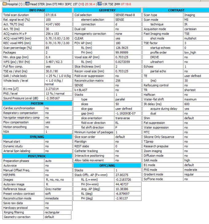
8 DTI: Diffusion Tensor imaging with 45 directions (Philips T3) Multitransmit = "no"; Nucleus = "H1"; Coil selection = "SENSEFlexS"; element selection = "12"; connection = "d"; Dual coil = "no"; CLEAR "yes"; body tuned = "no"; FOV RL (mm) = 160; AP (mm) = 160; FH (mm) = 90; Voxel size RL (mm) = 2; AP (mm) = 2; Slice thickness (mm) = 2; Recon voxel size (mm) =2; Small FOV imaging = "no"; Foldover suppression = "no"; Reconstruction matrix = 80; SENSE = "yes"; P reduction (AP) = ; P os factor = 1; kt BLAST = "no"; Stacks = 1; type = "parallel"; slices = 45; slice gap = "user defined"; gap (mm) = 0; slice orientation = "transverse"; foldover direction = "AP"; fat shift direction = "P"; Stack c. AP (P=+mm) = ; RL (L=+mm) = ; FH (H=+mm) = ; Ang. AP (deg) = ; RL (deg) = ; FH (deg) = ; Minimum number of packages = 1; Slice scan order = "default"; Large table movement ="no"; PlanAlign = "no"; REST slabs = 0; Shim Size AP (mm) = ; RL (mm) = ; FH (mm) = ; c. AP (P=+mm) = ; RL (L=+mm) = ; FH (H=+mm) = ; Ang. AP (deg) = ; RL (deg) = 0; FH (deg) = ; Catheter tracking = "no"; Interactive positioning = "no"; Allow table movement = "no"; Patient position = "head first"; orientation = "R decub"; Scan type = "Imaging"; Scan mode = "MS"; technique = "SE"; Modified SE = "no"; Acquisition mode = "cartesian"; Fast Imaging mode = "EPI"; shot mode = "singleshot"; Echoes = 1; partial echo = "no"; TE = "user defined"; (ms) = 80; Flip angle (deg) = 90;
9 TR = "user defined"; (ms) = 6500; Halfscan = "no"; Waterfat shift = "user defined"; (pixels) = 20; Shim = "PBvolume"; ShimAlign = "no"; mdixon = "no"; Fat suppression = "SPIR"; strength = "strong"; frequency offset = "user defined"; offset (Hz) = 200; Water suppression = "no"; Grad. rev. offres. supp. ="no"; BB pulse = "no"; MTC = "no"; Research prepulse = "no"; Diffusion mode = "DTI"; sequence = "SE"; gradient duration = "maximum"; gradient overplus = "no"; directional resolution ="user defined"; nr of directions = 45; user defined dirs = 1, (2) 0, , , 0, , , , , , , , , 0.588, , , , , , , , , , , , , , , , , , , , , , , , , , , , , , , , , , , , , , , , , , , , , , , , , , , , , , , , , , , , , , , , , , , , , , , , , , , , , , , , , , , , 0.912, , , ,
10 , , , , , , , , , , , , , , , , , , , , , , , , , , , , , , , , , (249) 0; nr of bfactors =2; bfactor order ="ascending"; max bfactor =800; average high b ="user defined"; bfactor averages =4, (15) 1; SAR mode = "high"; B1 mode = "default"; SAR Patient data = "auto"; PNS mode = "moderate"; Gradient mode = "enhanced"; SofTone mode = "no"; Cardiac synchronization ="no"; Heart rate > 250 bpm = "no"; Respiratory compensation ="no"; Navigator respiratory comp = "no"; Flow compensation = "no"; Temporal slice spacing = "default"; NSA = 1; Manual start = "no"; Dynamic study = "no"; dyn stabilization = "no"; Arterial Spin labeling = "no"; Preparation phases = "auto"; Interactive F0 = "no"; SmartPlan survey = "no"; B0 field map = "no"; B1 field map = "no"; MIP/MPR = "no"; Images = " M", (3) " no"; Autoview image = " M"; Calculated images = (4) " no"; Reference tissue = "User defined"; Tissue T1 = 2000; Tissue T2 = 150; Tissue rho = 100; EPI 2D phase correction = "no"; Preset window contrast = "soft"; Reconstruction mode = "immediate"; Save raw data = "no"; Hardcopy protocol = "no"; Ringing filtering = "default"; Geometry correction = "default"; IF_info_seperator = ; Total scan duration = "05:25.0"; Rel. signal level (%) = 100; Act. TR (ms) = "6500"; Act. TE (ms) = "80"; ACQ matrix M x P = "80 x 78"; ACQ voxel MPS (mm) = "2.00 / 2.03 / 2.00"; REC voxel MPS (mm) = "2.00 / 2.00 / 2.00"; Scan percentage (%) = ; Packages = 1; Min. slice gap (mm) = 0;
11 Diffusion gradient timing DELTA / delta (ms) = "38.8 / 9.5"; EPI factor = 61; WFS (pix) / BW (Hz) = " / 21.7"; BW in EPI freq. dir. (Hz) = "2638.7"; Min. TR (ms) = "5346"; SPIR offset act./default (Hz) = "200 [135]" SAR / local torso = "< 54 %"; Whole body / level = "< 0.5 W/kg / normal"; B1 rms = "1.18 ut"; PNS / level = "73 % / normal"; Sound Pressure Level (db) = ;
")
12 1.5T Philips Sagittal T1W SE (Philips T1.5)
: please change slice orientation in geometry from transverse into coronal: Inverse recovery: IR")
13 Coronal T2W (Philips T1.5): please change slice orientation in geometry from transverse into coronal: Inverse recovery: IR (Philips T1.5) please change slice orientation in geometry from transverse into coronal:
14 \

15 DWI: Diffusion weighted imaging (Philips T1.5)
 We prefer the settings that are explained above for the 3T, however if you will not manage to get nice")
16 DTI: diffusion tensor imaging please scan axial. (Philips T1.5) We prefer the settings that are explained above for the 3T, however if you will not manage to get nice images, please try the settings below for 1.5T
17 Simens Magnetom trio 3T
18 Properties Prio Recon Before measurement After measurement Load to viewer Inline movie Auto store images Load to stamp segments Load images to graphic segments Auto open inline display Start measurement without further preparation Wait for user to start Start measurements SIEMENS MAGNETOM TrioTim syngo MR B17 \\USER\Cerebrum\Projekter\SafeBoosC phase ll MRI.\localizer TA: 0:25 PAT: Voxel size: mm Rel. SNR: 1.00 SIEMENS: gre single Routine Slice group 1 Slices 4 Dist. factor 20 % Position R11.6 A103.7 H2.1 Orientation Transversal Phase enc. dir. A >> P Rotation 0.00 deg Slice group 2 Slices 3 Dist. factor 20 % Position R11.6 A104.7 F0.1 Orientation Coronal Phase enc. dir. R >> L Rotation 0.00 deg Slice group 3 Slices 5 Dist. factor 30 % Position R3.2 A104.7 F0.1 Orientation Sagittal Phase enc. dir. A >> P Rotation 0.00 deg Phase oversampling 0 % FoV read 250 mm FoV phase % Slice thickness 7.0 mm TR 8.6 ms TE 4.00 ms Averages 1 Concatenations 12 Filter Distortion Corr.(2D), Elliptical filter, B1 filter Coil elements NHA Contrast TD MTC Magn. preparation Flip angle Fat suppr. Water suppr. 0 ms 20 deg Averaging mode Short term Reconstruction Magnitude Measurements 1 Multiple series Each measurement Resolution Base resolution 256 Phase resolution 90 % Phase partial Fourier Interpolation PAT mode Matrix Coil Mode Auto (CP) Image Filter Distortion Corr. Mode Unfiltered images Prescan Normalize Normalize B1 filter Intensity Unfiltered images Raw filter Elliptical filter Mode Geometry Multislice mode Series 2D Medium Inplane Sequential Interleaved Saturation mode Standard Special sat. Tim CT mode System Body NBA NHA Positioning mode REF Table position H Table position 0 mm MSMA S C T Sagittal R >> L Coronal P >> A Transversal H >> F Save uncombined Coil Combine Mode Adaptive Combine AutoAlign Auto Coil Select Shim mode Tune up Adjust with body coil Confirm freq. adjustment Assume Silicone? Ref. amplitude 1H V Adjustment Tolerance Auto Adjust volume Position Isocenter Orientation Transversal Rotation 0.00 deg R >> L 350 mm A >> P 263 mm F >> H 350 mm Physio 1st Signal/Mode Segments 1 Tagging Dark blood Resp. control Inline 1/+
19 SIEMENS MAGNETOM TrioTim syngo MR B17 Subtract Liver registration StdDevSag StdDevCor StdDevTra StdDevTime MIPSag MIPCor MIPTra MIPTime Save original images Wash In Wash Out TTP PEI MIP time Sequence Introduction Dimension 2D Phase stabilisation Asymmetric echo Allowed Contrasts 1 Bandwidth 320 Hz/Px Flow comp. No Allowed delay 0 s RF pulse type Gradient mode Excitation RF spoiling Normal Normal Slicesel. 2/+
20 Properties Prio Recon Before measurement After measurement Load to viewer Inline movie Auto store images Load to stamp segments Load images to graphic segments Auto open inline display Start measurement without further preparation Wait for user to start Start measurements SIEMENS MAGNETOM TrioTim syngo MR B17 \\USER\Cerebrum\Projekter\SafeBoosC phase ll MRI.\t1_se_sag_3mm_200mm TA: 3:48 PAT: Voxel size: mm Rel. SNR: 1.00 SIEMENS: tse single Routine Slice group 1 Slices 31 Dist. factor 10 % Position R4.2 A96.9 F9.9 Orientation S > C5.2 > T3.4 Phase enc. dir. A >> P Rotation 0.00 deg Phase oversampling 0 % FoV read 200 mm FoV phase % Slice thickness 3.0 mm TR 550 ms TE 12 ms Averages 1 Concatenations 2 Filter Prescan Normalize, Elliptical filter Coil elements NHA Contrast TD MTC Magn. preparation Flip angle Fat suppr. Water suppr. Restore magn. 0.0 ms 90 deg Averaging mode Long term Reconstruction Magnitude Measurements 1 Multiple series Each measurement Resolution Base resolution 256 Phase resolution 80 % Phase partial Fourier Trajectory Cartesian Interpolation PAT mode Matrix Coil Mode Auto (CP) Image Filter Distortion Corr. Unfiltered images Prescan Normalize Normalize B1 filter Raw filter Elliptical filter Mode Geometry Multislice mode Series Inplane Interleaved Interleaved Special sat. Tim CT mode System Body NBA NHA Positioning mode REF Table position H Table position 0 mm MSMA S C T Sagittal R >> L Coronal P >> A Transversal H >> F Save uncombined Coil Combine Mode Adaptive Combine AutoAlign Auto Coil Select Default Shim mode Tune up Adjust with body coil Confirm freq. adjustment Assume Silicone? Ref. amplitude 1H V Adjustment Tolerance Auto Adjust volume Position Isocenter Orientation Transversal Rotation 0.00 deg R >> L 350 mm A >> P 263 mm F >> H 350 mm Physio 1st Signal/Mode Dark blood Resp. control Inline Subtract StdDevSag StdDevCor StdDevTra StdDevTime MIPSag MIPCor MIPTra MIPTime Save original images Sequence Introduction Dimension 2D Compensate T2 decay Reduce Motion Sens. Contrasts 1 3/+
21 SIEMENS MAGNETOM TrioTim syngo MR B17 Bandwidth 215 Hz/Px Flow comp. Slice Allowed delay 60 s Define Turbo factor Turbo factor 1 Echo trains per slice 205 RF pulse type Low SAR Gradient mode Normal 4/+
22 Properties Prio Recon Before measurement After measurement Load to viewer Inline movie Auto store images Load to stamp segments Load images to graphic segments Auto open inline display Start measurement without further preparation Wait for user to start Start measurements SIEMENS MAGNETOM TrioTim syngo MR B17 \\USER\Cerebrum\Projekter\SafeBoosC phase ll MRI.\t2_tse_cor_1,2mm_448_p2 TA: 5:10 PAT: 2 Voxel size: mm Rel. SNR: 1.00 SIEMENS: tse single Routine Slice group 1 Slices 110 Dist. factor 20 % Position R4.5 A101.2 F4.7 Orientation C > S3.5 > T1.5 Phase enc. dir. R >> L Rotation 0.00 deg Phase oversampling 0 % FoV read 180 mm FoV phase % Slice thickness 1.2 mm TR 4900 ms TE 148 ms Averages 2 Concatenations 7 Filter Prescan Normalize, Elliptical filter Coil elements NHA Contrast TD MTC Magn. preparation Flip angle Fat suppr. Water suppr. Restore magn. 0.0 ms 120 deg Averaging mode Long term Reconstruction Magnitude Measurements 1 Multiple series Each measurement Resolution Base resolution 256 Phase resolution 79 % Phase partial Fourier Trajectory Cartesian Interpolation PAT mode GRAPPA Accel. factor PE 2 Ref. lines PE 32 Matrix Coil Mode Auto (Triple) Reference scan mode Selfcalibration Image Filter Distortion Corr. Unfiltered images Prescan Normalize Normalize B1 filter Raw filter Elliptical filter Mode Geometry Multislice mode Series Inplane Interleaved Interleaved Special sat. Tim CT mode System Body NBA NHA Positioning mode REF Table position H Table position 0 mm MSMA S C T Sagittal R >> L Coronal P >> A Transversal H >> F Save uncombined Coil Combine Mode Adaptive Combine AutoAlign Auto Coil Select Default Shim mode Standard Adjust with body coil Confirm freq. adjustment Assume Silicone? Ref. amplitude 1H V Adjustment Tolerance Auto Adjust volume Position R4.5 A101.2 F4.7 Orientation C > S3.5 > T1.5 Rotation 0.00 deg F >> H 180 mm R >> L 180 mm A >> P 159 mm Physio 1st Signal/Mode Dark blood Resp. control Inline Subtract StdDevSag StdDevCor StdDevTra StdDevTime MIPSag MIPCor MIPTra MIPTime Save original images Sequence Introduction Dimension 5/+ 2D
23 SIEMENS MAGNETOM TrioTim syngo MR B17 Compensate T2 decay Reduce Motion Sens. Contrasts 1 Bandwidth 254 Hz/Px Flow comp. No Allowed delay 60 s Echo spacing 11.4 ms Define Turbo factor Turbo factor 26 Echo trains per slice 4 RF pulse type Normal Gradient mode Normal 6/+
24 SIEMENS MAGNETOM TrioTim syngo MR B17 \\USER\Cerebrum\Projekter\SafeBoosC phase ll MRI.\t1_mpr_cor TA: 4:05 PAT: 2 Voxel size: mm Rel. SNR: 1.00 SIEMENS: tfl Properties Prio Recon Before measurement After measurement Load to viewer Inline movie Auto store images Load to stamp segments Load images to graphic segments Auto open inline display Start measurement without further preparation Wait for user to start Start measurements single Routine Slab group 1 Slabs 1 Dist. factor 50 % Position R4.2 A96.9 F9.9 Orientation S > C5.2 > T3.4 Phase enc. dir. A >> P Rotation 0.00 deg Phase oversampling 20 % Slice oversampling 14.3 % Slices per slab 112 FoV read 200 mm FoV phase % Slice thickness 1.20 mm TR 1900 ms TE 2.94 ms Averages 1 Concatenations 1 Filter Prescan Normalize, Elliptical filter Coil elements NHA Contrast Magn. preparation TI Flip angle Fat suppr. Water suppr. Nonsel. IR 900 ms 9 deg Averaging mode Long term Reconstruction Magnitude Measurements 1 Multiple series Each measurement Resolution Base resolution 256 Phase resolution 86 % Slice resolution 100 % Phase partial Fourier 7/8 Slice partial Fourier Interpolation PAT mode Accel. factor PE 2 Ref. lines PE 24 Accel. factor 3D 1 GRAPPA Matrix Coil Mode Auto (Triple) Reference scan mode Integrated Image Filter Distortion Corr. Unfiltered images Prescan Normalize Normalize B1 filter Raw filter Elliptical filter Mode Geometry Multislice mode Series Inplane Single shot Ascending System Body NBA NHA Positioning mode FIX Table position H Table position 0 mm MSMA S C T Sagittal R >> L Coronal P >> A Transversal H >> F Save uncombined Coil Combine Mode Adaptive Combine AutoAlign Auto Coil Select Default Shim mode Tune up Adjust with body coil Confirm freq. adjustment Assume Silicone? Ref. amplitude 1H V Adjustment Tolerance Auto Adjust volume Position Isocenter Orientation Coronal Rotation 0.00 deg R >> L 350 mm A >> P 263 mm F >> H 350 mm Physio 1st Signal/Mode Dark blood Resp. control Inline Subtract StdDevSag StdDevCor StdDevTra StdDevTime MIPSag MIPCor MIPTra MIPTime Save original images Sequence Introduction Dimension Elliptical scanning Asymmetric echo 3D Allowed
25 SIEMENS MAGNETOM TrioTim syngo MR B17 Bandwidth 140 Hz/Px Flow comp. No Echo spacing 9 ms RF pulse type Gradient mode Excitation RF spoiling Normal Normal Nonsel. 8/+
26 SIEMENS MAGNETOM TrioTim syngo MR B17 \\USER\Cerebrum\Projekter\SafeBoosC phase ll MRI.\ep2d_diff_orth_p2_tra TA: 1:31 PAT: 2 Voxel size: mm Rel. SNR: 1.00 SIEMENS: ep2d_diff Properties Prio Recon Before measurement After measurement Load to viewer Inline movie Auto store images Load to stamp segments Load images to graphic segments Auto open inline display Start measurement without further preparation Wait for user to start Start measurements single Routine Slice group 1 Slices 28 Dist. factor 30 % Position R3.2 A106.3 H9.7 Orientation T > S4.5 > C2.9 Phase enc. dir. A >> P Rotation 0.00 deg Phase oversampling 0 % FoV read 230 mm FoV phase % Slice thickness 4.0 mm TR 4700 ms TE 102 ms Averages 4 Concatenations 1 Filter Raw filter, Distortion Corr.(2D), Prescan Normalize Coil elements NHA Contrast MTC Magn. preparation Fat suppr. Fat sat. Averaging mode Reconstruction Delay in TR Long term Magnitude 0 ms Resolution Base resolution 192 Phase resolution 100 % Phase partial Fourier 6/8 Interpolation PAT mode GRAPPA Accel. factor PE 2 Ref. lines PE 24 Matrix Coil Mode Auto (Triple) Reference scan mode Separate Distortion Corr. Mode 2D Unfiltered images Prescan Normalize Raw filter Intensity Weak Slope 25 Elliptical filter Hamming Geometry Multislice mode Series Interleaved Interleaved Special sat. System Body NBA NHA Positioning mode REF Table position H Table position 0 mm MSMA S C T Sagittal R >> L Coronal P >> A Transversal H >> F Coil Combine Mode Adaptive Combine AutoAlign Auto Coil Select Default Shim mode Standard Adjust with body coil Confirm freq. adjustment Assume Silicone? Ref. amplitude 1H V Adjustment Tolerance Auto Adjust volume Position R3.2 A106.3 H9.7 Orientation T > S4.5 > C2.9 Rotation 0.00 deg R >> L 230 mm A >> P 230 mm F >> H 145 mm Physio 1st Signal/Mode Resp. control Diff Diffusion mode Orthogonal Diff. weightings 2 bvalue 1 0 s/mm² bvalue s/mm² Diff. weighted images Trace weighted images Average ADC maps Individual ADC maps FA maps Mosaic Tensor Noise level 40 Diff. directions 3 Sequence Introduction Bandwidth Free echo spacing Echo spacing 1184 Hz/Px 0.93 ms 9/+ EPI factor 192 RF pulse type Normal Gradient mode Fast
27 SIEMENS MAGNETOM TrioTim syngo MR B17 \\USER\Cerebrum\Projekter\SafeBoosC phase ll MRI.\ep2d_diff_mddw_30_p2 TA: 9:28 PAT: 2 Voxel size: mm Rel. SNR: 1.00 SIEMENS: ep2d_diff Properties Prio Recon Before measurement After measurement Load to viewer Inline movie Auto store images Load to stamp segments Load images to graphic segments Auto open inline display Start measurement without further preparation Wait for user to start Start measurements single Routine Slice group 1 Slices 49 Dist. factor 30 % Position R3.2 A106.3 H9.7 Orientation T > S4.5 > C2.9 Phase enc. dir. A >> P Rotation 0.00 deg Phase oversampling 0 % FoV read 250 mm FoV phase % Slice thickness 2.0 mm TR 5900 ms TE 81 ms Averages 3 Concatenations 1 Filter Prescan Normalize Coil elements NHA Contrast MTC Magn. preparation Fat suppr. Fat sat. Averaging mode Reconstruction Delay in TR Multiple series Long term Magnitude 0 ms Resolution Base resolution 122 Phase resolution 100 % Phase partial Fourier 6/8 Interpolation PAT mode GRAPPA Accel. factor PE 2 Ref. lines PE 24 Matrix Coil Mode Auto (Triple) Reference scan mode Separate Distortion Corr. Prescan Normalize Raw filter Elliptical filter Hamming Geometry Multislice mode Series Interleaved Interleaved Special sat. System Body NBA NHA Positioning mode FIX Table position H Table position 0 mm MSMA S C T Sagittal R >> L Coronal P >> A Transversal H >> F Coil Combine Mode Adaptive Combine AutoAlign Auto Coil Select Default Shim mode Standard Adjust with body coil Confirm freq. adjustment Assume Silicone? Ref. amplitude 1H V Adjustment Tolerance Auto Adjust volume Position R3.2 A106.3 H9.7 Orientation T > S4.5 > C2.9 Rotation 0.00 deg R >> L 250 mm A >> P 250 mm F >> H 127 mm Physio 1st Signal/Mode Resp. control Diff Diffusion mode MDDW Diff. weightings 2 bvalue 1 0 s/mm² bvalue s/mm² Diff. weighted images Trace weighted images Average ADC maps Individual ADC maps FA maps Mosaic Tensor Noise level 30 Diff. directions 30 Sequence Introduction Bandwidth Free echo spacing Echo spacing 1518 Hz/Px 0.74 ms 10/+ EPI factor 122 RF pulse type Normal Gradient mode Fast
28 SIEMENS MAGNETOM TrioTim syngo MR B17 Table of contents \\USER Cerebrum Projekter SafeBoosC phase ll MRI. localizer t1_se_sag_3mm_200mm t2_tse_cor_1,2mm_448_p2 t1_mpr_sag ep2d_diff_orth_p2_tra ep2d_diff_mddw_30_p2
29 SafeBoosC phase II SOP Brain MRI Version
SIEMENS MAGNETOM Avanto syngo MR B15
 \\USER\INVESTIGATORS\Ravi\ADNI-Subject\Localizer TA: 0:10 PAT: Voxel size: 1.9 1.5 8.0 mm Rel. SNR: 1.00 SIEMENS: gre Properties Prio Recon Before measurement After measurement Load to viewer Inline movie
COBRE Scan Information
 COBRE Scan Information Below is more information on the directory structure for the COBRE imaging data. Also below are the imaging parameters for each series. Directory structure: var/www/html/dropbox/1139_anonymized/human:
SIEMENS MAGNETOM Skyra syngo MR D13
 Page 1 of 8 SIEMENS MAGNETOM Skyra syngo MR D13 \\USER\CIND\StudyProtocols\PTSA\*dm_ep2d_mono70_b0_p2_iso2.0 TA:1:05 PAT:2 Voxel size:2.0 2.0 2.0 mm Rel. SNR:1.00 :epse Properties Routine Prio Recon Load
SIEMENS MAGNETOM Avanto syngo MR B15
 \\USER\INVESTIGATORS\Ravi\ADNI-phantom\QC Phantom-Localizer TA: 0:10 PAT: Voxel size: 1.9 1.5 8.0 mm Rel. SNR: 1.00 SIEMENS: gre Properties Prio Recon Before measurement After measurement Load to viewer
SIEMENS MAGNETOM Verio syngo MR B15V
 \\USER\ZAHID_RESEARCH\MS\No Name\3D SWI TA: 6:39 PAT: 2 Voxel size: 1.0 0.5 2.0 mm Rel. SNR: 1.00 SIEMENS: gre Properties Prio Recon Before measurement After measurement Load to viewer Inline movie Auto
HST.583 Functional Magnetic Resonance Imaging: Data Acquisition and Analysis Fall 2008
 MIT OpenCourseWare http://ocw.mit.edu HST.583 Functional Magnetic Resonance Imaging: Data Acquisition and Analysis Fall 2008 For information about citing these materials or our Terms of Use, visit: http://ocw.mit.edu/terms.
SIEMENS MAGNETOM Verio syngo MR B17
 \\USER\Dr. Behrmann\routine\Ilan\ep2d_bold_PMU_resting TA: 8:06 PAT: Voxel size: 3.03.03.0 mm Rel. SNR: 1.00 USER: ep2d_bold_pmu Properties Special sat. Prio Recon System Before measurement Body After
Philips MRI Protocol Dump Created on Comment Software Stream
 Page 1 of 5 Philips MRI Protocol Dump Created on 2/17/2011 4:11:01 PM Comment Created by ExamCard_to_XML with inputs: "J:\ADNI GO - ADNI 2 Phantom5.ExamCard" on system (BU SCHOOL OF MEDICINE :: 192.168.71.10)
SIEMENS MAGNETOM TrioTim syngo MR B17
 \\USER\KNARRGROUP\MultiBand\LavretskyMultiBand\trufi localizer 3-plane TA: 5.1 s PAT: Voxel size: 1.2 1.2 5. Rel. SNR: 1.00 SIEMENS: trufi Load to stamp Slice group 1 Slices 1 Dist. factor 20 % Phase enc.
ADNI, ADNI_QH, SURVEY. Geometry. connection
 ADNI, ADNI_QH, SURVEY Geometry Coil selection = Head connection = d Multi coil Homogeneity correction ne FOV (mm) = 250.00 RFOV (%) = 100.00 Foldover suppression Matrix scan = 256 reconstruction = 256
HST.583 Functional Magnetic Resonance Imaging: Data Acquisition and Analysis Fall 2006
 MIT OpenCourseWare http://ocw.mit.edu HST.583 Functional Magnetic Resonance Imaging: Data Acquisition and Analysis Fall 2006 For information about citing these materials or our Terms of Use, visit: http://ocw.mit.edu/terms.
ADNI GO - ADNI 2 Human7 (9) 38:30.1
 38:30.1") Philips RI Protocol Dump Create on 11/25/2013 11:40:24 A Comment Create by ExamCar_to_XL with inputs: "K:\ADNI GO - ADNI 2 Human7.ExamCar" on system (BU SCHOOL OF EDICINE :: 192.168.71.10) Software Stream
Philips RI Protocol Dump Create on 11/25/2013 11:40:24 A Comment Create by ExamCar_to_XL with inputs: "K:\ADNI GO - ADNI 2 Human7.ExamCar" on system (BU SCHOOL OF EDICINE :: 192.168.71.10) Software Stream
Lab Location: MRI, B2, Cardinal Carter Wing, St. Michael s Hospital, 30 Bond Street
 Lab Location: MRI, B2, Cardinal Carter Wing, St. Michael s Hospital, 30 Bond Street MRI is located in the sub basement of CC wing. From Queen or Victoria, follow the baby blue arrows and ride the CC south
SIEMENS MAGNETOM Symphony syngo MR A30
 \\USER\ADNI STUDY\MAIN PROTOCOL\HUMAN PROTOCOL\localizer Scan Time: 9.2 [s] Voxel size: 2.2 1.1 10.0 [mm] Rel. SNR: 1.00 SIEMENS: gre Slice group 1 Slice group 2 Slice group 3 280 [mm] 10 [mm] 20 [ms]
Siemens AG, Healthcare Sector. syngo MR D13 0. Supplement - Parameters and image text 0.
 Siemens AG, Healthcare Sector 0 0 n.a. English Cs2 syngo Neuro Operator MR-05014 630 05/2010 01 02 Informatik, Manual D11 Cape syngo MR D13 0.0 Supplement - Parameters and image text 0. syngo MR D13 0.
Page 1 of 9. Protocol: adult_other_adni3basichumanprotocol25x_ _ _1. 3 Plane Localizer. 3 Plane Localizer PATIENT POSITION
 3 Localizer FOV 26.0 Slice Thickness 5.0 Slice Spacing 0.0 Freq 256 Phase 128 3-PLANE 3 Localizer Unswap Phase Correction Gradient Echo Imaging Options Seq, Fast Recon All Images 3 Localizer Pause / SCIC
Orthopedic MRI Protocols. Philips Panorama HFO
 Orthopedic MRI Protocols Philips Panorama HFO 1 2 Prepared in collaboration with Dr. John F. Feller, Medical Director of Desert Medical Imaging, Palm Springs, CA. Desert Medical Imaging will provide the
MRI. When to use What sequences. Outline 2012/09/19. Sequence: Definition. Basic Principles: Step 2. Basic Principles: Step 1. Govind Chavhan, MD
 MRI When to use What sequences Govind Chavhan, MD Assistant Professor and Staff Radiologist The Hospital For Sick Children, Toronto Planning Acquisition Post processing Interpretation Patient history and
Diffusion MRI Acquisition. Karla Miller FMRIB Centre, University of Oxford
 Diffusion MRI Acquisition Karla Miller FMRIB Centre, University of Oxford karla@fmrib.ox.ac.uk Diffusion Imaging How is diffusion weighting achieved? How is the image acquired? What are the limitations,
Applications Guide for Interleaved
 Applications Guide for Interleaved rephase/dephase MRAV Authors: Yongquan Ye, Ph.D. Dongmei Wu, MS. Tested MAGNETOM Systems : 7TZ, TRIO a Tim System, Verio MR B15A (N4_VB15A_LATEST_20070519) MR B17A (N4_VB17A_LATEST_20090307_P8)
Dynamic Contrast enhanced MRA
 Dynamic Contrast enhanced MRA Speaker: Yung-Chieh Chang Date : 106.07.22 Department of Radiology, Taichung Veterans General Hospital, Taichung, Taiwan 1 Outline Basic and advanced principles of Diffusion
MRI image formation 8/3/2016. Disclosure. Outlines. Chen Lin, PhD DABR 3. Indiana University School of Medicine and Indiana University Health
 MRI image formation Indiana University School of Medicine and Indiana University Health Disclosure No conflict of interest for this presentation 2 Outlines Data acquisition Spatial (Slice/Slab) selection
Slide 1. Technical Aspects of Quality Control in Magnetic Resonance Imaging. Slide 2. Annual Compliance Testing. of MRI Systems.
 Slide 1 Technical Aspects of Quality Control in Magnetic Resonance Imaging Slide 2 Compliance Testing of MRI Systems, Ph.D. Department of Radiology Henry Ford Hospital, Detroit, MI Slide 3 Compliance Testing
MRI Physics II: Gradients, Imaging
 MRI Physics II: Gradients, Imaging Douglas C., Ph.D. Dept. of Biomedical Engineering University of Michigan, Ann Arbor Magnetic Fields in MRI B 0 The main magnetic field. Always on (0.5-7 T) Magnetizes
CHAPTER 9: Magnetic Susceptibility Effects in High Field MRI
 Figure 1. In the brain, the gray matter has substantially more blood vessels and capillaries than white matter. The magnified image on the right displays the rich vasculature in gray matter forming porous,
A Virtual MR Scanner for Education
 A Virtual MR Scanner for Education Hackländer T, Schalla C, Trümper A, Mertens H, Hiltner J, Cramer BM Hospitals of the University Witten/Herdecke, Department of Radiology Wuppertal, Germany Purpose A
Motion Artifacts and Suppression in MRI At a Glance
 Motion Artifacts and Suppression in MRI At a Glance Xiaodong Zhong, PhD MR R&D Collaborations Siemens Healthcare MRI Motion Artifacts and Suppression At a Glance Outline Background Physics Common Motion
New Technology Allows Multiple Image Contrasts in a Single Scan
 These images were acquired with an investigational device. PD T2 T2 FLAIR T1 MAP T1 FLAIR PSIR T1 New Technology Allows Multiple Image Contrasts in a Single Scan MR exams can be time consuming. A typical
White Pixel Artifact. Caused by a noise spike during acquisition Spike in K-space <--> sinusoid in image space
 White Pixel Artifact Caused by a noise spike during acquisition Spike in K-space sinusoid in image space Susceptibility Artifacts Off-resonance artifacts caused by adjacent regions with different
Head motion in diffusion MRI
 Head motion in diffusion MRI Anastasia Yendiki HMS/MGH/MIT Athinoula A. Martinos Center for Biomedical Imaging 11/06/13 Head motion in diffusion MRI 0/33 Diffusion contrast Basic principle of diffusion
MR Advance Techniques. Vascular Imaging. Class III
 MR Advance Techniques Vascular Imaging Class III 1 Vascular Imaging There are several methods that can be used to evaluate the cardiovascular systems with the use of MRI. MRI will aloud to evaluate morphology
Scan Acceleration with Rapid Gradient-Echo
 Scan Acceleration with Rapid Gradient-Echo Hsiao-Wen Chung ( 鍾孝文 ), Ph.D., Professor Dept. Electrical Engineering, National Taiwan Univ. Dept. Radiology, Tri-Service General Hospital 1 of 214 The Need
Fast Imaging UCLA. Class Business. Class Business. Daniel B. Ennis, Ph.D. Magnetic Resonance Research Labs. Tuesday (3/7) from 6-9pm HW #1 HW #2
 from 6-9pm HW #1 HW #2") Fast Imaging Daniel B. Ennis, Ph.D. Magnetic Resonance Research Labs Class Business Tuesday (3/7) from 6-9pm 6:00-7:30pm Groups Avanto Sara Said, Yara Azar, April Pan Skyra Timothy Marcum, Diana Lopez,
Fast Imaging Daniel B. Ennis, Ph.D. Magnetic Resonance Research Labs Class Business Tuesday (3/7) from 6-9pm 6:00-7:30pm Groups Avanto Sara Said, Yara Azar, April Pan Skyra Timothy Marcum, Diana Lopez,
M R I Physics Course
 M R I Physics Course Multichannel Technology & Parallel Imaging Nathan Yanasak, Ph.D. Jerry Allison Ph.D. Tom Lavin, B.S. Department of Radiology Medical College of Georgia References: 1) The Physics of
(a Scrhon5 R2iwd b. P)jc%z 5. ivcr3. 1. I. ZOms Xn,s. 1E IDrAS boms. EE225E/BIOE265 Spring 2013 Principles of MRI. Assignment 8 Solutions
jc%z 5. ivcr3. 1. I. ZOms Xn,s. 1E IDrAS boms. EE225E/BIOE265 Spring 2013 Principles of MRI. Assignment 8 Solutions") EE225E/BIOE265 Spring 2013 Principles of MRI Miki Lustig Assignment 8 Solutions 1. Nishimura 7.1 P)jc%z 5 ivcr3. 1. I Due Wednesday April 10th, 2013 (a Scrhon5 R2iwd b 0 ZOms Xn,s r cx > qs 4-4 8ni6 4
EE225E/BIOE265 Spring 2013 Principles of MRI Miki Lustig Assignment 8 Solutions 1. Nishimura 7.1 P)jc%z 5 ivcr3. 1. I Due Wednesday April 10th, 2013 (a Scrhon5 R2iwd b 0 ZOms Xn,s r cx > qs 4-4 8ni6 4
Tina Pavlin 12 March 2013
 Table of Contents Chapter 1 General... 2 Spectrometer Reset... 2 Adjustment of DC- offset (Zipper Artifact)... 2 Removal of Zipper Artifact... 2 Prescription for Decreasing TE... 2 EPIs... 3 Chapter 2
MR-Encephalography (MREG)
") MR-Encephalography (MREG) C2P package Version 2.0 For SIEMENS Magnetom Verio/TRIO/Skyra/Prisma Installation and User s Guide NUMARIS/4 VB17, VD11, VD13 Jakob Assländer Pierre LeVan, Ph.D. 10.09.2014 Magnetic
MR-Encephalography (MREG) C2P package Version 2.0 For SIEMENS Magnetom Verio/TRIO/Skyra/Prisma Installation and User s Guide NUMARIS/4 VB17, VD11, VD13 Jakob Assländer Pierre LeVan, Ph.D. 10.09.2014 Magnetic
Fast Imaging Trajectories: Non-Cartesian Sampling (1)
") Fast Imaging Trajectories: Non-Cartesian Sampling (1) M229 Advanced Topics in MRI Holden H. Wu, Ph.D. 2018.05.03 Department of Radiological Sciences David Geffen School of Medicine at UCLA Class Business
Fast Imaging Trajectories: Non-Cartesian Sampling (1) M229 Advanced Topics in MRI Holden H. Wu, Ph.D. 2018.05.03 Department of Radiological Sciences David Geffen School of Medicine at UCLA Class Business
Field Maps. 1 Field Map Acquisition. John Pauly. October 5, 2005
 Field Maps John Pauly October 5, 25 The acquisition and reconstruction of frequency, or field, maps is important for both the acquisition of MRI data, and for its reconstruction. Many of the imaging methods
Functional MRI. Jerry Allison, Ph. D. Medical College of Georgia
 Functional MRI Jerry Allison, Ph. D. Medical College of Georgia BOLD Imaging Technique Blood Oxygen Level Dependent contrast can be used to map brain function Right Hand Motor Task Outline fmri BOLD Contrast
GE Healthcare CLINICAL GALLERY. Discovery * MR750w 3.0T. This brochure is intended for European healthcare professionals.
 GE Healthcare CLINICAL GALLERY Discovery * MR750w 3.0T This brochure is intended for European healthcare professionals. NEURO PROPELLER delivers high resolution, motion insensitive imaging in all planes.
Module 4. K-Space Symmetry. Review. K-Space Review. K-Space Symmetry. Partial or Fractional Echo. Half or Partial Fourier HASTE
 MRES 7005 - Fast Imaging Techniques Module 4 K-Space Symmetry Review K-Space Review K-Space Symmetry Partial or Fractional Echo Half or Partial Fourier HASTE Conditions for successful reconstruction Interpolation
8/11/2009. Common Areas of Motion Problem. Motion Compensation Techniques and Applications. Type of Motion. What s your problem
 Common Areas of Motion Problem Motion Compensation Techniques and Applications Abdominal and cardiac imaging. Uncooperative patient, such as pediatric. Dynamic imaging and time series. Chen Lin, PhD Indiana
Patient Specific. Protocol Identification Number (PID) Must be on each individual. MRI Image
 Must be on each individual. MRI Image") MRI PROCEDURE GUIDELINES Patient Specific Protocol Identification Number (PID) Must be on each individual MRI Image [Protocol 11/04/13, Version 1.4] (Revised 09/03/2014) Page 1 of 72 Abbreviations: BH:
MRI PROCEDURE GUIDELINES Patient Specific Protocol Identification Number (PID) Must be on each individual MRI Image [Protocol 11/04/13, Version 1.4] (Revised 09/03/2014) Page 1 of 72 Abbreviations: BH:
Parallel Imaging. Marcin.
 Parallel Imaging Marcin m.jankiewicz@gmail.com Parallel Imaging initial thoughts Over the last 15 years, great progress in the development of pmri methods has taken place, thereby producing a multitude
Breast MRI Accreditation Program Clinical Image Quality Guide
 Breast MRI Accreditation Program Clinical Image Quality Guide Introduction This document provides guidance on breast MRI clinical image quality and describes the criteria used by the ACR Breast MRI Accreditation
TOPICS 2/5/2006 8:17 PM. 2D Acquisition 3D Acquisition
 TOPICS 2/5/2006 8:17 PM 2D Acquisition 3D Acquisition 2D Acquisition Involves two main steps : Slice Selection Slice selection is accomplished by spatially saturating (single or multi slice imaging) or
Midterm Review
 Midterm Review - 2017 EE369B Concepts Noise Simulations with Bloch Matrices, EPG Gradient Echo Imaging 1 About the Midterm Monday Oct 30, 2017. CCSR 4107 Up to end of C2 1. Write your name legibly on this
syngo MR E11 Operator Manual Ortho Answers for life.
 www.siemens.com/healthcare syngo MR E11 Operator Manual Ortho Answers for life. syngo MR E11 Operator Manual Ortho Legend Indicates a hint Is used to provide information on how to avoid operating errors
MRI Image Quality Assessment
 in partnership with MRI Image Quality Assessment David Collins CR-UK Cancer Imaging Centre, The Institute of Cancer Research Making the discoveries that defeat cancer Overview Current Practice Quality
Sampling, Ordering, Interleaving
 Sampling, Ordering, Interleaving Sampling patterns and PSFs View ordering Modulation due to transients Temporal modulations Timing: cine, gating, triggering Slice interleaving Sequential, Odd/even, bit-reversed
Outline: Contrast-enhanced MRA
 Outline: Contrast-enhanced MRA Background Technique Clinical Indications Future Directions Disclosures: GE Health Care: Research support Consultant: Bracco, Bayer The Basics During rapid IV infusion, Gadolinium
An Introduction to Image Reconstruction, Processing, and their Effects in FMRI
 An Introduction to Image Reconstruction, Processing, and their Effects in FMRI Daniel B. Rowe Program in Computational Sciences Department of Mathematics, Statistics, and Computer Science Marquette University
TOF-MRA Using Multi-Oblique-Stack Acquisition (MOSA)
") JOURNAL OF MAGNETIC RESONANCE IMAGING 26:432 436 (2007) Technical Note TOF-MRA Using Multi-Oblique-Stack Acquisition (MOSA) Ed X. Wu, PhD, 1,2 * Edward S. Hui, BEng, 1,2 and Jerry S. Cheung, BEng 1,2 Purpose:
JOURNAL OF MAGNETIC RESONANCE IMAGING 26:432 436 (2007) Technical Note TOF-MRA Using Multi-Oblique-Stack Acquisition (MOSA) Ed X. Wu, PhD, 1,2 * Edward S. Hui, BEng, 1,2 and Jerry S. Cheung, BEng 1,2 Purpose:
Functional analysis with DTI and diffusion-neurography of cranial nerves
 Functional analysis with DTI and diffusion-neurography of cranial nerves Poster No.: C-1942 Congress: ECR 2013 Type: Educational Exhibit Authors: J. P. Martínez Barbero, T. Martín Noguerol, A. Luna Alcalá;
Measuring baseline whole-brain perfusion on GE 3.0T using arterial spin labeling (ASL) MRI
 MRI") Measuring baseline whole-brain perfusion on GE 3.0T using arterial spin labeling (ASL) MRI Revision date: 11/20/2006 Overview This document describes the procedure for measuring baseline whole-brain perfusion
Measuring baseline whole-brain perfusion on GE 3.0T using arterial spin labeling (ASL) MRI Revision date: 11/20/2006 Overview This document describes the procedure for measuring baseline whole-brain perfusion
ACQUIRING AND PROCESSING SUSCEPTIBILITY WEIGHTED IMAGING (SWI) DATA ON GE 3.0T
 DATA ON GE 3.0T") ACQUIRING AND PROCESSING SUSCEPTIBILITY WEIGHTED IMAGING (SWI) DATA ON GE 3.0T Revision date: 12/13/2010 Overview Susceptibility Weighted Imaging (SWI) is a relatively new data acquisition and processing
ACQUIRING AND PROCESSING SUSCEPTIBILITY WEIGHTED IMAGING (SWI) DATA ON GE 3.0T Revision date: 12/13/2010 Overview Susceptibility Weighted Imaging (SWI) is a relatively new data acquisition and processing
Supplementary methods
 Supplementary methods This section provides additional technical details on the sample, the applied imaging and analysis steps and methods. Structural imaging Trained radiographers placed all participants
Lucy Phantom MR Grid Evaluation
 Lucy Phantom MR Grid Evaluation Anil Sethi, PhD Loyola University Medical Center, Maywood, IL 60153 November 2015 I. Introduction: The MR distortion grid, used as an insert with Lucy 3D QA phantom, is
Role of Parallel Imaging in High Field Functional MRI
 Role of Parallel Imaging in High Field Functional MRI Douglas C. Noll & Bradley P. Sutton Department of Biomedical Engineering, University of Michigan Supported by NIH Grant DA15410 & The Whitaker Foundation
Release Notes. Multi-Band EPI C2P. Release HCP_v1 10 February 2014
 Release Notes Multi-Band EPI C2P Release HCP_v1 10 February 2014 Installation 1. Restart the system (reboot host and MRIR) 2. Extract the.zip file to a temporary directory 3. Run the installer.bat file
Clinical Importance. Aortic Stenosis. Aortic Regurgitation. Ultrasound vs. MRI. Carotid Artery Stenosis
 Clinical Importance Rapid cardiovascular flow quantitation using sliceselective Fourier velocity encoding with spiral readouts Valve disease affects 10% of patients with heart disease in the U.S. Most
syngo MR E11 Operator Manual Neuro Answers for life.
 www.siemens.com/healthcare syngo MR E11 Operator Manual Neuro Answers for life. syngo MR E11 Operator Manual Neuro Legend Indicates a hint Is used to provide information on how to avoid operating errors
Basic fmri Design and Analysis. Preprocessing
 Basic fmri Design and Analysis Preprocessing fmri Preprocessing Slice timing correction Geometric distortion correction Head motion correction Temporal filtering Intensity normalization Spatial filtering
Super-Resolution Reconstruction of Diffusion-Weighted Images from Distortion Compensated Orthogonal Anisotropic Acquisitions.
 Super-Resolution Reconstruction of Diffusion-Weighted Images from Distortion Compensated Orthogonal Anisotropic Acquisitions. Benoit Scherrer Ali Gholipour Simon K. Warfield Children s Hospital Boston,
Enhanced DICOM MR for spectroscopy, structural and functional imaging
 The Medicine Behind the Image Enhanced DICOM MR for spectroscopy, structural and functional imaging Dr. David A. Clunie, MB.,BS., FRACR Chief Technology Officer RadPharm, Inc. Acknowledgments Mark Day,
Image Acquisition Systems
 Image Acquisition Systems Goals and Terminology Conventional Radiography Axial Tomography Computer Axial Tomography (CAT) Magnetic Resonance Imaging (MRI) PET, SPECT Ultrasound Microscopy Imaging ITCS
Following on from the two previous chapters, which considered the model of the
 Chapter 5 Simulator validation Following on from the two previous chapters, which considered the model of the simulation process and how this model was implemented in software, this chapter is concerned
WE PROVIDE KNOWLEDGE, SERVICES AND PRODUCTS FOR ANY KIND OF PROCESS PLANTS
 MAGNETOM ESSENZA A Tim+Dot System Tim [25x8] www.siemens.com/magnetom-essenza 2 MAGNETOM ESSENZA Established 1.5 T performance. With Tim+Dot Lomisa Distribuciones y Proyectos S.L., socidad inscrita en
QIBA Profile: Magnetic Resonance Elastography of the Liver
 5 QIBA Profile: Magnetic Resonance Elastography of the Liver Stage: A. Initial Draft July 28, 2017 10 15 20 25 30 35 40 Table of Contents Change Log:...3 Open Issues:...4 Closed Issues:...4 1. Executive
Measuring baseline whole-brain perfusion on GE 3.0T using arterial spin labeling (ASL) MRI
 MRI") Measuring baseline whole-brain perfusion on GE 3.0T using arterial spin labeling (ASL) MRI Revision date: 09/15/2008 Overview This document describes the procedure for measuring baseline whole-brain perfusion
Measuring baseline whole-brain perfusion on GE 3.0T using arterial spin labeling (ASL) MRI Revision date: 09/15/2008 Overview This document describes the procedure for measuring baseline whole-brain perfusion
Qualitative Comparison of Conventional and Oblique MRI for Detection of Herniated Spinal Discs
 Qualitative Comparison of Conventional and Oblique MRI for Detection of Herniated Spinal Discs Doug Dean Final Project Presentation ENGN 2500: Medical Image Analysis May 16, 2011 Outline Review of the
Exam 8N080 - Introduction MRI
 Exam 8N080 - Introduction MRI Friday January 23 rd 2015, 13.30-16.30h For this exam you may use an ordinary calculator (not a graphical one). In total there are 6 assignments and a total of 65 points can
Blipped-Controlled Aliasing in Parallel Imaging for Simultaneous Multislice Echo Planar Imaging With Reduced g-factor Penalty
 IMAGING METHODOLOGY - Full Papers Magnetic Resonance in Medicine 67:1210 1224 (2012) Blipped-Controlled Aliasing in Parallel Imaging for Simultaneous Multislice Echo Planar Imaging With Reduced g-factor
SPM8 for Basic and Clinical Investigators. Preprocessing. fmri Preprocessing
 SPM8 for Basic and Clinical Investigators Preprocessing fmri Preprocessing Slice timing correction Geometric distortion correction Head motion correction Temporal filtering Intensity normalization Spatial
Advanced MRI Techniques (and Applications)
") Advanced MRI Techniques (and Applications) Jeffry R. Alger, PhD Department of Neurology Ahmanson-Lovelace Brain Mapping Center Brain Research Institute Jonsson Comprehensive Cancer Center University of
Advanced MRI Techniques (and Applications) Jeffry R. Alger, PhD Department of Neurology Ahmanson-Lovelace Brain Mapping Center Brain Research Institute Jonsson Comprehensive Cancer Center University of
3-D Gradient Shimming
 3-D Gradient Shimming 3-D Gradient Shimming on imaging and micro-imaging systems Technical Overview Introduction Advantage statement The GE3DSHIM method is a 3-D gradient shimming procedure developed for
HST.583 Functional Magnetic Resonance Imaging: Data Acquisition and Analysis Fall 2008
 MIT OpenCourseWare http://ocw.mit.edu HST.583 Functional Magnetic Resonance Imaging: Data Acquisition and Analysis Fall 2008 For information about citing these materials or our Terms of Use, visit: http://ocw.mit.edu/terms.
Error! Bookmark not defined. Error! Bookmark not defined.
 MRI-Neuro Protocols Brain Protocols... 2 WITH AND WITHOUT... 3 WITHOUT CONTRT... 4 HHT... 5 MS WITH AND WITHOUT CONTRT... 6 MS WITHOUT CONTRT... 7 PEDIATRIC... 8 PEDIATRIC WITH CONTRT... 9 PITUITARY NO
surface Image reconstruction: 2D Fourier Transform
 2/1/217 Chapter 2-3 K-space Intro to k-space sampling (chap 3) Frequenc encoding and Discrete sampling (chap 2) Point Spread Function K-space properties K-space sampling principles (chap 3) Basic Contrast
XI Signal-to-Noise (SNR)
") XI Signal-to-Noise (SNR) Lecture notes by Assaf Tal n(t) t. Noise. Characterizing Noise Noise is a random signal that gets added to all of our measurements. In D it looks like this: while in D
XI Signal-to-Noise (SNR) Lecture notes by Assaf Tal n(t) t. Noise. Characterizing Noise Noise is a random signal that gets added to all of our measurements. In D it looks like this: while in D
Functional MRI in Clinical Research and Practice Preprocessing
 Functional MRI in Clinical Research and Practice Preprocessing fmri Preprocessing Slice timing correction Geometric distortion correction Head motion correction Temporal filtering Intensity normalization
MRI Hot Topics Motion Correction for MR Imaging. medical
 MRI Hot Topics Motion Correction for MR Imaging s medical Motion Correction for MR Imaging Kyle A. Salem, PhD Motion Artifacts in a Clinical Setting Patient motion is probably the most common cause of
EPI Data Are Acquired Serially. EPI Data Are Acquired Serially 10/23/2011. Functional Connectivity Preprocessing. fmri Preprocessing
 Functional Connectivity Preprocessing Geometric distortion Head motion Geometric distortion Head motion EPI Data Are Acquired Serially EPI Data Are Acquired Serially descending 1 EPI Data Are Acquired
Super-resolution Reconstruction of Fetal Brain MRI
 Super-resolution Reconstruction of Fetal Brain MRI Ali Gholipour and Simon K. Warfield Computational Radiology Laboratory Children s Hospital Boston, Harvard Medical School Worshop on Image Analysis for
The SIMRI project A versatile and interactive MRI simulator *
 COST B21 Meeting, Lodz, 6-9 Oct. 2005 The SIMRI project A versatile and interactive MRI simulator * H. Benoit-Cattin 1, G. Collewet 2, B. Belaroussi 1, H. Saint-Jalmes 3, C. Odet 1 1 CREATIS, UMR CNRS
SPM8 for Basic and Clinical Investigators. Preprocessing
 SPM8 for Basic and Clinical Investigators Preprocessing fmri Preprocessing Slice timing correction Geometric distortion correction Head motion correction Temporal filtering Intensity normalization Spatial
A Novel Iterative Thresholding Algorithm for Compressed Sensing Reconstruction of Quantitative MRI Parameters from Insufficient Data
 A Novel Iterative Thresholding Algorithm for Compressed Sensing Reconstruction of Quantitative MRI Parameters from Insufficient Data Alexey Samsonov, Julia Velikina Departments of Radiology and Medical
Sampling, Ordering, Interleaving
 Sampling, Ordering, Interleaving Sampling patterns and PSFs View ordering Modulation due to transients Temporal modulations Slice interleaving Sequential, Odd/even, bit-reversed Arbitrary Other considerations:
Accelerated MRI Techniques: Basics of Parallel Imaging and Compressed Sensing
 Accelerated MRI Techniques: Basics of Parallel Imaging and Compressed Sensing Peng Hu, Ph.D. Associate Professor Department of Radiological Sciences PengHu@mednet.ucla.edu 310-267-6838 MRI... MRI has low
A Transmission Line Matrix Model for Shielding Effects in Stents
 A Transmission Line Matrix Model for hielding Effects in tents Razvan Ciocan (1), Nathan Ida (2) (1) Clemson University Physics and Astronomy Department Clemson University, C 29634-0978 ciocan@clemon.edu
T 1 MAPPING FOR DCE-MRI
 T 1 MAPPING FOR DCE-MRI A dissertation submitted to the Faculty of Medicine, University of Malaya in partial fulfillment of the requirements for the degree of Master of Medical Physics By NURUN NAJWA BINTI
Table of Contents MRI Neuro protocols
 Table of Contents MRI Neuro protocols 1. MRI Soft-tissue neck 2. MRI Head: a. Head with and without contrast b. Perfusion post processing c. CSF flow analysis d. Deep brain stimulator e. Brain spectroscopy
TARDIS Working Practice Document No: 035
 TARDIS Working Practice Document No: 035 Uploading CT or MR Images All stroke patients should have a baseline scan taken before randomisation. Any other CT scans or MRIs, taken up until completion of the
Module 5: Dynamic Imaging and Phase Sharing. (true-fisp, TRICKS, CAPR, DISTAL, DISCO, HYPR) Review. Improving Temporal Resolution.
 Review. Improving Temporal Resolution.") MRES 7005 - Fast Imaging Techniques Module 5: Dynamic Imaging and Phase Sharing (true-fisp, TRICKS, CAPR, DISTAL, DISCO, HYPR) Review Improving Temporal Resolution True-FISP (I) True-FISP (II) Keyhole
MRES 7005 - Fast Imaging Techniques Module 5: Dynamic Imaging and Phase Sharing (true-fisp, TRICKS, CAPR, DISTAL, DISCO, HYPR) Review Improving Temporal Resolution True-FISP (I) True-FISP (II) Keyhole
CT Basics Principles of Spiral CT Dose. Always Thinking Ahead.
 1 CT Basics Principles of Spiral CT Dose 2 Who invented CT? 1963 - Alan Cormack developed a mathematical method of reconstructing images from x-ray projections Sir Godfrey Hounsfield worked for the Central
Parallel Magnetic Resonance Imaging (pmri): How Does it Work, and What is it Good For?
: How Does it Work, and What is it Good For?") Parallel Magnetic Resonance Imaging (pmri): How Does it Work, and What is it Good For? Nathan Yanasak, Ph.D. Chair, AAPM TG118 Department of Radiology Georgia Regents University Overview Phased-array coils
Parallel Magnetic Resonance Imaging (pmri): How Does it Work, and What is it Good For? Nathan Yanasak, Ph.D. Chair, AAPM TG118 Department of Radiology Georgia Regents University Overview Phased-array coils
Short Tutorial for ODIN, the Object-oriented Development Interface for NMR
 Short Tutorial for ODIN, the Object-oriented Development Interface for NMR Thies H. Jochimsen November 8, 2016 Contents 1 Sequence Programming 2 1.1 Introduction..................................... 2
E. Mark Haacke, PhD. The MRI Institute for Biomedical Research Detroit, Michigan Wayne State University Detroit, Michigan 48201
 E. Mark Haacke, PhD The MRI Institute for Biomedical Research Detroit, Michigan 48202 Wayne State University Detroit, Michigan 48201 Acknowledgements The testing and establishment of these protocols has