Choosing and Commissioning a Video Based Motion Management System
|
|
|
- Elfreda Wells
- 6 years ago
- Views:
Transcription

1 Choosing and Commissioning a Video Based Motion Management System David Shepard Swedish Cancer Institute 2 1
2 Acknowledgments Daliang Cao, PhD, SCI Mohammed Jermoumi, PhD, SCI Roger Xie, PhD, SCI Malin Kügele, Skåne University Hospital Juergen Meyer, University of Washington Jonathan Rogers, Alyzen Medical Physics Fred Hieronimi - humediq 3 What s in a name? TG 147 defines photogrammetry as The extraction of three-dimensional information from data acquired by means of two-dimensional images. Names used to describe this technology: Video based motion management Surface guided radiotherapy (SGRT) Surface map imaging Optical localization 4 2

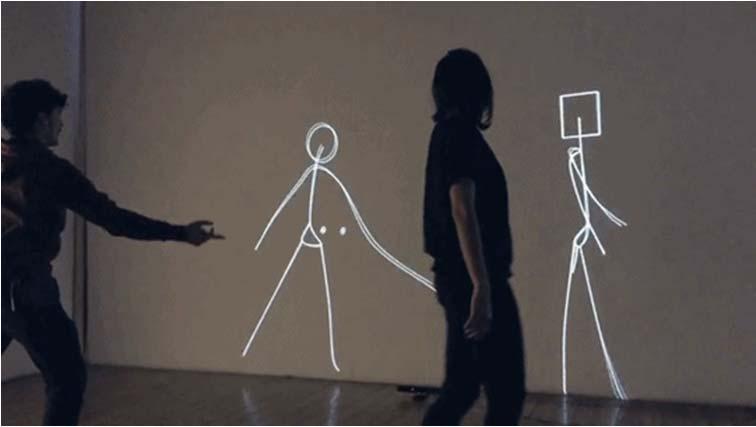
3 Outline What is video based motion management? What are the potential uses in radiation oncology? What commercial solutions exist? How to commission and perform ongoing QA? 5 Video Based Motion Management Radiation therapists can view the feed from closed circuit TV cameras Problem: Therapists cannot detect small shifts and are multitasking Question: Can modern technology improve the utility and accuracy of video based motion management?. 6 3


4 Video Based Motion Detection In 2010, Microsoft introduced the Xbox Kinect system that enables users to control and interact with their console without the need for a game controller through a natural user interface using gestures. It projects a known pattern in near-infrared light. An IR camera observes the scene and the system determines 3D surfaces

5 9 10 5


6 How Does X-Box Kinect Work? 11 How does it work? The infrared projector is an IR laser that passes through a diffraction grating that turns it into a set of IR dots. The relative geometry between the IR projector and IR camera is known as well as the projected dot pattern. A 3D image can be recreated using triangulation. 12 6
 has great value but has limitations: Only a snapshot.")
7 Potential Role for Video Based Motion Management Radiation Oncology Highly conformal delivery techniques (e.g. IMRT and VMAT) require very accurate patient positioning. Cone beam CT (CBCT) has great value but has limitations: Only a snapshot. Does not allow monitoring during treatment FOV is limited Not helpful for correction of the patient s posture Not useful for respiratory gating 13 How can video based monitoring be used in radiation oncology? Positioning: 3D image assists in accurate patient positioning. Intrafraction monitoring: Automated detection of changes in patient positioning. Respiratory gating: A tool for reducing the impact of respiratory induced tumor motion. Key feature is that video based techniques are non-invasive and non-ionizing. 14 7


8 Commercial Video Based Motion Management Solutions C-RAD Catalyst VisionRT AlignRT humediq Identify 15 C-RAD SCI Ceiling mounted projector-camera combination We have 3 Catalysts systems in our clinic. Each Catalyst is interfaced with the Elekta Response gating interface and provides SGRT. 16 8

9 C-RAD Catalyst: Under the Hood Projector-Camera Pair A sequence of patterns (structured light) is projected onto the surface measured. Software compares the projected and captured patterns to identify the coordinates of each pixel on the captured image. Courtesy of C-RAD 17 Non-rigid registration: Courtesy of C-Rad 18 9

10 C-RAD Catalyst at SCI 19 Catalyst Modules Positioning: Patient Setup 3D patient surface used to assist in initial setup Monitoring: Intra-fraction Patient Motion Monitoring Live image compared to reference image throughout the treatment session Out-of-tolerance shifts will trigger audio/visual alarm and can be setup to automatically pause the beam Gating: Tracking Patient s Breathing Signal can be used for gated delivery 20 10


11 Patient Setup The optical system scans the patient surface and compares it with the reference surface obtained from either the planning CT or BVS setup position. Automated couch shifts can be made using connectivity between the Catalyst and the linac. A color map is projected onto the patient s skin to indicate areas where the match is poor and to guide the needed changes in positioning. 21 Positioning Reference image Surface from CT Live image Patient on the couch Matched Images Shift determine System provides shifts in: lat, long, vert, pitch, roll, and yaw Courtesy of Malin Kügele 22 11


12 Color Map Courtesy of Malin Kügele


13 Patient Positioning Patient s arms not in the correct position. Breast position in longitudinal direction not within tolerance. Courtesy of Malin Kügele 25 Surface Map Imaging for Positioning Reduces risk of making an incorrect shift Provides extra check for a missed bolus Good check of arm position and breast board angle for breast patients Necessitates exposed skin in the treatment area Courtesy of Malin Kügele 26 13
 When motion threshold is exceeded it can trigger audio/visual alarm or automatically pause delivery")
14 Limitations Deep targets (lack of correlation with surface) Surfaces that move significantly (abdomen) Shadows 27 Intrafraction Patient for Motion Monitoring Provides real-time motion monitoring No additional radiation dose Millimeter sensitivity (much better than CCTV) When motion threshold is exceeded it can trigger audio/visual alarm or automatically pause delivery 28 14


15 Respiratory Gating A red gating point can be placed at any position on the patient. Point size / radius is adjustable. A secondary point can be placed on the patient for additional information. Amplitude given in absolute numbers. Courtesy of Malin Kügele 29 Respiratory Gating: C Rad Catalyst 30 15


16 Respiratory Gating The patient breathes as instructed: Audio Video Patient is coached to breathe with chest and not abdomen. Courtesy of Malin Kügele 31 Gating Workflow Example Breath hold : Audio-Visual Feedback Patient View Respiration Target Respiration Signal Exhale Phase (treatment beam off) Baseline Inhale Phase (treatment beam on) 32 16


17 Gated Beam Delivery Free Breathing Gating Breath hold gating Exception Gating 33 Catalyst Respiration Tracking/Gating: Visualized on A Phantom 34 17


18 Clinical Case with Catalyst: Tracking of Free Breathing SBRT 35 Gating: Catalyst vs. RPM Primary gating spot Breathing amplitude for Catalyst and RPM Amplitude [mm] Cata RPM Time [s] From: Sofie Ceberg och Charlotte Thornberg 18

19 Catalyst Tracking on an ABC Gated Left Breast Patient Delivery window coincides with ABC breath hold. 37 C-RAD Sentinel Device utilizing a laser scanning galvanometer and a video camera installed in a single ceiling mounted package. The scanning laser passes over the patient's surface and the apparent deformation of the laser line is used to render the surface. The surface position is compared to a reference surface to determine the patient s offset. Sentinel is installed in our CT simulation room. Used for 4DCT. Used to train patients on respiratory gating/dibh and used in acquired gated CT images
20 The C RAD Sentinel system acquires surface map images using which of the following: 20% 1. Scanning laser 20% 20% 20% 20% 2. Infrared cameras 3. Video cameras with projected light pattern 4. Implanted beacons with an RF panel 5. Spirometry 10 Answer: Scanning laser Reference: S. Pallotta, L. Marrazzo, M. Ceroti, P. Silli, and M. Bucciolini, A phantom evaluation of Sentinel, a commercial laser/camera surface imaging system for patient setup verification in radiotherapy, Med. Phys. 39, (2012). 20
 to dynamically capture and reconstruct maps of the patient s surface.")
 It is part of the package provided with the Varian Edge")
21 VisionRT AlignRT Uses stereoscopic video images in combination with patterns it projects onto the patient (structured light) to dynamically capture and reconstruct maps of the patient s surface. 41 VisionRT AlignRT More than 600 AlignRT systems have been ordered worldwide Installed in 37 of top 50 cancer centers (as ranked by US News) It is part of the package provided with the Varian Edge model 42 21


22 SURFACE GUIDED RADIOTHERAPY : PRINCIPLES Uses near infrared light Projects a pseudo random speckle pattern Matches all possible corresponding points Produces 3D triangulated mesh Maps out approx. 20,000 3D points of the patients anatomy 2016 Vision RT Ltd USER INTERFACE: EXAMPLE 2016 Vision RT Ltd 22


23 ALIGNRT: ACCESSORIES* Head adjuster Allows corrections of pitch, yaw and roll head rotations Adjustments to within a tenth of a degree Bolts on with quick release clamps to standard couch tops Real-time coach Contactless breath hold coaching: for DIBH 2016 Vision RT Ltd FRAMELESS SRS: BENEFITS Patient comfort: Open mask = reduced claustrophobia, non-invasive Safety: Continuous monitoring of any motion under the mask in real time Accuracy: <1mm Efficiency: Frameless SRS on conventional Linac Application: Can specify tracking region to avoid mask and increase accuracy 2016 Vision RT Ltd 23
")


24 CLINICAL APPLICATIONS: BREAST PATIENT SETUP DEEP INSPIRATION BREATH-HOLD (DIBH) IMRT Chest wall Multiple ROIs Real time tracking of breast during Deep Inspiration Breath Hold 2016 Vision RT Ltd humediq Identify Uses a medical RFID reader to automatically recognize each patient and the accessories while they re being positioned on the treatment couch. Two stereoscopic cameras use TOF imaging to create 3D surface map


![screen].](/docs-images/72/67393903/images/25-7.jpg "Patient self-positioning")
25 RFID to Check Accessories and Patient ID Headrest Jonathan Vacuum Cushion Jonathan KneeSTEP 8B 49 humediq Identify General Functionality Dual camera system o Time of flight and optical HD) RFID system for tracking patient ID and accessories. Patient virtual surface from the simulation is shown [on the wall screen and ceiling screen]. Patient self-positioning guidance. For gating, a gating marker is placed on patient surface


26 Patient s arms are down 26

27 Patient is shifted to the right Patient is shifted to the left 27
28 Features of the surface map imaging include all of the following except: 20% 1. Images are 3D in nature 20% 2. Uses radiofrequency tracking to track implanted markers 20% 3. Imaging is non-ionizing and non-invasive 20% 4. The patient s posture can be visualized 20% 5. Provides real-time feedback and can be used to monitor motion 10 Features of the surface map imaging include all of the following except: Answer: radiofrequency tracking is used by the Calypso system. Reference: Quality assurance for nonradiographic radiotherapy localization and positioning systems: report of Task Group 147, Med Phys, 39 (2012), pp

29 Considerations in Picking a Video Based Motion Monitoring System Determine your clinical need: What disease sites will be treated the most often? How will it be used: positioning, motion-management, and/or respiratory gating? Talk to current customers. Find customers with a similar configuration (Linac, EMR, TPS) Cost 57 Acceptance Testing, Commissioning, and QA of Your Video Based Monitoring System 58 29
30 Acceptance Testing (TG-147) Demonstrates that the equipment is meeting the specifications in the purchase contract between the facility and the vendor. Vendor must demonstrate localization consistent with the recommendations of TG-142, within 2mm for conventional delivery and 1mm for SRS/SBRT. Test connectivity between systems (e.g. EMR, TPS, treatment couch). 59 Commissioning and QA Start with your planned clinical utilization to establish QA procedures in conjunction with TG-147 recommendations. What disease sites will be treated the most often? Will it be used for positioning, motionmanagement, and/or respiratory gating? What tolerances will be needed during positioning and during motion monitoring? Prioritize your QA in terms of clinical need Courtesy of Jonathan Rogers 60 30
31 Commissioning and QA 61 Commissioning: Integration of Peripheral Equipment Check data transfer from simulation, treatment planning, and the record-and-verify system in different orientations (head first, feet first, prone, supine, etc.). Check communication Auto loading of patient information by surface mapping software Automated couch repositioning Automated beam interruption and impact on delivery accuracy The localization field of view should be measured and documented


32 Commissioning: Spatial Reproducibility and Drift Stability testing should be performed during the commissioning of a localization system by monitoring and recording a test pattern for at least 90 minutes or until sufficient stability is achieved. Safeguards or procedures should be developed to prevent clinical use during the camera warm up time. Reproducibility can be measured by the same device for an additional 60 minutes after establishing stability. Test should be done annually or after equipment changes or per manufacturer specifications
33 Commissioning: Spatial Reproducibility and Drift 3D displacement (mm) Time after power Up (min) 65 Commissioning: Static Localization Accuracy Localization displacement accuracy should be checked over a range that covers all clinical shifts to be expected for the system. Recommendation is that that at least 10cm shifts from isocenter or between two isocenters should be checked in all directions. Overall system accuracy should be measured using an end-to-end target test. This is typically performed using a phantom that has embedded radiographic markers visible on both CT and kv or MV imaging

34 Accuracy of Shifts Deviation (cm) Shift (cm) Lat (cm) Long (cm) Vert (cm) Maximum measured deviation 2mm Maximum measured rotation was 1 67 Map out your workflow and establish an end-to-end test that incorporates the complete process: Courtesy of Jonathan Rogers 68 34

35 Create a test that mimics the clinical use case (E2E): Courtesy of Jonathan Rogers 69 End-to-end Test Hidden Target Test Phantom marked with exterior visual cues for initial setup. A treatment planning CT is performed. Scans are sent to TPS where an isocenter can be defined relative to the location of the hidden target. At the linac, the phantom is positioned arbitrarily relative to the isocenter and surface map imaging is used restore the hidden target to the isocenter or its planned location relative to the isocenter. Imaging is performed to identify target localization accuracy relative to the machine s radiation isocenter

36 Commissioning: Dynamic Localization Accuracy If you are performing real-time patient tracking, testing should be performed with a 4D phantom. The accuracy during tracking is the difference between the phantom s programmed location and the measured location via surface map imaging. For example, you can program the phantom to move 1cm and stay at each location for 5 seconds. 71 Dynamic Localization Accuracy Test 72 36
37 CIRS Phantom Amplitude (mm) Catalyst Period (ms) Amplitude (mm) Period (ms) Maximum difference for amplitudes and periods are 0.54 mm and 260 ms respectively 73 Commissioning: Dynamic Localization Accuracy (2) Tests should be performed to evaluate any delay (latency) between the time a patient moves and when the localization system identifies the motion. Best performed using a motion phantom. Additionally use the motion phantom to test the beamhold mechanism that is employed when the patient moves beyond the specified tolerance. Delivered dose should be within 2% of radiation delivery without gating


38 Daily QA The agreement between the localization system isocenter and the treatment isocenter should be evaluated on the days that the system is used. The daily QA phantom should be recognized by the localization system as being within 2mm of the isocenter when it is positioned based upon the lasers. Motion tracking should be tested by moving the phantom a known amount (e.g. 5cm) and checking that the localization system indicates the correct shift within 2mm. 75 Daily QA Phantoms C-RAD Daily Check Device Align top of device to isocenter and side marks to lasers Device is scanned 5 times Provides system drift from previous daily QA and total drift since last isocenter adjustment AlignRT Calibration Plate Process identifies positions, orientations and optical properties of all camera sensors and lenses (for triangulation process) 38

 Surface can be imaged Gafchromic, pinpoint chamber, and/or OSL inserts Capable of full end-to-end testing Hidden target testing verifies radiation isocenter Courtesy of")
39 Monthly QA A localization accuracy test which relates the localization system directly to the treatment isocenter should be performed using films or portal imaging devices to verify alignment. Localization accuracy should be within 2mm of isocenter for standard fractionations and 1mm for SRS/SBRT. Evaluate tracking accuracy of the system. 77 QA Equipment Isocenter calibration phantom Used for correlating the MV isocenter to the surface imaging isocenter No dosimetric capabilities Any imaging SRS phantom (like MaxHD) Surface can be imaged Gafchromic, pinpoint chamber, and/or OSL inserts Capable of full end-to-end testing Hidden target testing verifies radiation isocenter Courtesy of Jonathan Rogers 78 39


40 Align RT QA Phantoms Calibration Phantom* Calibration to MV isocenter Allows verification of kv, AlignRT and laser isocenters Allows high accuracy tracking of surface at all couch rotations 79 Annual QA Camera stability Compare actual and predicted shifts over a range of distances. 4D/motion phantom to test accuracy of beam gating. Test data transfer for at least two patient/device configurations (head first supine, head first prone, etc.) A full end-to-end test from CT through treatment 80 40


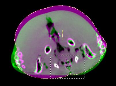
41 CBCT to Verify Surface Mapping Set-up to Breast Isocenter Frozen Turkey: Breast Isocenter Defrosted Turkey: Breast Isocenter 81 CBCT to Verify Surface Mapping Set up to Paraspinal Isocenter Frozen Turkey: Paraspinal Isocenter Defrosted Turkey: Paraspinal Isocenter 82 41
42 Near-Spine Isocenter Near-Surface Isocenter Frozen Turkey <2mm shifts in all directions <2mm shifts in all directions Defrosted Turkey ~10mm shifts <2mm shifts in all directions SGRT maintains its accuracy when the treatment isocenter is closer to the detected surface even when the body shaped is deformed. Surface mapping is less accurate for treatment sites deep inside the body when body deformation occurs. 83 According to Task Group 147, which of the following tests should be performed daily on non-radiographic radiotherapy localization and positioning systems (e.g. surface map imaging systems)? 20% 1. Positioning accuracy 20% 20% 20% 20% 2. End-to-end 3. Camera stability 4. Static localization 5. Winston-Lutz 10 42
43 According to Task Group 147, which of the following tests should be performed daily on non-radiographic radiotherapy localization and positioning systems (e.g. surface map imaging systems)? Answer: Static localization Reference: Quality assurance for nonradiographic radiotherapy localization and positioning systems: report of Task Group 147, Med Phys, 39 (2012), pp Gating Performance Test A MatriXX ion-chamber array mounted on a QUASAR Programmable Respiratory Motion Platform 6MV, 100MU, 10x10cm 2 field to a stationary MatriXX The same field was then delivered with the MatriXX platform programmed to move between -2 and +2 cm longitudinally. Gating signal was provided by Catalyst tracking a verticalmoving chest plate on QUASAR Gating windows were set between 10% and 100% at 10% increments


44 Quasar Respiratory Motion Platform 87 Results: Dose Distribution, Line Profile, Gamma Map 88 44
45 Results: Line Profile Comparison Stationary Phantom 100% Beam On 50% Gating Window 30% Gating Window 10% Gating Window 80 Dose (cgy) Position (cm) 89 Results: Gating Window vs. Gamma Rates and Delivery Time Time of Delivery (s) Gamma Passing Rate (%) Gating Window (%) 90 45
46 Conclusions SGRT is gaining wider acceptance and has the potential to significantly reduce the frequency of positioning errors in radiation oncology. It facilitates monitoring the patient s motion during the treatment and can be used for respiratory gating. TG147 provides guidance for commissioning and QA
Initial Clinical Experience with 3D Surface Image Guidance
 Initial Clinical Experience with 3D Surface Image Guidance Amanda Havnen-Smith, Ph.D. Minneapolis Radiation Oncology Ridges Radiation Therapy Center Burnsville, MN April 20 th, 2012 Non-funded research
7/31/2011. Learning Objective. Video Positioning. 3D Surface Imaging by VisionRT
 CLINICAL COMMISSIONING AND ACCEPTANCE TESTING OF A 3D SURFACE MATCHING SYSTEM Hania Al-Hallaq, Ph.D. Assistant Professor Radiation Oncology The University of Chicago Learning Objective Describe acceptance
Thank-You Members of TG147 TG 147: QA for nonradiographic
 Thank-You Members of TG147 TG 147: QA for nonradiographic localization and positioning systems Twyla Willoughby, M.S. Medical Physicist Clinical AAPM Meeting March 2013 Department of Radiation Oncology
Optical Guidance. Sanford L. Meeks. July 22, 2010
 Optical Guidance Sanford L. Meeks July 22, 2010 Optical Tracking Optical tracking is a means of determining in real-time the position of a patient relative to the treatment unit. Markerbased systems track
1. Learn to incorporate QA for surface imaging
 Hania Al-Hallaq, Ph.D. Assistant Professor Radiation Oncology The University of Chicago ***No disclosures*** 1. Learn to incorporate QA for surface imaging into current QA procedures for IGRT. 2. Understand
8/3/2016. Image Guidance Technologies. Introduction. Outline
 8/3/26 Session: Image Guidance Technologies and Management Strategies Image Guidance Technologies Jenghwa Chang, Ph.D.,2 Department of Radiation Medicine, Northwell Health 2 Hofstra Northwell School of
8/4/2016. Emerging Linac based SRS/SBRT Technologies with Modulated Arc Delivery. Disclosure. Introduction: Treatment delivery techniques
 Emerging Linac based SRS/SBRT Technologies with Modulated Arc Delivery Lei Ren, Ph.D. Duke University Medical Center 2016 AAPM 58 th annual meeting, Educational Course, Therapy Track Disclosure I have
Feasibility study of performing IGRT system daily QA using a commercial QA device
 JOURNAL OF APPLIED CLINICAL MEDICAL PHYSICS, VOLUME 12, NUMBER 3, summer 2011 Feasibility study of performing IGRT system daily QA using a commercial QA device Jean L.Peng, 1 Darren Kahler, 2 Jonathan
Commissioning and quality assurance of Calypso four dimensional target localization system in linear accelerator facility
 Technical Note Commissioning and quality assurance of Calypso four dimensional target localization system in linear accelerator facility K. R. Muralidhar, Krishna Komanduri, Birendra Kumar Rout, K. K.
Image Guidance and Beam Level Imaging in Digital Linacs
 Image Guidance and Beam Level Imaging in Digital Linacs Ruijiang Li, Ph.D. Department of Radiation Oncology Stanford University School of Medicine 2014 AAPM Therapy Educational Course Disclosure Research
Laser Systems for Patient Alignment
 Laser Systems for Patient Alignment GALAXY Patient TOPOGRAPHY Laser System Page: 2 Date: 28 March 2009 The GALAXY System NEW METHOD FOR HIGHER ALIGNMENT ACCURACY. Patient Positioning System for Linac Simulator
Commissioning and quality assurance of Calypso four dimensional target localization system in linear accelerator facility
 0 0 0 0 JMP R Commissioning and quality assurance of Calypso four dimensional target localization system in linear accelerator facility K. R. Muralidhar, Krishna Komanduri, Birendra Kumar Rout, K. K. D.
Performance and characteristics of an IR localizing system for radiation therapy
 JOURNAL OF APPLIED CLINICAL MEDICAL PHYSICS, VOLUME 7, NUMBER 2, SPRING 2006 Performance and characteristics of an IR localizing system for radiation therapy Yulia Lyatskaya, 1 Hsiao-Ming Lu, 2 and Lee
Image Quality Assessment and Quality Assurance of Advanced Imaging Systems for IGRT. AAPM Penn-Ohio Chapter Sep 25, 2015 Soyoung Lee, PhD
 Image Quality Assessment and Quality Assurance of Advanced Imaging Systems for IGRT AAPM Penn-Ohio Chapter Sep 25, 2015 Soyoung Lee, PhD 1 Outline q Introduction q Imaging performances in 4D-CBCT Image
8/2/2017. PerFRACTION from Sun Nuclear Automated Transit Dosimetry for In-Vivo QA
 PerFRACTION from Sun Nuclear Automated Transit Dosimetry for In-Vivo QA Jennifer R. Clark, M.S., DABR Conflict of Interest Statement: I am an employee of Sun Nuclear Corporation Outline: Platform overview
New Technology in Radiation Oncology. James E. Gaiser, Ph.D. DABR Physics and Computer Planning Charlotte, NC
 New Technology in Radiation Oncology James E. Gaiser, Ph.D. DABR Physics and Computer Planning Charlotte, NC Technology s s everywhere From the imaging chain To the planning system To the linac To QA..it..it
Tomotherapy Physics. Machine Twinning and Quality Assurance. Emilie Soisson, MS
 Tomotherapy Physics Machine Twinning and Quality Assurance Emilie Soisson, MS Tomotherapy at UW- Madison Treating for nearly 5 years Up to ~45 patients a day on 2 tomo units Units twinned to facilitate
Tumor motion during liver SBRT
 Tumor motion during liver SBRT - projects at Aarhus University Hospital - Per Poulsen, Esben Worm, Walther Fledelius, Morten Høyer Aarhus University Hospital, Denmark SBRT: Stereotactic Body Radiation
URGENT IMPORTANT FIELD SAFETY NOTIFICATION
 Subject: Incorrect Movement of the Treatment Table Product: MOSAIQ Scope: Sites affected will be those: 1. Running MOSAIQ and, 2. Treating on linear accelerators with the RATM license Notification Released:
IMRT and VMAT Patient Specific QA Using 2D and 3D Detector Arrays
 IMRT and VMAT Patient Specific QA Using 2D and 3D Detector Arrays Sotiri Stathakis Outline Why IMRT/VMAT QA AAPM TG218 UPDATE Tolerance Limits and Methodologies for IMRT Verification QA Common sources
Automated Quality Assurance for Image-Guided Radiation Therapy
 JOURNAL OF APPLIED CLINICAL MEDICAL PHYSICS, VOLUME 10, NUMBER 1, WINTER 2009 Automated Quality Assurance for Image-Guided Radiation Therapy Eduard Schreibmann, a Eric Elder, Tim Fox Department of Radiation
Raising the Bar in IMRT QA
 MapCHECK 2TM Raising the Bar in IMRT QA The leader in quick and precise measurement of modulated radiotherapy beams Benefits Proven solution for film-less rotational delivery and IMRT QA - More than 1500
Volumetric Modulated Arc Therapy - Clinical Implementation. Outline. Acknowledgement. History of VMAT. IMAT Basics of IMAT
 Volumetric Modulated Arc Therapy - Clinical Implementation Daliang Cao, PhD, DABR Swedish Cancer Institute, Seattle, WA Acknowledgement David M. Shepard, Ph.D. Muhammad K. N. Afghan, Ph.D. Fan Chen, Ph.D.
Position accuracy analysis of the stereotactic reference defined by the CBCT on Leksell Gamma Knife Icon
 Position accuracy analysis of the stereotactic reference defined by the CBCT on Leksell Gamma Knife Icon WHITE PAPER Introduction An image guidance system based on Cone Beam CT (CBCT) is included in Leksell
Comparison of Two Phantoms for End-to-End SRS/SBRT Testing. Vikren Sarkar, Ph.D. Associate Professor University of Utah Huntsman Cancer Center
 Comparison of Two Phantoms for End-to-End SRS/SBRT Testing Vikren Sarkar, Ph.D. Associate Professor University of Utah Huntsman Cancer Center Disclosure Travel and Speaker Honorarium from Sun Nuclear Corp.
State-of-the-Art IGRT
 in partnership with State-of-the-Art IGRT Exploring the Potential of High-Precision Dose Delivery and Real-Time Knowledge of the Target Volume Location Antje-Christin Knopf IOP Medical Physics Group Scientific
Artefakt-resistente Bewegungsschätzung für die bewegungskompensierte CT
 Artefakt-resistente Bewegungsschätzung für die bewegungskompensierte CT Marcus Brehm 1,2, Thorsten Heußer 1, Pascal Paysan 3, Markus Oehlhafen 3, and Marc Kachelrieß 1,2 1 German Cancer Research Center
VALIDATION OF DIR. Raj Varadhan, PhD, DABMP Minneapolis Radiation Oncology
 VALIDATION OF DIR Raj Varadhan, PhD, DABMP Minneapolis Radiation Oncology Overview Basics: Registration Framework, Theory Discuss Validation techniques Using Synthetic CT data & Phantoms What metrics to
A novel-integrated quality assurance phantom for radiographic and nonradiographic radiotherapy localization and positioning systems
 A novel-integrated quality assurance phantom for radiographic and nonradiographic radiotherapy localization and positioning systems Amy S. Yu a) Tyler L. Fowler, and Piotr Dubrowski Department of Radiation
DAILY LINAC QA BEAM QA
 BEAM QA DAILY LINAC QA The QA BeamChecker Plus allows for fast, reliable, and uncomplicated daily QA of Varian, Elekta, Siemens, and Accuray Treatment Machines. The QA BeamChecker Plus is specifically
Virtual Phantoms for IGRT QA
 TM Virtual Phantoms for IGRT QA Why ImSimQA? ImSimQA was developed to overcome the limitations of physical phantoms for testing modern medical imaging and radiation therapy software systems, when there
THE WIRELESS PHANTOM PERFORM ACCURATE PATIENT QA IN LESS TIME THAN EVER!
 THE WIRELESS PHANTOM PERFORM ACCURATE PATIENT QA IN LESS TIME THAN EVER! Confidence in complex treatments Modern radiation therapy uses complex plans with techniques such as IMRT, VMAT and Tomotherapy.
Real Time Tumor Motion Tracking with CyberKnife
 Real Time Tumor Motion Tracking with CyberKnife Martina Descovich, Ph.D University of California San Francisco July 16, 2015 Learning objectives Review the principles of real-time tumor motion tracking
Design and performance characteristics of a Cone Beam CT system for Leksell Gamma Knife Icon
 Design and performance characteristics of a Cone Beam CT system for Leksell Gamma Knife Icon WHITE PAPER Introduction Introducing an image guidance system based on Cone Beam CT (CBCT) and a mask immobilization
IHE Radiation Oncology Technical Framework Supplement. Treatment Delivery Plan Content (TDPC) Rev. 1.1 Trial Implementation
 Rev. 1.1 Trial Implementation") Integrating the Healthcare Enterprise 5 IHE Radiation Oncology Technical Framework Supplement 10 Treatment Delivery Plan Content 15 Rev. 1.1 Trial Implementation 20 Date: November 16, 2016 Author: IHE
Integrating the Healthcare Enterprise 5 IHE Radiation Oncology Technical Framework Supplement 10 Treatment Delivery Plan Content 15 Rev. 1.1 Trial Implementation 20 Date: November 16, 2016 Author: IHE
7/31/ D Cone-Beam CT: Developments and Applications. Disclosure. Outline. I have received research funding from NIH and Varian Medical System.
 4D Cone-Beam CT: Developments and Applications Lei Ren, PhD, DABR Department of Radiation Oncology Duke University Medical Center Disclosure I have received research funding from NIH and Varian Medical
Using a research real-time control interface to go beyond dynamic MLC tracking
 in partnership with Using a research real-time control interface to go beyond dynamic MLC tracking Dr. Simeon Nill Joint Department of Physics at The Institute of Cancer Research and the Royal Marsden
DEVELOPING AND IMPLEMENTING A HIGH PRECISION SETUP SYSTEM
 DEVELOPING AND IMPLEMENTING A HIGH PRECISION SETUP SYSTEM By LEE-CHENG PENG A DISSERTATION PRESENTED TO THE GRADUATE SCHOOL OF THE UNIVERSITY OF FLORIDA IN PARTIAL FULFILLMENT OF THE REQUIREMENTS FOR THE
Retrospective Spiral Respiratory Correlated Imaging with Varian RPM
 Retrospective Spiral Respiratory Correlated Imaging with Varian RPM This is a Quick Step Guide for Retrospective Spiral Respiratory Correlated Imaging (4D CT) using the Varian RPM device v1.7 with the
ICARO Vienna April Implementing 3D conformal radiotherapy and IMRT in clinical practice: Recommendations of IAEA- TECDOC-1588
 ICARO Vienna April 27-29 2009 Implementing 3D conformal radiotherapy and IMRT in clinical practice: Recommendations of IAEA- TECDOC-1588 M. Saiful Huq, Ph.D., Professor and Director, Dept. of Radiation
MR-guided radiotherapy: Vision, status and research at the UMC Utrecht. Dipl. Ing. Dr. Markus Glitzner
 MR-guided radiotherapy: Vision, status and research at the UMC Utrecht Dipl. Ing. Dr. Markus Glitzner About myself Training Medizintechnik TU Graz PhD UMC Utrecht Clinical work Software implementation
Version 5.6. Quick Start Guide
 WWW..COM Version 5.6 Quick Start Guide STANDARD IMAGING, INC. 3120 Deming Way Middleton, WI 53562-1461 Jul / 2018 2018 Standard Imaging, Inc. TEL 800.261.4446 TEL 608.831.0025 FAX 608.831.2202 DOC # 80714-05
Brilliance CT Big Bore.
 1 2 2 There are two methods of RCCT acquisition in widespread clinical use: cine axial and helical. In RCCT with cine axial acquisition, repeat CT images are taken each couch position while recording respiration.
ADVANCING CANCER TREATMENT
 3 ADVANCING CANCER TREATMENT SUPPORTING CLINICS WORLDWIDE RaySearch is advancing cancer treatment through pioneering software. We believe software has un limited potential, and that it is now the driving
Improvement and Evaluation of a Time-of-Flight-based Patient Positioning System
 Improvement and Evaluation of a Time-of-Flight-based Patient Positioning System Simon Placht, Christian Schaller, Michael Balda, André Adelt, Christian Ulrich, Joachim Hornegger Pattern Recognition Lab,
IAEA-TECDOC-1583 Commissioning of Radiotherapy Treatment Planning Systems: Testing for Typical External Beam Treatment Techniques
 IAEA-TECDOC-1583 Commissioning of Radiotherapy Treatment Planning Systems: Testing for Typical External Beam Treatment Techniques Report of the Coordinated Research Project (CRP) on Development of Procedures
ADVANCING CANCER TREATMENT
 The RayPlan treatment planning system makes proven, innovative RayStation technology accessible to clinics that need a cost-effective and streamlined solution. Fast, efficient and straightforward to use,
Investigation Of The Microsoft Kinect V2 Sensor As A Multi-Purpose Device For A Radiation Oncology Clinic
 Wayne State University Wayne State University Dissertations 1-1-2017 Investigation Of The Microsoft Kinect V2 Sensor As A Multi-Purpose Device For A Radiation Oncology Clinic Evan Asher Silverstein Wayne
TomoTherapy Related Projects. An image guidance alternative on Tomo Low dose MVCT reconstruction Patient Quality Assurance using Sinogram
 TomoTherapy Related Projects An image guidance alternative on Tomo Low dose MVCT reconstruction Patient Quality Assurance using Sinogram Development of A Novel Image Guidance Alternative for Patient Localization
IMSURE QA SOFTWARE FAST, PRECISE QA SOFTWARE
 QA SOFTWARE FAST, PRECISE Software for accurate and independent verification of monitor units, dose, and overall validity of standard, IMRT, VMAT, SRS and brachytherapy plans no film, no phantoms, no linac
Facility Questionnaire PART I (General Information for 3DCRT and IMRT)
") Facility Questionnaire PART I (General Information for 3DCRT and IMRT) The following items are required before you can enter cases on any RTOG protocol that requires data submission to the Image-Guided
Facility Questionnaire PART I (General Information for 3DCRT and IMRT) The following items are required before you can enter cases on any RTOG protocol that requires data submission to the Image-Guided
MapCHECK 2 & 3DVH. The Gold Standard for 2D Arrays
 MapCHECK 2 & 3DVH The Gold Standard for 2D Arrays Your Most Valuable QA and Dosimetry Tools THE GOLD STANDARD FOR 2D ARRAYS The MapCHECK 2 is the world s most selected independent 2D measurement array.
Acknowledgments. Ping Xia, Ph.D., UCSF. Pam Akazawa, CMD, UCSF. Cynthia Chuang, Ph.D., UCSF
 Page 1 Quality Assurance of IMRT Delivery Systems - Siemens Lynn J. Verhey, Ph.D. Professor and Vice-Chair UCSF Dept. of Radiation Oncology AAPM 22 Annual Meeting, Montreal Acknowledgments Ping Xia, Ph.D.,
GafChromic Protocol Multi-Channel Film Dosimetry + Gamma Map Analysis
 GafChromic Protocol Multi-Channel Film Dosimetry + Gamma Map Analysis Micke A. Ashland Inc. Advanced Materials Ashland proprietary patented technology GafChromic Film for Dose Measurement Radiotherapy
MapCHECK 2 & 3DVH The Gold Standard for 2D Arrays
 MapCHECK 2 & 3DVH The Gold Standard for 2D Arrays Your Most Valuable QA and Dosimetry Tools THE GOLD STANDARD FOR 2D ARRAYS The MapCHECK 2 is the world s most selected independent 2D measurement array.
Real-time monitoring of linac performance using RT plans and logfiles
 Real-time monitoring of linac performance using RT plans and logfiles NCCAAPM Wisconsin Dells 7 April 2017 Mark Wiesmeyer, PhD, DABR Technical Product Manager Technical talk with insights and results based
Simple quality assurance method of dynamic tumor tracking with the gimbaled linac system using a light field
 JOURNAL OF APPLIED CLINICAL MEDICAL PHYSICS, VOLUME 17, NUMBER 5, 2016 Simple quality assurance method of dynamic tumor tracking with the gimbaled linac system using a light field Hideharu Miura, 1a Shuichi
Overview of Proposed TG-132 Recommendations
 Overview of Proposed TG-132 Recommendations Kristy K Brock, Ph.D., DABR Associate Professor Department of Radiation Oncology, University of Michigan Chair, AAPM TG 132: Image Registration and Fusion Conflict
ph fax Deming Way Middleton WI USA
 www.standardimaging.com 800-261-4446. ph 608-831-0025. fax 608-831-2202 3120 Deming Way Middleton WI 53562-1461 USA 2007 Standard Imaging, Inc. 1239-21 (03.07) beam qa LINAC AND TOMOTHERAPY QA The NEW
3DVH : SUN NUCLEAR On The Accuracy Of The corporation Planned Dose Perturbation Algorithm Your Most Valuable QA and Dosimetry Tools *Patent Pending
 3DVH : On The Accuracy Of The Planned Dose Perturbation Algorithm SUN NUCLEAR corporation Your Most Valuable QA and Dosimetry Tools *Patent Pending introduction State-of-the-art IMRT QA of static gantry
Use of TrueBeam developer mode for imaging QA
 JOURNAL OF APPLIED CLINICAL MEDICAL PHYSICS, VOLUME 16, NUMBER 4, 2015 Use of TrueBeam developer mode for imaging QA Gilmer Valdes, 1,2a Olivier Morin, 1 Yanisley Valenciaga, 3 Niel Kirby, 1 Jean Pouliot,
radiotherapy Andrew Godley, Ergun Ahunbay, Cheng Peng, and X. Allen Li NCAAPM Spring Meeting 2010 Madison, WI
 GPU-Accelerated autosegmentation for adaptive radiotherapy Andrew Godley, Ergun Ahunbay, Cheng Peng, and X. Allen Li agodley@mcw.edu NCAAPM Spring Meeting 2010 Madison, WI Overview Motivation Adaptive
Estimating 3D Respiratory Motion from Orbiting Views
 Estimating 3D Respiratory Motion from Orbiting Views Rongping Zeng, Jeffrey A. Fessler, James M. Balter The University of Michigan Oct. 2005 Funding provided by NIH Grant P01 CA59827 Motivation Free-breathing
MRI-LINAC Dynamic Phantom
 MRI-LINAC Dynamic Phantom Model 008M IMAGE ACQUISITION TREATMENT PLANNING DOSE DELIVERY Strict QA procedures for the imaging, planning and delivery of radiotherapy using respiratory management devices
The team. Disclosures. Ultrasound Guidance During Radiation Delivery: Confronting the Treatment Interference Challenge.
 Ultrasound Guidance During Radiation Delivery: Confronting the Treatment Interference Challenge Dimitre Hristov Radiation Oncology Stanford University The team Renhui Gong 1 Magdalena Bazalova-Carter 1
Clinical implementation of photon beam flatness measurements to verify beam quality
 JOURNAL OF APPLIED CLINICAL MEDICAL PHYSICS, VOLUME 16, NUMBER 6, 2015 Clinical implementation of photon beam flatness measurements to verify beam quality Simon Goodall, a Nicholas Harding, Jake Simpson,
Automated Image Analysis Software for Quality Assurance of a Radiotherapy CT Simulator
 Automated Image Analysis Software for Quality Assurance of a Radiotherapy CT Simulator Andrew J Reilly Imaging Physicist Oncology Physics Edinburgh Cancer Centre Western General Hospital EDINBURGH EH4
Disclosure. Outline. Acknowledgments. Ping Xia, Ph.D., Page 1. LINAC and MLC QA for IMRT. Received research support from Siemens Medical Solutions
 LINAC and MLC QA for IMRT Ping Xia, Ph.D., Department of Radiation Oncology Disclosure Received research support from Siemens Medical Solutions University of California San Francisco Therapy Series (SAM)
IMRT site-specific procedure: Prostate (CHHiP)
") IMRT site-specific procedure: Prostate (CHHiP) Scope: To provide site specific instructions for the planning of CHHIP IMRT patients Responsibilities: Radiotherapy Physicists, HPC Registered Therapy Radiographers
IMRT site-specific procedure: Prostate (CHHiP) Scope: To provide site specific instructions for the planning of CHHIP IMRT patients Responsibilities: Radiotherapy Physicists, HPC Registered Therapy Radiographers
Use of Monte Carlo modelling in radiotherapy linac design. David Roberts, PhD Senior Physicist Elekta
 Use of Monte Carlo modelling in radiotherapy linac design David Roberts, PhD Senior Physicist Elekta Contents Overview of Elekta What we do Where we use Monte Carlo Codes and resources Example : Agility
FAST, precise. qa software
 qa software FAST, precise Software for accurate and independent verification of monitor units, dose, and overall validity of standard, IMRT, rotational or brachytherapy plans no film, no phantoms, no linac
Quick Reference Datasheet For All RIT113 Packages
 Quick Reference Datasheet For All RIT113 Packages For Rotational Therapies, IMRT & TG142 Highlights and selected product information only. A complete TG142 brochure is available. For more information on
Calibration of Video Cameras to the Coordinate System of a Radiation Therapy Treatment Machine
 Calibration of Video Cameras to the Coordinate System of a Radiation Therapy Treatment Machine Scott W. Hadley, L. Scott Johnson, and Charles A. Pelizzari University of Chicago The Department of Radiation
TG-148 overview. Introduction. System Overview. System Overview. QA for Helical Tomotherapy: Report of the AAPM Task Group 148. Conflict of Interest:
 QA or Helical Tomotherapy: Report o the AAPM Tas Group 148 Members: Conlict o Interest: r. John Balog owns TomoTherapy stoc. Katja Langen (Co-chair) Nio Papaniolaou (Co-chair) Walter Grant Richard Crilly
8/3/2016. MR-guided RT: Commissioning and Quality Control. Topics for MR-guided RT system commissioning & QC
 MR-guided RT: Commissioning and Quality Control T. Stanescu, PhD, MCCPM Medical Physicist, Princess Margaret Cancer Centre, UHN Assistant Professor, Radiation Oncology, University of Toronto Affiliated
An Automated Image-based Method for Multi-Leaf Collimator Positioning Verification in Intensity Modulated Radiation Therapy
 An Automated Image-based Method for Multi-Leaf Collimator Positioning Verification in Intensity Modulated Radiation Therapy Chenyang Xu 1, Siemens Corporate Research, Inc., Princeton, NJ, USA Xiaolei Huang,
Robust Real-Time 3D Respiratory Motion Detection Using Time-of-Flight Cameras
 Robust Real-Time 3D Respiratory Motion Detection Using Time-of-Flight Cameras Jochen Penne Chair of Pattern Recognition, Friedrich-Alexander-University Erlangen-Nuremberg Martensstr.3 91058 Erlangen, Germany
Delta 4 PT. True Volumetric Pre-treatment Verification. The difference is clear
 Delta 4 PT True Volumetric Pre-treatment Verification The difference is clear ScandiDos innovators in advanced volumetric dosimetry solutions ScandiDos ScandiDos is the innovative company that introduced
TG 132: Use of Image Registration and Fusion in RT
 TG 132: Use of Image Registration and Fusion in RT Kristy K Brock, PhD, DABR, FAAPM Associate Professor Department of Radiation Oncology, University of Michigan Chair, AAPM TG 132: Image Registration and
Advanced Radiotherapy
 Advanced Radiotherapy IBA Concept for IMRT Verification Dr. Lutz Müller 1 IMRT Verification BIS BIS ADAS Fluence Model Delivered fluence Target Volume Constraints Inverse Backprojection Fluence Map Leaf
Digital Tomosynthesis for Target Localization
 Digital Tomosynthesis for Target Localization Fang-Fang Yin, Devon Godfrey, Lei Ren Jacqueline Maurer, Jackie Q-L Wu Duke University Medical Center Acknowledgements Duke Radiation Oncology faculty and
An investigation of temporal resolution parameters in cine-mode four-dimensional computed tomography acquisition
 JOURNAL OF APPLIED CLINICAL MEDICAL PHYSICS, VOLUME 9, NUMBER 4, FALL 2008 An investigation of temporal resolution parameters in cine-mode four-dimensional computed tomography acquisition Yildirim D. Mutaf
REAL-TIME ADAPTIVITY IN HEAD-AND-NECK AND LUNG CANCER RADIOTHERAPY IN A GPU ENVIRONMENT
 REAL-TIME ADAPTIVITY IN HEAD-AND-NECK AND LUNG CANCER RADIOTHERAPY IN A GPU ENVIRONMENT Anand P Santhanam Assistant Professor, Department of Radiation Oncology OUTLINE Adaptive radiotherapy for head and
Qalitätssicherung an Multileafkollimatoren. Dr. Lutz Müller Würzburg jan 2004
 Qalitätssicherung an Multileafkollimatoren Dr. Lutz Müller Würzburg jan 2004 IMRT Verification - present Target Volume Constraints Inverse Planning Algorithm Fluence Map Leaf Sequencer Leaf & Gantry sequence
Evaluation of AutoQA Lite TM Image Quality Measurement Software
 Evaluation of AutoQA Lite TM Image Quality Measurement Software Andrew J Reilly Imaging Physicist Oncology Physics Edinburgh Cancer Centre Western General Hospital EDINBURGH EH4 2XU Phone: 0131 537 1161
The IORT Treatment Planning System. radiance. GMV, 2012 Property of GMV All rights reserved
 The IORT Treatment Planning System radiance Property of GMV All rights reserved WHY RADIANCE? JUSTIFICATION Property of GMV All rights reserved ADVANTAGES OF IORT PRECISION: RT guided by direct vision.
Monaco VMAT. The Next Generation in IMRT/VMAT Planning. Paulo Mathias Customer Support TPS Application
 Monaco VMAT The Next Generation in IMRT/VMAT Planning Paulo Mathias Customer Support TPS Application 11.05.2011 Background What is Monaco? Advanced IMRT/VMAT treatment planning system from Elekta Software
Novel methods and tools for MR Commissioning and Quality Control
 Novel methods and tools for MR Commissioning and Quality Control T. Stanescu, PhD, MCCPM Medical Physicist, RMP, Princess Margaret Cancer Centre Assistant Professor, Radiation Oncology, University of Toronto
A Study of Motion Tracking Accuracy of Robotic Radiosurgery Using a Novel CCD Camera Based End-to-end Test System
 A Study of Motion Tracking Accuracy of Robotic Radiosurgery Using a Novel CCD Camera Based End-to-end Test System Lei Wang 1, Brett Nelson 2,Youming Yang 1 1. Department of Radiation Oncology, Stanford
Introduction. Quality Assurance for Image- Guided Radiation Therapy. Justification for IGRT. Image-Guided Radiation Therapy
 Introduction Quality Assurance for Image- Guided Radiation Therapy Jean-Pierre Bissonnette, Ph.D., MCCPM Princess Margaret Hospital, Toronto, Canada IGRT What is it? Rationale Equipment Quality Assurance
Verification of Gated Radiation Therapy: Dosimetric Impact of Residual Motion
 Original Article PROGRESS in MEDICAL PHYSICS Vol. 25, No. 3, September, 2014 http://dx.doi.org/10.14316/pmp.2014.25.3.128 Verification of Gated Radiation Therapy: Dosimetric Impact of Residual Motion Inhwan
Data. ModuLeaf Mini Multileaf Collimator Precision Beam Shaping for Advanced Radiotherapy
 Data ModuLeaf Mini Multileaf Collimator Precision Beam Shaping for Advanced Radiotherapy ModuLeaf Mini Multileaf Collimator Precision Beam Shaping for Advanced Radiotherapy The ModuLeaf Mini Multileaf
Code of practice: Create a verification plan for OCTAVIUS Detector 729 in Philips Pinnacle³
 Technical Note D655.208.03/00 Code of practice: Create a verification plan for OCTAVIUS Detector 729 in There are basically three ways to carry out verification of IMRT fluences. For all options, it is
Image Co-Registration II: TG132 Quality Assurance for Image Registration. Image Co-Registration II: TG132 Quality Assurance for Image Registration
 Image Co-Registration II: TG132 Quality Assurance for Image Registration Preliminary Recommendations from TG 132* Kristy Brock, Sasa Mutic, Todd McNutt, Hua Li, and Marc Kessler *Recommendations are NOT
The MSKCC Approach to IMRT. Outline
 The MSKCC Approach to IMRT Spiridon V. Spirou, PhD Department of Medical Physics Memorial Sloan-Kettering Cancer Center New York, NY Outline Optimization Field splitting Delivery Independent verification
Quality assurance tests for the Gamma Knife Icon image guidance system
 Received: 11 May 2018 Revised: 19 June 2018 Accepted: 27 June 2018 DOI: 10.1002/acm2.12417 RADIATION ONCOLOGY PHYSICS Quality assurance tests for the Gamma Knife Icon image guidance system Ismail AlDahlawi
Table of Contents. About AQUA
 User Manual About AQUA Table of Contents 1 About AQUA... 6 1.1 Radiation Therapy... 6 1.1.1 Workflow manager... 6 1.1.2 Reports... 7 1.1.3 Tests... 7 1.1.4 Many Ways to Work... 7 1.2 Technology... 7 1.3
ROBUST OPTIMIZATION THE END OF PTV AND THE BEGINNING OF SMART DOSE CLOUD. Moe Siddiqui, April 08, 2017
 ROBUST OPTIMIZATION THE END OF PTV AND THE BEGINNING OF SMART DOSE CLOUD Moe Siddiqui, April 08, 2017 Agenda Background IRCU 50 - Disclaimer - Uncertainties Robust optimization Use Cases Lung Robust 4D
Patient Set-ups and Tumor Localizations
 Patient Set-ups and Tumor Localizations Amy S. Harrison Patient Positioning Prior to starting any localization or simulation procedure patients need to be positioned and immobilized Patients disease location
Photon beam dose distributions in 2D
 Photon beam dose distributions in 2D Sastry Vedam PhD DABR Introduction to Medical Physics III: Therapy Spring 2014 Acknowledgments! Narayan Sahoo PhD! Richard G Lane (Late) PhD 1 Overview! Evaluation