Medical Imaging Introduction
|
|
|
- Philip Atkins
- 6 years ago
- Views:
Transcription
1 Medical Imaging Introduction Jan Kybic February 16, 2010

2 Medical imaging: a collaborative paradigm picture from Atam P. Dhawan: Medical Imaging
3 From physiology to information processing (what we should learn) Understanding Imaging Medium Physics of Imaging Imaging Instrumentation Data Acquisition Methods Image Processing and Analysis
4 From physiology to information processing (what we should learn) Understanding Imaging Medium Physics of Imaging Imaging Instrumentation Data Acquisition Methods Image Processing and Analysis
5 From physiology to information processing (what we should learn) Understanding Imaging Medium Physics of Imaging Imaging Instrumentation Data Acquisition Methods Image Processing and Analysis
6 From physiology to information processing (what we should learn) Understanding Imaging Medium Physics of Imaging Imaging Instrumentation Data Acquisition Methods Image Processing and Analysis
7 From physiology to information processing (what we should learn) Understanding Imaging Medium Physics of Imaging Imaging Instrumentation Data Acquisition Methods Image Processing and Analysis
8 Medical imaging pipeline
9 Medical imaging pipeline Physical property Raw data Improved data Images/volumes Quantitative/qualitative inf. patient/subject Imaging device Preprocessing Reconstruction Interpretation physician Decision
10 Medical imaging pipeline Physical property Raw data Improved data Images/volumes Quantitative/qualitative inf. patient/subject Imaging device Preprocessing Reconstruction Interpretation physician Decision
11 Medical imaging pipeline Physical property Raw data Improved data Images/volumes Quantitative/qualitative inf. Statistical processing patient/subject Imaging device Preprocessing Reconstruction Interpretation physician Decision
12 Imaging device Physical property: density, absorbance, conductivity,... Imaging device Raw data Examples: CT, X-rays, MRI, ultrasound,...
13 Preprocessing A priori knowledge Raw data Preprocessing Preprocessed, cleaned, enhanced data Examples: Noise suppression, contract enhancement, intensity equalization, outlier elimination, bias compensation, time/space filtering,...
14 Reconstruction Physics of the acquisition device Preprocessed raw data Reconstruction Physically meaningfull images, volumes, sequences Inverse problem Examples: Tomographic reconstruction MRI, ultrasound...
15 Interpretation A priori knowledge Images, volumes, sequences Interpretation High-level processing Quantitative & qualitative inf. size, changes, metabolic inf. Examples: Registration, segmentation, classification, tracking,...
16 Modalities
17 Classification of medical modalities What information do we need? anatomical, physiological, functional
18 Classification of medical modalities What information do we need? anatomical, physiological, functional What is the energy source? external, internal, combined
19 Classification of medical modalities What information do we need? anatomical, physiological, functional What is the energy source? external, internal, combined How good is the image? resolution, noise
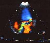
20 Classification of medical modalities What information do we need? anatomical, physiological, functional What is the energy source? external, internal, combined How good is the image? resolution, noise How much does it cost?
21 Energy source
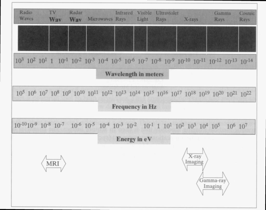
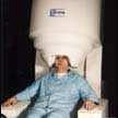
22 Electromagnetic spectrum
23 Examples


24 Microscopy Optical microscopy since 17th century; Jensen, van Leeuwenhoek, Galilei,...
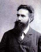
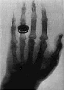
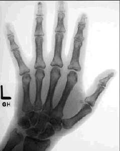
25 X-rays / Radiography 1895, W. Röntgen B. Röntgen hand modern hand

26 Computed tomography The machine:
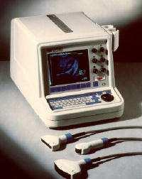
27 Ultrasound Equipment
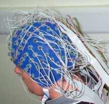
28 MEG and EEG Magnetoencephalography Electroencephalography
 Larmor,")

29 Magnetic resonance imaging (MRI) Larmor, Bloch, Purcell, Rabi, Lauterbur,... around 1973.
30 ... and many others SPECT (Single Photon Emission Computed Tomography) PET (Positron Emission Tomography) gamma camera termography...
31 OK, here ends the preview. Questions?... otherwise let us start with image processing
FIELD PARADIGM FOR 3D MEDICAL IMAGING: Safer, More Accurate, and Faster SPECT/PET, MRI, and MEG
 FIELD PARADIGM FOR 3D MEDICAL IMAGING: Safer, More Accurate, and Faster SPECT/PET, MRI, and MEG July 1, 2011 First, do no harm. --Medical Ethics (Hippocrates) Dr. Murali Subbarao, Ph. D. murali@fieldparadigm.com,
Medical Image Analysis
 Computer assisted Image Analysis VT04 29 april 2004 Medical Image Analysis Lecture 10 (part 1) Xavier Tizon Medical Image Processing Medical imaging modalities XRay,, CT Ultrasound MRI PET, SPECT Generic
MEDICAL IMAGE ANALYSIS
 SECOND EDITION MEDICAL IMAGE ANALYSIS ATAM P. DHAWAN g, A B IEEE Engineering in Medicine and Biology Society, Sponsor IEEE Press Series in Biomedical Engineering Metin Akay, Series Editor +IEEE IEEE PRESS
Medical Images Analysis and Processing
 Medical Images Analysis and Processing - 25642 Emad Course Introduction Course Information: Type: Graduated Credits: 3 Prerequisites: Digital Image Processing Course Introduction Reference(s): Insight
Reconstruction in CT and relation to other imaging modalities
 Reconstruction in CT and relation to other imaging modalities Jørgen Arendt Jensen November 1, 2017 Center for Fast Ultrasound Imaging, Build 349 Department of Electrical Engineering Center for Fast Ultrasound
Lecture 6: Medical imaging and image-guided interventions
 ME 328: Medical Robotics Winter 2019 Lecture 6: Medical imaging and image-guided interventions Allison Okamura Stanford University Updates Assignment 3 Due this Thursday, Jan. 31 Note that this assignment
Computational Medical Imaging Analysis
 Computational Medical Imaging Analysis Chapter 1: Introduction to Imaging Science Jun Zhang Laboratory for Computational Medical Imaging & Data Analysis Department of Computer Science University of Kentucky
CP467 Image Processing and Pattern Recognition
 CP467 Image Processing and Pattern Recognition Instructor: Hongbing Fan Introduction About DIP & PR About this course Lecture 1: an overview of DIP DIP&PR show What is Digital Image? We use digital image
Medical Imaging BMEN Spring 2016
 Name Medical Imaging BMEN 420-501 Spring 2016 Homework #4 and Nuclear Medicine Notes All questions are from the introductory Powerpoint (based on Chapter 7) and text Medical Imaging Signals and Systems,
BME I5000: Biomedical Imaging
 1 Lucas Parra, CCNY BME I5000: Biomedical Imaging Lecture 4 Computed Tomography Lucas C. Parra, parra@ccny.cuny.edu some slides inspired by lecture notes of Andreas H. Hilscher at Columbia University.
Computer Graphics. - Volume Rendering - Philipp Slusallek
 Computer Graphics - Volume Rendering - Philipp Slusallek Overview Motivation Volume Representation Indirect Volume Rendering Volume Classification Direct Volume Rendering Applications: Bioinformatics Image
Digital Image Processing
 Digital Image Processing SPECIAL TOPICS CT IMAGES Hamid R. Rabiee Fall 2015 What is an image? 2 Are images only about visual concepts? We ve already seen that there are other kinds of image. In this lecture
Image Restoration. Diffusion Denoising Deconvolution Super-resolution Tomographic Reconstruction
 Image Restoration Image Restoration Diffusion Denoising Deconvolution Super-resolution Tomographic Reconstruction Diffusion Term Consider only the regularization term E-L equation: (Laplace equation) Steepest
UNIVERSITY OF SOUTHAMPTON
 UNIVERSITY OF SOUTHAMPTON PHYS2007W1 SEMESTER 2 EXAMINATION 2014-2015 MEDICAL PHYSICS Duration: 120 MINS (2 hours) This paper contains 10 questions. Answer all questions in Section A and only two questions
Image Acquisition Systems
 Image Acquisition Systems Goals and Terminology Conventional Radiography Axial Tomography Computer Axial Tomography (CAT) Magnetic Resonance Imaging (MRI) PET, SPECT Ultrasound Microscopy Imaging ITCS
Index. aliasing artifacts and noise in CT images, 200 measurement of projection data, nondiffracting
 Index Algebraic equations solution by Kaczmarz method, 278 Algebraic reconstruction techniques, 283-84 sequential, 289, 293 simultaneous, 285-92 Algebraic techniques reconstruction algorithms, 275-96 Algorithms
DICOM. Supplement 188 Multi-Energy CT Imaging. DICOM Working Group 21 Computed Tomography
 DICOM Supplement 188 Multi-Energy CT Imaging DICOM Working Group 21 Computed Tomography Rationale Short introduction of Multi Energy (ME) s Overview: Imaging techniques, including scanning, reconstruction,
Emission Computed Tomography Notes
 Noll (24) ECT Notes: Page 1 Emission Computed Tomography Notes Introduction Emission computed tomography (ECT) is the CT applied to nuclear medicine. There are two varieties of ECT: 1. SPECT single-photon
Tomography. Forward projectionsp θ (r) are known as a Radon transform. Objective: reverse this process to form the original image
 are known as a Radon transform. Objective: reverse this process to form the original image") C. A. Bouman: Digital Image Processing - January 9, 217 1 Tomography Many medical imaging systems can only measure projections through an object with density f(x,y). Projections must be collected at every
C. A. Bouman: Digital Image Processing - January 9, 217 1 Tomography Many medical imaging systems can only measure projections through an object with density f(x,y). Projections must be collected at every
Reconstruction in CT and relation to other imaging modalities
 Reconstruction in CT and relation to other imaging modalities Jørgen Arendt Jensen November 16, 2015 Center for Fast Ultrasound Imaging, Build 349 Department of Electrical Engineering Center for Fast Ultrasound
Biomedical Imaging and Image Analysis
 Biomedical Imaging and Image Analysis Lecture in Medical Informatics Course Ewert Bengtsson Professor of computerized image analysis Centrum för bildanalys The theme Images are of central importance in
Computational Medical Imaging Analysis
 Computational Medical Imaging Analysis Chapter 2: Image Acquisition Systems Jun Zhang Laboratory for Computational Medical Imaging & Data Analysis Department of Computer Science University of Kentucky
DICOM DIRECTOR. User Manual for. DICOM Director Gateway. DICOM Director Team Version 1.0
 DICOM DIRECTOR User Manual for DICOM Director Gateway Version 1.0 DICOM Director Team support@dicomdirector.com Table of Contents How to Read the Manual... 3 Symbols used in the Manuals... 3 Notes... 3
CS 5630/6630 Scientific Visualization. Volume Rendering I: Overview
 CS 5630/6630 Scientific Visualization Volume Rendering I: Overview Motivation Isosurfacing is limited It is binary A hard, distinct boundary is not always appropriate Slice Isosurface Volume Rendering
Constructing System Matrices for SPECT Simulations and Reconstructions
 Constructing System Matrices for SPECT Simulations and Reconstructions Nirantha Balagopal April 28th, 2017 M.S. Report The University of Arizona College of Optical Sciences 1 Acknowledgement I would like
VISUALIZATION AND EXPLORATION OF TIME-VARYING AND DIFFUSION TENSOR MEDICAL IMAGE DATA SETS
 VISUALIZATION AND EXPLORATION OF TIME-VARYING AND DIFFUSION TENSOR MEDICAL IMAGE DATA SETS by Zhe Fang B.Sc. with Honours, Acadia University, 2003 a thesis submitted in partial fulfillment of the requirements
BME I5000: Biomedical Imaging
 BME I5000: Biomedical Imaging Lecture 1 Introduction Lucas C. Parra, parra@ccny.cuny.edu 1 Content Topics: Physics of medial imaging modalities (blue) Digital Image Processing (black) Schedule: 1. Introduction,
Introduction to Positron Emission Tomography
 Planar and SPECT Cameras Summary Introduction to Positron Emission Tomography, Ph.D. Nuclear Medicine Basic Science Lectures srbowen@uw.edu System components: Collimator Detector Electronics Collimator
RADIOMICS: potential role in the clinics and challenges
 27 giugno 2018 Dipartimento di Fisica Università degli Studi di Milano RADIOMICS: potential role in the clinics and challenges Dr. Francesca Botta Medical Physicist Istituto Europeo di Oncologia (Milano)
SonicDICOM PACS. Install Manual. https://sonicdicom.com/ December 14, JIUN Corporation. All rights reserved.
 SonicDICOM PACS Install Manual December 14, 2016 https://sonicdicom.com/ 2016 JIUN Corporation. All rights reserved. Contents Install Manual... 3 Version history... 3 1. Overview of software... 3 1.1 DICOM
11/18/ CPT Preauthorization Groupings Effective January 1, Computerized Tomography (CT) Abdomen 6. CPT Description SEGR CT01
 Abdomen 6. CPT Description SEGR CT01") Computerized Tomography (CT) 6 & 101 5 Upper Extremity 11 Lower Extremity 12 Head 3 Orbit 1 Sinus 2 Neck 4 7 Cervical Spine 8 Thoracic Spine 9 Lumbar Spine 10 Colon 13 CPT Description SEGR 74150 74160
Computerized Tomography (CT) 6 & 101 5 Upper Extremity 11 Lower Extremity 12 Head 3 Orbit 1 Sinus 2 Neck 4 7 Cervical Spine 8 Thoracic Spine 9 Lumbar Spine 10 Colon 13 CPT Description SEGR 74150 74160
Utilizing Salient Region Features for 3D Multi-Modality Medical Image Registration
 Utilizing Salient Region Features for 3D Multi-Modality Medical Image Registration Dieter Hahn 1, Gabriele Wolz 2, Yiyong Sun 3, Frank Sauer 3, Joachim Hornegger 1, Torsten Kuwert 2 and Chenyang Xu 3 1
Computed tomography (Item No.: P )
") Computed tomography (Item No.: P2550100) Curricular Relevance Area of Expertise: Biology Education Level: University Topic: Modern Imaging Methods Subtopic: X-ray Imaging Experiment: Computed tomography
Computed tomography (Item No.: P2550100) Curricular Relevance Area of Expertise: Biology Education Level: University Topic: Modern Imaging Methods Subtopic: X-ray Imaging Experiment: Computed tomography
Medical Image Registration
 Medical Image Registration Submitted by NAREN BALRAJ SINGH SB ID# 105299299 Introduction Medical images are increasingly being used within healthcare for diagnosis, planning treatment, guiding treatment
Multimodal Image Fusion Of The Human Brain
 Multimodal Image Fusion Of The Human Brain Isis Lázaro(1), Jorge Marquez(1), Juan Ortiz(2), Fernando Barrios(2) isislazaro@gmail.com Centro de Ciencias Aplicadas y Desarrollo Tecnológico, UNAM Circuito
THE DICOM 2013 INTERNATIONAL CONFERENCE & SEMINAR. DICOM Fields of Use. Klaus Neuner. Brainlab AG. Software Project Manager Feldkirchen, Germany
 THE DICOM 2013 INTERNATIONAL CONFERENCE & SEMINAR March 14-16 Bangalore, India DICOM Fields of Use Klaus Neuner Brainlab AG Software Project Manager Feldkirchen, Germany Introduction This presentation
Computed tomography of simple objects. Related topics. Principle. Equipment TEP Beam hardening, artefacts, and algorithms
 Related topics Beam hardening, artefacts, and algorithms Principle The CT principle is demonstrated with the aid of simple objects. In the case of very simple targets, only a few images need to be taken
An Introduction to Images
 An Introduction to Images CS6640/BIOENG6640/ECE6532 Ross Whitaker, Tolga Tasdizen SCI Institute, School of Computing, Electrical and Computer Engineering University of Utah 1 What Is An Digital Image?
Joint CI-JAI advanced accelerator lecture series Imaging and detectors for medical physics Lecture 1: Medical imaging
 Joint CI-JAI advanced accelerator lecture series Imaging and detectors for medical physics Lecture 1: Medical imaging Dr Barbara Camanzi barbara.camanzi@stfc.ac.uk Course layout Day AM 09.30 11.00 PM 15.30
A New Application for Displaying and Fusing Multimodal Data Sets
 A New Application for Displaying and Fusing Multimodal Data Sets Karl G. Baum* ac, María Helguera a, Andrzej Krol b a Rochester Institute of Technology, 54 Lomb Memorial Drive, Rochester, NY, USA 14623-5604;
Digital Volume Correlation for Materials Characterization
 19 th World Conference on Non-Destructive Testing 2016 Digital Volume Correlation for Materials Characterization Enrico QUINTANA, Phillip REU, Edward JIMENEZ, Kyle THOMPSON, Sharlotte KRAMER Sandia National
Medical Imaging Projects
 NSF REU MedIX Summer 2006 Medical Imaging Projects Daniela Stan Raicu, PhD http://facweb.cs.depaul.edu/research draicu@cs.depaul.edu Outline Medical Informatics Imaging Modalities Computed Tomography Medical
Mathematical methods and simulations tools useful in medical radiation physics
 Mathematical methods and simulations tools useful in medical radiation physics Michael Ljungberg, professor Department of Medical Radiation Physics Lund University SE-221 85 Lund, Sweden Major topic 1:
Introduction to Medical Image Processing
 Introduction to Medical Image Processing Δ Essential environments of a medical imaging system Subject Image Analysis Energy Imaging System Images Image Processing Feature Images Image processing may be
SPECT reconstruction
 Regional Training Workshop Advanced Image Processing of SPECT Studies Tygerberg Hospital, 19-23 April 2004 SPECT reconstruction Martin Šámal Charles University Prague, Czech Republic samal@cesnet.cz Tomography
Assessment of 3D performance metrics. X-ray based Volumetric imaging systems: Fourier-based imaging metrics. The MTF in CT
 Assessment of 3D performance metrics D and 3D Metrics of Performance Towards Quality Index: Volumetric imaging systems X-ray based Volumetric imaging systems: CBCT/CT Tomosynthesis Samuel Richard and Ehsan
Computer Assisted Image Analysis TF 3p and MN1 5p Lecture 1, (GW 1, )
") Centre for Image Analysis Computer Assisted Image Analysis TF p and MN 5p Lecture, 422 (GW, 2.-2.4) 2.4) 2 Why put the image into a computer? A digital image of a rat. A magnification of the rat s nose.
Centre for Image Analysis Computer Assisted Image Analysis TF p and MN 5p Lecture, 422 (GW, 2.-2.4) 2.4) 2 Why put the image into a computer? A digital image of a rat. A magnification of the rat s nose.
Interpolation of 3D magnetic resonance data
 Interpolation of 3D magnetic resonance data J. Mikulka 1, E. Gescheidtova 2 and K. Bartusek 3 1, 2 Department of Theoretical and Experimental Electrical Engineering, Brno University of Technology, Kolejni
An educational tool for demonstrating the TOF-PET technique
 Nuclear Instruments and Methods in Physics Research A 471 (2001) 200 204 An educational tool for demonstrating the TOF-PET technique T.Bȧack a, *, J. Cederkȧall a, B. Cederwall a, A. Johnson a, A. Kerek
1 Algorithms for Control and Image Reconstruction
 This is Al Hero s and Jeff Fessler s section to the DARPA MOSAIC proposal. 1 Algorithms for Control and Image Reconstruction The algorithm development and analysis team, led by Profs. Fessler and Hero,
Update ASA 4.8. Expand your research potential with ASA 4.8. Highly advanced 3D display of single channel coherence
 Update ASA 4.8 Expand your research potential with ASA 4.8. The ASA 4.8 software has everything needed for a complete analysis of EEG / ERP and MEG data. From features like (pre)processing of data, co-registration
Reconstruction from Projections
 Reconstruction from Projections M.C. Villa Uriol Computational Imaging Lab email: cruz.villa@upf.edu web: http://www.cilab.upf.edu Based on SPECT reconstruction Martin Šámal Charles University Prague,
ATLAS BASED IMAGE RECONSTRUCTION FOR DIFFUSE OPTICAL IMAGING OF THE HUMAN BRAIN
 ATLAS BASED IMAGE RECONSTRUCTION FOR DIFFUSE OPTICAL IMAGING OF THE HUMAN BRAIN By XUE WU A thesis submitted to the University of Birmingham For the degree of DOCTOR OF PHILOSOPHY School of Computer Science
A Comparative Study between Two Hybrid Medical Image Compression Methods
 A Comparative Study between Two Hybrid Medical Image Compression Methods Clarissa Philana Shopia Azaria 1, and Irwan Prasetya Gunawan 2 Abstract This paper aims to compare two hybrid medical image compression
Biomedical Image Processing for Human Elbow
 Biomedical Image Processing for Human Elbow Akshay Vishnoi, Sharad Mehta, Arpan Gupta Department of Mechanical Engineering Graphic Era University Dehradun, India akshaygeu001@gmail.com, sharadm158@gmail.com
Introduction to Neuroimaging Janaina Mourao-Miranda
 Introduction to Neuroimaging Janaina Mourao-Miranda Neuroimaging techniques have changed the way neuroscientists address questions about functional anatomy, especially in relation to behavior and clinical
MURDOCH RESEARCH REPOSITORY
 MURDOCH RESEARCH REPOSITORY http://dx.doi.org/10.1109/tencon.2000.893677 Xie, H. and Fung, C.C. (2000) Enhancing the performance of a BSP model-based parallel volume renderer with a profile visualiser.
Medical Image Fusion using Rayleigh Contrast Limited Adaptive Histogram Equalization and Ant Colony Edge Method
 Medical Image Fusion using Rayleigh Contrast Limited Adaptive Histogram Equalization and Ant Colony Edge Method Ramandeep 1, Rajiv Kamboj 2 1 Student, M. Tech (ECE), Doon Valley Institute of Engineering
Image Registration. Prof. Dr. Lucas Ferrari de Oliveira UFPR Informatics Department
 Image Registration Prof. Dr. Lucas Ferrari de Oliveira UFPR Informatics Department Introduction Visualize objects inside the human body Advances in CS methods to diagnosis, treatment planning and medical
The OpenGATE Collaboration
 The OpenGATE Collaboration GATE developments and future releases GATE Users meeting, IEEE MIC 2015, San Diego Sébastien JAN CEA - IMIV sebastien.jan@cea.fr Outline Theranostics modeling Radiobiology Optics
Medical Computer Vision
 Vienna Computer Vision Meetup 27.01.2016 sponsored by Medical Computer Vision Markus Holzer markus@radiology-explorer.com www.radiology-explorer.com www.meduniwien.ac.at www.cir.meduniwien.ac.at Overview
Advanced Visual Medicine: Techniques for Visual Exploration & Analysis
 Advanced Visual Medicine: Techniques for Visual Exploration & Analysis Interactive Visualization of Multimodal Volume Data for Neurosurgical Planning Felix Ritter, MeVis Research Bremen Multimodal Neurosurgical
Computed Tomography for Industry Needs and Status Umesh Kumar
 Computed Tomography for Industry Needs and Status Umesh Kumar Bhabha Atomic Research Centre, Mumbai, INDIA Industrial Computed Tomography (ICT) Needs Why industrial tomography is required when many conventional
Carl Zeiss Meditec AG
 OPMI PENTERO 900 Software Releases 1.0, 1.1, 1.2 and 1.3 OPMI PENTERO 800 Software Releases 1.2 and 1.3 DICOM GE-30-1870-en Version 1.3 MMN: EN_30_200_0029I 1 Networking 4 1.1 Introduction 4 1.2 Implementation
PACS and DICOM. Einar Heiberg,
 PACS and DICOM Einar Heiberg, einar@heiberg.se Why bother? Almost ALL medical images are stored in DICOM format. All hospitals use PACS (Picture Archive and Communication Systems) If you ever will work
Introduction to Medical Image Analysis
 Introduction to Medical Image Analysis Rasmus R. Paulsen DTU Compute rapa@dtu.dk http://courses.compute.dtu.dk/02511 http://courses.compute.dtu.dk/02511 Plenty of slides adapted from Thomas Moeslunds lectures
Version 7 DICOM Conformance Statement. Document Version 3.02, June 2009
 Version 7 DICOM Conformance Statement Document Version 3.02, June 2009 1 Conformance Statement Overview The application described in this Conformance Statement VEPRO EMR Manager PACS is a collection of
Preparing for Part 2 of the ABR Diagnostic Physics Exam
 Preparing for Part 2 of the ABR Diagnostic Physics Exam Joseph Zambelli, Ph.D. Spectrum Health, Grand Rapids, MI Outline Exam Background Exam Preparation Suggested Resources Location, Time, and Format
InstaPACS / InstaRISPACS / InstaViewer (v Release) DICOM Conformance Statement. Meddiff Technologies Pvt. Ltd.
 DICOM Conformance Statement. Meddiff Technologies Pvt. Ltd.") InstaPACS / InstaRISPACS / InstaViewer (v3.0.22 Release) DICOM Conformance Statement Meddiff Technologies Pvt. Ltd. Table of Contents 1. Introduction... 3 1.1 Scope and Field of Application:... 3 1.2 References
InstaPACS / InstaRISPACS / InstaViewer (v3.0.22 Release) DICOM Conformance Statement Meddiff Technologies Pvt. Ltd. Table of Contents 1. Introduction... 3 1.1 Scope and Field of Application:... 3 1.2 References
Medical Imaging Modalities
 Image Science Introduction Medical Imaging Modalities Ho Kyung Kim hokyung@pusan.ac.kr Pusan National University Projection Radiography Routine diagnostic radiography Chest x rays, fluoroscopy, mammography,
Gengsheng Lawrence Zeng. Medical Image Reconstruction. A Conceptual Tutorial
 Gengsheng Lawrence Zeng Medical Image Reconstruction A Conceptual Tutorial Gengsheng Lawrence Zeng Medical Image Reconstruction A Conceptual Tutorial With 163 Figures Author Prof. Dr. Gengsheng Lawrence
Introduction to Emission Tomography
 Introduction to Emission Tomography Gamma Camera Planar Imaging Robert Miyaoka, PhD University of Washington Department of Radiology rmiyaoka@u.washington.edu Gamma Camera: - collimator - detector (crystal
Carl Zeiss Surgical GmbH
 OPMI Pentero MediLive MindStream Software Release 2.1 GE-30-1612-en Version 1.2 Copyright Carl Zeiss Surgical GmbH, Germany 2006 All rights reserved 1 Networking 4 1.1 Introduction 4 1.2 Implementation
Carl Zeiss Surgical GmbH
 OPMI Pentero MediLive MindStream Software Release 2.2 GE-30-1712-en Version 1.0 Copyright Carl Zeiss Surgical GmbH, Germany 2007 All rights reserved 1 Networking 4 1.1 Introduction 4 1.2 Implementation
S93-8 Page 1 TOMOGRAPHIC APPROACHES TO NONWOVENS STRUCTURE DEFINITION
 S93-8 Page 1 TOMOGRAPHIC APPROACHES TO NONWOVENS STRUCTURE DEFINITION PIs: T. Gilmore, H. Davis, Z. Mi, North Carolina State University Code: S93-8 Date of Report: 9/94 RELEVANCE TO NTC MISSION AND GOALS:
Limitations of Projection Radiography. Stereoscopic Breast Imaging. Limitations of Projection Radiography. 3-D Breast Imaging Methods
 Stereoscopic Breast Imaging Andrew D. A. Maidment, Ph.D. Chief, Physics Section Department of Radiology University of Pennsylvania Limitations of Projection Radiography Mammography is a projection imaging
ANALYSIS OF CT AND PET/SPECT IMAGES FOR DOSIMETRY CALCULATION
 2009 International Nuclear Atlantic Conference - INAC 2009 Rio de Janeiro,RJ, Brazil, September27 to October 2, 2009 ASSOCIAÇÃO BRASILEIRA DE ENERGIA NUCLEAR - ABEN ISBN: 978-85-99141-03-8 ANALYSIS OF
Carl Zeiss Meditec AG
 OPMI PENTERO 900 Software Releases 1.0 and 1.1 GE-30-1870-en Version 1.1 1 Networking 4 1.1 Introduction 4 1.2 Implementation Model 4 1.2.1 Application Data Flow Diagram 5 1.2.2 Functional Definitions
Computer Aided Diagnosis Based on Medical Image Processing and Artificial Intelligence Methods
 International Journal of Information and Computation Technology. ISSN 0974-2239 Volume 3, Number 9 (2013), pp. 887-892 International Research Publications House http://www. irphouse.com /ijict.htm Computer
CURRICULUM COMMITTEE MEETING Friday, March 18, :00 p.m. Student Life Center, Faculty Dining Room (Building 23, First Floor) AGENDA
 AGENDA") CURRICULUM COMMITTEE MEETING Friday, March 18, 2016-2:00 p.m. Student Life Center, Faculty Dining Room (Building 23, First Floor) I. Call to Order AGENDA II. Roll Call III. Minutes of meeting of January
CURRICULUM COMMITTEE MEETING Friday, March 18, 2016-2:00 p.m. Student Life Center, Faculty Dining Room (Building 23, First Floor) I. Call to Order AGENDA II. Roll Call III. Minutes of meeting of January
The University of Chicago. Center for EPR Imaging in Vivo Physiology. Image Registration. Boris Epel
 The University of Chicago Center for EPR Imaging in Vivo Physiology Image Registration Boris Epel Imaging Methods are Complimentary CT MRI EPRI High resolution anatomic images Quantitative Poor soft tissue
Corso di laurea in Fisica A.A Fisica Medica 5 SPECT, PET
 Corso di laurea in Fisica A.A. 2007-2008 Fisica Medica 5 SPECT, PET Step 1: Inject Patient with Radioactive Drug Drug is labeled with positron (β + ) emitting radionuclide. Drug localizes
MULTIMODAL MEDICAL IMAGE FUSION BASED ON HYBRID FUSION METHOD
 MULTIMODAL MEDICAL IMAGE FUSION BASED ON HYBRID FUSION METHOD Sinija.T.S MTECH, Department of computer science Mohandas College of Engineering Karthik.M Assistant professor in CSE Mohandas College of Engineering
Workshop on Quantitative SPECT and PET Brain Studies January, 2013 PUCRS, Porto Alegre, Brasil Corrections in SPECT and PET
 Workshop on Quantitative SPECT and PET Brain Studies 14-16 January, 2013 PUCRS, Porto Alegre, Brasil Corrections in SPECT and PET Físico João Alfredo Borges, Me. Corrections in SPECT and PET SPECT and
Abstract. 1. Introduction
 A New Automated Method for Three- Dimensional Registration of Medical Images* P. Kotsas, M. Strintzis, D.W. Piraino Department of Electrical and Computer Engineering, Aristotelian University, 54006 Thessaloniki,
Radiology. Marta Anguiano Millán. Departamento de Física Atómica, Molecular y Nuclear Facultad de Ciencias. Universidad de Granada
 Departamento de Física Atómica, Molecular y Nuclear Facultad de Ciencias. Universidad de Granada Overview Introduction Overview Introduction Tecniques of imaging in Overview Introduction Tecniques of imaging
Integrating patient-oriented data processing into the PREPaRe virtual hospital using XML technology
 Integrating patient-oriented data processing into the PREPaRe virtual hospital using XML technology René Tschirley, Kai Köchy, Steffen Märkle Dept. for Computer Science and Computer Assisted Medicine,
Noise Reduction Techniques in Medical Imaging Data-A Review
 Noise Reduction Techniques in Medical Imaging Data-A Review Ajay Somkuwar and Shruti Bhargava Abstract Noise is an inherent property of medical imaging, and it generally tends to reduce the image resolution
SPECT QA and QC. Bruce McBride St. Vincent s Hospital Sydney.
 SPECT QA and QC Bruce McBride St. Vincent s Hospital Sydney. SPECT QA and QC What is needed? Why? How often? Who says? QA and QC in Nuclear Medicine QA - collective term for all the efforts made to produce
Fields Multi-Modality Imaging and Modeling. problem by Shuo Li (GE)
") problem by Shuo Li (GE) body part model geometry material properties physiological properties organ different tissue properties scans multiple modalities (ct, mri, pet,...) inverse problem different tissue
problem by Shuo Li (GE) body part model geometry material properties physiological properties organ different tissue properties scans multiple modalities (ct, mri, pet,...) inverse problem different tissue
An Introduction to Images. CS6640/BIOENG6640 Ross Whitaker SCI Institute, School of Computing University of Utah
 An Introduction to Images CS6640/BIOENG6640 Ross Whitaker SCI Institute, School of Computing University of Utah Module 1: Goals Understand images as mappings Understand the difference continuous vs discrete
Performance Enhancement In Accuracy and Imaging Time of a Hand-Held Probe-Based Optical Imager
 Florida International University FIU Digital Commons FIU Electronic Theses and Dissertations University Graduate School 2-21-2011 Performance Enhancement In Accuracy and Imaging Time of a Hand-Held Probe-Based
Tomographic Reconstruction
 Tomographic Reconstruction 3D Image Processing Torsten Möller Reading Gonzales + Woods, Chapter 5.11 2 Overview Physics History Reconstruction basic idea Radon transform Fourier-Slice theorem (Parallel-beam)
DICOM CONFORMANCE STATEMENT FOR ZIOBASE 4.0
 DICOM CONFORMANCE STATEMENT FOR ZIOBASE 4.0 DICOM Conformance Statement, Ziobase 4.0 1 Copyright 2006-2012, Ziosoft, Inc. 0 DICOM CONFORMANCE STATEMENT OVERVIEW... 4 1 IMPLEMENTATION MODEL... 5 1.1 APPLICATION
Version 9 DICOM Conformance Statement. Version 3.05, September 2015
 Version 9 DICOM Conformance Statement Version 3.05, September 2015 1 Conformance Statement Overview The application described in this conformance statement, VEPRO EMR Manager PACS, is a collection of processes
... user-friendly explanations for HL7, SNOMED, DICOM, XML, BDT
 ... user-friendly explanations for HL7, SNOMED, DICOM, XML, BDT HL 7 (Health Level 7) The quality of modern software can be measured by its ability to integrate with other systems. For this reason, an
Neuroimaging and mathematical modelling Lesson 2: Voxel Based Morphometry
 Neuroimaging and mathematical modelling Lesson 2: Voxel Based Morphometry Nivedita Agarwal, MD Nivedita.agarwal@apss.tn.it Nivedita.agarwal@unitn.it Volume and surface morphometry Brain volume White matter
Diagnostic imaging techniques. Krasznai Zoltán. University of Debrecen Medical and Health Science Centre Department of Biophysics and Cell Biology
 Diagnostic imaging techniques Krasznai Zoltán University of Debrecen Medical and Health Science Centre Department of Biophysics and Cell Biology 1. Computer tomography (CT) 2. Gamma camera 3. Single Photon
High Performance GPU-Based Preprocessing for Time-of-Flight Imaging in Medical Applications
 High Performance GPU-Based Preprocessing for Time-of-Flight Imaging in Medical Applications Jakob Wasza 1, Sebastian Bauer 1, Joachim Hornegger 1,2 1 Pattern Recognition Lab, Friedrich-Alexander University
Medical Imaging and Virtual Medicine
 Medical Imaging and Virtual Medicine Lecture 1: Volume data, Sampling theorem, Artifacts, Filtering Dirk Bartz, Visual Computing for Medicine bartz@gris.uni-tuebingen.de University of Tübingen 1 University