Atlas Based Segmentation of the prostate in MR images
|
|
|
- Easter Lane
- 6 years ago
- Views:
Transcription
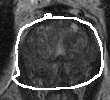
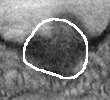
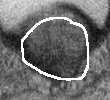
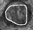
1 Atlas Based Segmentation of the prostate in MR images Albert Gubern-Merida and Robert Marti Universitat de Girona, Computer Vision and Robotics Group, Girona, Spain Abstract. The large variability and contrast differences between prostates make its segmentation difficult using traditional segmentation methods. In this paper we present an automatic method based on nonrigid registration of a set of prelabelled MR altas images. The algorithm consists of three stages. Firstly, the target image is nonrigidly registered with each atlas image, using mutual information as the similarity measure. Subsequently, the best registered image is selected comparing normalised mutual information measures after registration. Finally, the segmentation is obtained by deforming the corresponding atlas labelled image selected before using the transformation determined on the registration stage. The method is evaluated on 15 images using the Dice similarity coeficient and 95% Hausdorff distance, obtaining medians of 0.79 and 7.11 mm respectively. Key words: image segmentation, image registration, atlas matching, magnetic resonance imaging, prostate 1 Introduction In this work we have implemented an automatic segmentation method based on atlas matching that, in contrast to traditional segmentation techniques, allows to take the large variability and the contrast differences of the prostates into account. The atlas consists of a set of images with their respective labelled image manually provided by an expert. The segmentation method is based on the work of Klein et al. [1] but also introduces some modifications which are described in the Sec 2. The method is evaluated using 15 MRI T2 images (Sec. 3), following the guidelines of the prostate segmentation challenge held within the MICCAI 2009 conference. The aim of this work is to evaluate the atlas based segmentation methods using a common evaluation framework in order to be comparable to the other published approaches. The paper finally presents the discussion, conclusions and future directions. 2 Method The method consists of three stages: first a registration of all atlas images to the patient image that will be segmented, selection of atlas image that matches
2 2 Atlas Based Segmentation of the prostate in MR images best with the patient image, and finally the deformation of the corresponding atlas label image using the transformation obtained in the registration stage. Referring to the last two stages, due to the small number of images to test and the large variability among them, we have selected only one matching image so no combination of segmentations is used in contrast with the work presented by Klein et al. [1]. In the presented method, a set of N accurately labelled images, which serve as an atlas, are assumed to be available. The ith image in this atlas set is referred to as A i (x). The corresponding label image is called L i (x), a binary image where ones represent prostate tissue and zeros everything else. The new image to be segmented is denoted by P(x). The goal of the automatic segmentation method is to produce a label image ˆL P (x) that accurately defines the prostate of the patient. Ideally, this label image should be equal to a manual segmentation L P (x) created by an expert. 2.1 Registration In the registration stage, each atlas image A i is matched to the image P to be segmented. A coordinate transformation T i (x) is estimated that maximises the similarity of P and the deformed atlas A i T i. We use here mattes mutual information as a measure of similarity [2]. Figure 1 shows a schematic representation of this stage. Before registration, a rectangular region of interest the prostate where appears is extracted in the images in order to focus the registration process. In altas images, this region is delimited by a bounding box automatically obtained from its respective labelled image. The patient the region is selected manually that is the only step in the method that requires user intervention. Doing this extraction we have observed that even the effects of severe intensity inhomogeneity caused by magnetic field artifacts are minised, therefore bias field correction was not deemed to be needed. The registration process itself is performed in three steps. Firstly, global pose differences are compensated for by an affine registration algorithm. Subsequently, a nonrigid registration is performed, using a coordinate transformation that is parameterised by cubic B-Splines [3]. In this second step a small number of control grid points is defined to model global nonrigid deformations. Finally, a second nonrigid registration is performed in order to model highly local nonrigid deformations defining large number of control grid points. For the main test 5 and 10 control grid point values are the selected. The transformation that maximises the similarity measure is estimated by an iterative stochastic gradient descent optimisation. 2.2 Atlas selection In this stage, we need to select the registered atlas image which is more similar to the patient s image as shown in figure 2.
 taking into account for each pair of")
 = MI(A, B) H(A) (1) Fig.2. Second stage: selection of the image that best matches the patient image 2.")
3 Atlas Based Segmentation of the prostate in MR images 3 Fig.1. First stage: each atlas image is matched to the patient image The similarity measure used is a normalization of mattes mutual information (see equation 1) taking into account for each pair of registered images (A and B) the entropy of the fixed image H(A). The use of this metric has in consideration the differences of number of pixels between the registered images. The measure is computed on the extracted regions before registration stage. NMI(A, B) = MI(A, B) H(A) (1) Fig.2. Second stage: selection of the image that best matches the patient image 2.3 Label image deformation In the third stage, the label image corresponding to the chosen atlas image A i is deformed. Coordinate transformation T i obtained in the registration stage is applied L i T i in order to get the segmentation of the patient s image ˆL P (x). Figure 3 illustrates this stage.

4 4 Atlas Based Segmentation of the prostate in MR images Fig.3. Third stage: deformation of the labelled image 3 Results The proposed automatic segmentation method has been implemented using ITK libraries [4] and are evaluated using the set of 15 T2 images of the trainning data. Each image has been segmented building an atlas with the rest of the 14 MR images (following a leave-one-out scheme). Quantitative evaluation is done comparing the automatically generated segmentations with the manual segmentations provided by an expert. The measures to evaluate the segmentations are the Dice similarity coefficient (DSC) [5] and 95% Hausdorff distance (HD) [6]. Moreover, slices of MR images with their segmentation are shown providing a qualitative evaluation. The method has tested on a workstation with four Quad- Core AMD Opteron(tm) Processor 8378 and 128 GB RAM. The execution time to run each segmentation is about 60 minutes approximately. Figure 4.(a) compares the DSC and 4.(b) the HD values for each test volume. On one hand, having in mind it is generally accepted that a value of DSC > 0.7 represents excellent agreement [7], the median DSC value of 0.79 confirms the overall satisfactory results of the segmentations. On the other hand, the median HD value is 7.11 mm. We consider this value as acceptable, especially taking into account that the axial spacing in images are 5 mm and the average prostate volume is about 40 cm 3. Confirming and exemplifying the quantitative analysis above, figure 5 shows some slices of each segmentation with DSC values greater than 0.7 (13 out of 15). In most of images it is possible to observe that the contour correctly delimits the prostate in accordance with their respective DSC and HD values. Referring to unsatisfactory results with DSC < 0.7 (images 45 and 88), we have seen that the result in image 45 could be explained by the difficulty of finding a similar prostate in the atlas. This illustrates the fact that registration stage works correctly handling deformations up to a certain magnitude. On the other hand, the problem in image 88 seems to be explained by an incorrect registration result. In this case we have obtained a better segmentation (DSC = 0.76 and HD = 6.01 mm) using 15 control points in the last nonrigid registration step instead of 10. However, using these parameters for all the segmentations,

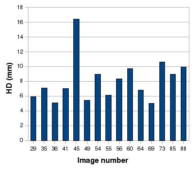
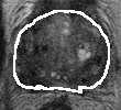
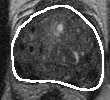
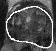
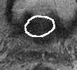
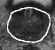
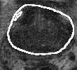
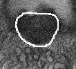
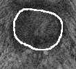
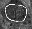
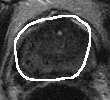
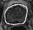
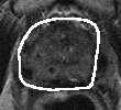
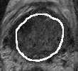
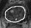
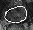
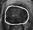
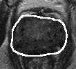
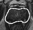
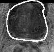
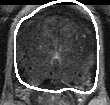
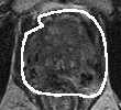
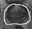
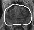
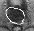
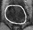
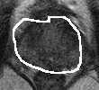
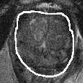
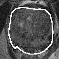
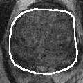
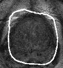
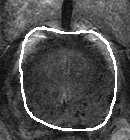
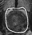
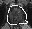
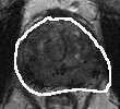
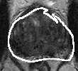
5 Atlas Based Segmentation of the prostate in MR images 5 (a) DSC (b) HD Fig.4. (a) DSC and (b) HD values for each test volume Fig.5. Example slices of each prostate segmentation with DSC > 0.7
6 6 Atlas Based Segmentation of the prostate in MR images the median DSC and HD values are slightly lower (DSC = 0.76 and HD = 8.48 mm). It should be noted that even with those registration parameters, the segmentation of image 45 was still unsatisfactory confirming the need of a larger atlas database as mentioned before. 4 Conclusions In this work an automatic prostate segmentation method of MR images has been investigated. The method is based on matching a set of manually segmented images taking into account the large variability and contrast differences of the prostates. Evaluation has been performed using the 15 T2 MR images of the training data. A leave one out methodology has been used for each segmentation: one image is segmented building an atlas with the rest of the 14 volumes. The obtained segmentations are compared to a manual segmentation using the Dice similarity coefficient and 95% Hausdorff distance (HD) achieving acceptable results with medians of 0.79 and 7.11 mm respectively. The visual results shown also confirm the DSC and HD values observing a good delineation of the prostate region. We have noted some important points concerning the behaviour of the prostate segmentation method based on atlas matching. Firstly, atlas needs to contain a representative variation of images. If the patient image to be segmented has a similar one into atlas, the segmentation result will be better because the registration stage will be able to handle the differences between prostates more effective and easily. For this reason, adding more images into the atlas and the optimization of the atlas composition are regarded as important future works. Moreover, we have observed different segmentation results depending on the number of control points in the nonrigid registration step, not achieving the best results with the same value. Therefore, future work also includes an optimitzation of registration stage to better adjust the registration parameters in order to obtain the best results. References 1. Klein, S., van der Heide, U., Raaymakers, B., Kotte, A., Staring, M., Pluim, J.: Segmentation of the prostate in MR images by atlas matching. In: International Symposium on Biomedical Imaging. (April 2007) Mattes, D., Haynor, D., Vesselle, H., Lewellen, T., Eubank, W.: Pet-ct image registration in the chest using free-form deformations. 22(1) (January 2003) Rueckert, D., Hayes, C., Studholme, C., Summers, P., Leach, M., Hawkes, D.J.: Non-rigid registration of breast mr images using mutual information. Proc. Medical Image Computing and Computer-Assisted Intervention MICCAI 98 (1998) Ibanez, L., Shcroeder, W., Ng, L., J.Cates, the Insight Software Consortium: ITK - Segmentation & Registration Toolkit. (May 2009)
7 Atlas Based Segmentation of the prostate in MR images 7 5. Bharatha, A., Hirose, M., Hata, N., Warfield, S.K., Ferrant, M., Zou, K.H., Suarezsantana, E., Ruiz-Alzola, J., D Amico, A., Cormack, R.A., Kikinis, R., Jolesz, F.A., Tempany, C.M.C.: Evaluation of three-dimensional finite element-based deformable registration of pre- and intra-operative prostate imaging (2001) 6. Archip, N., Clatz, O., Whalen, S., Kacher, D., Fedorov, A., Kot, A., Chrisochoides, N., Jolesz, F., Golby, A., Black, P., Warfield, S.: Non-rigid alignment of preoperative MRI, fmri, and DT-MRI with intra-operative MRI for enhanced visualization and navigation in image-guided neurosurgery. Neuroimage 35(2) (April 2007) Zijdenbos, A.P., Dawant, B.M., Margolin, R.A., Palmer, A.C.: Morphometric analysis of white matter lesions in mr images: method and validation. Medical Imaging, IEEE Transactions on 13(4) (1994)
Segmentation of the Pectoral Muscle in Breast MRI Using Atlas-Based Approaches
 Segmentation of the Pectoral Muscle in Breast MRI Using Atlas-Based Approaches Albert Gubern-Mérida 1, Michiel Kallenberg 2, Robert Martí 1, and Nico Karssemeijer 2 1 University of Girona, Spain {agubern,marly}@eia.udg.edu
Nonrigid Registration with Adaptive, Content-Based Filtering of the Deformation Field
 Nonrigid Registration with Adaptive, Content-Based Filtering of the Deformation Field Marius Staring*, Stefan Klein and Josien P.W. Pluim Image Sciences Institute, University Medical Center Utrecht, P.O.
Nonrigid Registration Using a Rigidity Constraint
 Nonrigid Registration Using a Rigidity Constraint Marius Staring, Stefan Klein and Josien P.W. Pluim Image Sciences Institute, University Medical Center Utrecht, P.O. Box 85500, 3508 GA, Room Q0S.459,
Using K-means Clustering and MI for Non-rigid Registration of MRI and CT
 Using K-means Clustering and MI for Non-rigid Registration of MRI and CT Yixun Liu 1,2 and Nikos Chrisochoides 2 1 Department of Computer Science, College of William and Mary, enjoywm@cs.wm.edu 2 Department
Adaptive Local Multi-Atlas Segmentation: Application to Heart Segmentation in Chest CT Scans
 Adaptive Local Multi-Atlas Segmentation: Application to Heart Segmentation in Chest CT Scans Eva M. van Rikxoort, Ivana Isgum, Marius Staring, Stefan Klein and Bram van Ginneken Image Sciences Institute,
Multi-Atlas Segmentation of the Cardiac MR Right Ventricle
 Multi-Atlas Segmentation of the Cardiac MR Right Ventricle Yangming Ou, Jimit Doshi, Guray Erus, and Christos Davatzikos Section of Biomedical Image Analysis (SBIA) Department of Radiology, University
A multi-atlas approach for prostate segmentation in MR images
 A multi-atlas approach for prostate segmentation in MR images Geert Litjens, Nico Karssemeijer, and Henkjan Huisman Diagnostic Image Analysis Group, Radboud University Nijmegen Medical Centre, Nijmegen,
Registration by continuous optimisation. Stefan Klein Erasmus MC, the Netherlands Biomedical Imaging Group Rotterdam (BIGR)
") Registration by continuous optimisation Stefan Klein Erasmus MC, the Netherlands Biomedical Imaging Group Rotterdam (BIGR) Registration = optimisation C t x t y 1 Registration = optimisation C t x t y
Registration by continuous optimisation Stefan Klein Erasmus MC, the Netherlands Biomedical Imaging Group Rotterdam (BIGR) Registration = optimisation C t x t y 1 Registration = optimisation C t x t y
Evaluation of Brain MRI Alignment with the Robust Hausdorff Distance Measures
 Evaluation of Brain MRI Alignment with the Robust Hausdorff Distance Measures Andriy Fedorov 1, Eric Billet 1, Marcel Prastawa 2, Guido Gerig 2, Alireza Radmanesh 3, Simon K. Warfield 4, Ron Kikinis 3,
Nonrigid Registration using Free-Form Deformations
 Nonrigid Registration using Free-Form Deformations Hongchang Peng April 20th Paper Presented: Rueckert et al., TMI 1999: Nonrigid registration using freeform deformations: Application to breast MR images
N. Archip, S. Tatli, PR Morrison, F Jolesz, SK Warfield, SG Silverman
 Non-rigid registration of pre-procedural MR images with intraprocedural unenhanced CT images for improved targeting of tumors during liver radiofrequency ablations N. Archip, S. Tatli, PR Morrison, F Jolesz,
Image Registration. Prof. Dr. Lucas Ferrari de Oliveira UFPR Informatics Department
 Image Registration Prof. Dr. Lucas Ferrari de Oliveira UFPR Informatics Department Introduction Visualize objects inside the human body Advances in CS methods to diagnosis, treatment planning and medical
Evaluation of Brain MRI Alignment with the Robust Hausdorff Distance Measures
 Evaluation of Brain MRI Alignment with the Robust Hausdorff Distance Measures Andriy Fedorov 1, Eric Billet 1, Marcel Prastawa 2, Guido Gerig 2, Alireza Radmanesh 3,SimonK.Warfield 4,RonKikinis 3, and
Attribute Similarity and Mutual-Saliency Weighting for Registration and Label Fusion
 Attribute Similarity and Mutual-Saliency Weighting for Registration and Label Fusion Yangming Ou, Jimit Doshi, Guray Erus, and Christos Davatzikos Section of Biomedical Image Analysis (SBIA) Department
FAST RECONSTRUCTION OF IMAGE DEFORMATION FIELD USING RADIAL BASIS FUNCTION
 FAST RECONSTRUCTION OF IMAGE DEFORMATION FIELD USING RADIAL BASIS FUNCTION Lukáš Ručka Igor Peterlík Faculty of Informatics, Masaryk University, Czech Republic Institute of Computer Science, Masaryk University,
Cluster of Workstation based Nonrigid Image Registration Using Free-Form Deformation
 Cluster of Workstation based Nonrigid Image Registration Using Free-Form Deformation Xiaofen Zheng, Jayaram K. Udupa, and Xinjian Chen Medical Image Processing Group, Department of Radiology 423 Guardian
Improved Non-rigid Registration of Prostate MRI
 Improved Non-rigid Registration of Prostate MRI Aloys du Bois d Aische 1,2,3, Mathieu De Craene 1,2,3, Steven Haker 2, Neil Weisenfeld 2,3, Clare Tempany 2, Benoit Macq 1, and Simon K. Warfield 2,3 1 Communications
Non-Rigid Multimodal Medical Image Registration using Optical Flow and Gradient Orientation
 M. HEINRICH et al.: MULTIMODAL REGISTRATION USING GRADIENT ORIENTATION 1 Non-Rigid Multimodal Medical Image Registration using Optical Flow and Gradient Orientation Mattias P. Heinrich 1 mattias.heinrich@eng.ox.ac.uk
The Insight Toolkit. Image Registration Algorithms & Frameworks
 The Insight Toolkit Image Registration Algorithms & Frameworks Registration in ITK Image Registration Framework Multi Resolution Registration Framework Components PDE Based Registration FEM Based Registration
Intraoperative Prostate Tracking with Slice-to-Volume Registration in MR
 Intraoperative Prostate Tracking with Slice-to-Volume Registration in MR Sean Gill a, Purang Abolmaesumi a,b, Siddharth Vikal a, Parvin Mousavi a and Gabor Fichtinger a,b,* (a) School of Computing, Queen
A Combined Statistical and Biomechanical Model for Estimation of Intra-operative Prostate Deformation
 A Combined Statistical and Biomechanical Model for Estimation of Intra-operative Prostate Deformation Ashraf Mohamed 1,2, Christos Davatzikos 1,2, and Russell Taylor 1 1 CISST NSF Engineering Research
A Ray-based Approach for Boundary Estimation of Fiber Bundles Derived from Diffusion Tensor Imaging
 A Ray-based Approach for Boundary Estimation of Fiber Bundles Derived from Diffusion Tensor Imaging M. H. A. Bauer 1,3, S. Barbieri 2, J. Klein 2, J. Egger 1,3, D. Kuhnt 1, B. Freisleben 3, H.-K. Hahn
A fully automatic multi-atlas based segmentation method for prostate MR images
 A fully automatic multi-atlas based segmentation method for prostate MR images Zhiqiang Tian, Emory University Lizhi Liu, Emory University Baowei Fei, Emory University Journal Title: Proceedings of SPIE
Automatic MS Lesion Segmentation by Outlier Detection and Information Theoretic Region Partitioning Release 0.00
 Automatic MS Lesion Segmentation by Outlier Detection and Information Theoretic Region Partitioning Release 0.00 Marcel Prastawa 1 and Guido Gerig 1 Abstract July 17, 2008 1 Scientific Computing and Imaging
Validation of Image Segmentation and Expert Quality with an Expectation-Maximization Algorithm
 Validation of Image Segmentation and Expert Quality with an Expectation-Maximization Algorithm Simon K. Warfield, Kelly H. Zou, and William M. Wells Computational Radiology Laboratory and Surgical Planning
Tetrahedral Mesh Generation for Medical Imaging
 Tetrahedral Mesh Generation for Medical Imaging Andriy Fedorov 1,2,3, Nikos Chrisochoides 1,2,3, Ron Kikinis 2, and Simon Warfield 2,3 1 Department of Computer Science, College of William and Mary, Williamsburg,
Automatic Subthalamic Nucleus Targeting for Deep Brain Stimulation. A Validation Study
 Automatic Subthalamic Nucleus Targeting for Deep Brain Stimulation. A Validation Study F. Javier Sánchez Castro a, Claudio Pollo a,b, Jean-Guy Villemure b, Jean-Philippe Thiran a a École Polytechnique
Tetrahedral Mesh Generation for Medical Imaging
 Tetrahedral Mesh Generation for Medical Imaging Andriy Fedorov 1,2,3, Nikos Chrisochoides 1,2,3, Ron Kikinis 2, and Simon Warfield 2,3 1 Department of Computer Science, College of William and Mary, Williamsburg,
Parallel Non-Rigid Registration on a Cluster of Workstations
 Parallel Non-Rigid Registration on a Cluster of Workstations Radu Stefanescu, Xavier Pennec, Nicholas Ayache INRIA Sophia, Epidaure, 004 Rte des Lucioles, F-0690 Sophia-Antipolis Cedex {Radu.Stefanescu,
Landmark-Guided Surface Matching and Volumetric Warping for Improved Prostate Biopsy Targeting and Guidance
 Landmark-Guided Surface Matching and Volumetric Warping for Improved Prostate Biopsy Targeting and Guidance Steven Haker, Simon K. Warfield, and Clare M.C. Tempany Surgical Planning Laboratory Harvard
Parallel Mutual Information Based 3D Non-Rigid Registration on a Multi-Core Platform
 Parallel Mutual Information Based 3D Non-Rigid Registration on a Multi-Core Platform Jonathan Rohrer 1, Leiguang Gong 2, and Gábor Székely 3 1 IBM Zurich Research Laboratory, 883 Rüschlikon, Switzerland
College of William & Mary Department of Computer Science
 Technical Report WM-CS-2009-11 College of William & Mary Department of Computer Science WM-CS-2009-11 A Point Based Non-Rigid Registration For Tumor Resection Using imri Yixun Liu Chengjun Yao Liangfu
Free-Form B-spline Deformation Model for Groupwise Registration
 Free-Form B-spline Deformation Model for Groupwise Registration The Harvard community has made this article openly available. Please share how this access benefits you. Your story matters Citation Balci
Automatic Segmentation of Parotids from CT Scans Using Multiple Atlases
 Automatic Segmentation of Parotids from CT Scans Using Multiple Atlases Jinzhong Yang, Yongbin Zhang, Lifei Zhang, and Lei Dong Department of Radiation Physics, University of Texas MD Anderson Cancer Center
Real-Time Prediction of Brain Shift Using Nonlinear Finite Element Algorithms
 Real-Time Prediction of Brain Shift Using Nonlinear Finite Element Algorithms Grand Roman Joldes 1, Adam Wittek 1, Mathieu Couton 1,3, Simon K. Warfield 2, and Karol Miller 1 1 Intelligent Systems for
Lesion Segmentation and Bias Correction in Breast Ultrasound B-mode Images Including Elastography Information
 Lesion Segmentation and Bias Correction in Breast Ultrasound B-mode Images Including Elastography Information Gerard Pons a, Joan Martí a, Robert Martí a, Mariano Cabezas a, Andrew di Battista b, and J.
A Generic Framework for Non-rigid Registration Based on Non-uniform Multi-level Free-Form Deformations
 A Generic Framework for Non-rigid Registration Based on Non-uniform Multi-level Free-Form Deformations Julia A. Schnabel 1, Daniel Rueckert 2, Marcel Quist 3, Jane M. Blackall 1, Andy D. Castellano-Smith
Incorporating Non-rigid Registration into Expectation Maximization Algorithm to Segment MR Images
 Incorporating Non-rigid Registration into Expectation Maximization Algorithm to Segment MR Images Kilian M Pohl 1, William M. Wells 2, Alexandre Guimond 3, Kiyoto Kasai 4, Martha E. Shenton 2,4, Ron Kikinis
Grid-Enabled Software Environment for Enhanced Dynamic Data-Driven Visualization and Navigation during Image-Guided Neurosurgery
 Grid-Enabled Software Environment for Enhanced Dynamic Data-Driven Visualization and Navigation during Image-Guided Neurosurgery Nikos Chrisochoides 1, Andriy Fedorov 1, Andriy Kot 1, Neculai Archip 2,
Automatic Construction of 3D Statistical Deformation Models Using Non-rigid Registration
 Automatic Construction of 3D Statistical Deformation Models Using Non-rigid Registration D. Rueckert 1, A.F. Frangi 2,3, and J.A. Schnabel 4 1 Visual Information Processing, Department of Computing, Imperial
arxiv: v1 [cs.cv] 20 Apr 2017
![arxiv: v1 [cs.cv] 20 Apr 2017](/thumbs/87/96775337.jpg "arxiv: v1 [cs.cv] 20 Apr 2017") End-to-End Unsupervised Deformable Image Registration with a Convolutional Neural Network Bob D. de Vos 1, Floris F. Berendsen 2, Max A. Viergever 1, Marius Staring 2, and Ivana Išgum 1 1 Image Sciences
End-to-End Unsupervised Deformable Image Registration with a Convolutional Neural Network Bob D. de Vos 1, Floris F. Berendsen 2, Max A. Viergever 1, Marius Staring 2, and Ivana Išgum 1 1 Image Sciences
Free-Form B-spline Deformation Model for Groupwise Registration
 Free-Form B-spline Deformation Model for Groupwise Registration Serdar K. Balci 1, Polina Golland 1, Martha Shenton 2, and William M. Wells 2 1 CSAIL, MIT, Cambridge, MA, USA, 2 Brigham & Women s Hospital,
Distance Transforms in Multi Channel MR Image Registration
 Distance Transforms in Multi Channel MR Image Registration Min Chen 1, Aaron Carass 1, John Bogovic 1, Pierre-Louis Bazin 2 and Jerry L. Prince 1 1 Image Analysis and Communications Laboratory, 2 The Laboratory
Methodological progress in image registration for ventilation estimation, segmentation propagation and multi-modal fusion
 Methodological progress in image registration for ventilation estimation, segmentation propagation and multi-modal fusion Mattias P. Heinrich Julia A. Schnabel, Mark Jenkinson, Sir Michael Brady 2 Clinical
Prototype of Silver Corpus Merging Framework
 www.visceral.eu Prototype of Silver Corpus Merging Framework Deliverable number D3.3 Dissemination level Public Delivery data 30.4.2014 Status Authors Final Markus Krenn, Allan Hanbury, Georg Langs This
Hierarchical Multi structure Segmentation Guided by Anatomical Correlations
 Hierarchical Multi structure Segmentation Guided by Anatomical Correlations Oscar Alfonso Jiménez del Toro oscar.jimenez@hevs.ch Henning Müller henningmueller@hevs.ch University of Applied Sciences Western
Hybrid Spline-based Multimodal Registration using a Local Measure for Mutual Information
 Hybrid Spline-based Multimodal Registration using a Local Measure for Mutual Information Andreas Biesdorf 1, Stefan Wörz 1, Hans-Jürgen Kaiser 2, Karl Rohr 1 1 University of Heidelberg, BIOQUANT, IPMB,
Multi-Atlas Brain MRI Segmentation with Multiway Cut
 Multi-Atlas Brain MRI Segmentation with Multiway Cut Duygu Sarikaya, Liang Zhao, and Jason J. Corso SUNY Buffalo, Computer Science and Engineering Department, 338 Davis Hall- Buffalo, New York, USA 14260-2500
Whole Body MRI Intensity Standardization
 Whole Body MRI Intensity Standardization Florian Jäger 1, László Nyúl 1, Bernd Frericks 2, Frank Wacker 2 and Joachim Hornegger 1 1 Institute of Pattern Recognition, University of Erlangen, {jaeger,nyul,hornegger}@informatik.uni-erlangen.de
Robust Linear Registration of CT images using Random Regression Forests
 Robust Linear Registration of CT images using Random Regression Forests Ender Konukoglu a Antonio Criminisi a Sayan Pathak b Duncan Robertson a Steve White b David Haynor c Khan Siddiqui b a Microsoft
Segmentation of 3D CT Volume Images Using a Single 2D Atlas
 Segmentation of 3D CT Volume Images Using a Single 2D Atlas Feng Ding 1, Wee Kheng Leow 1, and Shih-Chang Wang 2 1 Dept. of Computer Science, National University of Singapore, 3 Science Drive 2, Singapore
Comparison Study of Clinical 3D MRI Brain Segmentation Evaluation
 Comparison Study of Clinical 3D MRI Brain Segmentation Evaluation Ting Song 1, Elsa D. Angelini 2, Brett D. Mensh 3, Andrew Laine 1 1 Heffner Biomedical Imaging Laboratory Department of Biomedical Engineering,
Comparison of Different Metrics for Appearance-model-based 2D/3D-registration with X-ray Images
 Comparison of Different Metrics for Appearance-model-based 2D/3D-registration with X-ray Images Philipp Steininger 1, Karl D. Fritscher 1, Gregor Kofler 1, Benedikt Schuler 1, Markus Hänni 2, Karsten Schwieger
Non-rigid registration methods assessment of 3D CT images for head-neck radiotherapy
 Non-rigid registration methods assessment of 3D CT images for head-neck radiotherapy Adriane Parraga a,d, Johanna Pettersson b, Altamiro Susin a, Mathieu De Craene c and Benoît Macq d a Dept. of Electrical
An ITK Filter for Bayesian Segmentation: itkbayesianclassifierimagefilter
 An ITK Filter for Bayesian Segmentation: itkbayesianclassifierimagefilter John Melonakos 1, Karthik Krishnan 2 and Allen Tannenbaum 1 1 Georgia Institute of Technology, Atlanta GA 30332, USA {jmelonak,
Efficient population registration of 3D data
 Efficient population registration of 3D data Lilla Zöllei 1, Erik Learned-Miller 2, Eric Grimson 1, William Wells 1,3 1 Computer Science and Artificial Intelligence Lab, MIT; 2 Dept. of Computer Science,
Biomedical Image Processing
 Biomedical Image Processing Jason Thong Gabriel Grant 1 2 Motivation from the Medical Perspective MRI, CT and other biomedical imaging devices were designed to assist doctors in their diagnosis and treatment
BLUT : Fast and Low Memory B-spline Image Interpolation
 BLUT : Fast and Low Memory B-spline Image Interpolation David Sarrut a,b,c, Jef Vandemeulebroucke a,b,c a Université de Lyon, F-69622 Lyon, France. b Creatis, CNRS UMR 5220, F-69622, Villeurbanne, France.
PROSTATE CANCER DETECTION USING LABEL IMAGE CONSTRAINED MULTIATLAS SELECTION
 PROSTATE CANCER DETECTION USING LABEL IMAGE CONSTRAINED MULTIATLAS SELECTION Ms. Vaibhavi Nandkumar Jagtap 1, Mr. Santosh D. Kale 2 1 PG Scholar, 2 Assistant Professor, Department of Electronics and Telecommunication,
Scene-Based Segmentation of Multiple Muscles from MRI in MITK
 Scene-Based Segmentation of Multiple Muscles from MRI in MITK Yan Geng 1, Sebastian Ullrich 2, Oliver Grottke 3, Rolf Rossaint 3, Torsten Kuhlen 2, Thomas M. Deserno 1 1 Department of Medical Informatics,
Nonrigid Motion Compensation of Free Breathing Acquired Myocardial Perfusion Data
 Nonrigid Motion Compensation of Free Breathing Acquired Myocardial Perfusion Data Gert Wollny 1, Peter Kellman 2, Andrés Santos 1,3, María-Jesus Ledesma 1,3 1 Biomedical Imaging Technologies, Department
Medially Based Meshing with Finite Element Analysis of Prostate Deformation
 Medially Based Meshing with Finite Element Analysis of Prostate Deformation Jessica R. Crouch 1, Stephen M. Pizer 1, Edward L. Chaney 1, and Marco Zaider 2 1 Medical Image Display & Analysis Group, University
Spatio-Temporal Registration of Biomedical Images by Computational Methods
 Spatio-Temporal Registration of Biomedical Images by Computational Methods Francisco P. M. Oliveira, João Manuel R. S. Tavares tavares@fe.up.pt, www.fe.up.pt/~tavares Outline 1. Introduction 2. Spatial
Is deformable image registration a solved problem?
 Is deformable image registration a solved problem? Marcel van Herk On behalf of the imaging group of the RT department of NKI/AVL Amsterdam, the Netherlands DIR 1 Image registration Find translation.deformation
Non-rigid Image Registration using Electric Current Flow
 Non-rigid Image Registration using Electric Current Flow Shu Liao, Max W. K. Law and Albert C. S. Chung Lo Kwee-Seong Medical Image Analysis Laboratory, Department of Computer Science and Engineering,
Tissue Tracking: Applications for Brain MRI Classification
 Tissue Tracking: Applications for Brain MRI Classification John Melonakos a and Yi Gao a and Allen Tannenbaum a a Georgia Institute of Technology, 414 Ferst Dr, Atlanta, GA, USA; ABSTRACT Bayesian classification
Intraoperative Brain Resection Cavity Characterization with Conoscopic Holography
 Intraoperative Brain Resection Cavity Characterization with Conoscopic Holography Amber L. Simpson a, Jessica Burgner b, Ishita Chen a, Thomas S. Pheiffer a, Kay Sun a, Reid C. Thompson c, Robert J. Webster
Lecture 13 Theory of Registration. ch. 10 of Insight into Images edited by Terry Yoo, et al. Spring (CMU RI) : BioE 2630 (Pitt)
 : BioE 2630 (Pitt)") Lecture 13 Theory of Registration ch. 10 of Insight into Images edited by Terry Yoo, et al. Spring 2018 16-725 (CMU RI) : BioE 2630 (Pitt) Dr. John Galeotti The content of these slides by John Galeotti,
Lecture 13 Theory of Registration ch. 10 of Insight into Images edited by Terry Yoo, et al. Spring 2018 16-725 (CMU RI) : BioE 2630 (Pitt) Dr. John Galeotti The content of these slides by John Galeotti,
Good Morning! Thank you for joining us
 Good Morning! Thank you for joining us Deformable Registration, Contour Propagation and Dose Mapping: 101 and 201 Marc Kessler, PhD, FAAPM The University of Michigan Conflict of Interest I receive direct
Ensemble registration: Combining groupwise registration and segmentation
 PURWANI, COOTES, TWINING: ENSEMBLE REGISTRATION 1 Ensemble registration: Combining groupwise registration and segmentation Sri Purwani 1,2 sri.purwani@postgrad.manchester.ac.uk Tim Cootes 1 t.cootes@manchester.ac.uk
Rigid and Deformable Vasculature-to-Image Registration : a Hierarchical Approach
 Rigid and Deformable Vasculature-to-Image Registration : a Hierarchical Approach Julien Jomier and Stephen R. Aylward Computer-Aided Diagnosis and Display Lab The University of North Carolina at Chapel
FINITE element analysis is a powerful computational tool
 1 Automated Finite Element Analysis for Deformable Registration of Prostate Images Jessica R. Crouch, Member, IEEE, Stephen M. Pizer, Senior Member, IEEE, Edward L. Chaney, Yu-Chi Hu, Gig S. Mageras, Marco
ADAPTIVE GRAPH CUTS WITH TISSUE PRIORS FOR BRAIN MRI SEGMENTATION
 ADAPTIVE GRAPH CUTS WITH TISSUE PRIORS FOR BRAIN MRI SEGMENTATION Abstract: MIP Project Report Spring 2013 Gaurav Mittal 201232644 This is a detailed report about the course project, which was to implement
Non-rigid Registration of Preprocedural MRI and Intra-procedural CT for CT-guided Cryoablation Therapy of Liver Cancer
 NA-MIC Non-rigid Registration of Preprocedural MRI and Intra-procedural CT for CT-guided Cryoablation Therapy of Liver Cancer Atsushi Yamada, Dominik S. Meier and Nobuhiko Hata Brigham and Women s Hospital
Automatic Registration-Based Segmentation for Neonatal Brains Using ANTs and Atropos
 Automatic Registration-Based Segmentation for Neonatal Brains Using ANTs and Atropos Jue Wu and Brian Avants Penn Image Computing and Science Lab, University of Pennsylvania, Philadelphia, USA Abstract.
2D Rigid Registration of MR Scans using the 1d Binary Projections
 2D Rigid Registration of MR Scans using the 1d Binary Projections Panos D. Kotsas Abstract This paper presents the application of a signal intensity independent registration criterion for 2D rigid body
Preprocessing II: Between Subjects John Ashburner
 Preprocessing II: Between Subjects John Ashburner Pre-processing Overview Statistics or whatever fmri time-series Anatomical MRI Template Smoothed Estimate Spatial Norm Motion Correct Smooth Coregister
Geometrical Modeling of the Heart
 Geometrical Modeling of the Heart Olivier Rousseau University of Ottawa The Project Goal: Creation of a precise geometrical model of the heart Applications: Numerical calculations Dynamic of the blood
Knowledge-Based Segmentation of Brain MRI Scans Using the Insight Toolkit
 Knowledge-Based Segmentation of Brain MRI Scans Using the Insight Toolkit John Melonakos 1, Ramsey Al-Hakim 1, James Fallon 2 and Allen Tannenbaum 1 1 Georgia Institute of Technology, Atlanta GA 30332,
Auto-contouring the Prostate for Online Adaptive Radiotherapy
 Auto-contouring the Prostate for Online Adaptive Radiotherapy Yan Zhou 1 and Xiao Han 1 Elekta Inc., Maryland Heights, MO, USA yan.zhou@elekta.com, xiao.han@elekta.com, Abstract. Among all the organs under
Demons Methods for Digital Mammography Registration
 Demons Methods for Digital Mammography Registration Yago Díez 1, Meritxell Tortajada 1,SergiGanau 2, Lidia Tortajada 2, Melcior Sentís 2, and Robert Martí 1 1 Computer Vision and Robotics Group, University
Elastic Registration of Prostate MR Images Based on State Estimation of Dynamical Systems
 Elastic Registration of Prostate MR Images Based on State Estimation of Dynamical Systems Bahram Marami a,b,c, Suha Ghoul a, Shahin Sirouspour c, David W. Capson d, Sean R. H. Davidson e, John Trachtenberg
Multi-subject Registration for Unbiased Statistical Atlas Construction
 Multi-subject Registration for Unbiased Statistical Atlas Construction Mathieu De Craene 1,2,3, Aloys du Bois d Aische 1,2,3, Benoît Macq 1, and Simon K. Warfield 2,3 1 Communications and Remote Sensing
Multi-atlas Propagation Whole Heart Segmentation from MRI and CTA Using a Local Normalised Correlation Coefficient Criterion
 Multi-atlas Propagation Whole Heart Segmentation from MRI and CTA Using a Local Normalised Correlation Coefficient Criterion Maria A. Zuluaga, M. Jorge Cardoso, Marc Modat, and Sébastien Ourselin Centre
Assessing Accuracy Factors in Deformable 2D/3D Medical Image Registration Using a Statistical Pelvis Model
 Assessing Accuracy Factors in Deformable 2D/3D Medical Image Registration Using a Statistical Pelvis Model Jianhua Yao National Institute of Health Bethesda, MD USA jyao@cc.nih.gov Russell Taylor The Johns
Fast CT-CT Fluoroscopy Registration with Respiratory Motion Compensation for Image-Guided Lung Intervention
 Fast CT-CT Fluoroscopy Registration with Respiratory Motion Compensation for Image-Guided Lung Intervention Po Su a,b, Zhong Xue b*, Kongkuo Lu c, Jianhua Yang a, Stephen T. Wong b a School of Automation,
Overview of Proposed TG-132 Recommendations
 Overview of Proposed TG-132 Recommendations Kristy K Brock, Ph.D., DABR Associate Professor Department of Radiation Oncology, University of Michigan Chair, AAPM TG 132: Image Registration and Fusion Conflict
Automated Brain-Tissue Segmentation by Multi-Feature SVM Classification
 Automated Brain-Tissue Segmentation by Multi-Feature SVM Classification Annegreet van Opbroek 1, Fedde van der Lijn 1 and Marleen de Bruijne 1,2 1 Biomedical Imaging Group Rotterdam, Departments of Medical
Image Registration + Other Stuff
 Image Registration + Other Stuff John Ashburner Pre-processing Overview fmri time-series Motion Correct Anatomical MRI Coregister m11 m 21 m 31 m12 m13 m14 m 22 m 23 m 24 m 32 m 33 m 34 1 Template Estimate
arxiv: v2 [cs.cv] 15 Oct 2018
![arxiv: v2 [cs.cv] 15 Oct 2018](/thumbs/92/109958293.jpg "arxiv: v2 [cs.cv] 15 Oct 2018") AIRNet: Self-Supervised Affine Registration for 3D Medical Images using Neural Networks Evelyn Chee evelyn.chee@biomind.ai Zhenzhou Wu joe.wu@biomind.ai arxiv:1810.02583v2 [cs.cv] 15 Oct 2018 Abstract
AIRNet: Self-Supervised Affine Registration for 3D Medical Images using Neural Networks Evelyn Chee evelyn.chee@biomind.ai Zhenzhou Wu joe.wu@biomind.ai arxiv:1810.02583v2 [cs.cv] 15 Oct 2018 Abstract
HST.582J / 6.555J / J Biomedical Signal and Image Processing Spring 2007
 MIT OpenCourseWare http://ocw.mit.edu HST.582J / 6.555J / 16.456J Biomedical Signal and Image Processing Spring 2007 For information about citing these materials or our Terms of Use, visit: http://ocw.mit.edu/terms.
White Matter Lesion Segmentation (WMLS) Manual
 Manual") White Matter Lesion Segmentation (WMLS) Manual 1. Introduction White matter lesions (WMLs) are brain abnormalities that appear in different brain diseases, such as multiple sclerosis (MS), head injury,
White Matter Lesion Segmentation (WMLS) Manual 1. Introduction White matter lesions (WMLs) are brain abnormalities that appear in different brain diseases, such as multiple sclerosis (MS), head injury,
Automatic Optimization of Segmentation Algorithms Through Simultaneous Truth and Performance Level Estimation (STAPLE)
") Automatic Optimization of Segmentation Algorithms Through Simultaneous Truth and Performance Level Estimation (STAPLE) Mahnaz Maddah, Kelly H. Zou, William M. Wells, Ron Kikinis, and Simon K. Warfield
Automatic Optimization of Segmentation Algorithms Through Simultaneous Truth and Performance Level Estimation (STAPLE) Mahnaz Maddah, Kelly H. Zou, William M. Wells, Ron Kikinis, and Simon K. Warfield
Non-rigid Image Registration
 Overview Non-rigid Image Registration Introduction to image registration - he goal of image registration - Motivation for medical image registration - Classification of image registration - Nonrigid registration
VOLUMETRIC HARMONIC MAP
 COMMUNICATIONS IN INFORMATION AND SYSTEMS c 2004 International Press Vol. 3, No. 3, pp. 191-202, March 2004 004 VOLUMETRIC HARMONIC MAP YALIN WANG, XIANFENG GU, AND SHING-TUNG YAU Abstract. We develop
Non-Parametric Bayesian Registration (NParBR) on CT Lungs Data - EMPIRE10 Challenge
 on CT Lungs Data - EMPIRE10 Challenge") Non-Parametric Bayesian Registration (NParBR) on CT Lungs Data - EMPIRE10 Challenge David Pilutti 1, Maddalena Strumia 1, Stathis Hadjidemetriou 2 1 University Medical Center Freiburg, 79106 Freiburg,
Non-Parametric Bayesian Registration (NParBR) on CT Lungs Data - EMPIRE10 Challenge David Pilutti 1, Maddalena Strumia 1, Stathis Hadjidemetriou 2 1 University Medical Center Freiburg, 79106 Freiburg,
Measurement and Analysis of Brain Deformation During Neurosurgery
 82 IEEE TRANSACTIONS ON MEDICAL IMAGING, VOL. 22, NO. 1, JANUARY 2003 Measurement and Analysis of Brain Deformation During Neurosurgery T. Hartkens, D. L. G. Hill*, A. D. Castellano-Smith, D. J. Hawkes,
Fast Free-Form Deformation using the Normalised Mutual Information gradient and Graphics Processing Units
 Fast Free-Form Deformation using the Normalised Mutual Information gradient and Graphics Processing Units Marc Modat 1, Zeike A. Taylor 1, Josephine Barnes 2, David J. Hawkes 1, Nick C. Fox 2, and Sébastien
Multi-Modal Volume Registration Using Joint Intensity Distributions
 Multi-Modal Volume Registration Using Joint Intensity Distributions Michael E. Leventon and W. Eric L. Grimson Artificial Intelligence Laboratory, Massachusetts Institute of Technology, Cambridge, MA leventon@ai.mit.edu
Biomedical Imaging Registration Trends and Applications. Francisco P. M. Oliveira, João Manuel R. S. Tavares
 Biomedical Imaging Registration Trends and Applications Francisco P. M. Oliveira, João Manuel R. S. Tavares tavares@fe.up.pt, www.fe.up.pt/~tavares Outline 1. Introduction 2. Spatial Registration of (2D