FROM IMAGE RECONSTRUCTION TO CONNECTIVITY ANALYSIS: A JOURNEY THROUGH THE BRAIN'S WIRING. Francesca Pizzorni Ferrarese
|
|
|
- Lizbeth Gibbs
- 6 years ago
- Views:
Transcription
1 FROM IMAGE RECONSTRUCTION TO CONNECTIVITY ANALYSIS: A JOURNEY THROUGH THE BRAIN'S WIRING Francesca Pizzorni Ferrarese


2 Pipeline overview WM and GM Segmentation Registration Data reconstruction Tractography Network construction


3 Morphological data - T1
4 Freesurfer overview
5 Freesurfer overview
6 Freesurfer - MGZ File Format 001.mgz mgz = compressed MGH file Can store 4D (like NIFTI) cols, rows, slices, frames Generic: volumes and Surfaces Eg, Typical Anatomical volume: 256 x 256 x 128 x 1 Volume-encoded Surface Files lh.thickness.sm10.mgz nvertices, 1, 1, frames (eg, x 1 x 1 x 40) No geometry information

7 Freesurfer Talairach coordinate system The Talairach coordinate system is defined by making two points, the anterior commissure and posterior commissure, lie on a straight horizontal line. Since these two points lie on the midsagittal plane, the coordinate system is completely defined by requiring this plane to be vertical. Distances in Talairach coordinates are measured from the anterior commissure as origin. Talairach atlas is based upon postmortem sections of a 60-year-old French female who had a smaller than average brain size. This means that most individual brains must be considerably warped to fit the small size of the atlas, inducing some error. Another disadvantage is the approximate method of labeling a tissue-specific Brodmann area based on gross visual inspection rather than histological examination.
8 Freesurfer MNI coordinate system The MNI defined a new standard brain by using a large series of MRI scans on normal controls. The MNI wanted to define a brain that is more representative of the population. They created a new template that was approximately matched to the Talairach brain in a two-stage procedure. First, they took 250 normal MRI scans, and manually defined various landmarks, in order to identify a line very similar to the AC-PC line, and the edges of the brain. Each brain was scaled to match the landmarks to equivalent positions on the Talairach atlas. This resulted in the 250 atlas brain that is very rarely used. They then took an extra 55 images, and registered them to the 250 atlas using an automatic linear registration method. They averaged the registered 55 brains with the 250 manually registered brains to create the MNI 305 atlas. The MNI 305 brain is made up of all right handed subjects, 239 M, 66 F, age /- 4.1.
9 Freesurfer - GUI Tksurfer

10 Freesurfer - GUI Tkmedit

11 Diffusion Weighted MRI (DWI)


12 Diffusion MRI Molecular diffusion, or brownian motion, was first formally described by Einstein in The term molecular diffusion refers to the notion that any type of molecule in a fluid (eg. water) is randomly displaced as the molecule is agitated by thermal energy


13 Diffusion Weighted Imaging When the PDF of the displacement of water molecules is Gaussian and behaves according to the Einstein equation, the attenuation of the MR signal due to diffusion is b=0 b=1000 where ADC

14 Diffusion Tensor MRI (DTI)
 The diffusion tensor can be estimated by the")
15 DTI Assumption: the diffusion pdf is an anisotropic Gaussian (influenced by the tissue architecture). It is fully characterized by its covariance matrix, a 3x3 symmetric matrix called the Diffusion Tensor (6 degrees of freedom) The diffusion tensor can be estimated by the acquisition of diffusion images in 6 different directions of the diffusion gradient
16 DTI segmentation The necessity of developing new methods for the segmentation of tensor images arises from the fact that traditional segmentation techniques operate with scalars or vectors, and therefore cannot be directly applied to tensors. Initially, most of the methods intended for the analysis of tensor fields were based on scalar or vector values extracted from the tensors.
17 DTI segmentation
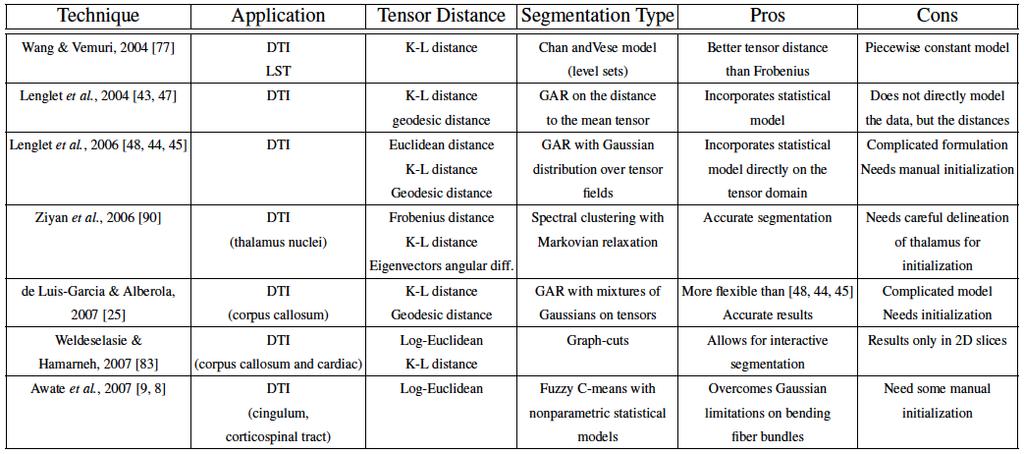
18 DTI segmentation [LuisGarcia_2009]
19 19 Initial goal: LGN identification 1. We wanted to find an automated way to identify LGN 2. Clustering had initially being used in the thalamus. Behrens et al Use thamalic-cerebral connections to segment the thalamic nuclei Wiegell et al Use similarity between tensor and coordinates information to identify clusters
20 20 First paper to use coordinate + diffusion Wiegell et al E( j, k) = x j x k +γ D j D k y Where x is the coordinate vector D is the diffusion tensor j is the pixel under investigation k is the centroid of the cluster γ is a weighting factor x
21 21 First paper to use coordinate + diffusion Wiegell et al E( j, k) = x j x k +γ D j D k y Given a set of pixels in a ROI Prior knowledge of clusters # x
22 22 First paper to use coordinate + diffusion Wiegell et al E( j, k) = x j x k +γ D j D k y Given a set of pixels in a ROI Prior knowledge of clusters # Manual cluster initialization x
 = x j x k +γ D j D k y Given a set of pixels in a ROI E(1,1) E(1,2) Prior knowledge of clusters # Manual cluster")
23 23 First paper to use coordinate + diffusion Wiegell et al E( j, k) = x j x k +γ D j D k y Given a set of pixels in a ROI E(1,1) E(1,2) Prior knowledge of clusters # Manual cluster initialization Distance voxel-centroids k-means clustering x New centroids calculation
24 24 First paper to use coordinate + diffusion Wiegell et al E( j, k) = x j x k +γ D j D k y Given a set of pixels in a ROI Prior knowledge of clusters # Manual cluster initialization Distance voxel-centroids k-means clustering x New centroids calculation Pixels labeling
25 25 Improvements from previous work Previous work relied heavily on manual initialization by an expert, had a fixed # of clusters and used only space and diffusivity information We wanted to create an automatic and scalable segmentation method and run it on a set of subjects We modified the Wiegell algorithm: Automatic: no manual initialization needed Scalable: to scale up different measurements, not constrained to space and diffusion information
 x j x k E( j, k) = 1 n")

 Tensors elements similarity T1")
26 26 A scalable distance measure Coords distance (mm) x j x k E( j, k) = 1 n Where j is the pixel under investigation k is the centroid of the cluster n is the number of measures considered n d i=1 i ( j, k) Tensors elements similarity T1 pixel intensity
27 27 Avoid manual initialization Data driven approach: voxels dissimilarity is used to describe the distances of one voxel j to all the other voxels in the ROI. y Dissimilarity matrix representation (DMR) E '( j, j) = 1 n n d i=1 i ( j, j) x
28 28 Avoid manual initialization Data driven approach: voxels dissimilarity is used to describe the distances of one voxel j to all the other voxels in the ROI. y Dissimilarity matrix representation (DMR) E '( j, j) = 1 n n d i=1 i ( j, j) x
29 29 Avoid manual initialization Data driven approach: voxels dissimilarity is used to describe the distances of one voxel j to all the other voxels in the ROI. y Dissimilarity matrix representation (DMR) E '( j, j) = 1 n n d i=1 i ( j, j) x
30 30 Avoid manual initialization Data driven approach: voxels dissimilarity is used to describe the distances of one voxel j to all the other voxels in the ROI. y Dissimilarity matrix representation (DMR) E '( j, j) = 1 n n d i=1 i ( j, j) x
31 31 Avoid manual initialization Data driven approach: voxels dissimilarity is used to describe the distances of one voxel j to all the other voxels in the ROI. y Dissimilarity matrix representation (DMR) E '( j, j) = 1 n n d i=1 i ( j, j) x
32 32 Avoid manual initialization Data driven approach: voxels dissimilarity is used to describe the distances of one voxel j to all the other voxels in the ROI. y Dissimilarity matrix representation (DMR) E '( j, j) = 1 n n d i=1 i ( j, j) x
33 33 Avoid manual initialization Data driven approach: voxels dissimilarity is used to describe the distances of one voxel j to all the other voxels in the ROI. y Dissimilarity matrix representation (DMR) E '( j, j) = 1 n n d i=1 i ( j, j) x
34 34 Avoid manual initialization Data driven approach: voxels dissimilarity is used to describe the distances of one voxel j to all the other voxels in the ROI. y Dissimilarity matrix representation (DMR) E '( j, j) = 1 n n d i=1 i ( j, j) x
35 35 Thalamus: clustering results Load Freesurfer Thalamus ROI Extraction of different measures (tensors, T1) Measures of the distance from each voxel to every voxel E Multidimensional scaling: E k-means clustering

36 36 Thalamus: clustering results Wiegell et al. 2003

37 37 Thalamus: clustering results Behrens et al. 2003

38 Thalamus: clustering results
 Deterministic vs. probabilistic tractography: Deterministic assumes a single orientation at each voxel Probabilistic assumes a distribution of orientations Local vs.")
39 Tractography Use local diffusion orientation at each voxel to determine pathway between distant brain regions? Local orientation comes from diffusion model fit (tensor, ball-andstick, etc.) Deterministic vs. probabilistic tractography: Deterministic assumes a single orientation at each voxel Probabilistic assumes a distribution of orientations Local vs. global tractography: Local fits the pathway to the data one step at a time Global fits the entire pathway at once
40 Deterministic vs. probabilistic Deterministic methods give you an estimate of model parameters 5 Probabilistic methods give you the uncertainty (probability distribution) of the estimate 5
41 Deterministic vs. probabilistic Sample 1 Sample 2 Deterministic tractography: One streamline per seed voxel Probabilistic tractography: Multiple streamline samples per seed voxel (drawn from probability distribution)

42 Deterministic vs. probabilistic Deterministic tractography: One streamline per seed voxel Probabilistic tractography: A probability distribution (sum of all streamline samples from all seed voxels)
43 Local vs. global Local tractography: Fits pathway step-by-step, using local diffusion orientation at each step Global tractography: Fits the entire pathway, using diffusion orientation at all voxels along pathway length
44 Local vs. global Local tractography: - Deterministic: One direction at each step [DTIStudio, trackvis] - Probabilistic: A distribution of directions at each step [FSL/ probtrack] Global tractography: - Deterministic: Fit path to diffusion orientations along its length - Probabilistic: Fit path to diffusion orientation distributions along its length
45 Local tractography Best suited for exploratory study of connections All connections from a seed region, not constrained to a specific target region How do we isolate a specific white-matter pathway? Thresholding? Intermediate masks? Non-dominant connections are hard to reconstruct Results are not symmetric between seed and target regions Sensitive to areas of high local uncertainty in orientation (e.g., pathaway crossings), errors propagate from those areas
46 Global tractography Best suited for reconstruction of known white-matter pathways Constrained to connection of two specific end regions Not sensitive to areas of high local uncertainty in orientation, integrates over entire pathway Symmetric between seed and target regions Need to search through a large solution space of all possible connections between two regions: Computationally expensive Sensitive to initialization

47 Tractography

48 Tractography: ipsilateral fibers


49 Connectivity

50 Connectivity

51 Connectivity
52 THANK YOU!
Diffusion model fitting and tractography: A primer
 Diffusion model fitting and tractography: A primer Anastasia Yendiki HMS/MGH/MIT Athinoula A. Martinos Center for Biomedical Imaging 03/18/10 Why n how Diffusion model fitting and tractography 0/18 Why
Quantitative MRI of the Brain: Investigation of Cerebral Gray and White Matter Diseases
 Quantities Measured by MR - Quantitative MRI of the Brain: Investigation of Cerebral Gray and White Matter Diseases Static parameters (influenced by molecular environment): T, T* (transverse relaxation)
HST.583 Functional Magnetic Resonance Imaging: Data Acquisition and Analysis Fall 2008
 MIT OpenCourseWare http://ocw.mit.edu HST.583 Functional Magnetic Resonance Imaging: Data Acquisition and Analysis Fall 2008 For information about citing these materials or our Terms of Use, visit: http://ocw.mit.edu/terms.
Surface-based Analysis: Inter-subject Registration and Smoothing
 Surface-based Analysis: Inter-subject Registration and Smoothing Outline Exploratory Spatial Analysis Coordinate Systems 3D (Volumetric) 2D (Surface-based) Inter-subject registration Volume-based Surface-based
HST.583 Functional Magnetic Resonance Imaging: Data Acquisition and Analysis Fall 2008
 MIT OpenCourseWare http://ocw.mit.edu HST.583 Functional Magnetic Resonance Imaging: Data Acquisition and Analysis Fall 2008 For information about citing these materials or our Terms of Use, visit: http://ocw.mit.edu/terms.
An Introduction To Automatic Tissue Classification Of Brain MRI. Colm Elliott Mar 2014
 An Introduction To Automatic Tissue Classification Of Brain MRI Colm Elliott Mar 2014 Tissue Classification Tissue classification is part of many processing pipelines. We often want to classify each voxel
Lilla Zöllei A.A. Martinos Center, MGH; Boston, MA
 Lilla Zöllei lzollei@nmr.mgh.harvard.edu A.A. Martinos Center, MGH; Boston, MA Bruce Fischl Gheorghe Postelnicu Jean Augustinack Anastasia Yendiki Allison Stevens Kristen Huber Sita Kakonoori + the FreeSurfer
Atelier 2 : Calcul Haute Performance et Sciences du Vivant Forum er juillet, Paris, France
 From Diffusion MR Image Analysis to Whole Brain Connectivity Simulation Jean-Philippe Thiran EPFL Lausanne, Switzerland EPFL - Lausanne HPC in life sciences at EPFL The Blue Brain project: create a biologically
TRACULA: Troubleshooting, visualization, and group analysis
 TRACULA: Troubleshooting, visualization, and group analysis Anastasia Yendiki HMS/MGH/MIT Athinoula A. Martinos Center for Biomedical Imaging 18/11/13 TRACULA: troubleshooting, visualization, group analysis
Fiber Selection from Diffusion Tensor Data based on Boolean Operators
 Fiber Selection from Diffusion Tensor Data based on Boolean Operators D. Merhof 1, G. Greiner 2, M. Buchfelder 3, C. Nimsky 4 1 Visual Computing, University of Konstanz, Konstanz, Germany 2 Computer Graphics
Anatomic parcellation based on DTI data with FSL taking the example of SMA/preSMA
 Groupe de Travail IRMf/MEG 02/2014 Anatomic parcellation based on DTI data with FSL taking the example of SMA/preSMA Magdalena Wutte, Lucile Brun & Boris Burle SMA/preSMA Parcellation - what for? - connectivity
ECE1778 Final Report MRI Visualizer
 ECE1778 Final Report MRI Visualizer David Qixiang Chen Alex Rodionov Word Count: 2408 Introduction We aim to develop a mobile phone/tablet based neurosurgical MRI visualization application with the goal
ASAP_2.0 (Automatic Software for ASL Processing) USER S MANUAL
 USER S MANUAL") ASAP_2.0 (Automatic Software for ASL Processing) USER S MANUAL ASAP was developed as part of the COST Action "Arterial Spin Labelling Initiative in Dementia (AID)" by: Department of Neuroimaging, Institute
ASAP_2.0 (Automatic Software for ASL Processing) USER S MANUAL ASAP was developed as part of the COST Action "Arterial Spin Labelling Initiative in Dementia (AID)" by: Department of Neuroimaging, Institute
NEURO M203 & BIOMED M263 WINTER 2014
 NEURO M203 & BIOMED M263 WINTER 2014 MRI Lab 2: Neuroimaging Connectivity Lab In today s lab we will work with sample diffusion imaging data and the group averaged fmri data collected during your scanning
Diffusion-MRI processing for group analysis
 Diffusion-MRI processing for group analysis Felix Renard IRMaGe: Inserm US 17 / CNRS UMS 3552 University Hospital of Grenoble - France 25/09/2015 felixrenard@gmail.com 1 Diffusion-MRI processing for group
BDP: BrainSuite Diffusion Pipeline. Chitresh Bhushan
 BDP: BrainSuite Diffusion Pipeline Chitresh Bhushan Why diffusion MRI? T 2 weighted MPRAGE FA map Fiber track Quantify microstructural tissue characteristics Structural connectivity Connectome Clinical
Fmri Spatial Processing
 Educational Course: Fmri Spatial Processing Ray Razlighi Jun. 8, 2014 Spatial Processing Spatial Re-alignment Geometric distortion correction Spatial Normalization Smoothing Why, When, How, Which Why is
Supplementary methods
 Supplementary methods This section provides additional technical details on the sample, the applied imaging and analysis steps and methods. Structural imaging Trained radiographers placed all participants
Normalization for clinical data
 Normalization for clinical data Christopher Rorden, Leonardo Bonilha, Julius Fridriksson, Benjamin Bender, Hans-Otto Karnath (2012) Agespecific CT and MRI templates for spatial normalization. NeuroImage
Computational Neuroanatomy
 Computational Neuroanatomy John Ashburner john@fil.ion.ucl.ac.uk Smoothing Motion Correction Between Modality Co-registration Spatial Normalisation Segmentation Morphometry Overview fmri time-series kernel
Evaluation of Local Filter Approaches for Diffusion Tensor based Fiber Tracking
 Evaluation of Local Filter Approaches for Diffusion Tensor based Fiber Tracking D. Merhof 1, M. Buchfelder 2, C. Nimsky 3 1 Visual Computing, University of Konstanz, Konstanz 2 Department of Neurosurgery,
Saturn User Manual. Rubén Cárdenes. 29th January 2010 Image Processing Laboratory, University of Valladolid. Abstract
 Saturn User Manual Rubén Cárdenes 29th January 2010 Image Processing Laboratory, University of Valladolid Abstract Saturn is a software package for DTI processing and visualization, provided with a graphic
This Time. fmri Data analysis
 This Time Reslice example Spatial Normalization Noise in fmri Methods for estimating and correcting for physiologic noise SPM Example Spatial Normalization: Remind ourselves what a typical functional image
Norbert Schuff VA Medical Center and UCSF
 Norbert Schuff Medical Center and UCSF Norbert.schuff@ucsf.edu Medical Imaging Informatics N.Schuff Course # 170.03 Slide 1/67 Objective Learn the principle segmentation techniques Understand the role
PANDA Manual Zaixu Cui & Suyu Zhong & Gaolang Gong
 PANDA Manual Zaixu Cui & Suyu Zhong & Gaolang Gong National Key Laboratory of Cognitive Neuroscience and Learning Beijing Normal University, China Contents Overview Setup Files/Directories selection Preparing
BDP: BrainSuite Diffusion Pipeline. Chitresh Bhushan
 BDP: BrainSuite Diffusion Pipeline Chitresh Bhushan Why diffusion MRI? T 2 weighted MPRAGE FA map Fiber track Quantify microstructural tissue characteristics Structural connectivity Connectome Clinical
Diffusion Imaging Visualization
 Diffusion Imaging Visualization Thomas Schultz URL: http://cg.cs.uni-bonn.de/schultz/ E-Mail: schultz@cs.uni-bonn.de 1 Outline Introduction to Diffusion Imaging Basic techniques Glyph-based Visualization
Complex Fiber Visualization
 Annales Mathematicae et Informaticae 34 (2007) pp. 103 109 http://www.ektf.hu/tanszek/matematika/ami Complex Fiber Visualization Henrietta Tomán a, Róbert Tornai b, Marianna Zichar c a Department of Computer
Preprocessing II: Between Subjects John Ashburner
 Preprocessing II: Between Subjects John Ashburner Pre-processing Overview Statistics or whatever fmri time-series Anatomical MRI Template Smoothed Estimate Spatial Norm Motion Correct Smooth Coregister
Automated MR Image Analysis Pipelines
 Automated MR Image Analysis Pipelines Andy Simmons Centre for Neuroimaging Sciences, Kings College London Institute of Psychiatry. NIHR Biomedical Research Centre for Mental Health at IoP & SLAM. Neuroimaging
Last Time. This Time. Thru-plane dephasing: worse at long TE. Local susceptibility gradients: thru-plane dephasing
 Motion Correction Last Time Mutual Information Optimiation Decoupling Translation & Rotation Interpolation SPM Example (Least Squares & MI) A Simple Derivation This Time Reslice example SPM Example : Remind
Diffusion Tensor Imaging and Reading Development
 Diffusion Tensor Imaging and Reading Development Bob Dougherty Stanford Institute for Reading and Learning Reading and Anatomy Every brain is different... Not all brains optimized for highly proficient
Reconstruction of Fiber Trajectories via Population-Based Estimation of Local Orientations
 IDEA Reconstruction of Fiber Trajectories via Population-Based Estimation of Local Orientations Pew-Thian Yap, John H. Gilmore, Weili Lin, Dinggang Shen Email: ptyap@med.unc.edu 2011-09-21 Poster: P2-46-
Diffusion MRI. Introduction and Modern Methods. John Plass. Department Of Psychology
 Diffusion MRI Introduction and Modern Methods John Plass Department Of Psychology Diffusion MRI Introduction and Modern Methods John Plass Department Of Psychology Overview I. Why use diffusion MRI? II.
A Novel Contrast for DTI Visualization for Thalamus Delineation
 A Novel Contrast for DTI Visualization for Thalamus Delineation Xian Fan a, Meredith Thompson a,b, John A. Bogovic a, Pierre-Louis Bazin c, Jerry L. Prince a,c a Johns Hopkins University, Baltimore, MD,
Reproducibility of Whole-brain Structural Connectivity Networks
 Reproducibility of Whole-brain Structural Connectivity Networks Christopher Parker Thesis for Masters of Research in Medical and Biomedical Imaging Supervised by Prof. Sebastien Ourselin and Dr Jonathan
A Stochastic Tractography System and Applications. Tri M. Ngo
 A Stochastic Tractography System and Applications by Tri M. Ngo Submitted to the Department of Electrical Engineering and Computer Science in partial fulfillment of the requirements for the degree of Master
Head motion in diffusion MRI
 Head motion in diffusion MRI Anastasia Yendiki HMS/MGH/MIT Athinoula A. Martinos Center for Biomedical Imaging 11/06/13 Head motion in diffusion MRI 0/33 Diffusion contrast Basic principle of diffusion
Statistical Analysis of Neuroimaging Data. Phebe Kemmer BIOS 516 Sept 24, 2015
 Statistical Analysis of Neuroimaging Data Phebe Kemmer BIOS 516 Sept 24, 2015 Review from last time Structural Imaging modalities MRI, CAT, DTI (diffusion tensor imaging) Functional Imaging modalities
Brain Extraction, Registration & EPI Distortion Correction
 Brain Extraction, Registration & EPI Distortion Correction What use is Registration? Some common uses of registration: Combining across individuals in group studies: including fmri & diffusion Quantifying
DIFFUSION TENSOR IMAGING ANALYSIS. Using Analyze
 DIFFUSION TENSOR IMAGING ANALYSIS Using Analyze 2 Table of Contents 1. Introduction page 3 2. Loading DTI Data page 4 3. Computing DTI Maps page 5 4. Defining ROIs for Fiber Tracking page 6 5. Visualizing
EMSegment Tutorial. How to Define and Fine-Tune Automatic Brain Compartment Segmentation and the Detection of White Matter Hyperintensities
 EMSegment Tutorial How to Define and Fine-Tune Automatic Brain Compartment Segmentation and the Detection of White Matter Hyperintensities This documentation serves as a tutorial to learn to customize
MR IMAGE SEGMENTATION
 MR IMAGE SEGMENTATION Prepared by : Monil Shah What is Segmentation? Partitioning a region or regions of interest in images such that each region corresponds to one or more anatomic structures Classification
Measuring longitudinal brain changes in humans and small animal models. Christos Davatzikos
 Measuring longitudinal brain changes in humans and small animal models Christos Davatzikos Section of Biomedical Image Analysis University of Pennsylvania (Radiology) http://www.rad.upenn.edu/sbia Computational
TractoR and Other Software
 TractoR and Other Software Jon Clayden DIBS Teaching Seminar, 11 Dec 2015 Photo by José Martín Ramírez Carrasco https://www.behance.net/martini_rc TractoR A set of R packages Additional
n o r d i c B r a i n E x Tutorial DTI Module
 m a k i n g f u n c t i o n a l M R I e a s y n o r d i c B r a i n E x Tutorial DTI Module Please note that this tutorial is for the latest released nordicbrainex. If you are using an older version please
MITK-DI. A new Diffusion Imaging Component for MITK. Klaus Fritzsche, Hans-Peter Meinzer
 MITK-DI A new Diffusion Imaging Component for MITK Klaus Fritzsche, Hans-Peter Meinzer Division of Medical and Biological Informatics, DKFZ Heidelberg k.fritzsche@dkfz-heidelberg.de Abstract. Diffusion-MRI
Input tab. Insert the path to the DTI Image that you want to tract.
 Tutorial: AutoTract Input tab Insert the path to the DTI Image that you want to tract. Input tab (optional)if you already have a WM/CSF mask you want to use to process the tracts, insert the path here.
Advanced Visual Medicine: Techniques for Visual Exploration & Analysis
 Advanced Visual Medicine: Techniques for Visual Exploration & Analysis Interactive Visualization of Multimodal Volume Data for Neurosurgical Planning Felix Ritter, MeVis Research Bremen Multimodal Neurosurgical
The organization of the human cerebral cortex estimated by intrinsic functional connectivity
 1 The organization of the human cerebral cortex estimated by intrinsic functional connectivity Journal: Journal of Neurophysiology Author: B. T. Thomas Yeo, et al Link: https://www.ncbi.nlm.nih.gov/pubmed/21653723
Issues Regarding fmri Imaging Workflow and DICOM
 Issues Regarding fmri Imaging Workflow and DICOM Lawrence Tarbox, Ph.D. Fred Prior, Ph.D Mallinckrodt Institute of Radiology Washington University in St. Louis What is fmri fmri is used to localize functions
MRI Segmentation MIDAS, 2007, 2010
 MRI Segmentation MIDAS, 2007, 2010 Lawrence O. Hall, Dmitry Goldgof, Yuhua Gu, Prodip Hore Dept. of Computer Science & Engineering University of South Florida CONTENTS: 1. Introduction... 1 2. Installing
Where are we now? Structural MRI processing and analysis
 Where are we now? Structural MRI processing and analysis Pierre-Louis Bazin bazin@cbs.mpg.de Leipzig, Germany Structural MRI processing: why bother? Just use the standards? SPM FreeSurfer FSL However:
Functional analysis with DTI and diffusion-neurography of cranial nerves
 Functional analysis with DTI and diffusion-neurography of cranial nerves Poster No.: C-1942 Congress: ECR 2013 Type: Educational Exhibit Authors: J. P. Martínez Barbero, T. Martín Noguerol, A. Luna Alcalá;
Fiber Selection from Diffusion Tensor Data based on Boolean Operators
 Fiber Selection from Diffusion Tensor Data based on Boolean Operators D. Merhofl, G. Greiner 2, M. Buchfelder 3, C. Nimsky4 1 Visual Computing, University of Konstanz, Konstanz, Germany 2 Computer Graphics
FSL Workshop Session 3 David Smith & John Clithero
 FSL Workshop 12.09.08 Session 3 David Smith & John Clithero What is MELODIC? Probabilistic ICA Improves upon standard ICA Allows for inference Avoids over-fitting Three stage process ( ppca ) 1.) Dimension
BrainSpace V2.1: Geometric mapping and diffusion-based software for cross-subject multimodality brain imaging informatics
 Brief Introduction to BrainSpace V2.1 BrainSpace V2.1: Geometric mapping and diffusion-based software for cross-subject multimodality brain imaging informatics Graphics and Imaging Laboratory Wayne State
CHAPTER 2. Morphometry on rodent brains. A.E.H. Scheenstra J. Dijkstra L. van der Weerd
 CHAPTER 2 Morphometry on rodent brains A.E.H. Scheenstra J. Dijkstra L. van der Weerd This chapter was adapted from: Volumetry and other quantitative measurements to assess the rodent brain, In vivo NMR
Knowledge-Based Segmentation of Brain MRI Scans Using the Insight Toolkit
 Knowledge-Based Segmentation of Brain MRI Scans Using the Insight Toolkit John Melonakos 1, Ramsey Al-Hakim 1, James Fallon 2 and Allen Tannenbaum 1 1 Georgia Institute of Technology, Atlanta GA 30332,
Detecting Changes In Non-Isotropic Images
 Detecting Changes In Non-Isotropic Images K.J. Worsley 1, M. Andermann 1, T. Koulis 1, D. MacDonald, 2 and A.C. Evans 2 August 4, 1999 1 Department of Mathematics and Statistics, 2 Montreal Neurological
FSL Pre-Processing Pipeline
 The Art and Pitfalls of fmri Preprocessing FSL Pre-Processing Pipeline Mark Jenkinson FMRIB Centre, University of Oxford FSL Pre-Processing Pipeline Standard pre-processing: Task fmri Resting-state fmri
Groupwise Shape Correspondence with Local Features
 Groupwise Shape Correspondence with Local Features İpek Oğuz A dissertation submitted to the faculty of the University of North Carolina at Chapel Hill in partial fulfillment of the requirements for the
Functional MRI data preprocessing. Cyril Pernet, PhD
 Functional MRI data preprocessing Cyril Pernet, PhD Data have been acquired, what s s next? time No matter the design, multiple volumes (made from multiple slices) have been acquired in time. Before getting
syngo.mr Neuro 3D: Your All-In-One Post Processing, Visualization and Reporting Engine for BOLD Functional and Diffusion Tensor MR Imaging Datasets
 syngo.mr Neuro 3D: Your All-In-One Post Processing, Visualization and Reporting Engine for BOLD Functional and Diffusion Tensor MR Imaging Datasets Julien Gervais; Lisa Chuah Siemens Healthcare, Magnetic
Math in image processing
 Math in image processing Math in image processing Nyquist theorem Math in image processing Discrete Fourier Transformation Math in image processing Image enhancement: scaling Math in image processing Image
Multimodal Imaging Brain Connectivity Analysis (MIBCA)
") Multimodal Imaging Brain Connectivity Analysis (MIBCA) Andre Santos Ribeiro, Luis Miguel Lacerda, Hugo Ferreira April 23, 2015 Abstract In recent years, connectivity studies using neuroimaging data have
Multimodal Imaging Brain Connectivity Analysis (MIBCA) Andre Santos Ribeiro, Luis Miguel Lacerda, Hugo Ferreira April 23, 2015 Abstract In recent years, connectivity studies using neuroimaging data have
Basic principles of MR image analysis. Basic principles of MR image analysis. Basic principles of MR image analysis
 Basic principles of MR image analysis Basic principles of MR image analysis Julien Milles Leiden University Medical Center Terminology of fmri Brain extraction Registration Linear registration Non-linear
Generation of Hulls Encompassing Neuronal Pathways Based on Tetrahedralization and 3D Alpha Shapes
 Generation of Hulls Encompassing Neuronal Pathways Based on Tetrahedralization and 3D Alpha Shapes Dorit Merhof 1,2, Martin Meister 1, Ezgi Bingöl 1, Peter Hastreiter 1,2, Christopher Nimsky 2,3, Günther
腦部結構影像 標準化 組織分割 體素型態 本週課程內容. Analysis Softwares. A Course of MRI
 本週課程內容 腦部結構影像 A Course of MRI 盧家鋒助理教授國立陽明大學物理治療暨輔助科技學系 alvin4016@ym.edu.tw 腦部結構影像 空間標準化 (Spatial normalization) 均勻度校正 (Bias correction) 組織分割 (Segmentation) 體素形態學分析 (Voxel-based morphometry, VBM) 影像平滑化
MITK-DI. A new Diffusion Imaging Component for MITK. Klaus Fritzsche, Hans-Peter Meinzer
 MITK-DI A new Diffusion Imaging Component for MITK Klaus Fritzsche, Hans-Peter Meinzer Division of Medical and Biological Informatics, DKFZ Heidelberg k.fritzsche@dkfz-heidelberg.de Abstract. Diffusion-MRI
Introduc)on to FreeSurfer h0p://surfer.nmr.mgh.harvard.edu. Jenni Pacheco.
on to FreeSurfer h0p://surfer.nmr.mgh.harvard.edu. Jenni Pacheco.") Introduc)on to FreeSurfer h0p://surfer.nmr.mgh.harvard.edu Jenni Pacheco jpacheco@mail.utexas.edu Analyzing the individual subject What happens? How do I do that? Now what? Tools you will use recon all
Introduc)on to FreeSurfer h0p://surfer.nmr.mgh.harvard.edu Jenni Pacheco jpacheco@mail.utexas.edu Analyzing the individual subject What happens? How do I do that? Now what? Tools you will use recon all
Acceleration of Probabilistic Tractography Using Multi-GPU Parallel Processing. Jungsoo Lee, Sun Mi Park, Dae-Shik Kim
 Acceleration of Probabilistic Tractography Using Multi-GPU Parallel Processing Jungsoo Lee, Sun Mi Park, Dae-Shik Kim Introduction In particular, probabilistic tractography requires relatively long computation
Appendix E1. Supplementary Methods. MR Image Acquisition. MR Image Analysis
 RSNA, 2015 10.1148/radiol.2015150532 Appendix E1 Supplementary Methods MR Image Acquisition By using a 1.5-T system (Avanto, Siemens Medical, Erlangen, Germany) under a program of regular maintenance (no
Introduc)on to FreeSurfer h0p://surfer.nmr.mgh.harvard.edu. Jenni Pacheco.
on to FreeSurfer h0p://surfer.nmr.mgh.harvard.edu. Jenni Pacheco.") Introduc)on to FreeSurfer h0p://surfer.nmr.mgh.harvard.edu Jenni Pacheco jpacheco@mail.utexas.edu fmri Integra)on Visualize individual fmri results on surface volume ROI Volume Study: Count number of voxels
Introduc)on to FreeSurfer h0p://surfer.nmr.mgh.harvard.edu Jenni Pacheco jpacheco@mail.utexas.edu fmri Integra)on Visualize individual fmri results on surface volume ROI Volume Study: Count number of voxels
Automatic segmentation of the cortical grey and white matter in MRI using a Region Growing approach based on anatomical knowledge
 Automatic segmentation of the cortical grey and white matter in MRI using a Region Growing approach based on anatomical knowledge Christian Wasserthal 1, Karin Engel 1, Karsten Rink 1 und André Brechmann
MIDAS Processing of Segmentation Data for Brain Lesions
 MIDAS Processing of Segmentation Data for Brain Lesions A. A. Maudsley 4/6/2012 For studies that contain lesions, the MIDAS processing has two steps that benefit from a modified processing pipeline to
EMSegmenter Tutorial (Advanced Mode)
") EMSegmenter Tutorial (Advanced Mode) Dominique Belhachemi Section of Biomedical Image Analysis Department of Radiology University of Pennsylvania 1/65 Overview The goal of this tutorial is to apply the
EMSegmenter Tutorial (Advanced Mode) Dominique Belhachemi Section of Biomedical Image Analysis Department of Radiology University of Pennsylvania 1/65 Overview The goal of this tutorial is to apply the
ABSTRACT 1. INTRODUCTION 2. METHODS
 Finding Seeds for Segmentation Using Statistical Fusion Fangxu Xing *a, Andrew J. Asman b, Jerry L. Prince a,c, Bennett A. Landman b,c,d a Department of Electrical and Computer Engineering, Johns Hopkins
NIH Public Access Author Manuscript Proc Soc Photo Opt Instrum Eng. Author manuscript; available in PMC 2011 September 7.
 NIH Public Access Author Manuscript Published in final edited form as: Proc Soc Photo Opt Instrum Eng. 2011 March ; 7962: 796225-1 796225-7. doi:10.1117/12.878405. Automatic Skull-stripping of Rat MRI/DTI
Automatic Registration-Based Segmentation for Neonatal Brains Using ANTs and Atropos
 Automatic Registration-Based Segmentation for Neonatal Brains Using ANTs and Atropos Jue Wu and Brian Avants Penn Image Computing and Science Lab, University of Pennsylvania, Philadelphia, USA Abstract.
Histograms. h(r k ) = n k. p(r k )= n k /NM. Histogram: number of times intensity level rk appears in the image
 = n k. p(r k )= n k /NM. Histogram: number of times intensity level rk appears in the image") Histograms h(r k ) = n k Histogram: number of times intensity level rk appears in the image p(r k )= n k /NM normalized histogram also a probability of occurence 1 Histogram of Image Intensities Create
Histograms h(r k ) = n k Histogram: number of times intensity level rk appears in the image p(r k )= n k /NM normalized histogram also a probability of occurence 1 Histogram of Image Intensities Create
A Ray-based Approach for Boundary Estimation of Fiber Bundles Derived from Diffusion Tensor Imaging
 A Ray-based Approach for Boundary Estimation of Fiber Bundles Derived from Diffusion Tensor Imaging M. H. A. Bauer 1,3, S. Barbieri 2, J. Klein 2, J. Egger 1,3, D. Kuhnt 1, B. Freisleben 3, H.-K. Hahn
Structural connectivity of the brain measured by diffusion tensor imaging [DTI]
![Structural connectivity of the brain measured by diffusion tensor imaging [DTI]](/thumbs/74/71065400.jpg "Structural connectivity of the brain measured by diffusion tensor imaging [DTI]") Structural connectivity of the brain measured by diffusion tensor imaging [DTI] Lars T. Westlye CSHC / Centre for Advanced Study PSY4320 06.05.2012 l.t.westlye@psykologi.uio.no ~ 40 % of the brain is white
Structural connectivity of the brain measured by diffusion tensor imaging [DTI] Lars T. Westlye CSHC / Centre for Advanced Study PSY4320 06.05.2012 l.t.westlye@psykologi.uio.no ~ 40 % of the brain is white
Using the Face Masking Pipeline
 1 Using the Face Masking Pipeline We have now made available a Face Masking Pipeline to mask DICOMs to prevent possible patient identification from reconstructed structural images. The pipeline uses a
MriCloud DTI Processing Pipeline. DTI processing can be initiated by choosing DTI Processing in the top menu bar.
 MriCloud DTI Processing Pipeline 1: Data Upload 1-1: Web interface DTI processing can be initiated by choosing DTI Processing in the top menu bar. To avoid any HIPPA issues, data need to be first converted
Technische Universiteit Eindhoven Department of Mathematics and Computer Science. Master s Thesis HIERARCHICAL VISUALIZATION USING FIBER CLUSTERING
 Technische Universiteit Eindhoven Department of Mathematics and Computer Science Master s Thesis HIERARCHICAL VISUALIZATION USING FIBER CLUSTERING by Ing. B. Moberts Supervisors: Dr. A. Vilanova Prof.dr.ir.
Neuroimage Processing
 Neuroimage Processing Instructor: Moo K. Chung mkchung@wisc.edu Lecture 2-3. General Linear Models (GLM) Voxel-based Morphometry (VBM) September 11, 2009 What is GLM The general linear model (GLM) is a
BrainSuite Lab Exercises. presented at the UCLA/NITP Advanced Neuroimaging Summer Program 29 July 2014
 BrainSuite Lab Exercises presented at the UCLA/NITP Advanced Neuroimaging Summer Program 29 July 2014 1. Opening and Displaying an MRI Start BrainSuite Drag and drop the T1 image from the native space
How to create a head model
 How to create a head model This document describes the command line tools: mri2mesh: Central tool to reconstruct a head model from T1w and T2w data dwi2cond: Reconstruct conductivity tensors for brain
Transforming Datasets to Talairach-Tournoux Coordinates
 -1- Transforming Datasets to Talairach-Tournoux Coordinates The original purpose of AFNI was to perform the transformation of datasets to Talairach-Tournoux (stereotaxic) coordinates The transformation
Playing with data from lab
 Playing with data from lab Getting data off the scanner From the Patient Browser, select the folder for the study you want (or within that study, the set of images you want), and then from the Transfer
ADAPTIVE GRAPH CUTS WITH TISSUE PRIORS FOR BRAIN MRI SEGMENTATION
 ADAPTIVE GRAPH CUTS WITH TISSUE PRIORS FOR BRAIN MRI SEGMENTATION Abstract: MIP Project Report Spring 2013 Gaurav Mittal 201232644 This is a detailed report about the course project, which was to implement
Function-Structure Integration in FreeSurfer
 Function-Structure Integration in FreeSurfer Outline Function-Structure Integration Function-Structure Registration in FreeSurfer fmri Analysis Preprocessing First-Level Analysis Higher-Level (Group) Analysis
A validation of the biexponential model in diffusion MRI signal attenuation using diffusion Monte Carlo simulator
 A validation of the biexponential model in diffusion MRI signal attenuation using diffusion Monte Carlo simulator Poster No.: C-0331 Congress: ECR 2014 Type: Scientific Exhibit Authors: D. Nishigake, S.
Automatic Subthalamic Nucleus Targeting for Deep Brain Stimulation. A Validation Study
 Automatic Subthalamic Nucleus Targeting for Deep Brain Stimulation. A Validation Study F. Javier Sánchez Castro a, Claudio Pollo a,b, Jean-Guy Villemure b, Jean-Philippe Thiran a a École Polytechnique
Methods for data preprocessing
 Methods for data preprocessing John Ashburner Wellcome Trust Centre for Neuroimaging, 12 Queen Square, London, UK. Overview Voxel-Based Morphometry Morphometry in general Volumetrics VBM preprocessing
Introduction to fmri. Pre-processing
 Introduction to fmri Pre-processing Tibor Auer Department of Psychology Research Fellow in MRI Data Types Anatomical data: T 1 -weighted, 3D, 1/subject or session - (ME)MPRAGE/FLASH sequence, undistorted
A diffusion tensor imaging software comparison and between control subjects and subjects with known anatomical diagnosis
 UNLV Theses, Dissertations, Professional Papers, and Capstones 12-2011 A diffusion tensor imaging software comparison and between control subjects and subjects with known anatomical diagnosis Michael C.
Analysis of Functional MRI Timeseries Data Using Signal Processing Techniques
 Analysis of Functional MRI Timeseries Data Using Signal Processing Techniques Sea Chen Department of Biomedical Engineering Advisors: Dr. Charles A. Bouman and Dr. Mark J. Lowe S. Chen Final Exam October
Spatial Filtering Methods in MEG. Part 3: Template Normalization and Group Analysis"
 Spatial Filtering Methods in MEG Part 3: Template Normalization and Group Analysis" Douglas Cheyne, PhD" Program in Neurosciences and Mental Health" Hospital for Sick Children Research Institute " &" Department