Functional MRI data preprocessing. Cyril Pernet, PhD
|
|
|
- Hope Melton
- 6 years ago
- Views:
Transcription
1 Functional MRI data preprocessing Cyril Pernet, PhD

2 Data have been acquired, what s s next? time No matter the design, multiple volumes (made from multiple slices) have been acquired in time. Before getting data out, we need to make sure the signal from each voxel contains the right temporal and spatial information. Picture credit:


3 fmri time-series kernel Slice timing and Realignment smoothing Statistics normalisation Anatomical reference
4 Slice Timing Correction R. Henson, C. Buechel, O. Josephs, K. Friston The slice-timing problem in event-related fmri NeuroImage, 9 (1999), p
5 Slice Timing Correction Most of the time, fmri data are acquired using sequential 2D imaging like single shot EPI. Since fmri statistics are about analyzing the time course of the BOLD signal, exact timing with regard to the stimulus presentation is crucial. For instance, if you set a TR of 2 sec and acquire 30 slices, the acquisition time of 1 slice is ~66.66 ms (2000/30) and STC compensates for these sampling differences. Sladky et al. (2011) NeuroImage, 58,
6 Slice Timing Correction STC consists in shifting the signal phase by a given amount to temporally align data. It is therefore mandatory to select a reference slice. The reference slice is usually the slice acquired in the middle of the sequence (maximum interpolation of TR/2) but any slice can be used. Data are acquired either in sequential or interleaved mode and the middle of the sequence is not the middle of the brain! Sequential acquisition Ref slice Interleaved acquisition Ref slice
7 Slice Timing Correction TR of e.g. 2 sec with the middle temporal slice as reference is comparable to a dataset with a TR of 1 sec when the first (or last) slice acquired are used as a reference slice. This can be a reasonable practice, if the region of interest and putative activations are located near the first (or last) slice because it suppresses temporal interpolation effects in these areas. Note that all regressors in the GLM also need to be adjusted for this shift in time according to the reference slice (e.g. TR / 2). Sladky et al. (2011) NeuroImage, 58,
8 Slice Timing Correction Possible introduction of aliasing effects for signals at frequencies above the Nyquist sampling limit. Given a typical TR of 2 s (f = 0.25 Hz) a minimal inter-stimulus interval (ISI) of more than 4 s is recommended. When using the slice acquired in the middle of the acquisition period (TR) as a reference slice, the signal needs to be shifted by TR / 2, therefore reducing the suggested minimal ISI to 2 s. It is sometimes advocated to not do the STC especially for TR<2 sec. However, Sladky et al. showed that is always beneficial. When not performed, the reduction of parameter estimates (effects) were more pronounced for long TRs, event-related designs and designs with shorter SOA (up to 63%!). Sladky et al. (2011) NeuroImage, 58,
9 motion correction (realignment) JV. Hajnal, R. Myers, A. Oatridge, JE. Schwieso, IR. Young, GM. Bydder Artifacts due to stimulus-correlated motion in functional imaging of the brain. Magn Reson Med, 3 (1994), p
10 Motion artefacts Subjects will always move in the scanner: swallowing for instance lead to motion along the x axis or some movements may be related to the tasks performed. Motion will results in a mismatch of the location of subsequent images in the time-series. Since the sensitivity of the statistical analysis is determined by the amount of residual noise in the image series, mismatch of the location will add to this noise and reduce the sensitivity. This type of motion problem corresponds to wholesale movements ( (bulk-motion)) and is well corrected by realignment algorithms.
11 Motion correction: How? Determine the rigid body transformation that minimises some cost function (a way to define the difference between 2 images as e.g. least square (SPM) or normalized correlation ratio (FSL)). Rigid body transformation is defined by: 3 translations in X, Y & Z directions and 3 rotations around the X, Y & Z axes. Transform co-ordinate θ x For each voxel-centre co-ordinate Illustration taken from Jesper Andersson x=10mm y=0 θ=10º Find co-ordinate in original image
12 Motion correction: Quality check Plots the estimates from the head motion algorithm (might be useful to change angles into degrees and also plot the 1 st derivative outlier detection can then be run to identify bad scans) Compute whole head distance between volumes and to the mean (mean square difference) Inspect the realigned data as a movie Lots of tools available on websites For SPM users, I wrote a script to do this automatically see
13 Motion correction: Quality check Plots of head motion Translation Rotation mm scans scan to scan translation degrees scans scan to scan rotation mm scans degrees Mean square displacement scans 0.4 displacement scans
14 Motion correction: Quality check Whole head distances Mean quared distance to the average 700 squared difference scans Volume to volume mean squared distance squared difference scans


15 Motion correction: Quality check Motion correction: Quality check Movies
16 Even more motion artefacts Motion can also alter the MR signal because protons that moves into a voxel from a neighbouring slice have an excitation different from that expected by the scanner and the signal will not reflect well the tissue in that voxel.. This spin history effect is not corrected using motion correction algorithms and Independent Component Analysis (ICA) or dedicated methods must be used. The spin history effect is seen as alternating bright and dark stripes with interleaved acquisitions. One can mitigate this by modelling bad images as dummy regressors and include movement parameter estimates into the analysis. Picture credit:


17 Even more motion artefacts Physiological artefacts respiratory-induced noise is dominant near the edges of the brain as well as near in the larger veins and in the ventricles. cardiac-induced noise is dominant near larger vessels (e.g. medial cerebral artery and Circle of Willis) Physio monitoring Don t trust those regions Lund et al. (2006) NeuroImage, 29, 54-66
18 Spatial Normalization M, Holden A review of geometric transformations for nonrigid body registration. IEEE Trans Med Imaging, 27 (2008), p
19 Normalization: Why? Inter-subject averaging extrapolate findings to the population as a whole increase activation signal above that obtained from single subject increase number of possible degrees of freedom allowed in statistical model Enable reporting of activations as co-ordinates ordinates within a known standard space e.g. the space described by Talairach & Tournoux, or the MNI space (SPM, FSL)
, so in practice one uses a 3 steps approach in which one i)")

 and iii)")
20 Normalization: How? Current methods works mainly on T1 weighted images (i.e. anatomical), so in practice one uses a 3 steps approach in which one i) coregister T1 and T2* (fmri) data so that they are aligned and in the same space and ii) normalize the anatomical image (i.e. transform it to match the template) and iii) apply the parameters obtained to the fmri images Mean EPI T1-weighted Template coregister normalize apply normalization parameters
21 T1 image preprocessing The anatomical image is 1 st preprocessed,, depending on the method this includes: i) Noise reduction: : improves local features using local smoothing (SUSAN algorithm in FSL), ii) Bias correction: : from 3 Teslas,, there is often broad intensity variations in space that need to be corrected, iii) Brain extraction: : remove non brain tissue, iv) Segmentation: : separate grey matter, white matter and CSF In SPM, the unified segmentation approach (Ashburner and Friston (2005) NeuroImage, 26, ) combines/optimizes bias correction, segmentation, and normalization all in one, such as the prior probability of a voxel to belong to a tissue class is determined using a probabilistic atlas. In this framework 2 voxels with the same values can be classified differently using anatomical knowledge.
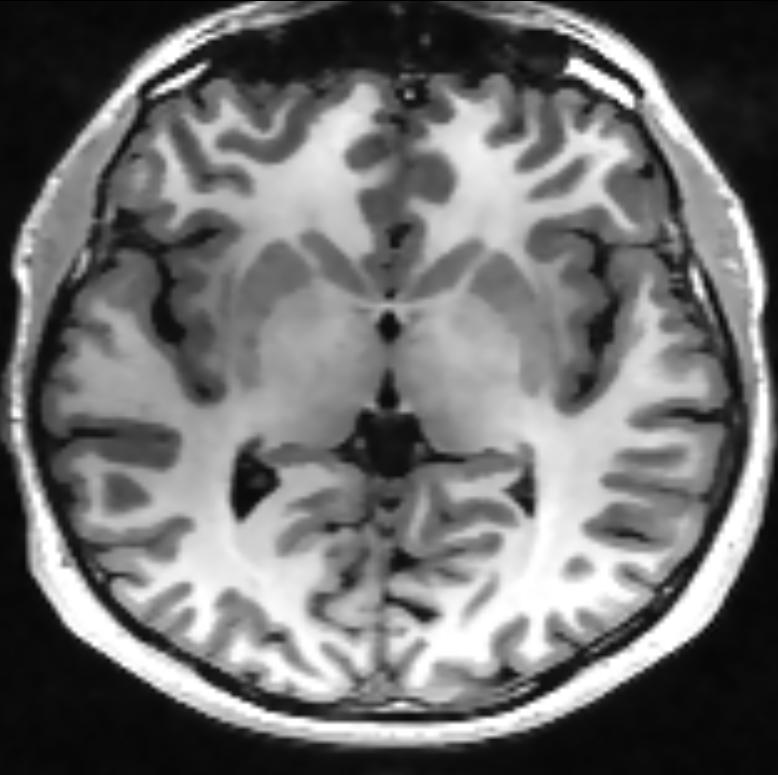
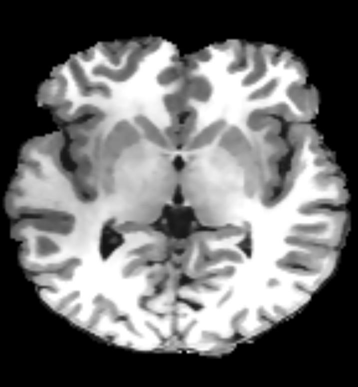
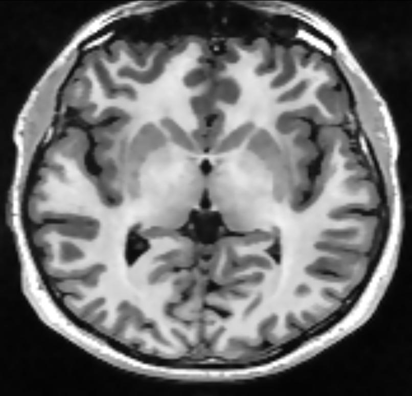
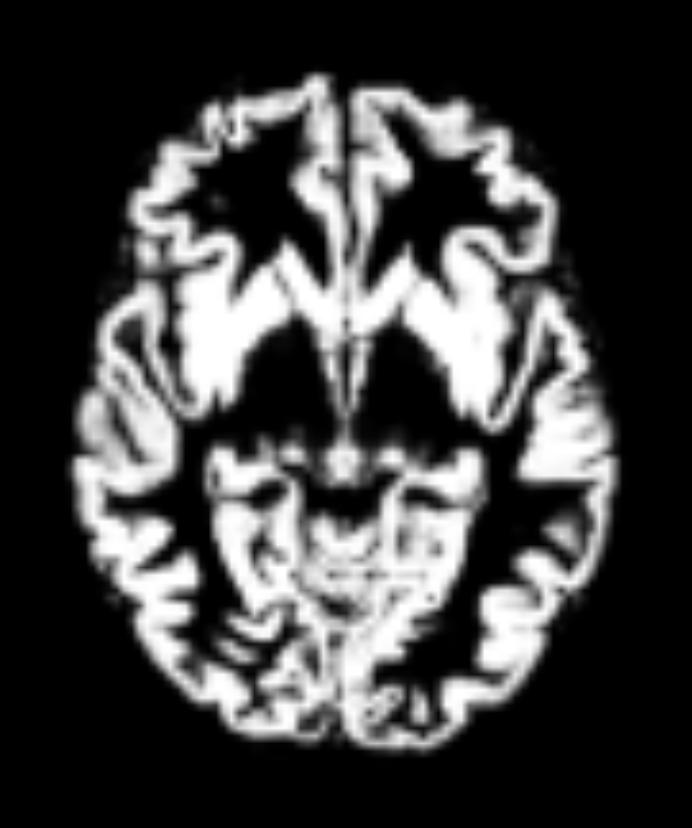
22 T1 image preprocessing Original T1-weighted Bias correction Noise reduction Segmentation Brain extraction
23 Volume-based normalization The first part of spatial normalisation is a 12 parameter affine transformation: 3 translations 3 rotations 3 scaling 3 shears } Rigid } Allow body transformation (realignment) does not change the size or shape of images change in overall size and shape Affine transformation: any set of points that fell on a line prior transformation will continue to fall on a line after the transformation

24 Volume-based normalization The second part of the normalization uses non- linear deformations. DARTEL toolbox in SPM or FNIRT in FSL rely on diffeomorphism,, that is the transformation from one image to the other can be represented as a vector field, describing the movements to apply at each voxel.


 is available in")

25 Surface-based Normalization Often more accurate to register cortical features than old volume based methods. Often limited to cortical surface. A combined method (surface based for cortical and volume based for deep brain structures) is available in Freesurfer. Image credit: Sarah Whittle & Dominic Dwyer free-surfer talk original white pial orig white pial inflated sphere patch


26 Normalization: quality check Visual inspection using e.g. CheckReg in SPM Mean T2*, T1, Template Allows checking the coregistration and normalization worked
 gray")

")

27 Normalization: quality check Segmented (normalized) gray matter and white matter vs. priors (i.e. the gray and white matter images used with the template) Sometimes the overall shape looks ok but the segmentation was not too good best to check this as well
28 Normalization: quality check Outlines of template vs. normalized T1 Average normalized images Distance of normalized data to the template Movie Lots of tools available on websites For SPM users, I wrote a script to do this automatically see


29 Normalization: quality check Average of Normalized T2* Image Outlines Average/T1
30 Interpolation methods P. Thevenaz,, T. Blu & M Unser Interpolation revised IEEE Trans Med Imaging, 19 (2000), p
31 Write down the new images Each transformation (realignment, segment, normalize) comes with different methods of interpolation Each time an image is transformed, voxels don t t align with the original ones and new voxels have to be created their value is inferred from neighbours. There is no need to apply each transformation. Parameters are stored in the header and for a new transform, previous parameters are applied and the new transform computed. Only when all is computed, one has to write images. At this stage all parameters are applied. This allows to interpolate voxel values only once.
32 Write down the new images Interpolation methods include i) nearest neighbour ii) linear interpolation iii) higher-order interpolation like sinc interpolation or spline. Loss of resolution Tends to blur images
33 Write down the new images Interpolation methods include i) nearest neighbour ii) linear interpolation iii) higher-order interpolation like sinc interpolation or spline.
34 Smoothing
35 Smoothing: Why? Increase signal to noise by removing high-frequency information (small-scale scale changes in the image) Inter-subject averaging as spatial normalization cannot perfectly align all structures Increase validity of statistics when using random field theory.


36 Smoothing: How? Each voxel, after smoothing, effectively becomes the result of applying a weighted region of interest. In SPM, smoothing is a convolution with a 3D Gaussian kernel, and the kernel is defined in terms of FWHM (full width at half maximum) Before convolution Convolved with a circle Convolved with a Gaussian 2D illustration using the conv2 function in Matlab
) Picture credit: http://fcp-indi.")
37 Smoothing: How much? Depends on effects to be detected Matched filter theorem: smoothing kernel = expected signal. Practically FWHM 2 times the voxel size is appropriate for random fields theory, whilst improving SNR. May consider varying kernel size if interested in different brain regions (e.g. hippocampus -vs- parietal cortex)) Picture credit:
SPM8 for Basic and Clinical Investigators. Preprocessing. fmri Preprocessing
 SPM8 for Basic and Clinical Investigators Preprocessing fmri Preprocessing Slice timing correction Geometric distortion correction Head motion correction Temporal filtering Intensity normalization Spatial
Basic fmri Design and Analysis. Preprocessing
 Basic fmri Design and Analysis Preprocessing fmri Preprocessing Slice timing correction Geometric distortion correction Head motion correction Temporal filtering Intensity normalization Spatial filtering
Functional MRI in Clinical Research and Practice Preprocessing
 Functional MRI in Clinical Research and Practice Preprocessing fmri Preprocessing Slice timing correction Geometric distortion correction Head motion correction Temporal filtering Intensity normalization
EPI Data Are Acquired Serially. EPI Data Are Acquired Serially 10/23/2011. Functional Connectivity Preprocessing. fmri Preprocessing
 Functional Connectivity Preprocessing Geometric distortion Head motion Geometric distortion Head motion EPI Data Are Acquired Serially EPI Data Are Acquired Serially descending 1 EPI Data Are Acquired
SPM8 for Basic and Clinical Investigators. Preprocessing
 SPM8 for Basic and Clinical Investigators Preprocessing fmri Preprocessing Slice timing correction Geometric distortion correction Head motion correction Temporal filtering Intensity normalization Spatial
Introduction to fmri. Pre-processing
 Introduction to fmri Pre-processing Tibor Auer Department of Psychology Research Fellow in MRI Data Types Anatomical data: T 1 -weighted, 3D, 1/subject or session - (ME)MPRAGE/FLASH sequence, undistorted
Image Registration + Other Stuff
 Image Registration + Other Stuff John Ashburner Pre-processing Overview fmri time-series Motion Correct Anatomical MRI Coregister m11 m 21 m 31 m12 m13 m14 m 22 m 23 m 24 m 32 m 33 m 34 1 Template Estimate
Computational Neuroanatomy
 Computational Neuroanatomy John Ashburner john@fil.ion.ucl.ac.uk Smoothing Motion Correction Between Modality Co-registration Spatial Normalisation Segmentation Morphometry Overview fmri time-series kernel
Fmri Spatial Processing
 Educational Course: Fmri Spatial Processing Ray Razlighi Jun. 8, 2014 Spatial Processing Spatial Re-alignment Geometric distortion correction Spatial Normalization Smoothing Why, When, How, Which Why is
FMRI Pre-Processing and Model- Based Statistics
 FMRI Pre-Processing and Model- Based Statistics Brief intro to FMRI experiments and analysis FMRI pre-stats image processing Simple Single-Subject Statistics Multi-Level FMRI Analysis Advanced FMRI Analysis
White Pixel Artifact. Caused by a noise spike during acquisition Spike in K-space <--> sinusoid in image space
 White Pixel Artifact Caused by a noise spike during acquisition Spike in K-space sinusoid in image space Susceptibility Artifacts Off-resonance artifacts caused by adjacent regions with different
FSL Pre-Processing Pipeline
 The Art and Pitfalls of fmri Preprocessing FSL Pre-Processing Pipeline Mark Jenkinson FMRIB Centre, University of Oxford FSL Pre-Processing Pipeline Standard pre-processing: Task fmri Resting-state fmri
Preprocessing II: Between Subjects John Ashburner
 Preprocessing II: Between Subjects John Ashburner Pre-processing Overview Statistics or whatever fmri time-series Anatomical MRI Template Smoothed Estimate Spatial Norm Motion Correct Smooth Coregister
Image Processing for fmri John Ashburner. Wellcome Trust Centre for Neuroimaging, 12 Queen Square, London, UK.
 Iage Processing for fmri John Ashburner Wellcoe Trust Centre for Neuroiaging, 12 Queen Square, London, UK. Contents * Preliinaries * Rigid-Body and Affine Transforations * Optiisation and Objective Functions
This Time. fmri Data analysis
 This Time Reslice example Spatial Normalization Noise in fmri Methods for estimating and correcting for physiologic noise SPM Example Spatial Normalization: Remind ourselves what a typical functional image
Methods for data preprocessing
 Methods for data preprocessing John Ashburner Wellcome Trust Centre for Neuroimaging, 12 Queen Square, London, UK. Overview Voxel-Based Morphometry Morphometry in general Volumetrics VBM preprocessing
Journal of Articles in Support of The Null Hypothesis
 Data Preprocessing Martin M. Monti, PhD UCLA Psychology NITP 2016 Typical (task-based) fmri analysis sequence Image Pre-processing Single Subject Analysis Group Analysis Journal of Articles in Support
fmri pre-processing Juergen Dukart
 fmri pre-processing Juergen Dukart Outline Why do we need pre-processing? fmri pre-processing Slice time correction Realignment Unwarping Coregistration Spatial normalisation Smoothing Overview fmri time-series
Basic principles of MR image analysis. Basic principles of MR image analysis. Basic principles of MR image analysis
 Basic principles of MR image analysis Basic principles of MR image analysis Julien Milles Leiden University Medical Center Terminology of fmri Brain extraction Registration Linear registration Non-linear
FSL Pre-Processing Pipeline
 The Art and Pitfalls of fmri Preprocessing FSL Pre-Processing Pipeline Mark Jenkinson FMRIB Centre, University of Oxford FSL Pre-Processing Pipeline Standard pre-processing: Task fmri Resting-state fmri
Spatial Preprocessing
 Spatial Preprocessing Overview of SPM Analysis fmri time-series Design matrix Statistical Parametric Map John Ashburner john@fil.ion.ucl.ac.uk Motion Correction Smoothing General Linear Model Smoothing
The organization of the human cerebral cortex estimated by intrinsic functional connectivity
 1 The organization of the human cerebral cortex estimated by intrinsic functional connectivity Journal: Journal of Neurophysiology Author: B. T. Thomas Yeo, et al Link: https://www.ncbi.nlm.nih.gov/pubmed/21653723
Analysis of fmri data within Brainvisa Example with the Saccades database
 Analysis of fmri data within Brainvisa Example with the Saccades database 18/11/2009 Note : All the sentences in italic correspond to informations relative to the specific dataset under study TP participants
Surface-based Analysis: Inter-subject Registration and Smoothing
 Surface-based Analysis: Inter-subject Registration and Smoothing Outline Exploratory Spatial Analysis Coordinate Systems 3D (Volumetric) 2D (Surface-based) Inter-subject registration Volume-based Surface-based
Preprocessing I: Within Subject John Ashburner
 Preprocessing I: Within Subject John Ashburner Pre-processing Overview Statistics or whatever fmri tie-series Anatoical MRI Teplate Soothed Estiate Spatial Nor Motion Correct Sooth Coregister 11 21 31
fmri Preprocessing & Noise Modeling
 Translational Neuromodeling Unit fmri Preprocessing & Noise Modeling Lars Kasper September 25 th / October 17 th, 2015 MR-Technology Group & Translational Neuromodeling Unit An SPM Tutorial Institute for
An Introduction To Automatic Tissue Classification Of Brain MRI. Colm Elliott Mar 2014
 An Introduction To Automatic Tissue Classification Of Brain MRI Colm Elliott Mar 2014 Tissue Classification Tissue classification is part of many processing pipelines. We often want to classify each voxel
Neuroimaging and mathematical modelling Lesson 2: Voxel Based Morphometry
 Neuroimaging and mathematical modelling Lesson 2: Voxel Based Morphometry Nivedita Agarwal, MD Nivedita.agarwal@apss.tn.it Nivedita.agarwal@unitn.it Volume and surface morphometry Brain volume White matter
AFNI Preprocessing: Outline, Recommendations, and New(ish) Stuff. Robert W Cox SSCC / NIMH & NINDS / NIH / DHHS / USA / EARTH
 Stuff. Robert W Cox SSCC / NIMH & NINDS / NIH / DHHS / USA / EARTH") AFNI Preprocessing: Outline, Recommendations, and New(ish) Stuff Robert W Cox SSCC / NIMH & NINDS / NIH / DHHS / USA / EARTH HBM 2016 As a work of a US Government official, this presentation is not copyrighted
AFNI Preprocessing: Outline, Recommendations, and New(ish) Stuff Robert W Cox SSCC / NIMH & NINDS / NIH / DHHS / USA / EARTH HBM 2016 As a work of a US Government official, this presentation is not copyrighted
GLM for fmri data analysis Lab Exercise 1
 GLM for fmri data analysis Lab Exercise 1 March 15, 2013 Medical Image Processing Lab Medical Image Processing Lab GLM for fmri data analysis Outline 1 Getting Started 2 AUDIO 1 st level Preprocessing
Function-Structure Integration in FreeSurfer
 Function-Structure Integration in FreeSurfer Outline Function-Structure Integration Function-Structure Registration in FreeSurfer fmri Analysis Preprocessing First-Level Analysis Higher-Level (Group) Analysis
SPM Introduction. SPM : Overview. SPM: Preprocessing SPM! SPM: Preprocessing. Scott Peltier. FMRI Laboratory University of Michigan
 SPM Introduction Scott Peltier FMRI Laboratory University of Michigan! Slides adapted from T. Nichols SPM! SPM : Overview Library of MATLAB and C functions Graphical user interface Four main components:
SPM Introduction SPM! Scott Peltier. FMRI Laboratory University of Michigan. Software to perform computation, manipulation and display of imaging data
 SPM Introduction Scott Peltier FMRI Laboratory University of Michigan Slides adapted from T. Nichols SPM! Software to perform computation, manipulation and display of imaging data 1 1 SPM : Overview Library
Cocozza S., et al. : ALTERATIONS OF FUNCTIONAL CONNECTIVITY OF THE MOTOR CORTEX IN FABRY'S DISEASE: AN RS-FMRI STUDY
 ALTERATIONS OF FUNCTIONAL CONNECTIVITY OF THE MOTOR CORTEX IN FABRY'S DISEASE: AN RS-FMRI STUDY SUPPLEMENTARY MATERIALS Sirio Cocozza, MD 1*, Antonio Pisani, MD, PhD 2, Gaia Olivo, MD 1, Francesco Saccà,
Statistical Analysis of Neuroimaging Data. Phebe Kemmer BIOS 516 Sept 24, 2015
 Statistical Analysis of Neuroimaging Data Phebe Kemmer BIOS 516 Sept 24, 2015 Review from last time Structural Imaging modalities MRI, CAT, DTI (diffusion tensor imaging) Functional Imaging modalities
Data pre-processing framework in SPM. Bogdan Draganski
 Data pre-processing fraework in SPM Bogdan Draganski Outline Why do we need pre-processing? Overview Structural MRI pre-processing fmri pre-processing Why do we need pre-processing? What do we want? Reason
fmri Image Preprocessing
 fmri Image Preprocessing Rick Hoge, Ph.D. Laboratoire de neuroimagerie vasculaire (LINeV) Centre de recherche de l institut universitaire de gériatrie de Montréal, Université de Montréal Outline Motion
Basic Introduction to Data Analysis. Block Design Demonstration. Robert Savoy
 Basic Introduction to Data Analysis Block Design Demonstration Robert Savoy Sample Block Design Experiment Demonstration Use of Visual and Motor Task Separability of Responses Combined Visual and Motor
Introduction to Neuroimaging Janaina Mourao-Miranda
 Introduction to Neuroimaging Janaina Mourao-Miranda Neuroimaging techniques have changed the way neuroscientists address questions about functional anatomy, especially in relation to behavior and clinical
Supplementary methods
 Supplementary methods This section provides additional technical details on the sample, the applied imaging and analysis steps and methods. Structural imaging Trained radiographers placed all participants
Preprocessing of fmri data
 Preprocessing of fmri data Pierre Bellec CRIUGM, DIRO, UdM Flowchart of the NIAK fmri preprocessing pipeline fmri run 1 fmri run N individual datasets CIVET NUC, segmentation, spatial normalization slice
Single Subject Demo Data Instructions 1) click "New" and answer "No" to the "spatially preprocess" question.
 click New and answer No to the spatially preprocess question.") (1) conn - Functional connectivity toolbox v1.0 Single Subject Demo Data Instructions 1) click "New" and answer "No" to the "spatially preprocess" question. 2) in "Basic" enter "1" subject, "6" seconds
(1) conn - Functional connectivity toolbox v1.0 Single Subject Demo Data Instructions 1) click "New" and answer "No" to the "spatially preprocess" question. 2) in "Basic" enter "1" subject, "6" seconds
SPM Course! Single Subject Analysis
 SPM Course! Single Subject Analysis Practical Session Dr. Jakob Heinzle & Dr. Frederike Petzschner & Dr. Lionel Rigoux Hands up: Who has programming experience with Matlab? Who has analyzed an fmri experiment
Math in image processing
 Math in image processing Math in image processing Nyquist theorem Math in image processing Discrete Fourier Transformation Math in image processing Image enhancement: scaling Math in image processing Image
Last Time. This Time. Thru-plane dephasing: worse at long TE. Local susceptibility gradients: thru-plane dephasing
 Motion Correction Last Time Mutual Information Optimiation Decoupling Translation & Rotation Interpolation SPM Example (Least Squares & MI) A Simple Derivation This Time Reslice example SPM Example : Remind
Brain Extraction, Registration & EPI Distortion Correction
 Brain Extraction, Registration & EPI Distortion Correction What use is Registration? Some common uses of registration: Combining across individuals in group studies: including fmri & diffusion Quantifying
Characterization and Correction of Interpolation Effects in the Realignment of fmri Time Series
 NeuroImage 11, 49 57 (2000) doi:10.1006/nimg.1999.0515, available online at http://www.idealibrary.com on Characterization and Correction of Interpolation Effects in the Realignment of fmri Time Series
Preprocessing of fmri data (basic)
") Preprocessing of fmri data (basic) Practical session SPM Course 2016, Zurich Andreea Diaconescu, Maya Schneebeli, Jakob Heinzle, Lars Kasper, and Jakob Sieerkus Translational Neuroodeling Unit (TNU) Institute
Preprocessing of fmri data (basic) Practical session SPM Course 2016, Zurich Andreea Diaconescu, Maya Schneebeli, Jakob Heinzle, Lars Kasper, and Jakob Sieerkus Translational Neuroodeling Unit (TNU) Institute
Supplementary Figure 1
 Supplementary Figure 1 BOLD and CBV functional maps showing EPI versus line-scanning FLASH fmri. A. Colored BOLD and CBV functional maps are shown in the highlighted window (green frame) of the raw EPI
Artifact detection and repair in fmri
 Artifact detection and repair in fmri Paul K. Mazaika, Ph.D. Center for Interdisciplinary Brain Sciences Research (CIBSR) Division of Interdisciplinary Behavioral Sciences Stanford University School of
Tutorial BOLD Module
 m a k i n g f u n c t i o n a l M R I e a s y n o r d i c B r a i n E x Tutorial BOLD Module Please note that this tutorial is for the latest released nordicbrainex. If you are using an older version please
Pre-processing of ASL data T CT
 Wed October 2, 2013 Image Processing Pre-processing: motion correction, denoising, outlier detection Alessandra Bertoldo Pre-processing of ASL data T CT C T C Single TI ASL T T T T C CCC average Pre-processing
Automatic segmentation of the cortical grey and white matter in MRI using a Region Growing approach based on anatomical knowledge
 Automatic segmentation of the cortical grey and white matter in MRI using a Region Growing approach based on anatomical knowledge Christian Wasserthal 1, Karin Engel 1, Karsten Rink 1 und André Brechmann
Noise and Artifacts in FMRI
 Noise and Artifacts in FMRI Instructor: Luis Hernandez-Garcia, Ph.D. Associate Research Professor FMRI Laboratory, Biomedical Engineering FMRI analysis - synopsis of what you ll do next week 1. Formulate
SPM99 fmri Data Analysis Workbook
 SPM99 fmri Data Analysis Workbook This file is a description of the steps needed to use SPM99 analyze a fmri data set from a single subject using a simple on/off activation paradigm. There are two parts
HST.583 Functional Magnetic Resonance Imaging: Data Acquisition and Analysis Fall 2008
 MIT OpenCourseWare http://ocw.mit.edu HST.583 Functional Magnetic Resonance Imaging: Data Acquisition and Analysis Fall 2008 For information about citing these materials or our Terms of Use, visit: http://ocw.mit.edu/terms.
Normalization for clinical data
 Normalization for clinical data Christopher Rorden, Leonardo Bonilha, Julius Fridriksson, Benjamin Bender, Hans-Otto Karnath (2012) Agespecific CT and MRI templates for spatial normalization. NeuroImage
腦部結構影像 標準化 組織分割 體素型態 本週課程內容. Analysis Softwares. A Course of MRI
 本週課程內容 腦部結構影像 A Course of MRI 盧家鋒助理教授國立陽明大學物理治療暨輔助科技學系 alvin4016@ym.edu.tw 腦部結構影像 空間標準化 (Spatial normalization) 均勻度校正 (Bias correction) 組織分割 (Segmentation) 體素形態學分析 (Voxel-based morphometry, VBM) 影像平滑化
The simulator can be applied in a number of diverse applications which span both
 Chapter 6 Simulator applications The simulator can be applied in a number of diverse applications which span both MRI and FMRI fields These applications include the simulation and removal of various imaging
Correction of Partial Volume Effects in Arterial Spin Labeling MRI
 Correction of Partial Volume Effects in Arterial Spin Labeling MRI By: Tracy Ssali Supervisors: Dr. Keith St. Lawrence and Udunna Anazodo Medical Biophysics 3970Z Six Week Project April 13 th 2012 Introduction
2. Creating Field Maps Using the Field Map GUI (Version 2.0) in SPM5
 in SPM5") 1. Introduction This manual describes how to use the Field Map Toolbox Version 2.0 for creating unwrapped field maps that can be used to do geometric distortion correction of EPI images in SPM5. 1. 1.
1. Introduction This manual describes how to use the Field Map Toolbox Version 2.0 for creating unwrapped field maps that can be used to do geometric distortion correction of EPI images in SPM5. 1. 1.
Human Connectom Project : The minimal processing Pipeline
 Human Connectom Project : The minimal processing Pipeline Human Connectom Project : The minimal processing Pipeline Van Essen DC, The WU-Minn Human Connectome Project: an overview. Neuroimage. 2013 Marcus
Structural Segmentation
 Structural Segmentation FAST tissue-type segmentation FIRST sub-cortical structure segmentation FSL-VBM voxelwise grey-matter density analysis SIENA atrophy analysis FAST FMRIB s Automated Segmentation
ASAP_2.0 (Automatic Software for ASL Processing) USER S MANUAL
 USER S MANUAL") ASAP_2.0 (Automatic Software for ASL Processing) USER S MANUAL ASAP was developed as part of the COST Action "Arterial Spin Labelling Initiative in Dementia (AID)" by: Department of Neuroimaging, Institute
ASAP_2.0 (Automatic Software for ASL Processing) USER S MANUAL ASAP was developed as part of the COST Action "Arterial Spin Labelling Initiative in Dementia (AID)" by: Department of Neuroimaging, Institute
Structural Segmentation
 Structural Segmentation FAST tissue-type segmentation FIRST sub-cortical structure segmentation FSL-VBM voxelwise grey-matter density analysis SIENA atrophy analysis FAST FMRIB s Automated Segmentation
FSL Workshop Session 3 David Smith & John Clithero
 FSL Workshop 12.09.08 Session 3 David Smith & John Clithero What is MELODIC? Probabilistic ICA Improves upon standard ICA Allows for inference Avoids over-fitting Three stage process ( ppca ) 1.) Dimension
A Model-Independent, Multi-Image Approach to MR Inhomogeneity Correction
 Tina Memo No. 2007-003 Published in Proc. MIUA 2007 A Model-Independent, Multi-Image Approach to MR Inhomogeneity Correction P. A. Bromiley and N.A. Thacker Last updated 13 / 4 / 2007 Imaging Science and
Where are we now? Structural MRI processing and analysis
 Where are we now? Structural MRI processing and analysis Pierre-Louis Bazin bazin@cbs.mpg.de Leipzig, Germany Structural MRI processing: why bother? Just use the standards? SPM FreeSurfer FSL However:
Automated MR Image Analysis Pipelines
 Automated MR Image Analysis Pipelines Andy Simmons Centre for Neuroimaging Sciences, Kings College London Institute of Psychiatry. NIHR Biomedical Research Centre for Mental Health at IoP & SLAM. Neuroimaging
fmri Analysis Sackler Ins2tute 2011
 fmri Analysis Sackler Ins2tute 2011 How do we get from this to this? How do we get from this to this? And what are those colored blobs we re all trying to see, anyway? Raw fmri data straight from the scanner
Diffusion MRI Acquisition. Karla Miller FMRIB Centre, University of Oxford
 Diffusion MRI Acquisition Karla Miller FMRIB Centre, University of Oxford karla@fmrib.ox.ac.uk Diffusion Imaging How is diffusion weighting achieved? How is the image acquired? What are the limitations,
MR IMAGE SEGMENTATION
 MR IMAGE SEGMENTATION Prepared by : Monil Shah What is Segmentation? Partitioning a region or regions of interest in images such that each region corresponds to one or more anatomic structures Classification
Analysis of Functional MRI Timeseries Data Using Signal Processing Techniques
 Analysis of Functional MRI Timeseries Data Using Signal Processing Techniques Sea Chen Department of Biomedical Engineering Advisors: Dr. Charles A. Bouman and Dr. Mark J. Lowe S. Chen Final Exam October
Image Registration I
 Image Registration I Comp 254 Spring 2002 Guido Gerig Image Registration: Motivation Motivation for Image Registration Combine images from different modalities (multi-modality registration), e.g. CT&MRI,
Signal, Noise and Preprocessing*
 Translational Neuroodeling Unit SNR & Preproc Teporal Spatial General Realign Coreg Noralise Sooth Signal, Noise and Preprocessing* Methods and Models for fmri Analysis Septeber 25 th, 2015 Lars Kasper,
HST.583 Functional Magnetic Resonance Imaging: Data Acquisition and Analysis Fall 2008
 MIT OpenCourseWare http://ocw.mit.edu HST.583 Functional Magnetic Resonance Imaging: Data Acquisition and Analysis Fall 2008 For information about citing these materials or our Terms of Use, visit: http://ocw.mit.edu/terms.
Structural MRI analysis
 Structural MRI analysis volumetry and voxel-based morphometry cortical thickness measurements structural covariance network mapping Boris Bernhardt, PhD Department of Social Neuroscience, MPI-CBS bernhardt@cbs.mpg.de
HST.583 Functional Magnetic Resonance Imaging: Data Acquisition and Analysis Fall 2006
 MIT OpenCourseWare http://ocw.mit.edu HST.583 Functional Magnetic Resonance Imaging: Data Acquisition and Analysis Fall 2006 For information about citing these materials or our Terms of Use, visit: http://ocw.mit.edu/terms.
Neuroimage Processing
 Neuroimage Processing Instructor: Moo K. Chung mkchung@wisc.edu Lecture 2-3. General Linear Models (GLM) Voxel-based Morphometry (VBM) September 11, 2009 What is GLM The general linear model (GLM) is a
Spatial Special Preprocessing
 Spatial Special Preprocessing Methods & Models for fmri Data Analysis Septeber 26, 2014 Lars Kasper, PhD TNU & MR-Technology Group Institute for Bioedical Engineering, UZH & ETHZ Generous slide support:
Data Visualisation in SPM: An introduction
 Data Visualisation in SPM: An introduction Alexa Morcom Edinburgh SPM course, April 2015 SPMmip [-30, 3, -9] 3 Visualising results remembered vs. fixation contrast(s) < < After the results table - what
Data Visualisation in SPM: An introduction
 Data Visualisation in SPM: An introduction Alexa Morcom Edinburgh SPM course, April 2010 Centre for Cognitive & Neural Systems/ Department of Psychology University of Edinburgh Visualising results remembered
First-level fmri modeling
 First-level fmri modeling Monday, Lecture 3 Jeanette Mumford University of Wisconsin - Madison What do we need to remember from the last lecture? What is the general structure of a t- statistic? How about
Locating Motion Artifacts in Parametric fmri Analysis
 Tina Memo No. 200-002 Presented at MICCAI 999 Locating Motion Artifacts in Parametric fmri Analysis A.J.Lacey, N.A.Thacker, E. Burton, and A.Jackson Last updated 2 / 02 / 2002 Imaging Science and Biomedical
Artifact Detection and Repair: Overview and Sample Outputs
 Artifact Detection and Repair: Overview and Sample Outputs Paul Mazaika February 2007 Programs originated in Gabrieli Neuroscience Laboratory, updated and enhanced at Center for Interdisciplinary Brain
A Registration-Based Atlas Propagation Framework for Automatic Whole Heart Segmentation
 A Registration-Based Atlas Propagation Framework for Automatic Whole Heart Segmentation Xiahai Zhuang (PhD) Centre for Medical Image Computing University College London Fields-MITACS Conference on Mathematics
Zurich SPM Course Voxel-Based Morphometry. Ged Ridgway (Oxford & UCL) With thanks to John Ashburner and the FIL Methods Group
 With thanks to John Ashburner and the FIL Methods Group") Zurich SPM Course 2015 Voxel-Based Morphometry Ged Ridgway (Oxford & UCL) With thanks to John Ashburner and the FIL Methods Group Examples applications of VBM Many scientifically or clinically interesting
Zurich SPM Course 2015 Voxel-Based Morphometry Ged Ridgway (Oxford & UCL) With thanks to John Ashburner and the FIL Methods Group Examples applications of VBM Many scientifically or clinically interesting
Serial Correlations in Single-Subject fmri with Sub-Second TR
 Serial Correlations in Single-Subject fmri with Sub-Second TR Saskia Bollmann 1, Alexander M. Puckett 2, Ross Cunnington 2,3, Markus Barth 1 1Centre for Advanced Imaging, The University of Queensland,
Politecnico di Torino. Porto Institutional Repository
 Politecnico di Torino Porto Institutional Repository [Proceeding] Motion artifact correction in ASL images: automated procedure an improved Original Citation: Di Cataldo S., Ficarra E., Acquaviva A., Macii
Nonrigid Registration using Free-Form Deformations
 Nonrigid Registration using Free-Form Deformations Hongchang Peng April 20th Paper Presented: Rueckert et al., TMI 1999: Nonrigid registration using freeform deformations: Application to breast MR images
CHAPTER 2. Morphometry on rodent brains. A.E.H. Scheenstra J. Dijkstra L. van der Weerd
 CHAPTER 2 Morphometry on rodent brains A.E.H. Scheenstra J. Dijkstra L. van der Weerd This chapter was adapted from: Volumetry and other quantitative measurements to assess the rodent brain, In vivo NMR
RIGID IMAGE REGISTRATION
 RIGID IMAGE REGISTRATION Duygu Tosun-Turgut, Ph.D. Center for Imaging of Neurodegenerative Diseases Department of Radiology and Biomedical Imaging duygu.tosun@ucsf.edu What is registration? Image registration
HST.583 Functional Magnetic Resonance Imaging: Data Acquisition and Analysis Fall 2006
 MIT OpenCourseWare http://ocw.mit.edu HST.583 Functional Magnetic Resonance Imaging: Data Acquisition and Analysis Fall 2006 For information about citing these materials or our Terms of Use, visit: http://ocw.mit.edu/terms.
Image Registration. Prof. Dr. Lucas Ferrari de Oliveira UFPR Informatics Department
 Image Registration Prof. Dr. Lucas Ferrari de Oliveira UFPR Informatics Department Introduction Visualize objects inside the human body Advances in CS methods to diagnosis, treatment planning and medical
Measuring baseline whole-brain perfusion on GE 3.0T using arterial spin labeling (ASL) MRI
 MRI") Measuring baseline whole-brain perfusion on GE 3.0T using arterial spin labeling (ASL) MRI Revision date: 09/15/2008 Overview This document describes the procedure for measuring baseline whole-brain perfusion
Measuring baseline whole-brain perfusion on GE 3.0T using arterial spin labeling (ASL) MRI Revision date: 09/15/2008 Overview This document describes the procedure for measuring baseline whole-brain perfusion
INDEPENDENT COMPONENT ANALYSIS APPLIED TO fmri DATA: A GENERATIVE MODEL FOR VALIDATING RESULTS
 INDEPENDENT COMPONENT ANALYSIS APPLIED TO fmri DATA: A GENERATIVE MODEL FOR VALIDATING RESULTS V. Calhoun 1,2, T. Adali, 2 and G. Pearlson 1 1 Johns Hopkins University Division of Psychiatric Neuro-Imaging,
Clinical Importance. Aortic Stenosis. Aortic Regurgitation. Ultrasound vs. MRI. Carotid Artery Stenosis
 Clinical Importance Rapid cardiovascular flow quantitation using sliceselective Fourier velocity encoding with spiral readouts Valve disease affects 10% of patients with heart disease in the U.S. Most
Evaluation of multiple voxel-based morphometry approaches and applications in the analysis of white matter changes in temporal lobe epilepsy
 Evaluation of multiple voxel-based morphometry approaches and applications in the analysis of white matter changes in temporal lobe epilepsy Wenjing Li a, Huiguang He a, Jingjing Lu b, Bin Lv a, Meng Li
Voxel-Based Morphometry & DARTEL. Ged Ridgway, London With thanks to John Ashburner and the FIL Methods Group
 Zurich SPM Course 2012 Voxel-Based Morphometry & DARTEL Ged Ridgway, London With thanks to John Ashburner and the FIL Methods Group Aims of computational neuroanatomy * Many interesting and clinically
Getting Started Guide
 Getting Started Guide Version 2.5 for BVQX 1.9 Rainer Goebel, Henk Jansma and Jochen Seitz Copyright 2007 Brain Innovation B.V. 2 Contents Preface...4 The Objects Tutorial...5 Scanning session information...5