Brain Extraction, Registration & EPI Distortion Correction
|
|
|
- Claribel Hunter
- 6 years ago
- Views:
Transcription


1 Brain Extraction, Registration & EPI Distortion Correction


2 What use is Registration? Some common uses of registration: Combining across individuals in group studies: including fmri & diffusion Quantifying structural change Correcting for motion time
3 Overview Brain Extraction (BET) Registration concepts (FLIRT & FNIRT) Practical applications (FLIRT & FNIRT) Single-stage registration Multi-stage registrations EPI distortion correction Pathological image registration
4 Overview Brain Extraction (BET) Registration concepts (FLIRT & FNIRT) Practical applications (FLIRT & FNIRT) Single-stage registration Multi-stage registrations EPI distortion correction Pathological image registration
 - works best if some fat sat is used Robust to bias fields (by using local intensity changes) S.M. Smith; Fast robust automated brain extraction; HBM 17(3), 2002.")
5 BET: Brain Extraction Tool Brain / non-brain segmentation Preparation step for registration and segmentation Eliminates non-brain tissues with highly variable contrast and geometry (e.g. scalp, marrow, etc.) - works best if some fat sat is used Robust to bias fields (by using local intensity changes) S.M. Smith; Fast robust automated brain extraction; HBM 17(3), Works with a wide range of MRI sequences (T1, T2, etc.) and resolutions

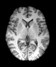
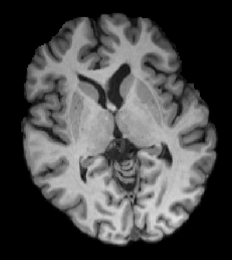
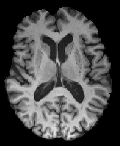
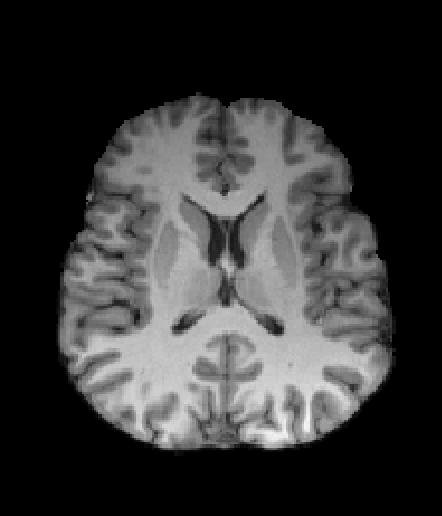
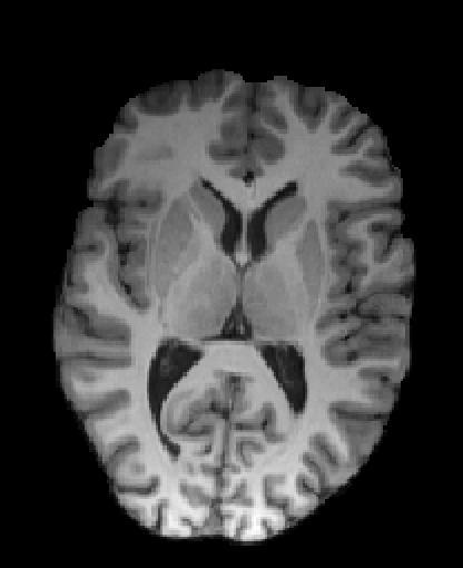
6 Example Results Brain Surface Model Extracted Brain Surface (not what we aim for here)

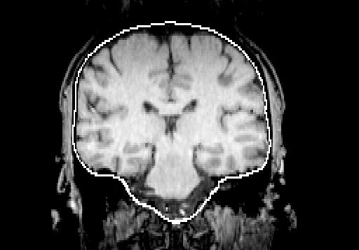
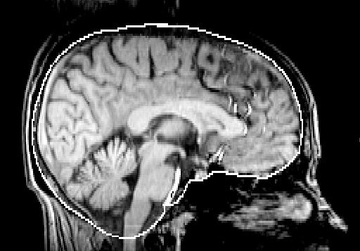
7 Example Results

8 Example Results Original Brain Extracted Brain Mask


9 Difficulties Marrow Membranes Blood (sinus) Marrow


10 Example Results Want to remove the majority of non-brain structures, leaving all the brain intact. Leaving small pieces of non-brain is unimportant for linear registration, but it is important for segmentation.
11 Overview Brain Extraction (BET) Registration concepts (FLIRT & FNIRT) Practical applications (FLIRT & FNIRT) Single-stage registration Multi-stage registrations EPI distortion correction Pathological image registration
12 What is Registration?
13 What is Registration? Voxel location = anatomical location with accurate intensity values
14 Basic Registration Concepts Need to understand: Image spaces Spatial Transformations Cost Functions Interpolation
15 Basic Registration Concepts Need to understand: Image spaces Spatial Transformations Cost Functions Interpolation
 MNI is not quite")
16 Standard Space Common reference coordinate system for reporting/describing Register all members of a group to this space for group studies Original Talairach & Tournoux coords based on one postmortem brain Now use standard images based on non-linear group average (MNI152) MNI is not quite Talairach
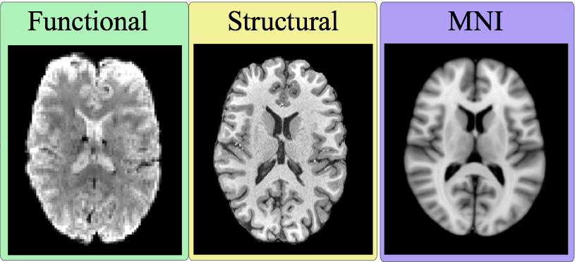
17 Other Spaces FMRI Structural Standard All images in the same space are aligned Different images different spaces e.g. standard space, structural space, functional space Can have different resolution images in the same space e.g. 1mm and 2mm versions of standard space images Want to move image-related info between spaces e.g. a mask from standard space to structural space
18 Other Spaces Transform Transform FMRI Structural Standard Need to registration between spaces (via images) and get the transformations before transforming/moving/resampling any image-related info (like masks or atlas ROIs) Can have versions of the same image (e.g. a mask) in several different spaces FSL tools (e.g. FEAT) often move things between spaces
19 Other Spaces Transform Transform FMRI Structural Standard Inverse Transform Inverse Transform Need to registration between spaces (via images) and get the transformations before transforming/moving/resampling any image-related info (like masks or atlas ROIs) Can have versions of the same image (e.g. a mask) in several different spaces FSL tools (e.g. FEAT) often move things between spaces
20 Image (Voxel) Coordinates Confusingly, there are many types of coordinates Voxel coordinates in FSL: Integers between 0 and N-1 inclusive Refer to the whole voxel Origin in the lower-left corner: (0,0,0) Axes are not aligned with the anatomy Cannot distinguish left from right by voxel coordinate values FSLeyes reports these Used by FSL commands & same as NIfTI coords z Nz-1 0 Nx-1 x y Ny-1
21 Standard Space Coordinates Standard Space coordinates in FSL: Real numbers, in units of mm Origin (0,0,0) near centre of image (anatomical landmark; e.g. anterior commisure) Axes aligned with anatomy (left and right specified) z Several standard spaces exist: MNI, Talairach, BrainWeb, etc FSLeyes also reports these when possible P L S I 0 A R y x
22 Basic Registration Concepts Need to understand: Image spaces Spatial Transformations Cost Functions Interpolation
23 Spatial Transformations To align images must transform them Many types of transformation Degrees of Freedom (DOF) partially describe transform Examples: Rigid Body (6 DOF) Affine (12 DOF) Non-linear (12 - millions DOF)
24 Rigid-Body Transformations 6 DOF in 3D Includes: 3 Rotations


25 Rigid-Body Transformations 6 DOF in 3D Includes: 3 Rotations 3 Translations Used for within-subject registrations


26 Affine Transformations 12 DOF in 3D Linear Transf. Includes: 3 Rotations 3 Translations 3 Scalings
27 Affine Transformations 12 DOF in 3D Linear Transf. Includes: 3 Rotations 3 Translations 3 Scalings 3 Skews/Shears Used for eddy current correction and initialising non-linear registration
28 Non-Linear Transformations More than 12 DOF Can be purely local Subject to constraints: Basis Functions e.g. B-Splines Regularisation Topology-preservation Used for good quality between-subject registrations
29 Non-Linear Transformations Before Registration Linear Registration Nonlinear Registration Reference (MNI152)
30 What transform/dof do I use? Rigid body (6 DOF) - within-subject motion Non-linear (lots of DOF!) - high-quality image (resolution, contrast) & same modality of reference/template - better with a non-linear template (e.g. MNI152_T1_2mm) Affine (12 DOF) - needed as a starting point for non-linear - align to affine template, or using lower quality images, or eddy current correction Global scaling (7 DOF) - within-subject but with global scaling (equal in x,y,z) - corrects for scanner scaling drift in longitudinal studies More DOF is NOT always better (e.g. within-subject)
31 What do the transformations look like? A = a 11 a 12 a 13 a 14 a 21 a 22 a 23 a 24 a 31 a 32 a 33 a An affine transformation is represented by these 12 numbers. This matrix multiplies coordinate vectors to define the transformed coordinates. A non-linear transformation can be represented by a deformation field.


 x-component Deformation-field")
32 Non-linear deformation The deformation-field (also called a displacement-field or just warp ) is stored using images with one 3D image per component of the vector NB: this is not the default output in FSL (the default is a coefficient file) x-component Deformation-field y-component mm
33 Non-linear deformation Regularisation, Warp Resolution and DOF Various ways of controlling warp smoothness Less DOF = smoother Spacing of points = warp resolution = regularisation = DOF Lower warp resolution = smoother Higher regularisation = smoother
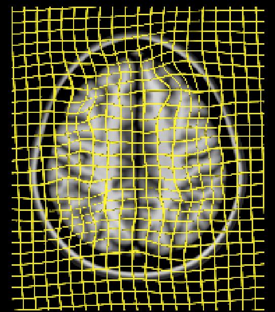
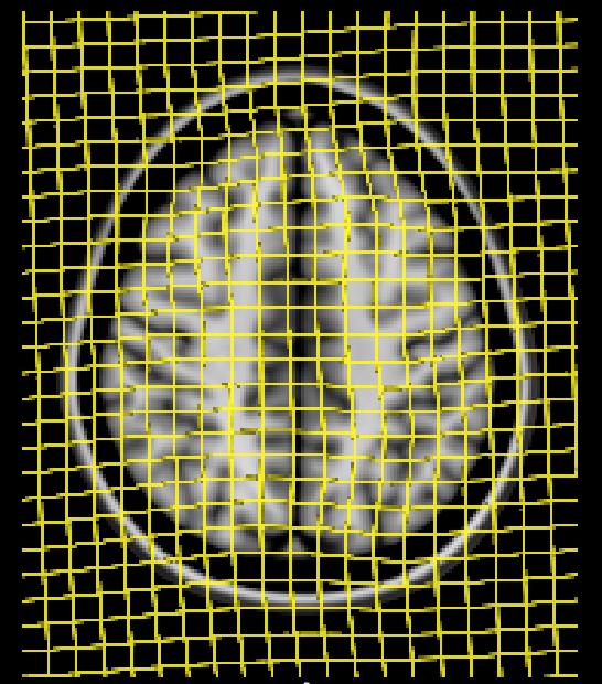
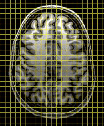
34 Non-linear deformation High Regularisation Lower Regularisation Input MNI
35 Non-linear deformation Regularisation, Warp Resolution and DOF Various ways of controlling warp smoothness Less DOF = smoother Spacing of points = warp resolution = regularisation = DOF Lower warp resolution = smoother Higher regularisation = smoother Default warp resolution of 10mm is a good compromise for MNI152 Between two subjects can use less smooth warps (less regularisation, higher warp resolution, more DOF)
36 Basic Registration Concepts Need to understand: Image spaces Spatial Transformations Cost Functions Interpolation


37 Cost Function Measures goodness of alignment Seek the minimum value Several main varieties _ = Similarity function is opposite (maximum sought)
38 FLIRT: Cost Functions FMRIB s Linear Image Registration Tool
39 FLIRT: Cost Functions Important: Allowable image modalities Less important: Details Least Squares Normalised Correlation Correlation Ratio Mutual Information Normalised Mutual Info. BBR Same modality (exact sequence parameters) Same modality (can change brightness & contrast) Any MR modalities Any modalities (including CT, PET, etc.) Any modalities (including CT, PET, etc.) Within-subject EPI to structural (see later)
40 FNIRT: Cost Functions FMRIB s Non-linear Image Registration Tool

 Without RF modelling FNIRT: Cost Functions")
41 Only uses Least Squares as cost function so images must be of the same modality/sequence Also includes an explicit model for bias field (RF inhomog.) Estimate displacement field and RF bias field together Options exist to control bias field (turn off/on, smoothness) Without RF modelling FNIRT: Cost Functions Template With RF modelling
42 Basic Registration Concepts Need to understand: Image spaces Spatial Transformations Cost Functions Interpolation
43 Interpolation Finds intensity values between grid points Various types include Nearest Neighbour Trilinear Spline Sinc k-space methods Fast, but blocky - can be used for discrete labels
44 Interpolation Finds intensity values between grid points Various types include Nearest Neighbour Trilinear Spline Sinc k-space methods Fast, with some blurring - most common option
45 Interpolation Finds intensity values between grid points Various types include Nearest Neighbour Trilinear Spline Sinc k-space methods Slower (spline is fairly fast) - creates sharp images but can create values outside the original range
46 Interpolation Nearest Neighbour Trilinear Spline Affects accuracy of subsequent analysis Important for quantitative imaging Can affect size of artefacts

47 Applying Transformations Step 1: Estimating a transformation finding the transformation no resampling transformation
48 Applying Transformations Step 1: Estimating a transformation finding the transformation no resampling Step 2: Resampling applying a transformation thus creating a new, modified image Registration can mean either transformation Usually delay resampling as it reduces image quality Other terms: coregistration & spatial normalisation
49 Transforming Masks Mask values are normally 0 and 1 (integer format) Interpolation gives values in between if rounded to integer mask "shrinks Ensure output datatype = float (applywarp & flirt default) Re-threshold (binarize) the transformed mask "Correct" thresholding depends on the particular case Threshold near 0.0 to include partial-volume edges Threshold near 1.0 to exclude partial-volume edges Threshold at 0.5 to keep the same size (approx)

50 0.1 Threshold 0.5 Threshold 0.9 Threshold Transforming Masks
51 Overview Brain Extraction (BET) Registration concepts (FLIRT & FNIRT) Practical applications (FLIRT & FNIRT) Single-stage registration Multi-stage registrations EPI distortion correction Pathological image registration
52 Registration with FSL Two main tools: FNIRT & FLIRT (FMRIB s Non-Linear/Linear Image Registration Tool) Both tools used by FMRI and Diffusion tools (FEAT, MELODIC & FDT)
 Bias-field correction (FAST - see later) Note that labels are correct in both")
53 Preliminary Steps Recommended steps: Reorientation (fslreorient2std) Brain Extraction (BET) Bias-field correction (FAST - see later) Note that labels are correct in both cases
54 Overview Brain Extraction (BET) Registration concepts (FLIRT & FNIRT) Practical applications (FLIRT & FNIRT) Single-stage registration Multi-stage registrations EPI distortion correction Pathological image registration
55 Case Study Scenario: Have two (or more) different types of images from the same subject For example, T1-weighted and T2-weighted images Objective: Have images aligned so that, for example, they can be used for multi-modal segmentation Solution: FLIRT with 6 DOF (rigid-body)
56 Single-stage Registration Input Reference Single subject 6 DOF = FLIRT T2-wt to T1-wt multi-modal cost function (e.g. default of correlation ratio) Run brain extraction on both images Choose image with better resolution or contrast as the reference Always check your output BET
 slices for a static view use (as in FEAT) slices")
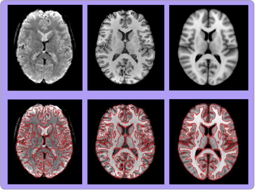
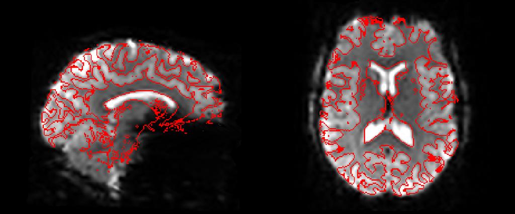
57 Visual Check Always assess registration quality visually! Can use: FSLeyes (using overlay or flicking between images) slices for a static view use (as in FEAT) slices T2_to_T1im T1im Grayscale from first image Red edges from second image
58 Registration in FSL Input In FSL the reference image controls the FOV and resolution of the output image Transformations are given: from input space to reference space Reference Inverse transformations can easily be calculated to go from reference space to input space when needed Can overlay images in FSLeyes with different FOV or resolution: i.e. images can be in different spaces and resolutions Images can be resampled into a different space by applying a previously derived transformation
59 Troubleshooting Check that voxel size is correct for both images Check if brain extraction is OK: small, isolated errors are fine, but large or consistent shifts are not Limit search (if initially fairly well aligned) Try different (but still appropriate) cost functions Check for artefacts and pathologies (see later - cost function weighting)
60 Overview Brain Extraction (BET) Registration concepts (FLIRT & FNIRT) Practical applications (FLIRT & FNIRT) Single-stage registration Multi-stage registrations EPI distortion correction Pathological image registration
61 Case Study Scenario: Doing a functional (or diffusion) study Have EPI and T1-weighted of each subject Objective: Need to register images to a common (standard) space to allow the group study to be performed Solution: 2-stage registration with FLIRT & FNIRT (in FEAT)
62 Two Stage Registration Registering very different images is difficult due to: Differences in individual anatomies Different contrasts in various modalities Distortions which differ between images To register an EPI to a standard space template (e.g. MNI152) use a structural intermediate image Automatically done by FEAT GUI (some user control) Need to manually run brain extraction (not on EPI usually*) 6 DOF FLIRT >12 DOF FNIRT**
63 Registration for FMRI Analysis (FEAT) FMRI (implicit) FLIRT Main Structural FLIRT + FNIRT Standard NB: actually need brain extracted and original images for FNIRT
64 Registration Registration MNI Space
65 Registration for FMRI Analysis
66 Registration for FMRI Analysis Functional image in standard space: fmri in grey + red lines from MNI (standard space template)
67 Registration for FMRI Analysis Example func (fmri) in highres (structural) space: top line = fmri in grey + red lines from structural bottom line = structural in grey + red lines from fmri Also: fsleyes highres example_func2highres (in reg subdirectory of feat directory)
68
69 Registration for FMRI Analysis Highres (structural) in standard space (MNI) top line = structural in grey + red lines from MNI bottom line = MNI in grey + red lines from structural Also: fsleyes standard highres2standard
70
71 Registration for FMRI Analysis Example func (fmri) in standard space (MNI) top line = fmri in grey + red lines from MNI bottom line = MNI in grey + red lines from fmri Also: fsleyes standard example_func2standard
72 Registration for FMRI Analysis Core registrations are: example_func 2 highres + highres 2 standard The example_func2standard is derived from these Any failures need to be fixed in the core registrations (and then can run updatefeatreg)
73 Overview Brain Extraction (BET) Registration concepts (FLIRT & FNIRT) Practical applications (FLIRT & FNIRT) Single-stage registration Multi-stage registrations EPI distortion correction Pathological image registration
74 Case Study Scenario: Doing a functional (or diffusion) study Objective: Want to correct for distortions in EPI as otherwise the registrations are inaccurate Solution: Fieldmap-based correction using FUGUE/FEAT
 EPI T1-weighted")
75 Registration of EPI Problem: - EPI images distorted and suffer signal loss - standard registration does not work well Solution: - undo distortion by unwarping - ignore areas of high signal loss - needs a fieldmap (special acquisition) EPI T1-weighted anatomical

76 T1-weighted (aligned) Signal Loss Distortion


77 B0 Field Inhomogeneities EPI is very sensitive to any deviations from a perfectly uniform B0 field Air-tissue interfaces cause magnetic disturbances A separate fieldmap image measures the B0 deviations fieldmap EPI distortion signal loss Courtesy of D. Greve, MGH
78 Using Fieldmaps From the fieldmap image we get: Magnitude of spatial distortions (phase-encode direction only) Estimate of signal loss PE Only takes a few minutes to acquire one fieldmap - and it massively improves registration Need a new fieldmap for each scanning session as it changes (e.g. it depends on head orientation) EPI B 0 Fieldmap
79 Unwarping with Fieldmaps Used to improve registration of EPI and structural scan It does not restore signal in the frontal lobe Fieldmap Original EPI Unwarped EPI
80 Unwarping with Fieldmaps Used to improve registration of EPI and structural scan Fieldmap Original EPI It does not restore signal in the frontal lobe It does not do anything about motion correction It does use fieldmap image to calculate distortion and unwarp EPI It does deweight areas with substantial signal loss in the registration Unwarped EPI
81 Fieldmap Acquisition Fieldmaps are becoming standard sequences Only takes a few minutes to acquire - best either immediately before or after EPI scans (but this is not crucial) Four main types of acquisitions: Gradient Echo Asymmetric Spin Echo EPI Blip-reversed b=0 pair (EPI) Each based on a pair of images with different TE (record these TE values) Distortion & Signal Loss Magnitude part of fieldmap Phase difference of images Crucially requires the phase information (not only the magnitude, unlike the vast majority of other images)
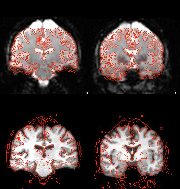
 Need good structurals (not too much bias field) Also requires anatomical contrast in the EPI Driven by intensity difference across boundary (samples) More robust to")
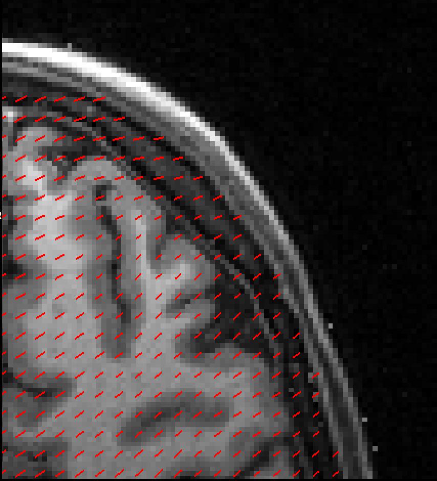
82 Boundary-Based Registration (BBR) EPI to structural registration (Greve & Fischl, NeuroImage, 2009) incorporates fieldmap correction (previously FUGUE) used in FEAT (B0 unwarping) Uses white-matter boundaries (via T1w segmentation) Need good structurals (not too much bias field) Also requires anatomical contrast in the EPI Driven by intensity difference across boundary (samples) More robust to pathologies and artefacts in EPI T1w T1w + boundaries EPI + boundaries EPI

83 Distortion Correction Structural Image Registration without Distortion Correction Registration with Distortion Correction
84 Distortion Correction within FEAT
85 Distortion Correction within FEAT Fieldmap in rad/s TE difference (sec) = Phase difference (rad) B 0 Field (rad/s) Need to prepare the fieldmap image: Fsl_prepare_fieldmap (for Siemens)
86 Distortion Correction within FEAT Fieldmap in rad/s Fieldmap Magnitude... needs this and aggressive BET (leave no non-brain) for best performance Input file = brain extracted file... but also needs to find original*
87 Distortion Correction within FEAT Fieldmap in rad/s Fieldmap Magnitude EPI echo spacing (ms) Also called dwell time Normally about ms v v Divide value by any acceleration factor x v Time between echos in k- space
88 Distortion Correction within FEAT Fieldmap in rad/s Fieldmap Magnitude EPI echo spacing (ms) EPI echo time (ms) Normally about 30-40ms at 3T
89 Distortion Correction within FEAT Fieldmap in rad/s Fieldmap Magnitude EPI echo spacing (ms) EPI echo time (ms) Unwarp (PE) direction - Often A-P but can be anything - Cannot tell if it is + or - - Try both and see what works (see practical)
90 Distortion Correction within FEAT Fieldmap in rad/s Fieldmap Magnitude EPI echo spacing (ms) EPI echo time (ms) Unwarp (PE) direction Signal loss thresh % Ignore voxels with more than this signal loss in registration
91 Fieldmap use in FEAT
92 Fieldmap use in FEAT This should be fairly uniform everywhere except where the field is not uniform - inferior temporal and frontal lobes
93 Fieldmap use in FEAT This should be mostly yellow - red voxels get ignored in the registration (lots of red is bad)
94 Fieldmap use in FEAT This shows how much each voxel moves - check that the range is sensible (anything from +/- 3 to +/- 20 is common)
95 Fieldmap use in FEAT This shows the white matter edges from the structural on the fieldmap (to check fieldmap to structural registration - not EPI)
96 Fieldmap use in FEAT Fieldmap to highres (structural) Functional (EPI) to highres (structural) - no correction Functional (EPI) to highres (structural) - with fieldmap correction
97 Fieldmap use in FEAT Functional (EPI) to highres (structural) - no correction Functional (EPI) to highres (structural) - with fieldmap correction Movie of EPI with and without correction
98 Fieldmap use in FEAT Look for areas where unwarping (correction) changes brain shape Functional (EPI) to highres (structural) - no correction Functional (EPI) to highres (structural) - with fieldmap correction Movie of EPI with and without correction
 to NB: Using FSLeyes is often better highres (structural) - with fieldmap correction Functional (EPI) to highres (structural) - no correction Movie of EPI with and without")
99 Fieldmap use in FEAT Look for areas where unwarping (correction) changes brain shape See if these areas are better aligned with or without correction but don t trust borders with signal loss areas Functional (EPI) to NB: Using FSLeyes is often better highres (structural) - with fieldmap correction Functional (EPI) to highres (structural) - no correction Movie of EPI with and without correction
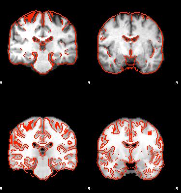
100 Standard FLIRT BBR and Fieldmaps
101 BBR FLIRT BBR and Fieldmaps
102 BBR FLIRT with Fieldmap BBR and Fieldmaps
103 BBR and Fieldmaps Standard FLIRT
104 BBR and Fieldmaps BBR FLIRT
105 BBR and Fieldmaps BBR FLIRT with Fieldmap
106 Overview Brain Extraction (BET) Registration concepts (FLIRT & FNIRT) Practical applications (FLIRT & FNIRT) Single-stage registration Multi-stage registrations EPI distortion correction Pathological image registration
107 Case Study Scenario: Have images containing a known pathology (or artefact) which looks different in different images For example, some sequences (e.g. FLAIR) highlight lesions that are hard to see in other sequences Objective: Align the images based on the healthy tissue, but ignoring the area of the pathology (or artefact) Solution: Cost-Function Weighting (FLIRT or FNIRT)


108 Cost Function Weighting Artefacts and pathologies introduce non-matching image regions weighting image Cost (similarity) functions assume that all of the images can be matched weighting image black=0; white=1 Use a weighting image to down-weight nonmatching regions
109 Cost Function Weighting All FLIRT & FNIRT cost functions can be weighted Weighting for reference image, input image or both Voxel weights are relative, reflecting its importance in overall matching - Zero, or small, values for corrupted areas e.g. gross pathology or artefact - Large values for important areas/regions e.g. ventricular matching Do not assign zero to the background as then the brain/background contrast is lost
110 Troubleshooting Registrations Check the images: voxel sizes, artefacts, large bias field Check the brain extraction: look for large/consistent errors For EPI: acquire and use fieldmap to unwarp distortion For FMRI or diffusion: use multi-stage registration (e.g. via GUIs) with a structural image for best results If pathologies/artefacts exist: use cost-function deweighting If images are nearly aligned: try limiting the search For FLIRT: can try different cost functions For FNIRT: check initial affine alignment is OK For small FOV: acquire whole-brain EPI for multi-stage reg


111 Advanced Registration 2D - 3D Registration Severe Pathology Surface-based Registration e.g. connectivity-driven (FMRI) Other Image Modalities e.g. diffusion imaging data MR Spectroscopic Imaging (MRSI)

112 That s all folks
113 Advanced Topics Small FOV Advanced non-linear registration (FNIRT)
114 Case Study Scenario: Functional study using a small FOV (e.g. a few slices) Often done to obtain better resolution scans over ROI Objective: Get activation results registered well to the full brain (and standard space) Solution: Scan one whole-brain EPI and use a 3-stage registration

115 Registration within FEAT (small FOV) If your FMRI scans only cover part of the brain... Acquire one wholebrain EPI volume: it only takes a few seconds to scan but makes registration work much better Then use the 3-stage approach
116 Partial Brain EPI & Unwarping In partial FOV studies, registration is massively improved by multi-stage registration: 1. Partial Brain to Full Brain EPI Desirable for full brain to contain exactly the same slices so that registration is simple 2. Full Brain EPI to Structural apply unwarping (full brain field map) 3. Structural to Standard All taken care of when using the FEAT GUI Partial Brain FMRI timeseries Full Brain Single Image (an extra acquisition - but only takes seconds!)

117 Registration within FEAT (small FOV)

118 Registration within FEAT (small FOV)

119 Registration within FEAT (small FOV)
120 Advanced Topics Small FOV Advanced non-linear registration (FNIRT)
121 Case Study Scenario: Structural study where you want to non-linearly register good quality images of different subjects Objective: Achieve accurate registration for each image pair so that images or deformation fields can be analysed Solution: Use a customised FNIRT configuration file
122 FNIRT Controls FNIRT has many different controls available to the user: warp resolution (control/knot point spacing) cost function (bias correction or not) desired outputs (deformation field, Jacobian, etc) config file advanced stuff! multi-resolution scales regularization strength cost-function weighting number of iterations...
123 Multi-resolution Why use lower resolutions? Faster calculations 8mm - 80 thousand calcs 1mm - 40 million calcs 8mm Insensitive to fine structure Progressively improve resolution during registration 1mm


124 Sub-sampling in FNIRT Allows optimization to avoid distracting detail Ref Sub-sample by 4 Sub-sample by 2 Sub-sample by FLIRT matrix M 1 Input Yielding these warps


125 Regularization Need to prevent deformation from changing topology (folding/tearing/etc) Control strength of regularization vs image similarity (cost) by a constant, λ - often linked with multi-res Final results not too sensitive to λ Affine λ=1000 λ=100 λ=10 Deformation -fields
FSL Pre-Processing Pipeline
 The Art and Pitfalls of fmri Preprocessing FSL Pre-Processing Pipeline Mark Jenkinson FMRIB Centre, University of Oxford FSL Pre-Processing Pipeline Standard pre-processing: Task fmri Resting-state fmri
Introduction to MRI data processing with FSL. Anna Blazejewska
 Introduction to MRI data processing with FSL Anna Blazejewska FSL = FMRIB Software Library FMRIB = Functional Magnetic Resonance Imaging of the Brain @ Oxford since 2000, last stable FSL 5.0, free! for
Introduction to fmri. Pre-processing
 Introduction to fmri Pre-processing Tibor Auer Department of Psychology Research Fellow in MRI Data Types Anatomical data: T 1 -weighted, 3D, 1/subject or session - (ME)MPRAGE/FLASH sequence, undistorted
Basic principles of MR image analysis. Basic principles of MR image analysis. Basic principles of MR image analysis
 Basic principles of MR image analysis Basic principles of MR image analysis Julien Milles Leiden University Medical Center Terminology of fmri Brain extraction Registration Linear registration Non-linear
Last Time. This Time. Thru-plane dephasing: worse at long TE. Local susceptibility gradients: thru-plane dephasing
 Motion Correction Last Time Mutual Information Optimiation Decoupling Translation & Rotation Interpolation SPM Example (Least Squares & MI) A Simple Derivation This Time Reslice example SPM Example : Remind
Computational Neuroanatomy
 Computational Neuroanatomy John Ashburner john@fil.ion.ucl.ac.uk Smoothing Motion Correction Between Modality Co-registration Spatial Normalisation Segmentation Morphometry Overview fmri time-series kernel
This Time. fmri Data analysis
 This Time Reslice example Spatial Normalization Noise in fmri Methods for estimating and correcting for physiologic noise SPM Example Spatial Normalization: Remind ourselves what a typical functional image
Basic fmri Design and Analysis. Preprocessing
 Basic fmri Design and Analysis Preprocessing fmri Preprocessing Slice timing correction Geometric distortion correction Head motion correction Temporal filtering Intensity normalization Spatial filtering
Image Registration + Other Stuff
 Image Registration + Other Stuff John Ashburner Pre-processing Overview fmri time-series Motion Correct Anatomical MRI Coregister m11 m 21 m 31 m12 m13 m14 m 22 m 23 m 24 m 32 m 33 m 34 1 Template Estimate
Functional MRI data preprocessing. Cyril Pernet, PhD
 Functional MRI data preprocessing Cyril Pernet, PhD Data have been acquired, what s s next? time No matter the design, multiple volumes (made from multiple slices) have been acquired in time. Before getting
SPM8 for Basic and Clinical Investigators. Preprocessing. fmri Preprocessing
 SPM8 for Basic and Clinical Investigators Preprocessing fmri Preprocessing Slice timing correction Geometric distortion correction Head motion correction Temporal filtering Intensity normalization Spatial
fmri pre-processing Juergen Dukart
 fmri pre-processing Juergen Dukart Outline Why do we need pre-processing? fmri pre-processing Slice time correction Realignment Unwarping Coregistration Spatial normalisation Smoothing Overview fmri time-series
FSL Pre-Processing Pipeline
 The Art and Pitfalls of fmri Preprocessing FSL Pre-Processing Pipeline Mark Jenkinson FMRIB Centre, University of Oxford FSL Pre-Processing Pipeline Standard pre-processing: Task fmri Resting-state fmri
Functional MRI in Clinical Research and Practice Preprocessing
 Functional MRI in Clinical Research and Practice Preprocessing fmri Preprocessing Slice timing correction Geometric distortion correction Head motion correction Temporal filtering Intensity normalization
Fmri Spatial Processing
 Educational Course: Fmri Spatial Processing Ray Razlighi Jun. 8, 2014 Spatial Processing Spatial Re-alignment Geometric distortion correction Spatial Normalization Smoothing Why, When, How, Which Why is
EPI Data Are Acquired Serially. EPI Data Are Acquired Serially 10/23/2011. Functional Connectivity Preprocessing. fmri Preprocessing
 Functional Connectivity Preprocessing Geometric distortion Head motion Geometric distortion Head motion EPI Data Are Acquired Serially EPI Data Are Acquired Serially descending 1 EPI Data Are Acquired
Diffusion MRI Acquisition. Karla Miller FMRIB Centre, University of Oxford
 Diffusion MRI Acquisition Karla Miller FMRIB Centre, University of Oxford karla@fmrib.ox.ac.uk Diffusion Imaging How is diffusion weighting achieved? How is the image acquired? What are the limitations,
Playing with data from lab
 Playing with data from lab Getting data off the scanner From the Patient Browser, select the folder for the study you want (or within that study, the set of images you want), and then from the Transfer
Normalization for clinical data
 Normalization for clinical data Christopher Rorden, Leonardo Bonilha, Julius Fridriksson, Benjamin Bender, Hans-Otto Karnath (2012) Agespecific CT and MRI templates for spatial normalization. NeuroImage
SPM8 for Basic and Clinical Investigators. Preprocessing
 SPM8 for Basic and Clinical Investigators Preprocessing fmri Preprocessing Slice timing correction Geometric distortion correction Head motion correction Temporal filtering Intensity normalization Spatial
Automated MR Image Analysis Pipelines
 Automated MR Image Analysis Pipelines Andy Simmons Centre for Neuroimaging Sciences, Kings College London Institute of Psychiatry. NIHR Biomedical Research Centre for Mental Health at IoP & SLAM. Neuroimaging
Manual image registration in BrainVoyager QX Table of Contents
 Manual image registration in BrainVoyager QX Table of Contents Manual image registration in BrainVoyager QX......1 Performing manual alignment for functional to anatomical images......2 Step 1: preparation......2
Structural Segmentation
 Structural Segmentation FAST tissue-type segmentation FIRST sub-cortical structure segmentation FSL-VBM voxelwise grey-matter density analysis SIENA atrophy analysis FAST FMRIB s Automated Segmentation
Methods for data preprocessing
 Methods for data preprocessing John Ashburner Wellcome Trust Centre for Neuroimaging, 12 Queen Square, London, UK. Overview Voxel-Based Morphometry Morphometry in general Volumetrics VBM preprocessing
A Model-Independent, Multi-Image Approach to MR Inhomogeneity Correction
 Tina Memo No. 2007-003 Published in Proc. MIUA 2007 A Model-Independent, Multi-Image Approach to MR Inhomogeneity Correction P. A. Bromiley and N.A. Thacker Last updated 13 / 4 / 2007 Imaging Science and
Journal of Articles in Support of The Null Hypothesis
 Data Preprocessing Martin M. Monti, PhD UCLA Psychology NITP 2016 Typical (task-based) fmri analysis sequence Image Pre-processing Single Subject Analysis Group Analysis Journal of Articles in Support
Preprocessing II: Between Subjects John Ashburner
 Preprocessing II: Between Subjects John Ashburner Pre-processing Overview Statistics or whatever fmri time-series Anatomical MRI Template Smoothed Estimate Spatial Norm Motion Correct Smooth Coregister
Structural Segmentation
 Structural Segmentation FAST tissue-type segmentation FIRST sub-cortical structure segmentation FSL-VBM voxelwise grey-matter density analysis SIENA atrophy analysis FAST FMRIB s Automated Segmentation
BDP: BrainSuite Diffusion Pipeline. Chitresh Bhushan
 BDP: BrainSuite Diffusion Pipeline Chitresh Bhushan Why diffusion MRI? T 2 weighted MPRAGE FA map Fiber track Quantify microstructural tissue characteristics Structural connectivity Connectome Clinical
SPM Introduction. SPM : Overview. SPM: Preprocessing SPM! SPM: Preprocessing. Scott Peltier. FMRI Laboratory University of Michigan
 SPM Introduction Scott Peltier FMRI Laboratory University of Michigan! Slides adapted from T. Nichols SPM! SPM : Overview Library of MATLAB and C functions Graphical user interface Four main components:
Supplementary methods
 Supplementary methods This section provides additional technical details on the sample, the applied imaging and analysis steps and methods. Structural imaging Trained radiographers placed all participants
1 Introduction Motivation and Aims Functional Imaging Computational Neuroanatomy... 12
 Contents 1 Introduction 10 1.1 Motivation and Aims....... 10 1.1.1 Functional Imaging.... 10 1.1.2 Computational Neuroanatomy... 12 1.2 Overview of Chapters... 14 2 Rigid Body Registration 18 2.1 Introduction.....
BDP: BrainSuite Diffusion Pipeline. Chitresh Bhushan
 BDP: BrainSuite Diffusion Pipeline Chitresh Bhushan Why diffusion MRI? T 2 weighted MPRAGE FA map Fiber track Quantify microstructural tissue characteristics Structural connectivity Connectome Clinical
SPM Introduction SPM! Scott Peltier. FMRI Laboratory University of Michigan. Software to perform computation, manipulation and display of imaging data
 SPM Introduction Scott Peltier FMRI Laboratory University of Michigan Slides adapted from T. Nichols SPM! Software to perform computation, manipulation and display of imaging data 1 1 SPM : Overview Library
2. Creating Field Maps Using the Field Map GUI (Version 2.0) in SPM5
 in SPM5") 1. Introduction This manual describes how to use the Field Map Toolbox Version 2.0 for creating unwrapped field maps that can be used to do geometric distortion correction of EPI images in SPM5. 1. 1.
1. Introduction This manual describes how to use the Field Map Toolbox Version 2.0 for creating unwrapped field maps that can be used to do geometric distortion correction of EPI images in SPM5. 1. 1.
Image Processing for fmri John Ashburner. Wellcome Trust Centre for Neuroimaging, 12 Queen Square, London, UK.
 Iage Processing for fmri John Ashburner Wellcoe Trust Centre for Neuroiaging, 12 Queen Square, London, UK. Contents * Preliinaries * Rigid-Body and Affine Transforations * Optiisation and Objective Functions
FROM IMAGE RECONSTRUCTION TO CONNECTIVITY ANALYSIS: A JOURNEY THROUGH THE BRAIN'S WIRING. Francesca Pizzorni Ferrarese
 FROM IMAGE RECONSTRUCTION TO CONNECTIVITY ANALYSIS: A JOURNEY THROUGH THE BRAIN'S WIRING Francesca Pizzorni Ferrarese Pipeline overview WM and GM Segmentation Registration Data reconstruction Tractography
White Pixel Artifact. Caused by a noise spike during acquisition Spike in K-space <--> sinusoid in image space
 White Pixel Artifact Caused by a noise spike during acquisition Spike in K-space sinusoid in image space Susceptibility Artifacts Off-resonance artifacts caused by adjacent regions with different
FMRI Pre-Processing and Model- Based Statistics
 FMRI Pre-Processing and Model- Based Statistics Brief intro to FMRI experiments and analysis FMRI pre-stats image processing Simple Single-Subject Statistics Multi-Level FMRI Analysis Advanced FMRI Analysis
Lilla Zöllei A.A. Martinos Center, MGH; Boston, MA
 Lilla Zöllei lzollei@nmr.mgh.harvard.edu A.A. Martinos Center, MGH; Boston, MA Bruce Fischl Gheorghe Postelnicu Jean Augustinack Anastasia Yendiki Allison Stevens Kristen Huber Sita Kakonoori + the FreeSurfer
Analysis of fmri data within Brainvisa Example with the Saccades database
 Analysis of fmri data within Brainvisa Example with the Saccades database 18/11/2009 Note : All the sentences in italic correspond to informations relative to the specific dataset under study TP participants
The simulator can be applied in a number of diverse applications which span both
 Chapter 6 Simulator applications The simulator can be applied in a number of diverse applications which span both MRI and FMRI fields These applications include the simulation and removal of various imaging
Neuroimaging and mathematical modelling Lesson 2: Voxel Based Morphometry
 Neuroimaging and mathematical modelling Lesson 2: Voxel Based Morphometry Nivedita Agarwal, MD Nivedita.agarwal@apss.tn.it Nivedita.agarwal@unitn.it Volume and surface morphometry Brain volume White matter
CHAPTER 9: Magnetic Susceptibility Effects in High Field MRI
 Figure 1. In the brain, the gray matter has substantially more blood vessels and capillaries than white matter. The magnified image on the right displays the rich vasculature in gray matter forming porous,
Spatial Preprocessing
 Spatial Preprocessing Overview of SPM Analysis fmri time-series Design matrix Statistical Parametric Map John Ashburner john@fil.ion.ucl.ac.uk Motion Correction Smoothing General Linear Model Smoothing
Automatic segmentation of the cortical grey and white matter in MRI using a Region Growing approach based on anatomical knowledge
 Automatic segmentation of the cortical grey and white matter in MRI using a Region Growing approach based on anatomical knowledge Christian Wasserthal 1, Karin Engel 1, Karsten Rink 1 und André Brechmann
User s Guide Neuroimage Processing ToolKit (NPTK) Version.1.7 (beta) fmri Registration Software Pipeline for Functional Localization
 Version.1.7 (beta) fmri Registration Software Pipeline for Functional Localization") User s Guide Neuroimage Processing ToolKit (NPTK) Version.1.7 (beta) fmri Registration Software Pipeline for Functional Localization Software Written by Ali Gholipour SIP Lab, UTD, 2005-2007 Revision 1.7
User s Guide Neuroimage Processing ToolKit (NPTK) Version.1.7 (beta) fmri Registration Software Pipeline for Functional Localization Software Written by Ali Gholipour SIP Lab, UTD, 2005-2007 Revision 1.7
Statistical Analysis of Neuroimaging Data. Phebe Kemmer BIOS 516 Sept 24, 2015
 Statistical Analysis of Neuroimaging Data Phebe Kemmer BIOS 516 Sept 24, 2015 Review from last time Structural Imaging modalities MRI, CAT, DTI (diffusion tensor imaging) Functional Imaging modalities
RIGID IMAGE REGISTRATION
 RIGID IMAGE REGISTRATION Duygu Tosun-Turgut, Ph.D. Center for Imaging of Neurodegenerative Diseases Department of Radiology and Biomedical Imaging duygu.tosun@ucsf.edu What is registration? Image registration
How to create a head model
 How to create a head model This document describes the command line tools: mri2mesh: Central tool to reconstruct a head model from T1w and T2w data dwi2cond: Reconstruct conductivity tensors for brain
Preprocessing of fmri data
 Preprocessing of fmri data Pierre Bellec CRIUGM, DIRO, UdM Flowchart of the NIAK fmri preprocessing pipeline fmri run 1 fmri run N individual datasets CIVET NUC, segmentation, spatial normalization slice
Sources of Distortion in Functional MRI Data
 Human Brain Mapping 8:80 85(1999) Sources of Distortion in Functional MRI Data Peter Jezzard* and Stuart Clare FMRIB Centre, Department of Clinical Neurology, University of Oxford, Oxford, UK Abstract:
PHILIPS B0 UNWARPING IN FSL FEAT ON SHARK (OR LOCALLY)
") PHILIPS B0 UNWARPING IN FSL FEAT ON SHARK (OR LOCALLY) LATEST VERSION: MARCH, 2017 ANNA VAN T VEER & ANNE HAFKEMEIJER HENK VAN STEENBERGEN & JANNA MARIE BAS-HOOGENDAM This le is updated every now and.
PHILIPS B0 UNWARPING IN FSL FEAT ON SHARK (OR LOCALLY) LATEST VERSION: MARCH, 2017 ANNA VAN T VEER & ANNE HAFKEMEIJER HENK VAN STEENBERGEN & JANNA MARIE BAS-HOOGENDAM This le is updated every now and.
The organization of the human cerebral cortex estimated by intrinsic functional connectivity
 1 The organization of the human cerebral cortex estimated by intrinsic functional connectivity Journal: Journal of Neurophysiology Author: B. T. Thomas Yeo, et al Link: https://www.ncbi.nlm.nih.gov/pubmed/21653723
Nonrigid Registration using Free-Form Deformations
 Nonrigid Registration using Free-Form Deformations Hongchang Peng April 20th Paper Presented: Rueckert et al., TMI 1999: Nonrigid registration using freeform deformations: Application to breast MR images
Whole Body MRI Intensity Standardization
 Whole Body MRI Intensity Standardization Florian Jäger 1, László Nyúl 1, Bernd Frericks 2, Frank Wacker 2 and Joachim Hornegger 1 1 Institute of Pattern Recognition, University of Erlangen, {jaeger,nyul,hornegger}@informatik.uni-erlangen.de
Table of Contents. IntroLab < SPMLabs < Dynevor TWiki
 Table of Contents Lab 1: Introduction to SPM and data checking...1 Goals of this Lab...1 Prerequisites...1 An SPM Installation...1 SPM Defaults...2 L/R Brain Orientation...2 Memory Use for Data Processing...2
HST.583 Functional Magnetic Resonance Imaging: Data Acquisition and Analysis Fall 2008
 MIT OpenCourseWare http://ocw.mit.edu HST.583 Functional Magnetic Resonance Imaging: Data Acquisition and Analysis Fall 2008 For information about citing these materials or our Terms of Use, visit: http://ocw.mit.edu/terms.
Image Registration I
 Image Registration I Comp 254 Spring 2002 Guido Gerig Image Registration: Motivation Motivation for Image Registration Combine images from different modalities (multi-modality registration), e.g. CT&MRI,
HST.583 Functional Magnetic Resonance Imaging: Data Acquisition and Analysis Fall 2008
 MIT OpenCourseWare http://ocw.mit.edu HST.583 Functional Magnetic Resonance Imaging: Data Acquisition and Analysis Fall 2008 For information about citing these materials or our Terms of Use, visit: http://ocw.mit.edu/terms.
An Introduction To Automatic Tissue Classification Of Brain MRI. Colm Elliott Mar 2014
 An Introduction To Automatic Tissue Classification Of Brain MRI Colm Elliott Mar 2014 Tissue Classification Tissue classification is part of many processing pipelines. We often want to classify each voxel
MRI Physics II: Gradients, Imaging
 MRI Physics II: Gradients, Imaging Douglas C., Ph.D. Dept. of Biomedical Engineering University of Michigan, Ann Arbor Magnetic Fields in MRI B 0 The main magnetic field. Always on (0.5-7 T) Magnetizes
Introduction to Neuroimaging Janaina Mourao-Miranda
 Introduction to Neuroimaging Janaina Mourao-Miranda Neuroimaging techniques have changed the way neuroscientists address questions about functional anatomy, especially in relation to behavior and clinical
Shape-based Diffeomorphic Registration on Hippocampal Surfaces Using Beltrami Holomorphic Flow
 Shape-based Diffeomorphic Registration on Hippocampal Surfaces Using Beltrami Holomorphic Flow Abstract. Finding meaningful 1-1 correspondences between hippocampal (HP) surfaces is an important but difficult
GLM for fmri data analysis Lab Exercise 1
 GLM for fmri data analysis Lab Exercise 1 March 15, 2013 Medical Image Processing Lab Medical Image Processing Lab GLM for fmri data analysis Outline 1 Getting Started 2 AUDIO 1 st level Preprocessing
Human Connectom Project : The minimal processing Pipeline
 Human Connectom Project : The minimal processing Pipeline Human Connectom Project : The minimal processing Pipeline Van Essen DC, The WU-Minn Human Connectome Project: an overview. Neuroimage. 2013 Marcus
HST.583 Functional Magnetic Resonance Imaging: Data Acquisition and Analysis Fall 2006
 MIT OpenCourseWare http://ocw.mit.edu HST.583 Functional Magnetic Resonance Imaging: Data Acquisition and Analysis Fall 2006 For information about citing these materials or our Terms of Use, visit: http://ocw.mit.edu/terms.
Function-Structure Integration in FreeSurfer
 Function-Structure Integration in FreeSurfer Outline Function-Structure Integration Function-Structure Registration in FreeSurfer fmri Analysis Preprocessing First-Level Analysis Higher-Level (Group) Analysis
CHAPTER 2. Morphometry on rodent brains. A.E.H. Scheenstra J. Dijkstra L. van der Weerd
 CHAPTER 2 Morphometry on rodent brains A.E.H. Scheenstra J. Dijkstra L. van der Weerd This chapter was adapted from: Volumetry and other quantitative measurements to assess the rodent brain, In vivo NMR
Where are we now? Structural MRI processing and analysis
 Where are we now? Structural MRI processing and analysis Pierre-Louis Bazin bazin@cbs.mpg.de Leipzig, Germany Structural MRI processing: why bother? Just use the standards? SPM FreeSurfer FSL However:
Math in image processing
 Math in image processing Math in image processing Nyquist theorem Math in image processing Discrete Fourier Transformation Math in image processing Image enhancement: scaling Math in image processing Image
HST.583 Functional Magnetic Resonance Imaging: Data Acquisition and Analysis Fall 2008
 MIT OpenCourseWare http://ocw.mit.edu HST.583 Functional Magnetic Resonance Imaging: Data Acquisition and Analysis Fall 2008 For information about citing these materials or our Terms of Use, visit: http://ocw.mit.edu/terms.
AFNI Preprocessing: Outline, Recommendations, and New(ish) Stuff. Robert W Cox SSCC / NIMH & NINDS / NIH / DHHS / USA / EARTH
 Stuff. Robert W Cox SSCC / NIMH & NINDS / NIH / DHHS / USA / EARTH") AFNI Preprocessing: Outline, Recommendations, and New(ish) Stuff Robert W Cox SSCC / NIMH & NINDS / NIH / DHHS / USA / EARTH HBM 2016 As a work of a US Government official, this presentation is not copyrighted
AFNI Preprocessing: Outline, Recommendations, and New(ish) Stuff Robert W Cox SSCC / NIMH & NINDS / NIH / DHHS / USA / EARTH HBM 2016 As a work of a US Government official, this presentation is not copyrighted
MR IMAGE SEGMENTATION
 MR IMAGE SEGMENTATION Prepared by : Monil Shah What is Segmentation? Partitioning a region or regions of interest in images such that each region corresponds to one or more anatomic structures Classification
Implementation of Advanced Image Guided Radiation Therapy
 Image Acquisition Course Outline Principles, characteristics& applications of the available modalities Image Processing in the T x room Image guided treatment delivery What can / can t we do in the room
fmri Basics: Spatial Pre-processing Workshop
 fmri Basics: Spatial Pre-processing Workshop Starting a VNC session: Most of your fmri analysis will be done on the central Linux machines accessed via a VNC (Virtual Network Computing) server. This is
Surface-based Analysis: Inter-subject Registration and Smoothing
 Surface-based Analysis: Inter-subject Registration and Smoothing Outline Exploratory Spatial Analysis Coordinate Systems 3D (Volumetric) 2D (Surface-based) Inter-subject registration Volume-based Surface-based
Appendix E1. Supplementary Methods. MR Image Acquisition. MR Image Analysis
 RSNA, 2015 10.1148/radiol.2015150532 Appendix E1 Supplementary Methods MR Image Acquisition By using a 1.5-T system (Avanto, Siemens Medical, Erlangen, Germany) under a program of regular maintenance (no
ASAP_2.0 (Automatic Software for ASL Processing) USER S MANUAL
 USER S MANUAL") ASAP_2.0 (Automatic Software for ASL Processing) USER S MANUAL ASAP was developed as part of the COST Action "Arterial Spin Labelling Initiative in Dementia (AID)" by: Department of Neuroimaging, Institute
ASAP_2.0 (Automatic Software for ASL Processing) USER S MANUAL ASAP was developed as part of the COST Action "Arterial Spin Labelling Initiative in Dementia (AID)" by: Department of Neuroimaging, Institute
Preprocessing I: Within Subject John Ashburner
 Preprocessing I: Within Subject John Ashburner Pre-processing Overview Statistics or whatever fmri tie-series Anatoical MRI Teplate Soothed Estiate Spatial Nor Motion Correct Sooth Coregister 11 21 31
COBRE Scan Information
 COBRE Scan Information Below is more information on the directory structure for the COBRE imaging data. Also below are the imaging parameters for each series. Directory structure: var/www/html/dropbox/1139_anonymized/human:
Issues Regarding fmri Imaging Workflow and DICOM
 Issues Regarding fmri Imaging Workflow and DICOM Lawrence Tarbox, Ph.D. Fred Prior, Ph.D Mallinckrodt Institute of Radiology Washington University in St. Louis What is fmri fmri is used to localize functions
The Insight Toolkit. Image Registration Algorithms & Frameworks
 The Insight Toolkit Image Registration Algorithms & Frameworks Registration in ITK Image Registration Framework Multi Resolution Registration Framework Components PDE Based Registration FEM Based Registration
Transforming Datasets to Talairach-Tournoux Coordinates
 -1- Transforming Datasets to Talairach-Tournoux Coordinates The original purpose of AFNI was to perform the transformation of datasets to Talairach-Tournoux (stereotaxic) coordinates The transformation
Data pre-processing framework in SPM. Bogdan Draganski
 Data pre-processing fraework in SPM Bogdan Draganski Outline Why do we need pre-processing? Overview Structural MRI pre-processing fmri pre-processing Why do we need pre-processing? What do we want? Reason
FSL Workshop Session 3 David Smith & John Clithero
 FSL Workshop 12.09.08 Session 3 David Smith & John Clithero What is MELODIC? Probabilistic ICA Improves upon standard ICA Allows for inference Avoids over-fitting Three stage process ( ppca ) 1.) Dimension
Automatic Registration-Based Segmentation for Neonatal Brains Using ANTs and Atropos
 Automatic Registration-Based Segmentation for Neonatal Brains Using ANTs and Atropos Jue Wu and Brian Avants Penn Image Computing and Science Lab, University of Pennsylvania, Philadelphia, USA Abstract.
Pre-processing of ASL data T CT
 Wed October 2, 2013 Image Processing Pre-processing: motion correction, denoising, outlier detection Alessandra Bertoldo Pre-processing of ASL data T CT C T C Single TI ASL T T T T C CCC average Pre-processing
Measuring longitudinal brain changes in humans and small animal models. Christos Davatzikos
 Measuring longitudinal brain changes in humans and small animal models Christos Davatzikos Section of Biomedical Image Analysis University of Pennsylvania (Radiology) http://www.rad.upenn.edu/sbia Computational
Geometric Transformations and Image Warping
 Geometric Transformations and Image Warping Ross Whitaker SCI Institute, School of Computing University of Utah Univ of Utah, CS6640 2009 1 Geometric Transformations Greyscale transformations -> operate
Advanced Visual Medicine: Techniques for Visual Exploration & Analysis
 Advanced Visual Medicine: Techniques for Visual Exploration & Analysis Interactive Visualization of Multimodal Volume Data for Neurosurgical Planning Felix Ritter, MeVis Research Bremen Multimodal Neurosurgical
Methodological progress in image registration for ventilation estimation, segmentation propagation and multi-modal fusion
 Methodological progress in image registration for ventilation estimation, segmentation propagation and multi-modal fusion Mattias P. Heinrich Julia A. Schnabel, Mark Jenkinson, Sir Michael Brady 2 Clinical
Slide 1. Technical Aspects of Quality Control in Magnetic Resonance Imaging. Slide 2. Annual Compliance Testing. of MRI Systems.
 Slide 1 Technical Aspects of Quality Control in Magnetic Resonance Imaging Slide 2 Compliance Testing of MRI Systems, Ph.D. Department of Radiology Henry Ford Hospital, Detroit, MI Slide 3 Compliance Testing
Lecture 4: Spatial Domain Transformations
 # Lecture 4: Spatial Domain Transformations Saad J Bedros sbedros@umn.edu Reminder 2 nd Quiz on the manipulator Part is this Fri, April 7 205, :5 AM to :0 PM Open Book, Open Notes, Focus on the material
Diffusion-MRI processing for group analysis
 Diffusion-MRI processing for group analysis Felix Renard IRMaGe: Inserm US 17 / CNRS UMS 3552 University Hospital of Grenoble - France 25/09/2015 felixrenard@gmail.com 1 Diffusion-MRI processing for group
Ensemble registration: Combining groupwise registration and segmentation
 PURWANI, COOTES, TWINING: ENSEMBLE REGISTRATION 1 Ensemble registration: Combining groupwise registration and segmentation Sri Purwani 1,2 sri.purwani@postgrad.manchester.ac.uk Tim Cootes 1 t.cootes@manchester.ac.uk
ECSE 626 Project Report Multimodality Image Registration by Maximization of Mutual Information
 ECSE 626 Project Report Multimodality Image Registration by Maximization of Mutual Information Emmanuel Piuze McGill University Montreal, Qc, Canada. epiuze@cim.mcgill.ca Abstract In 1997, Maes et al.
Registration Techniques
 EMBO Practical Course on Light Sheet Microscopy Junior-Prof. Dr. Olaf Ronneberger Computer Science Department and BIOSS Centre for Biological Signalling Studies University of Freiburg Germany O. Ronneberger,
ACCURATE NONLINEAR MAPPING BETWEEN MNI152/COLIN27 VOLUMETRIC AND FREESURFER SURFACE COORDINATE SYSTEMS
 ACCURATE NONLINEAR MAPPING BETWEEN MNI152/COLIN27 VOLUMETRIC AND FREESURFER SURFACE COORDINATE SYSTEMS WU JIANXIAO (B.Eng. (Hons.), NUS) A THESIS SUBMITTED FOR THE DEGREE OF MASTER OF ENGINEERING DEPARTMENT
syngo.mr Neuro 3D: Your All-In-One Post Processing, Visualization and Reporting Engine for BOLD Functional and Diffusion Tensor MR Imaging Datasets
 syngo.mr Neuro 3D: Your All-In-One Post Processing, Visualization and Reporting Engine for BOLD Functional and Diffusion Tensor MR Imaging Datasets Julien Gervais; Lisa Chuah Siemens Healthcare, Magnetic