Nuclear Medicine Imaging Systems
|
|
|
- Ezra Doyle
- 6 years ago
- Views:
Transcription

1 Nuclear Medicine Imaging Systems Position estimation based on 2-D centroid calculation called Anger logic or Anger camera PMT signal Posi/on op/cal photons Scin/lla/on gamma-ray Collimator
2 Analogies with X-ray Imaging X-rays photon formation bremsstrahlung characteristic x-rays attenuation by tissues photoelectric absorption Compton scatter detection systems scintillators 2-D projection imaging radiographs tomographic imaging CT collect projections from around the patient Nuclear medicine photon formation positron emission isomeric transition attenuation by tissues photoelectric absorption Compton scatter detection systems scintillators 2-D projection imaging scintigraphs tomographic imaging SPECT and PET collect projections from around the patient

")
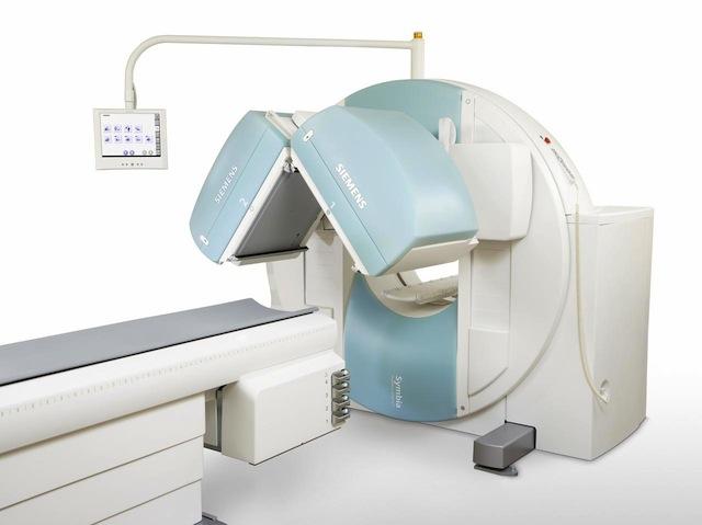
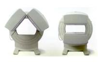
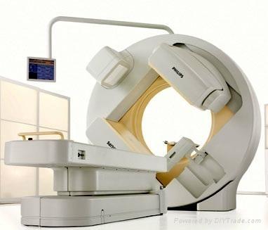
3 Gamma Camera Imaging (SPECT) Siemens Healthcare, Symbia Philips Medical, Brightview XCT GE Healthcare, MG
4 Single Photon Emission Computed Tomography SPECT is to scin/graphy as CT is to radiography We collect projec/on views all the way around the pa/ent We use tomographic principles (e.g. FBP) to reconstruct an image One difference is that we are using emission rather than transmission Another difference is that agenua/on is a headache rather than what we want (as for CT) ScaGered photons, however, remain a nuisance
5 SPECT Imaging Equa/on Using parallel-hole collimators, and ignoring depth-dependent collimator burring etc., we have A(x, y, z) φ(z,l) = R exp{ R µ(x, " 2 4π (y R) y, z, E)d y" } dy y Our inverse problem is: given φ(z,l) for all angles θ, what is A(x,y,z)? - Like poly-energe/c CT problem this is hard - Complicated by agenua/on - Solu/on discovered in 2000 integra/on geometry
6 SPECT Image Reconstruc/on For the approximate solu/on used in prac/ce, we make several assump/ons ignore collimator effects parameterize x and y as func/ons of distance along line integra/on geometry x(s) = l cosθ ssinθ y(s) = l sinθ + s cosθ This changes the imaging equa/on to: φ(l,θ) = R { } ds A(x(s), y(s)) R exp µ(x( s %), y( % 4π(s R) s );E)d s% 2 s
7 SPECT Image Reconstruc/on Addi/onal assump/ons ignore inverse-square dependence of fluence assume we can correct for agenua/on effects later This changes the imaging equa/on to φ(l,θ) = A(x(s), y(s))ds dy = A(x, y)δ(x cosθ + ysinθ l)dx which is the 2-D x-ray or Radon transform that we can solve with FBP A(x, y) π 0 ( )* ρ Φ(ρ,θ)e j 2πρl dρ+,- dθ where Φ(ρ,θ) = F 1D { φ(l,θ) }

8 Angular asymmetry +p)
9 Comparison of Imaging Equa/ons
10 Image reconstruction

11 Point source reconstrction by Filtered back projection (FBP) Filtering the projection data with a ramp filter creates the negative values in back-projected profile necessary for cancelling the contributions from other angles in the region about the reconstructed point. The back projections from different angles are added together to form the reconstructed image Image reconstruction Cullom SJ, In: Cardiac SPECT Imaging, 2001

12 Image reconstruction Star (or streak) artifact of FBP reconstruction A = object B through G = {1, 3, 4, 16, 32, 64} number of projections Star artifact decreases with number of projections Bruyant PP, J Nucl Med 2002; 43:

13 Reconstruction Filters used to reduce image noise A ramp filter of the form r used to correct for 1/r tomographic blurring ar/fact Ohen, the ramp filter is further modified by addi/onal frequency filters, such as: the Hann, Shepp-Logan or BuGerworth filters. In filtering, we see a tradoff of image noise vs. spa/al resolu/on Higher cutoff frequencies maintain spa/al resolu/on at the expense of more noise Filtered FBP is a very rapid technique for reconstruc/ng SPECT data Itera/ve reconstruc/on techniques, such as ordered subset will be covered in later lecture Modified Frequency Filters Effect of BuGerworth-filter cutoff Koch W. J Nucl Med 2005; 46:
14 Iterative reconstruction a generic example f (0 ) ini/al image es/mate system model noise p measured data f (k) p (k) = Hf (k) + n p p (k) f (k) f (k+1) Es/mate of projec/on Compare measured to es/mated projec/on Update image based on ra/o or difference Model the system (and the noise) used in projector Image update is typically based on differences or ratio of measured and estimated projection data Must decide when to stop iterating
15 Projector can account for Signal probability model Physics model of SPECT imaging System (intrinsic + collimator) spa/al resolu/on Pa/ent agenua/on Compton ScaGer (in pa/ent, collimator, crystal) Collimator septal penetra/on
16 Various Itera/ve Recon. Methods Algebraic reconstruc/on technique (ART) Mul/plica/ve ART (MART) Weighted least-squares conjugate gradient (WLS-CG) Expecta/on maximiza/on (EM)
17 Expecta/on Maximiza/on (EM) Es/mates parameters of the sta/s/cal distribu/ons underlying the measured data Maximize marginal likelihood by itera/ng two steps: Expecta/on: Marginalize log likelihood with respect to the missing data given observed data for the current es/mate of parameters Maximiza/on: Find set of parameters that maximizes this quan/ty In the case of SPECT Observed data, F = {f j, j=1,,j) are the projec/ons onto detector elements Parameters, A={A i, i=1,,i) are the true count rate in a voxel at {x i, y i, z i } The parameters {A i } are independent and Poisson distributed  = argmax{pr(φ A)} = A I i=1 J j=1 A i φ j e A i φ j!
18 Ordered subset EM (OS-EM) Performs EM sequen/ally on non-overlapping subsets of the projec/on data un/l all projec/ons are considered Must decide # of subsets (n) and # of itera/ons (m) m n EM itera/ons, but only m projec/ons of data Time for 1 OSEM itera/on (all subsets) > 1 EM itera/on However, OSEM increases convergence rate
19 Image Acquisition

20 Number of detectors and orbits Single head vs 360 acquisition: speed vs. uniformity tradeoff - Conjugate views - Non-circular orbits: improved resolution Two headed - Smaller rotation needed: 360!180 or 180!90 - H vs. L modes: region of interest and attenuation Circular vs. contouring orbits

21 Number of detectors and orbits Single head vs 360 acquisition: speed vs. uniformity tradeoff - Conjugate views - Non-circular orbits: improved resolution Two headed - Smaller rotation needed: 360!180 or 180!90 - H vs. L modes: region of interest and attenuation Circular vs. contouring orbits
22 Image Acquisition Modes Frame mode (data stored as an image) - static - single image acquisition - can have multiple energy windows - dynamic - series of images acquired sequentially - gated - repetitive, dynamic imaging - used for cardiac imaging List-mode (data stored event by event) - time stamps are included within data stream - allows for flexible post-acquisition binning - can result in very large data files
23 Dynamic acquisition Regions of interests (ROIs) Time-ac/vity curves (TACs) From: The Essen/al Physics of Medical Imaging (Bushberg, et al)
24 Cardiac Gated Acquisition From: The Essential Physics of Medical Imaging (Bushberg, et al)
 left ventricular (LV) function vs. phase (i.e. systolic and diastolic) Gated cardiac SPECT")
25 Images obtained during individual phases of cardiac cycle Gating based on EKG 8-16 frames per cycle Assessment of global and regional (apex, mid, base) left ventricular (LV) function vs. phase (i.e. systolic and diastolic) Gated cardiac SPECT


26 Attenuation Effects Energy dependance Object size Cherry SR, In: Physics in Nuclear Medicine, 2003, p 308 & 311
27 Methods of Attenuation Correction Transmission measurement - Scanned line source - Moving energy window: different energy for transmission & emission photons - Low signal-to-noise due to practical transmission activity - Direct measure of attenuation coefficient
28 Methods of Attenuation Correction Conjugate views - Arithmetic mean: (L 1 +L 2 )/2 - Geometric mean: (L 12 +L 22 ) L 12
29 Methods of Attenuation Correction Chang correction - Assumes constant μ within anatomical boundary - Requires accurate anatomical boundary definition - Empirically adjusted for out-of-plane scatter - Accurate boundaries difficult to obtain Where will this work well? Brain SPECT Where is this problematic? Variable μ values (esp. in thorax)
30 X-ray CT Methods of Attenuation Correction - Co-located and co-registered - Lower energy than transmission delivers lower dose - Conversion from CT Hounsfield units to equivalent linear attenuation coefficient

31 Converting Hounsfield Units to attenuation coefficients
32 Examples of SPECT imaging

33 Thyroid Imaging Sample nuclear medicine thyroid images. The main characteristics used for interpretation of the images are size of the thyroids and whether there is uniform uptake between the left and right thyroids.
 CT, SPECT, and fused SPECT/CT images localize increased tracer uptake to the posterior aspect of right lobe of the thyroid confirming a right superior parathyroid")
34 SPECT/CT Thyroid Image Example A 37 YOF with elevated Ca, decreased phosphorus and increased parathyroid hormone (a), (b) early and delayed Tc99m-sestamibi scin/graphs showing uptake near thyroid (c) CT, SPECT, and fused SPECT/CT images localize increased tracer uptake to the posterior aspect of right lobe of the thyroid confirming a right superior parathyroid adenoma

35 Renal Imaging A renogram provides a timeactivity curve of the uptake and excretion of a radiotracer by the kidneys. It is used to both evaluate renal function and if there are any bilateral differences between the kidneys. There is often a perfusion phase and a functional phase of the exam. A standard protocol is 80 one second frames to visualize kidney perfusion and 120 twenty second frames to evaluate function. Robert Miyaoka, PhD., BioEng 420, Spring 2013
, while the abnormal period scan is for a patient with PS (lower panel).")
36 Brain Scan In this example, an imaging agent called DaTscan is used to differentiate between Parkinsonian syndromes (PS) and essential tremor. The comma image is for a patient without PS (upper panel), while the abnormal period scan is for a patient with PS (lower panel). DaTscan is a radiopharmaceutical indicated for striatal dopamine transporter visualization.
.")
37 SPECT/CT Lung Perfusion Imaging Tc-99 MAA lung perfusion with planar perfusion (upper left) and SPECT/CT with attenuation correction (upper right). The estimated perfusion contribution of each lung obtained by both methods is nearly identical (lower left). However, the SPECT/CT method provides more anatomically accurate lobar perfusion quantitation.
Reconstruction from Projections
 Reconstruction from Projections M.C. Villa Uriol Computational Imaging Lab email: cruz.villa@upf.edu web: http://www.cilab.upf.edu Based on SPECT reconstruction Martin Šámal Charles University Prague,
SPECT reconstruction
 Regional Training Workshop Advanced Image Processing of SPECT Studies Tygerberg Hospital, 19-23 April 2004 SPECT reconstruction Martin Šámal Charles University Prague, Czech Republic samal@cesnet.cz Tomography
Introduction to Emission Tomography
 Introduction to Emission Tomography Gamma Camera Planar Imaging Robert Miyaoka, PhD University of Washington Department of Radiology rmiyaoka@u.washington.edu Gamma Camera: - collimator - detector (crystal
Introduction to Positron Emission Tomography
 Planar and SPECT Cameras Summary Introduction to Positron Emission Tomography, Ph.D. Nuclear Medicine Basic Science Lectures srbowen@uw.edu System components: Collimator Detector Electronics Collimator
Emission Computed Tomography Notes
 Noll (24) ECT Notes: Page 1 Emission Computed Tomography Notes Introduction Emission computed tomography (ECT) is the CT applied to nuclear medicine. There are two varieties of ECT: 1. SPECT single-photon
Introduc)on to PET Image Reconstruc)on. Tomographic Imaging. Projec)on Imaging. Types of imaging systems
on to PET Image Reconstruc)on. Tomographic Imaging. Projec)on Imaging. Types of imaging systems") Introduc)on to PET Image Reconstruc)on Adam Alessio http://faculty.washington.edu/aalessio/ Nuclear Medicine Lectures Imaging Research Laboratory Division of Nuclear Medicine University of Washington Fall
Introduc)on to PET Image Reconstruc)on Adam Alessio http://faculty.washington.edu/aalessio/ Nuclear Medicine Lectures Imaging Research Laboratory Division of Nuclear Medicine University of Washington Fall
Medical Imaging BMEN Spring 2016
 Name Medical Imaging BMEN 420-501 Spring 2016 Homework #4 and Nuclear Medicine Notes All questions are from the introductory Powerpoint (based on Chapter 7) and text Medical Imaging Signals and Systems,
Workshop on Quantitative SPECT and PET Brain Studies January, 2013 PUCRS, Porto Alegre, Brasil Corrections in SPECT and PET
 Workshop on Quantitative SPECT and PET Brain Studies 14-16 January, 2013 PUCRS, Porto Alegre, Brasil Corrections in SPECT and PET Físico João Alfredo Borges, Me. Corrections in SPECT and PET SPECT and
Corso di laurea in Fisica A.A Fisica Medica 5 SPECT, PET
 Corso di laurea in Fisica A.A. 2007-2008 Fisica Medica 5 SPECT, PET Step 1: Inject Patient with Radioactive Drug Drug is labeled with positron (β + ) emitting radionuclide. Drug localizes
3/27/2012 WHY SPECT / CT? SPECT / CT Basic Principles. Advantages of SPECT. Advantages of CT. Dr John C. Dickson, Principal Physicist UCLH
 3/27/212 Advantages of SPECT SPECT / CT Basic Principles Dr John C. Dickson, Principal Physicist UCLH Institute of Nuclear Medicine, University College London Hospitals and University College London john.dickson@uclh.nhs.uk
Implementation and evaluation of a fully 3D OS-MLEM reconstruction algorithm accounting for the PSF of the PET imaging system
 Implementation and evaluation of a fully 3D OS-MLEM reconstruction algorithm accounting for the PSF of the PET imaging system 3 rd October 2008 11 th Topical Seminar on Innovative Particle and Radiation
Philips SPECT/CT Systems
 Philips SPECT/CT Systems Ling Shao, PhD Director, Imaging Physics & System Analysis Nuclear Medicine, Philips Healthcare June 14, 2008 *Presented SNM08 Categorical Seminar - Quantitative SPECT and PET
Constructing System Matrices for SPECT Simulations and Reconstructions
 Constructing System Matrices for SPECT Simulations and Reconstructions Nirantha Balagopal April 28th, 2017 M.S. Report The University of Arizona College of Optical Sciences 1 Acknowledgement I would like
Multi-slice CT Image Reconstruction Jiang Hsieh, Ph.D.
 Multi-slice CT Image Reconstruction Jiang Hsieh, Ph.D. Applied Science Laboratory, GE Healthcare Technologies 1 Image Generation Reconstruction of images from projections. textbook reconstruction advanced
BME I5000: Biomedical Imaging
 1 Lucas Parra, CCNY BME I5000: Biomedical Imaging Lecture 4 Computed Tomography Lucas C. Parra, parra@ccny.cuny.edu some slides inspired by lecture notes of Andreas H. Hilscher at Columbia University.
Assessment of OSEM & FBP Reconstruction Techniques in Single Photon Emission Computed Tomography Using SPECT Phantom as Applied on Bone Scintigraphy
 Assessment of OSEM & FBP Reconstruction Techniques in Single Photon Emission Computed Tomography Using SPECT Phantom as Applied on Bone Scintigraphy Physics Department, Faculty of Applied Science,Umm Al-Qura
Ch. 4 Physical Principles of CT
 Ch. 4 Physical Principles of CT CLRS 408: Intro to CT Department of Radiation Sciences Review: Why CT? Solution for radiography/tomography limitations Superimposition of structures Distinguishing between
SPECT/CT Basics, Technology Updates, Quality Assurance, and Applications
 SPECT/CT Basics, Technology Updates, Quality Assurance, and Applications S. Cheenu Kappadath, PhD Department of Imaging Physics University of Texas M D Anderson Cancer Center, Houston, Texas skappadath@mdanderson.org
GPU implementation for rapid iterative image reconstruction algorithm
 GPU implementation for rapid iterative image reconstruction algorithm and its applications in nuclear medicine Jakub Pietrzak Krzysztof Kacperski Department of Medical Physics, Maria Skłodowska-Curie Memorial
Development and Performance of a Sparsity- Exploiting Algorithm for Few-View Single Photon Emission Computed Tomogrpahy (SPECT) Reconstruction
 Reconstruction") Marquette University e-publications@marquette Master's Theses (2009 -) Dissertations, Theses, and Professional Projects Development and Performance of a Sparsity- Exploiting Algorithm for Few-View Single
Marquette University e-publications@marquette Master's Theses (2009 -) Dissertations, Theses, and Professional Projects Development and Performance of a Sparsity- Exploiting Algorithm for Few-View Single
Tomographic Reconstruction
 Tomographic Reconstruction 3D Image Processing Torsten Möller Reading Gonzales + Woods, Chapter 5.11 2 Overview Physics History Reconstruction basic idea Radon transform Fourier-Slice theorem (Parallel-beam)
A Comparison of the Uniformity Requirements for SPECT Image Reconstruction Using FBP and OSEM Techniques
 IMAGING A Comparison of the Uniformity Requirements for SPECT Image Reconstruction Using FBP and OSEM Techniques Lai K. Leong, Randall L. Kruger, and Michael K. O Connor Section of Nuclear Medicine, Department
Deviceless respiratory motion correction in PET imaging exploring the potential of novel data driven strategies
 g Deviceless respiratory motion correction in PET imaging exploring the potential of novel data driven strategies Presented by Adam Kesner, Ph.D., DABR Assistant Professor, Division of Radiological Sciences,
Image Acquisition Systems
 Image Acquisition Systems Goals and Terminology Conventional Radiography Axial Tomography Computer Axial Tomography (CAT) Magnetic Resonance Imaging (MRI) PET, SPECT Ultrasound Microscopy Imaging ITCS
Spiral ASSR Std p = 1.0. Spiral EPBP Std. 256 slices (0/300) Kachelrieß et al., Med. Phys. 31(6): , 2004
 Kachelrieß et al., Med. Phys. 31(6): , 2004") Spiral ASSR Std p = 1.0 Spiral EPBP Std p = 1.0 Kachelrieß et al., Med. Phys. 31(6): 1623-1641, 2004 256 slices (0/300) Advantages of Cone-Beam Spiral CT Image quality nearly independent of pitch Increase
Spiral ASSR Std p = 1.0 Spiral EPBP Std p = 1.0 Kachelrieß et al., Med. Phys. 31(6): 1623-1641, 2004 256 slices (0/300) Advantages of Cone-Beam Spiral CT Image quality nearly independent of pitch Increase
Validation of GEANT4 for Accurate Modeling of 111 In SPECT Acquisition
 Validation of GEANT4 for Accurate Modeling of 111 In SPECT Acquisition Bernd Schweizer, Andreas Goedicke Philips Technology Research Laboratories, Aachen, Germany bernd.schweizer@philips.com Abstract.
Educational Objectives SPECT/CT: Basics, Quality Assurance, and Clinical Applications
 Educational Objectives SPECT/CT: Basics, Quality Assurance, and Clinical Applications 1. To understand the physics principles underlying SPECT/CT image acquisition and reconstruction 2. To understand the
ATTENUATION CORRECTION IN SPECT DURING IMAGE RECONSTRUCTION USING INVERSE MONTE CARLO METHOD A SIMULATION STUDY *
 Romanian Reports in Physics, Vol. 66, No. 1, P. 200 211, 2014 ATTENUATION CORRECTION IN SPECT DURING IMAGE RECONSTRUCTION USING INVERSE MONTE CARLO METHOD A SIMULATION STUDY * S. AHMADI 1, H. RAJABI 2,
Conflicts of Interest Nuclear Medicine and PET physics reviewer for the ACR Accreditation program
 James R Halama, PhD Loyola University Medical Center Conflicts of Interest Nuclear Medicine and PET physics reviewer for the ACR Accreditation program Learning Objectives 1. Be familiar with recommendations
Small Angle Gamma Ray Scatter: What Is The Impact On Image Quality
 ISPUB.COM The Internet Journal of Medical Technology Volume 4 Number 2 Small Angle Gamma Ray Scatter: What Is The Impact On Image Quality G CUrrie, J Wheat Citation G CUrrie, J Wheat.. The Internet Journal
V/Q Imaging Protocols
 Elizabeth ailey ppsc(mrs) M Department of Nuclear Medicine Royal North Shore Hospital, Sydney, ustralia email: ebailey@nsccahs.health.nsw.gov.au The V/Q scan has traditionally used planar imaging techniques.
Iterative and analytical reconstruction algorithms for varying-focal-length cone-beam
 Home Search Collections Journals About Contact us My IOPscience Iterative and analytical reconstruction algorithms for varying-focal-length cone-beam projections This content has been downloaded from IOPscience.
(RMSE). Reconstructions showed that modeling the incremental blur improved the resolution of the attenuation map and quantitative accuracy.
. Reconstructions showed that modeling the incremental blur improved the resolution of the attenuation map and quantitative accuracy.") Modeling the Distance-Dependent Blurring in Transmission Imaging in the Ordered-Subset Transmission (OSTR) Algorithm by Using an Unmatched Projector/Backprojector Pair B. Feng, Member, IEEE, M. A. King,
Modeling the Distance-Dependent Blurring in Transmission Imaging in the Ordered-Subset Transmission (OSTR) Algorithm by Using an Unmatched Projector/Backprojector Pair B. Feng, Member, IEEE, M. A. King,
Basics of treatment planning II
 Basics of treatment planning II Sastry Vedam PhD DABR Introduction to Medical Physics III: Therapy Spring 2015 Dose calculation algorithms! Correction based! Model based 1 Dose calculation algorithms!
Diagnostic imaging techniques. Krasznai Zoltán. University of Debrecen Medical and Health Science Centre Department of Biophysics and Cell Biology
 Diagnostic imaging techniques Krasznai Zoltán University of Debrecen Medical and Health Science Centre Department of Biophysics and Cell Biology 1. Computer tomography (CT) 2. Gamma camera 3. Single Photon
Review of PET Physics. Timothy Turkington, Ph.D. Radiology and Medical Physics Duke University Durham, North Carolina, USA
 Review of PET Physics Timothy Turkington, Ph.D. Radiology and Medical Physics Duke University Durham, North Carolina, USA Chart of Nuclides Z (protons) N (number of neutrons) Nuclear Data Evaluation Lab.
Moscow-Bavarian Joint Advanced Student School 2006 / Medical Imaging Principles of Computerized Tomographic Imaging and Cone-Beam Reconstruction
 Line Integrals Line integrals represent the integral of some parameter of the object along the line (e.g. attenuation of x-rays) Object: f(x,y) Line: x cosθ + y sinθ = t Line integral / Radon transform:
Medical Image Processing in Nuclear Medicine. Andrew Todd-Pokropek University College London INSERM U494 Paris. Aims and Objectives
 Medical Image Processing in Nuclear Medicine Andrew Todd-Pokropek University College London INSERM U494 Paris Aims and Objectives The aim of this module is to provide an understanding of the special nature
Quantitative capabilities of four state-of-the-art SPECT-CT cameras
 Seret et al. EJNMMI Research 2012, 2:45 ORIGINAL RESEARCH Open Access Quantitative capabilities of four state-of-the-art SPECT-CT cameras Alain Seret 1,2*, Daniel Nguyen 1 and Claire Bernard 3 Abstract
James R Halama, PhD Loyola University Medical Center
 James R Halama, PhD Loyola University Medical Center Conflicts of Interest Nuclear Medicine and PET physics reviewer for the ACR Accreditation program Learning Objectives Be familiar with the tests recommended
Concepts, Applications, and Requirements for Quantitative SPECT/CT. Conflict of Interest Disclosure
 Concepts, Applications, and Requirements for Quantitative SPECT/CT Eric C. Frey, Ph.D. (efrey@jhmi.edu) Division of Medical Imaging Physics Russell H. Morgan Department of Radiology and Radiological Science
Monte-Carlo-Based Scatter Correction for Quantitative SPECT Reconstruction
 Monte-Carlo-Based Scatter Correction for Quantitative SPECT Reconstruction Realization and Evaluation Rolf Bippus 1, Andreas Goedicke 1, Henrik Botterweck 2 1 Philips Research Laboratories, Aachen 2 Fachhochschule
Tomography. Forward projectionsp θ (r) are known as a Radon transform. Objective: reverse this process to form the original image
 are known as a Radon transform. Objective: reverse this process to form the original image") C. A. Bouman: Digital Image Processing - January 9, 217 1 Tomography Many medical imaging systems can only measure projections through an object with density f(x,y). Projections must be collected at every
C. A. Bouman: Digital Image Processing - January 9, 217 1 Tomography Many medical imaging systems can only measure projections through an object with density f(x,y). Projections must be collected at every
SPECT QA and QC. Bruce McBride St. Vincent s Hospital Sydney.
 SPECT QA and QC Bruce McBride St. Vincent s Hospital Sydney. SPECT QA and QC What is needed? Why? How often? Who says? QA and QC in Nuclear Medicine QA - collective term for all the efforts made to produce
Radiology. Marta Anguiano Millán. Departamento de Física Atómica, Molecular y Nuclear Facultad de Ciencias. Universidad de Granada
 Departamento de Física Atómica, Molecular y Nuclear Facultad de Ciencias. Universidad de Granada Overview Introduction Overview Introduction Tecniques of imaging in Overview Introduction Tecniques of imaging
Digital Image Processing
 Digital Image Processing SPECIAL TOPICS CT IMAGES Hamid R. Rabiee Fall 2015 What is an image? 2 Are images only about visual concepts? We ve already seen that there are other kinds of image. In this lecture
AN ELLIPTICAL ORBIT BACKPROJECTION FILTERING ALGORITHM FOR SPECT
 1102 IEEE TRANSACTIONS ON NUCLEAR SCIENCE, VOL. 40, NO. 4, AUGUST 1993 AN ELLIPTICAL ORBIT BACKPROJECTION FILTERING ALGORITHM FOR SPECT Grant. T. Gullberg and Gengsheng L. Zeng, Department of Radiology,
Attenuation map reconstruction from TOF PET data
 Attenuation map reconstruction from TOF PET data Qingsong Yang, Wenxiang Cong, Ge Wang* Department of Biomedical Engineering, Rensselaer Polytechnic Institute, Troy, NY 80, USA *Ge Wang (ge-wang@ieee.org)
Fits you like no other
 Fits you like no other BrightView X and XCT specifications The new BrightView X system is a fully featured variableangle camera that is field-upgradeable to BrightView XCT without any increase in room
Mathematical methods and simulations tools useful in medical radiation physics
 Mathematical methods and simulations tools useful in medical radiation physics Michael Ljungberg, professor Department of Medical Radiation Physics Lund University SE-221 85 Lund, Sweden Major topic 1:
Computational Medical Imaging Analysis
 Computational Medical Imaging Analysis Chapter 2: Image Acquisition Systems Jun Zhang Laboratory for Computational Medical Imaging & Data Analysis Department of Computer Science University of Kentucky
Medical Image Analysis
 Computer assisted Image Analysis VT04 29 april 2004 Medical Image Analysis Lecture 10 (part 1) Xavier Tizon Medical Image Processing Medical imaging modalities XRay,, CT Ultrasound MRI PET, SPECT Generic
MEDICAL IMAGE ANALYSIS
 SECOND EDITION MEDICAL IMAGE ANALYSIS ATAM P. DHAWAN g, A B IEEE Engineering in Medicine and Biology Society, Sponsor IEEE Press Series in Biomedical Engineering Metin Akay, Series Editor +IEEE IEEE PRESS
Biomedical Imaging. Computed Tomography. Patrícia Figueiredo IST
 Biomedical Imaging Computed Tomography Patrícia Figueiredo IST 2013-2014 Overview Basic principles X ray attenuation projection Slice selection and line projections Projection reconstruction Instrumentation
Index. aliasing artifacts and noise in CT images, 200 measurement of projection data, nondiffracting
 Index Algebraic equations solution by Kaczmarz method, 278 Algebraic reconstruction techniques, 283-84 sequential, 289, 293 simultaneous, 285-92 Algebraic techniques reconstruction algorithms, 275-96 Algorithms
Some reference material
 Some reference material Physics reference book on medical imaging: A good one is The Essential Physics of Medical Imaging, 3 rd Ed. by Bushberg et al. ($170! new). However, there are several similar books
Introduction to Biomedical Imaging
 Alejandro Frangi, PhD Computational Imaging Lab Department of Information & Communication Technology Pompeu Fabra University www.cilab.upf.edu X-ray Projection Imaging Computed Tomography Digital X-ray
REMOVAL OF THE EFFECT OF COMPTON SCATTERING IN 3-D WHOLE BODY POSITRON EMISSION TOMOGRAPHY BY MONTE CARLO
 REMOVAL OF THE EFFECT OF COMPTON SCATTERING IN 3-D WHOLE BODY POSITRON EMISSION TOMOGRAPHY BY MONTE CARLO Abstract C.S. Levin, Y-C Tai, E.J. Hoffman, M. Dahlbom, T.H. Farquhar UCLA School of Medicine Division
Principles of Computerized Tomographic Imaging
 Principles of Computerized Tomographic Imaging Parallel CT, Fanbeam CT, Helical CT and Multislice CT Marjolein van der Glas August 29, 2000 Abstract The total attenuation suffered by one beam of x-rays
A closer look at CT scanning
 Vet Times The website for the veterinary profession https://www.vettimes.co.uk A closer look at CT scanning Author : Charissa Lee, Natalie Webster Categories : General, Vets Date : April 3, 2017 A basic
Digital Image Processing
 Digital Image Processing Image Restoration and Reconstruction (Image Reconstruction from Projections) Christophoros Nikou cnikou@cs.uoi.gr University of Ioannina - Department of Computer Science and Engineering
Effects of attenuation in single slow rotation dynamic SPECT
 Effects of attenuation in single slow rotation dynamic SPECT T Humphries 1, A Celler 2 and M Trummer 1 1 Department of Mathematics, Simon Fraser University, 8888 University Ave, Burnaby, British Columbia,
Modeling and Incorporation of System Response Functions in 3D Whole Body PET
 Modeling and Incorporation of System Response Functions in 3D Whole Body PET Adam M. Alessio, Member IEEE, Paul E. Kinahan, Senior Member IEEE, and Thomas K. Lewellen, Senior Member IEEE University of
Fits you like no other
 Fits you like no other Philips BrightView X and XCT specifications The new BrightView X system is a fully featured variableangle camera that is field-upgradeable to BrightView XCT without any increase
Cardiac Dual Energy CT: Technique
 RSNA 2013, VSCA51-01, Chicago, Dec. 5, 2013 Cardiac Radiology Series Cardiac Dual Energy CT: Technique Willi A. Kalender, Ph.D. Institute of Medical Physics University of Erlangen www.imp.uni-erlangen.de
Image reconstruction for PET/CT scanners: past achievements and future challenges
 Review Image reconstruction for PET/CT scanners: past achievements and future challenges PET is a medical imaging modality with proven clinical value for disease diagnosis and treatment monitoring. The
A Weighted Least Squares PET Image Reconstruction Method Using Iterative Coordinate Descent Algorithms
 A Weighted Least Squares PET Image Reconstruction Method Using Iterative Coordinate Descent Algorithms Hongqing Zhu, Huazhong Shu, Jian Zhou and Limin Luo Department of Biological Science and Medical Engineering,
Master of Science Thesis. Activity quantification of planar gamma camera images
 Master of Science Thesis Activity quantification of planar gamma camera images Marie Sydoff Supervisor: Sigrid Leide-Svegborn The work has been performed at Department of Radiation Physics Malmö University
An FDK-like cone-beam SPECT reconstruction algorithm for non-uniform attenuated
 Home Search Collections Journals About Contact us My IOPscience An FK-like cone-beam SPECT reconstruction algorithm for non-uniform attenuated projections acquired using a circular trajectory This content
Reconstruction in CT and relation to other imaging modalities
 Reconstruction in CT and relation to other imaging modalities Jørgen Arendt Jensen November 1, 2017 Center for Fast Ultrasound Imaging, Build 349 Department of Electrical Engineering Center for Fast Ultrasound
A novel coupled transmission-reflection tomography and the V-line Radon transform
 A novel coupled transmission-reflection tomography and the V-line Radon transform Rémi Regnier, M.K. Nguyen To cite this version: Rémi Regnier, M.K. Nguyen. A novel coupled transmission-reflection tomography
Medical Image Reconstruction Term II 2012 Topic 6: Tomography
 Medical Image Reconstruction Term II 2012 Topic 6: Tomography Professor Yasser Mostafa Kadah Tomography The Greek word tomos means a section, a slice, or a cut. Tomography is the process of imaging a cross
Development of Spect and Ct Tomographic Image Reconstruction
 Syracuse University SURFACE Physics - Dissertations College of Arts and Sciences 6-2012 Development of Spect and Ct Tomographic Image Reconstruction Levon Orion Vogelsang Syracuse University Follow this
Evaluation of Spectrum Mismatching using Spectrum Binning Approach for Statistical Polychromatic Reconstruction in CT
 Evaluation of Spectrum Mismatching using Spectrum Binning Approach for Statistical Polychromatic Reconstruction in CT Qiao Yang 1,4, Meng Wu 2, Andreas Maier 1,3,4, Joachim Hornegger 1,3,4, Rebecca Fahrig
ML reconstruction for CT
 ML reconstruction for CT derivation of MLTR rigid motion correction resolution modeling polychromatic ML model dual energy ML model Bruno De Man, Katrien Van Slambrouck, Maarten Depypere, Frederik Maes,
Absolute quantification in brain SPECT imaging
 Absolute quantification in brain SPECT imaging Thesis presented by Albert Cot Sanz 15 June 2003 Thesis directors: Dr. Francisco Calviño Dr. Domènec Ros Dr. Josep Sempau CONTENTS Part I Thesis 1 1. Introduction.....................................
Q.Clear. Steve Ross, Ph.D.
 Steve Ross, Ph.D. Accurate quantitation (SUV - Standardized Uptake Value) is becoming more important as clinicians seek to utilize PET imaging for more than just diagnosing and staging disease, but also
Acknowledgments and financial disclosure
 AAPM 2012 Annual Meeting Digital breast tomosynthesis: basic understanding of physics principles James T. Dobbins III, Ph.D., FAAPM Director, Medical Physics Graduate Program Ravin Advanced Imaging Laboratories
NIH Public Access Author Manuscript Int J Imaging Syst Technol. Author manuscript; available in PMC 2010 September 1.
 NIH Public Access Author Manuscript Published in final edited form as: Int J Imaging Syst Technol. 2009 September 1; 19(3): 271 276. doi:10.1002/ima.20200. Attenuation map estimation with SPECT emission
8/7/2017. Disclosures. MECT Systems Overview and Quantitative Opportunities. Overview. Computed Tomography (CT) CT Numbers. Polyenergetic Acquisition
 CT Numbers. Polyenergetic Acquisition") Quantitative Multi-Energy Computed Tomography: Imaging and Therapy Advancements Disclosures MECT Systems Overview and Quantitative Opportunities The speaker receives research funding from GE Healthcare
Quantitative Multi-Energy Computed Tomography: Imaging and Therapy Advancements Disclosures MECT Systems Overview and Quantitative Opportunities The speaker receives research funding from GE Healthcare
Fast Timing and TOF in PET Medical Imaging
 Fast Timing and TOF in PET Medical Imaging William W. Moses Lawrence Berkeley National Laboratory October 15, 2008 Outline: Time-of-Flight PET History Present Status Future This work was supported in part
Iterative SPECT reconstruction with 3D detector response
 Iterative SPECT reconstruction with 3D detector response Jeffrey A. Fessler and Anastasia Yendiki COMMUNICATIONS & SIGNAL PROCESSING LABORATORY Department of Electrical Engineering and Computer Science
Noise weighting with an exponent for transmission CT
 doi:10.1088/2057-1976/2/4/045004 RECEIVED 13 January 2016 REVISED 4 June 2016 ACCEPTED FOR PUBLICATION 21 June 2016 PUBLISHED 27 July 2016 PAPER Noise weighting with an exponent for transmission CT Gengsheng
ELEG404/604: Digital Imaging & Photography
 : Digital Imaging & Photography Gonzalo R. Arce Department of Electrical and Computer Engineering University of Delaware Chapter VIII History X-Ray discovery In 1895 Wilhelm Rontgen discovered the X-rays,
3-D PET Scatter Correction
 Investigation of Accelerated Monte Carlo Techniques for PET Simulation and 3-D PET Scatter Correction C.H. Holdsworth, Student Member, IEEE, C.S. Levin", Member, IEEE, T.H. Farquhar, Student Member, IEEE,
Joint CI-JAI advanced accelerator lecture series Imaging and detectors for medical physics Lecture 1: Medical imaging
 Joint CI-JAI advanced accelerator lecture series Imaging and detectors for medical physics Lecture 1: Medical imaging Dr Barbara Camanzi barbara.camanzi@stfc.ac.uk Course layout Day AM 09.30 11.00 PM 15.30
Positron. MillenniumVG. Emission Tomography Imaging with the. GE Medical Systems
 Positron Emission Tomography Imaging with the MillenniumVG GE Medical Systems Table of Contents Introduction... 3 PET Imaging With Gamma Cameras PET Overview... 4 Coincidence Detection on Gamma Cameras...
Corridor4DM Feature List
 Software Services Features Corridor4DM Feature List 4DM v2013 FDG PET Standard Uptake Value (SUV) Calculation Auto-Selection of Report Template Motion Correction of SPECT datasets New Normals Databases
COMPARATIVE STUDIES OF DIFFERENT SYSTEM MODELS FOR ITERATIVE CT IMAGE RECONSTRUCTION
 COMPARATIVE STUDIES OF DIFFERENT SYSTEM MODELS FOR ITERATIVE CT IMAGE RECONSTRUCTION BY CHUANG MIAO A Thesis Submitted to the Graduate Faculty of WAKE FOREST UNIVERSITY GRADUATE SCHOOL OF ARTS AND SCIENCES
4DM Packages. 4DM Packages & License Types. Information to help you order the appropriate licenses for your site.
 4DM Packages 4DM Packages & License Types. Information to help you order the appropriate licenses for your site. Nuclear Cardiac Quantification, Review, and Reporting Select Your 4DM Package and corresponding
Nuclear Medicine Imaging
 Introduction to Medical Engineering (Medical Imaging) Suetens 5 Nuclear Medicine Imaging Ho Kyung Kim Pusan National University Introduction Use of radioactive isotopes for medical purposes since 1920
Computational Medical Imaging Analysis
 Computational Medical Imaging Analysis Chapter 1: Introduction to Imaging Science Jun Zhang Laboratory for Computational Medical Imaging & Data Analysis Department of Computer Science University of Kentucky
Medical Images Analysis and Processing
 Medical Images Analysis and Processing - 25642 Emad Course Introduction Course Information: Type: Graduated Credits: 3 Prerequisites: Digital Image Processing Course Introduction Reference(s): Insight
STA 4273H: Sta-s-cal Machine Learning
 STA 4273H: Sta-s-cal Machine Learning Russ Salakhutdinov Department of Statistics! rsalakhu@utstat.toronto.edu! h0p://www.cs.toronto.edu/~rsalakhu/ Lecture 3 Parametric Distribu>ons We want model the probability
Hybrid Imaging for Patient-Specific Dosimetry in Radionuclide Therapy
 Hybrid Imaging for Patient-Specific Dosimetry in Radionuclide Therapy Ljungberg, Michael; Sjögreen Gleisner, Katarina Published in: Diagnostics DOI: 10.3390/diagnostics5030296 Published: 2015-01-01 Link
Venus Explorer Processing Technical specifications
 24, rue du champ de l Alouette F 75013 Paris, France Venus Explorer Processing Technical specifications Venus Explorer Processing Server software Web base application sever; tomcat Apache technology. Include
Performance Evaluation of radionuclide imaging systems
 Performance Evaluation of radionuclide imaging systems Nicolas A. Karakatsanis STIR Users meeting IEEE Nuclear Science Symposium and Medical Imaging Conference 2009 Orlando, FL, USA Geant4 Application
PET Quantification using STIR
 PET Quantification using STIR STIR User s Meeting Charalampos Tsoumpas, PhD King s College London Hammersmith Imanet 1 PET Quantification Image elements should correspond to concentration of the injected
A Projection Access Scheme for Iterative Reconstruction Based on the Golden Section
 A Projection Access Scheme for Iterative Reconstruction Based on the Golden Section Thomas Köhler Philips Research Laboratories Roentgenstrasse - Hamburg Germany Abstract A new access scheme for projections
Compression of Dynamic PET Based on Principal Component Analysis and JPEG 2000 in Sinogram Domain
 Compression of Dynamic PET Based on Principal Component Analysis and JPEG 2000 in Sinogram Domain Zhe Chen 1 and David Dagan Feng 1,2 1 Biomedical and Multimedia Information Technology (BMIT) Group School