Advanced Targeting Using Image Deformation. Justin Keister, MS DABR Aurora Health Care Kenosha, WI
|
|
|
- Eugene Warner
- 6 years ago
- Views:
Transcription

1 Advanced Targeting Using Image Deformation Justin Keister, MS DABR Aurora Health Care Kenosha, WI
2 History of Targeting The advance of IMRT and CT simulation has changed how targets are identified in radiation therapy. GTV, CTV, PTV, and ITVs are now commonly used in radiation therapy. How does a physician identify their targets? Many use previous diagnostic imaging studies to help identify their targets at the time of CT simulation.
3
4 PET/CT for CT simulation Many clinics have moved to using PET/CT studies for diagnostic imaging workup. PET has become the gold standard for metabolic diagnostic information with certain types of cancers. PET/CT provides challenges for CT simulation because patient immobilization setup can be uncomfortable. The long scan time, smaller bore sizes, and the devices used for immobilization can make PET/CT prohibitive for use in simulation.
5 Diagnostic Information from PET PET has proven to be a critically useful tool for target identification in Radiation Therapy. PET provides information about the metabolically active tissues that comprise targets. PET also provides limited information about motion management. PET studies take into account tissue motion throughout the length of study, usually about 30 mins. This can function as a time elongated ITV.
6
7
8 Use of PET for Targeting With metabolic information so useful, it is natural for physicians to want to incorporate it in targeting. The gold standard for this is PET/CT simulation. If PET/CT simulation study is not an option for the patient, fusion of PET data to the CT simulation study is an alternative for physicians. Limitations with this method for target identification is it does not take into account changes to the patient between the diagnostic study and the CT simulation.

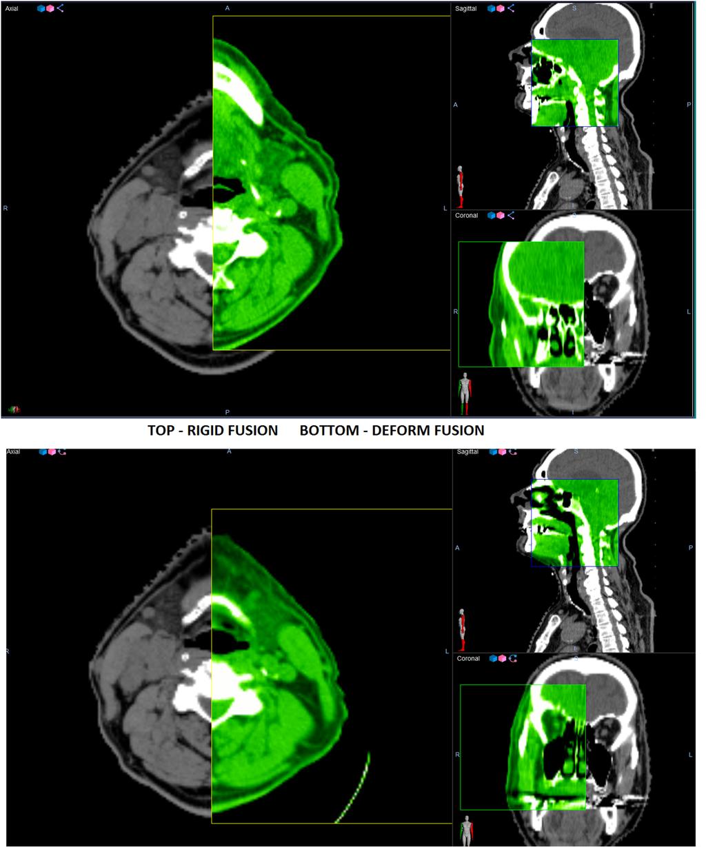
9 Fusion of Studysets Rigid Fusion of diagnostic studysets historically has been the only option and is the standard for gleaning diagnostic information onto the CT simulation data. Immobilization in treatment position necessitates that, frequently, the diagnostic imaging will not align with rigid fusion onto the CT simulation dataset. RIGID FUSION

10 How does a physician resolve this dilemma?
11 Deformation is an alternative When a patient is imaged in a different position for a diagnostic image study than the CT simulation study, deformation can be used to derive information about the target volume from the diagnostic studyset.
12 What is Deformation? Deformation is the process of applying an algorithm to an image studyset or a defined volume within an image studyset to stretch and move the voxels so that it matches the overlayed images of an equal volume.

13 Deformation Not Applied Outer Box CT Sim Inner Box Diagnostic CT

14 Deformation Applied Outer Box CT Sim Inner Box Diagnostic CT
15
16 B Splines algorithm The B Splines algorithm is most commonly used for imaging CT studysets. The two most commonly used commercial software platforms for applying deformations in radiation therapy are MimVista TM and Velocity TM. Each software vendor has proprietary application of the B Splines algorithm for image deformation.
17 Applying Deformation Our experience with applying deformation is within the Velocity TM software platform using their proprietary Deformable Multipass algorithm. The algorithm has two input basis from the user, the rigid registration and the deformation volume. Rigid registration and choice of deformation volume substantially effect the output and success of deformation.
18 Medical Physicist s Role in Deformation Applying our mathematical background, knowledge of cross sectional anatomy, and clinical background is where the physicist becomes the medical physicist. Deformation evaluation is as much an art as it is a science and medical physicist has a skillset that is suitable to this process.
19 Issues Surrounding Deformation The first thing to consider when fusing multiple studysets is whether or not to use deformation. When considering whether or not to use deformation, one must consider the location of the volume of interest, the position of the patient in both image sets, and the size of the volume of interest. The goal should always be rigid fusion, as it reduces uncertainty. Example of this is intercranial fusion.
20 Issues Surrounding Deformation (Con t) Because deformation involves changing one image set to match another, it assumes that there are no changes between anatomy between studysets (no major weight loss/gain, no major volumetric change to target, etc) This is frequently not the case, especially with a long duration between a PET workup and CT simulation. Surgery and chemotherapy in between can have major anatomic effects on the patient.
21 Issues Surrounding Deformation Due to difference between patient studies, deformation can lead to artifact, creation of false structures, blurring, and mismatch of known structures in the body. The physicist has to understand the deformation process to be the expert in evaluating and answering questions regarding deformation as it applies to fusion.


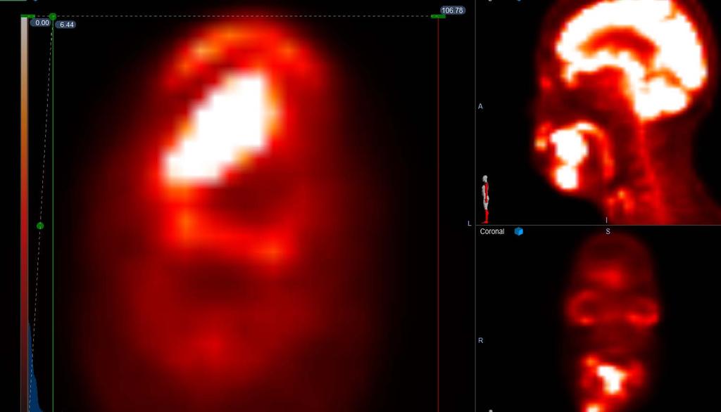
22 Examples of deformation failure Inside Box Deformed PET/CT Outside Box CT simulation Massive stomach/intestinal volume changes. Deformation fails


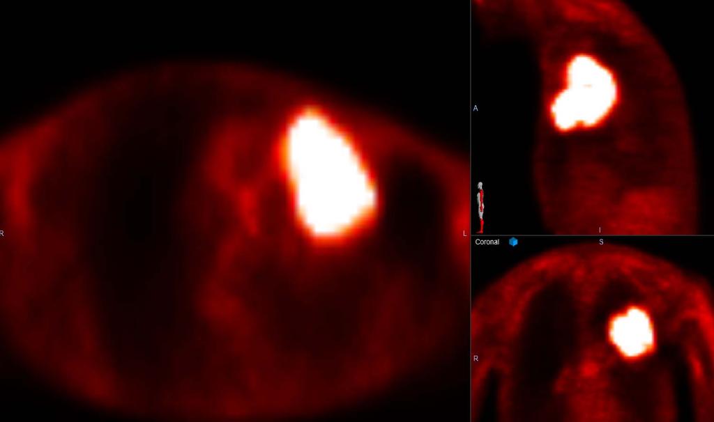
23 Diagnostic CT/PET Scan Deformed PET on CT Simulation The patient s arms were up in CT sim vs. down in CT/PET scan. Deformation clearly fails.
24 Evaluating Deformation Evaluating deformation is an evolving skillset and like anything else, it is a learning process. There are many considerations that go into fusing multiple images and the use of deformation.
25 Quantitative vs. Qualitative Measure of Success Physicists tend to want to apply a quantitative measure of success to an evaluation process. With deformation, there is no proven quantitative measure to currently apply and the evaluation process necessitates a subjective qualitative review by the physicist.
26 Qualitative Components to Evaluation Visual Analysis of Input Parameters (Rigid Fusion and Deformation Volume) Vector and Grid Review of Deformation Grid Alignment of Identifiable Anatomic Structures (best done with CT to CT overlay) Visual Analysis of CT resolution and artifact
27 Qualitative Components to Evaluation Comparison of Volumes between rigid and deformed scans for critical structures and target volumes. Conformality Index of Comparible Structures
28 Managing the Imperfect Art of Deformation Despite the components to evaluation, there is no perfect way to evaluate deformation to verify certainty of the volumes and positioning of deformed targets. Uncertainty must be accepted with target structures.

29 Example of issues surrounding deformation evaluation Blue structure is 5.0 SUV from PET/CT on 6/1/16 Green structure is 5.0 SUV deformed structure to CT sim on 10/17/16 Rigid volume is 15 cc. Deformed is 6.7 cc.

30 Uncertainty in Targeting How is uncertainty best managed in targeting? MARGINS!! By comparing volumes and using a spherical volume assumption, one can margin for uncertainty.
31 Potential Future Considerations For deformation that creates a volume to volume overlay of CT studysets, a gamma comparison analysis of HU values between the two volumes of interest may be useful in the future. This may provide another quantitative component to analysis, but qualitative measures are still needed to make sure that deformation doesn t ignore crucial patient data that can negate postive quantitative results.
32 In Summary Deformation has become an important tool that physics and dosimetry can use for assistance in targeting. The physicist has an important role in defining the use of deformation. There is further development to be done with deformation to ensure safe and accurate use in patient targeting.

33 THANK YOU FOR YOUR ATTENTION AND FEEDBACK!
ICARO Vienna April Implementing 3D conformal radiotherapy and IMRT in clinical practice: Recommendations of IAEA- TECDOC-1588
 ICARO Vienna April 27-29 2009 Implementing 3D conformal radiotherapy and IMRT in clinical practice: Recommendations of IAEA- TECDOC-1588 M. Saiful Huq, Ph.D., Professor and Director, Dept. of Radiation
VALIDATION OF DIR. Raj Varadhan, PhD, DABMP Minneapolis Radiation Oncology
 VALIDATION OF DIR Raj Varadhan, PhD, DABMP Minneapolis Radiation Oncology Overview Basics: Registration Framework, Theory Discuss Validation techniques Using Synthetic CT data & Phantoms What metrics to
REAL-TIME ADAPTIVITY IN HEAD-AND-NECK AND LUNG CANCER RADIOTHERAPY IN A GPU ENVIRONMENT
 REAL-TIME ADAPTIVITY IN HEAD-AND-NECK AND LUNG CANCER RADIOTHERAPY IN A GPU ENVIRONMENT Anand P Santhanam Assistant Professor, Department of Radiation Oncology OUTLINE Adaptive radiotherapy for head and
ROBUST OPTIMIZATION THE END OF PTV AND THE BEGINNING OF SMART DOSE CLOUD. Moe Siddiqui, April 08, 2017
 ROBUST OPTIMIZATION THE END OF PTV AND THE BEGINNING OF SMART DOSE CLOUD Moe Siddiqui, April 08, 2017 Agenda Background IRCU 50 - Disclaimer - Uncertainties Robust optimization Use Cases Lung Robust 4D
Brilliance CT Big Bore.
 1 2 2 There are two methods of RCCT acquisition in widespread clinical use: cine axial and helical. In RCCT with cine axial acquisition, repeat CT images are taken each couch position while recording respiration.
Head and Neck Lymph Node Region Delineation with Auto-segmentation and Image Registration
 Head and Neck Lymph Node Region Delineation with Auto-segmentation and Image Registration Chia-Chi Teng Department of Electrical Engineering University of Washington 1 Outline Introduction Related Work
Auto-Segmentation Using Deformable Image Registration. Disclosure. Objectives 8/4/2011
 Auto-Segmentation Using Deformable Image Registration Lei Dong, Ph.D. Dept. of Radiation Physics University of Texas MD Anderson Cancer Center, Houston, Texas AAPM Therapy Educational Course Aug. 4th 2011
UvA-DARE (Digital Academic Repository) Motion compensation for 4D PET/CT Kruis, M.F. Link to publication
 Motion compensation for 4D PET/CT Kruis, M.F. Link to publication") UvA-DARE (Digital Academic Repository) Motion compensation for 4D PET/CT Kruis, M.F. Link to publication Citation for published version (APA): Kruis, M. F. (2014). Motion compensation for 4D PET/CT General
UvA-DARE (Digital Academic Repository) Motion compensation for 4D PET/CT Kruis, M.F. Link to publication Citation for published version (APA): Kruis, M. F. (2014). Motion compensation for 4D PET/CT General
Image Co-Registration II: TG132 Quality Assurance for Image Registration. Image Co-Registration II: TG132 Quality Assurance for Image Registration
 Image Co-Registration II: TG132 Quality Assurance for Image Registration Preliminary Recommendations from TG 132* Kristy Brock, Sasa Mutic, Todd McNutt, Hua Li, and Marc Kessler *Recommendations are NOT
Medical Image Analysis
 Computer assisted Image Analysis VT04 29 april 2004 Medical Image Analysis Lecture 10 (part 1) Xavier Tizon Medical Image Processing Medical imaging modalities XRay,, CT Ultrasound MRI PET, SPECT Generic
Use of MRI in Radiotherapy: Technical Consideration
 Use of MRI in Radiotherapy: Technical Consideration Yanle Hu, PhD Department of Radiation Oncology, Mayo Clinic Arizona 04/07/2018 2015 MFMER slide-1 Conflict of Interest: None 2015 MFMER slide-2 Objectives
Help Guide. mm Copyright Mirada Medical Ltd, Mirada Medical RTx 1
 Help Guide mm3237-1.6-1 Copyright Mirada Medical Ltd, 2000-2014. Mirada Medical RTx 1 Contents Help Guide... 1 Contents... 2 Introduction to RTx... 4 Regulatory Statement... 6 Notes... 15 Data Supported...
Virtual Phantoms for IGRT QA
 TM Virtual Phantoms for IGRT QA Why ImSimQA? ImSimQA was developed to overcome the limitations of physical phantoms for testing modern medical imaging and radiation therapy software systems, when there
ADVANCING CANCER TREATMENT
 The RayPlan treatment planning system makes proven, innovative RayStation technology accessible to clinics that need a cost-effective and streamlined solution. Fast, efficient and straightforward to use,
Using Pinnacle 16 Deformable Image registration in a re-treat scenario
 Introduction Using Pinnacle 16 Deformable Image registration in a re-treat scenario This short Hands On exercise will introduce how the Deformable Image Registration (DIR) tools in Pinnacle can be used
PET/CT multimodality imaging for radiotherapy planning in lung cancer The medical physicist point of view Isabelle Gardin Rouen CHB and Quant.I.
 PET/CT multimodality imaging for radiotherapy planning in lung cancer The medical physicist point of view Isabelle Gardin Rouen CHB and Quant.I.F (EA4108 FR CNRS 3638) Outline Specific acquisition conditions
iplan RT Image Advanced Contouring Workstation - Driving Physician Collaboration
 iplan RT Image Advanced Contouring Workstation - Driving Physician Collaboration The iplan Contouring Workstation offers unique and innovative capabilities for faster contouring and consistent segmentation
Motion artifact detection in four-dimensional computed tomography images
 Motion artifact detection in four-dimensional computed tomography images G Bouilhol 1,, M Ayadi, R Pinho, S Rit 1, and D Sarrut 1, 1 University of Lyon, CREATIS; CNRS UMR 5; Inserm U144; INSA-Lyon; University
8/4/2016. Emerging Linac based SRS/SBRT Technologies with Modulated Arc Delivery. Disclosure. Introduction: Treatment delivery techniques
 Emerging Linac based SRS/SBRT Technologies with Modulated Arc Delivery Lei Ren, Ph.D. Duke University Medical Center 2016 AAPM 58 th annual meeting, Educational Course, Therapy Track Disclosure I have
Is deformable image registration a solved problem?
 Is deformable image registration a solved problem? Marcel van Herk On behalf of the imaging group of the RT department of NKI/AVL Amsterdam, the Netherlands DIR 1 Image registration Find translation.deformation
Overview of Proposed TG-132 Recommendations
 Overview of Proposed TG-132 Recommendations Kristy K Brock, Ph.D., DABR Associate Professor Department of Radiation Oncology, University of Michigan Chair, AAPM TG 132: Image Registration and Fusion Conflict
Methodological progress in image registration for ventilation estimation, segmentation propagation and multi-modal fusion
 Methodological progress in image registration for ventilation estimation, segmentation propagation and multi-modal fusion Mattias P. Heinrich Julia A. Schnabel, Mark Jenkinson, Sir Michael Brady 2 Clinical
ADVANCING CANCER TREATMENT
 3 ADVANCING CANCER TREATMENT SUPPORTING CLINICS WORLDWIDE RaySearch is advancing cancer treatment through pioneering software. We believe software has un limited potential, and that it is now the driving
Interactive Deformable Registration Visualization and Analysis of 4D Computed Tomography
 Interactive Deformable Registration Visualization and Analysis of 4D Computed Tomography Burak Erem 1, Gregory C. Sharp 2, Ziji Wu 2, and David Kaeli 1 1 Department of Electrical and Computer Engineering,
3D Voxel-Based Volumetric Image Registration with Volume-View Guidance
 3D Voxel-Based Volumetric Image Registration with Volume-View Guidance Guang Li*, Huchen Xie, Holly Ning, Deborah Citrin, Jacek Copala, Barbara Arora, Norman Coleman, Kevin Camphausen, and Robert Miller
Coverage based treatment planning to accommodate organ deformable motions and contouring uncertainties for prostate treatment. Huijun Xu, Ph.D.
 Coverage based treatment planning to accommodate organ deformable motions and contouring uncertainties for prostate treatment Huijun Xu, Ph.D. Acknowledgement and Disclosure Dr. Jeffrey Siebers Dr. DJ
Dosimetric Analysis Report
 RT-safe 48, Artotinis str 116 33, Athens Greece +30 2107563691 info@rt-safe.com Dosimetric Analysis Report SAMPLE, for demonstration purposes only Date of report: ----------- Date of irradiation: -----------
Automated segmentation methods for liver analysis in oncology applications
 University of Szeged Department of Image Processing and Computer Graphics Automated segmentation methods for liver analysis in oncology applications Ph. D. Thesis László Ruskó Thesis Advisor Dr. Antal
Use of image registration and fusion algorithms and techniques in radiotherapy: Report of the AAPM Radiation Therapy Committee Task Group No.
 Use of image registration and fusion algorithms and techniques in radiotherapy: Report of the AAPM Radiation Therapy Committee Task Group No. 132 Kristy K. Brock a) Department of Imaging Physics, The University
Technical aspects of SPECT and SPECT-CT. John Buscombe
 Technical aspects of SPECT and SPECT-CT John Buscombe What does the clinician need to know? For SPECT What factors affect SPECT How those factors should be sought Looking for artefacts For SPECT-CT Issues
radiotherapy Andrew Godley, Ergun Ahunbay, Cheng Peng, and X. Allen Li NCAAPM Spring Meeting 2010 Madison, WI
 GPU-Accelerated autosegmentation for adaptive radiotherapy Andrew Godley, Ergun Ahunbay, Cheng Peng, and X. Allen Li agodley@mcw.edu NCAAPM Spring Meeting 2010 Madison, WI Overview Motivation Adaptive
Good Morning! Thank you for joining us
 Good Morning! Thank you for joining us Deformable Registration, Contour Propagation and Dose Mapping: 101 and 201 Marc Kessler, PhD, FAAPM The University of Michigan Conflict of Interest I receive direct
If it matters to you, it matters to us
 If it matters to you, it matters to us Philips clinical innovations in nuclear medicine Innovation with insight We understand that clinical innovations are only as valuable as the day-to-day difference
Automatic Segmentation of Parotids from CT Scans Using Multiple Atlases
 Automatic Segmentation of Parotids from CT Scans Using Multiple Atlases Jinzhong Yang, Yongbin Zhang, Lifei Zhang, and Lei Dong Department of Radiation Physics, University of Texas MD Anderson Cancer Center
2 Michael E. Leventon and Sarah F. F. Gibson a b c d Fig. 1. (a, b) Two MR scans of a person's knee. Both images have high resolution in-plane, but ha
 Two MR scans of a person's knee. Both images have high resolution in-plane, but ha") Model Generation from Multiple Volumes using Constrained Elastic SurfaceNets Michael E. Leventon and Sarah F. F. Gibson 1 MIT Artificial Intelligence Laboratory, Cambridge, MA 02139, USA leventon@ai.mit.edu
Model Generation from Multiple Volumes using Constrained Elastic SurfaceNets Michael E. Leventon and Sarah F. F. Gibson 1 MIT Artificial Intelligence Laboratory, Cambridge, MA 02139, USA leventon@ai.mit.edu
TG 132: Use of Image Registration and Fusion in RT
 TG 132: Use of Image Registration and Fusion in RT Kristy K Brock, PhD, DABR, FAAPM Associate Professor Department of Radiation Oncology, University of Michigan Chair, AAPM TG 132: Image Registration and
Michael Speiser, Ph.D.
 IMPROVED CT-BASED VOXEL PHANTOM GENERATION FOR MCNP MONTE CARLO Michael Speiser, Ph.D. Department of Radiation Oncology UT Southwestern Medical Center Dallas, TX September 1 st, 2012 CMPWG Workshop Medical
Design and performance characteristics of a Cone Beam CT system for Leksell Gamma Knife Icon
 Design and performance characteristics of a Cone Beam CT system for Leksell Gamma Knife Icon WHITE PAPER Introduction Introducing an image guidance system based on Cone Beam CT (CBCT) and a mask immobilization
Basic principles of MR image analysis. Basic principles of MR image analysis. Basic principles of MR image analysis
 Basic principles of MR image analysis Basic principles of MR image analysis Julien Milles Leiden University Medical Center Terminology of fmri Brain extraction Registration Linear registration Non-linear
CREATION AND VISUALIZATION OF ANATOMICAL MODELS WITH AMIRA CREATION ET VISUALISATION DES MODELES ANATOMIQUES AVEC AMIRA
 CREATION AND VISUALIZATION OF ANATOMICAL MODELS WITH AMIRA CREATION ET VISUALISATION DES MODELES ANATOMIQUES AVEC AMIRA Summary 3D imaging methods are widely used in medicine and biology, mainly for image-guided
Tomographic Reconstruction
 Tomographic Reconstruction 3D Image Processing Torsten Möller Reading Gonzales + Woods, Chapter 5.11 2 Overview Physics History Reconstruction basic idea Radon transform Fourier-Slice theorem (Parallel-beam)
Image Quality Assessment and Quality Assurance of Advanced Imaging Systems for IGRT. AAPM Penn-Ohio Chapter Sep 25, 2015 Soyoung Lee, PhD
 Image Quality Assessment and Quality Assurance of Advanced Imaging Systems for IGRT AAPM Penn-Ohio Chapter Sep 25, 2015 Soyoung Lee, PhD 1 Outline q Introduction q Imaging performances in 4D-CBCT Image
Clinical Prospects and Technological Challenges for Multimodality Imaging Applications in Radiotherapy Treatment Planning
 Clinical Prospects and Technological Challenges for Multimodality Imaging Applications in Radiotherapy Treatment Planning Issam El Naqa, PhD Assistant Professor Department of Radiation Oncology Washington
TomoTherapy Related Projects. An image guidance alternative on Tomo Low dose MVCT reconstruction Patient Quality Assurance using Sinogram
 TomoTherapy Related Projects An image guidance alternative on Tomo Low dose MVCT reconstruction Patient Quality Assurance using Sinogram Development of A Novel Image Guidance Alternative for Patient Localization
Mutual Information Based Methods to Localize Image Registration
 Mutual Information Based Methods to Localize Image Registration by Kathleen P. Wilkie A thesis presented to the University of Waterloo in fulfilment of the thesis requirement for the degree of Master of
The IORT Treatment Planning System. radiance. GMV, 2012 Property of GMV All rights reserved
 The IORT Treatment Planning System radiance Property of GMV All rights reserved WHY RADIANCE? JUSTIFICATION Property of GMV All rights reserved ADVANTAGES OF IORT PRECISION: RT guided by direct vision.
Quantitative imaging for clinical dosimetry
 Quantitative imaging for clinical dosimetry Irène Buvat Laboratoire d Imagerie Fonctionnelle U678 INSERM - UPMC CHU Pitié-Salpêtrière, Paris buvat@imed.jussieu.fr http://www.guillemet.org/irene Methodology
ATC Conformance Statement Overview
 Advanced-Technology QA Consortium DICOM Workshop: ATC Conformance Statement Overview Walter R. Bosch, D.Sc. ATC DICOM Conformance Statement Overview DICOM Part 10 File Set Reader Application (ITC) Defines
From Image Data to Three-Dimensional Geometric Models Case Studies on the Impact of 3D Patient Models
 From Image Data to Three-Dimensional Geometric Models Case Studies on the Impact of 3D Patient Models Hans-Christian HEGE 1,2), Hartmut SCHIRMACHER 2), Malte WESTERHOFF 1,2), Hans LAMECKER 1), Steffen
Medicale Image Analysis
 Medicale Image Analysis Registration Validation Prof. Dr. Philippe Cattin MIAC, University of Basel Prof. Dr. Philippe Cattin: Registration Validation Contents 1 Validation 1.1 Validation of Registration
!"# $ # # $ $ % $ &% $ '"# $ ()&*&)+(( )+(( )
&*&)+(( )+(( )") !"# # # % &% '"# ) !#, ' "# " "# -. / # 0 0 0 0 0 "0 "# " # 1 #! " " 0 0 0 0 0 0 2# 0 # # 3 ' 4 56 7-56 87 9# 5 6 7 6 & 0 " : 9 ; 4 " #! 0 - '% # % "# " "# " < 4 "! % " % 4 % % 9# 4 56 87 = 4 > 0 " %!#
!"# # # % &% '"# ) !#, ' "# " "# -. / # 0 0 0 0 0 "0 "# " # 1 #! " " 0 0 0 0 0 0 2# 0 # # 3 ' 4 56 7-56 87 9# 5 6 7 6 & 0 " : 9 ; 4 " #! 0 - '% # % "# " "# " < 4 "! % " % 4 % % 9# 4 56 87 = 4 > 0 " %!#
Initial Clinical Experience with 3D Surface Image Guidance
 Initial Clinical Experience with 3D Surface Image Guidance Amanda Havnen-Smith, Ph.D. Minneapolis Radiation Oncology Ridges Radiation Therapy Center Burnsville, MN April 20 th, 2012 Non-funded research
MR-guided radiotherapy: Vision, status and research at the UMC Utrecht. Dipl. Ing. Dr. Markus Glitzner
 MR-guided radiotherapy: Vision, status and research at the UMC Utrecht Dipl. Ing. Dr. Markus Glitzner About myself Training Medizintechnik TU Graz PhD UMC Utrecht Clinical work Software implementation
New Technology in Radiation Oncology. James E. Gaiser, Ph.D. DABR Physics and Computer Planning Charlotte, NC
 New Technology in Radiation Oncology James E. Gaiser, Ph.D. DABR Physics and Computer Planning Charlotte, NC Technology s s everywhere From the imaging chain To the planning system To the linac To QA..it..it
Annales UMCS Informatica AI 1 (2003) UMCS. Registration of CT and MRI brain images. Karol Kuczyński, Paweł Mikołajczak
 UMCS. Registration of CT and MRI brain images. Karol Kuczyński, Paweł Mikołajczak") Annales Informatica AI 1 (2003) 149-156 Registration of CT and MRI brain images Karol Kuczyński, Paweł Mikołajczak Annales Informatica Lublin-Polonia Sectio AI http://www.annales.umcs.lublin.pl/ Laboratory
Annales Informatica AI 1 (2003) 149-156 Registration of CT and MRI brain images Karol Kuczyński, Paweł Mikołajczak Annales Informatica Lublin-Polonia Sectio AI http://www.annales.umcs.lublin.pl/ Laboratory
Leksell SurgiPlan Overview. Powerful planning for surgical success
 Leksell SurgiPlan Overview Powerful planning for surgical success Making a Difference in Surgical Planning Leksell SurgiPlan Leksell SurgiPlan is an advanced image-based neuro surgical planning software,
Image-based Monte Carlo calculations for dosimetry
 Image-based Monte Carlo calculations for dosimetry Irène Buvat Imagerie et Modélisation en Neurobiologie et Cancérologie UMR 8165 CNRS Universités Paris 7 et Paris 11 Orsay, France buvat@imnc.in2p3.fr
Tumor motion during liver SBRT
 Tumor motion during liver SBRT - projects at Aarhus University Hospital - Per Poulsen, Esben Worm, Walther Fledelius, Morten Høyer Aarhus University Hospital, Denmark SBRT: Stereotactic Body Radiation
Multimodality Imaging for Tumor Volume Definition in Radiation Oncology
 81 There are several commercial and academic software tools that support different segmentation algorithms. In general, commercial software packages have better implementation (with a user-friendly interface
A fluence convolution method to account for respiratory motion in three-dimensional dose calculations of the liver: A Monte Carlo study
 A fluence convolution method to account for respiratory motion in three-dimensional dose calculations of the liver: A Monte Carlo study Indrin J. Chetty, a) Mihaela Rosu, Neelam Tyagi, Lon H. Marsh, Daniel
1. Learn to incorporate QA for surface imaging
 Hania Al-Hallaq, Ph.D. Assistant Professor Radiation Oncology The University of Chicago ***No disclosures*** 1. Learn to incorporate QA for surface imaging into current QA procedures for IGRT. 2. Understand
Fast CT-CT Fluoroscopy Registration with Respiratory Motion Compensation for Image-Guided Lung Intervention
 Fast CT-CT Fluoroscopy Registration with Respiratory Motion Compensation for Image-Guided Lung Intervention Po Su a,b, Zhong Xue b*, Kongkuo Lu c, Jianhua Yang a, Stephen T. Wong b a School of Automation,
IMRT site-specific procedure: Prostate (CHHiP)
") IMRT site-specific procedure: Prostate (CHHiP) Scope: To provide site specific instructions for the planning of CHHIP IMRT patients Responsibilities: Radiotherapy Physicists, HPC Registered Therapy Radiographers
IMRT site-specific procedure: Prostate (CHHiP) Scope: To provide site specific instructions for the planning of CHHIP IMRT patients Responsibilities: Radiotherapy Physicists, HPC Registered Therapy Radiographers
Use of Deformable Image Registration in Radiation Therapy. Colin Sims, M.Sc. Accuray Incorporated 1
 Use of Deformable Image Registration in Radiation Therapy Colin Sims, M.Sc. Accuray Incorporated 1 Overview of Deformable Image Registration (DIR) Algorithms that can deform one dataset to another have
Medical Image Registration
 Medical Image Registration Submitted by NAREN BALRAJ SINGH SB ID# 105299299 Introduction Medical images are increasingly being used within healthcare for diagnosis, planning treatment, guiding treatment
8/3/2017. Contour Assessment for Quality Assurance and Data Mining. Objective. Outline. Tom Purdie, PhD, MCCPM
 Contour Assessment for Quality Assurance and Data Mining Tom Purdie, PhD, MCCPM Objective Understand the state-of-the-art in contour assessment for quality assurance including data mining-based techniques
Patient Set-ups and Tumor Localizations
 Patient Set-ups and Tumor Localizations Amy S. Harrison Patient Positioning Prior to starting any localization or simulation procedure patients need to be positioned and immobilized Patients disease location
Object Identification in Ultrasound Scans
 Object Identification in Ultrasound Scans Wits University Dec 05, 2012 Roadmap Introduction to the problem Motivation Related Work Our approach Expected Results Introduction Nowadays, imaging devices like
UvA-DARE (Digital Academic Repository) Motion compensation for 4D PET/CT Kruis, M.F. Link to publication
 Motion compensation for 4D PET/CT Kruis, M.F. Link to publication") UvA-DARE (Digital Academic Repository) Motion compensation for 4D PET/CT Kruis, M.F. Link to publication Citation for published version (APA): Kruis, M. F. (2014). Motion compensation for 4D PET/CT General
UvA-DARE (Digital Academic Repository) Motion compensation for 4D PET/CT Kruis, M.F. Link to publication Citation for published version (APA): Kruis, M. F. (2014). Motion compensation for 4D PET/CT General
Development and validation of clinically feasible 4D-MRI in radiotherapy
 MSc. Applied Physics Thesis Development and validation of clinically feasible 4D-MRI in radiotherapy Daniel Tekelenburg By: Daniel Ruphay Tekelenburg In partial fulfilment of the requirements for the degree
PCRT 3D. Scalable Architecture System. User-Friendly. Traceable. Continuos Development
 PCRT 3D The PCRT3D is a versatile 3D radiation treatment planning system featuring the most accurate algorithm calculations, the latest techniques in virtual simulation and the most advanced radiotherapy
Using a research real-time control interface to go beyond dynamic MLC tracking
 in partnership with Using a research real-time control interface to go beyond dynamic MLC tracking Dr. Simeon Nill Joint Department of Physics at The Institute of Cancer Research and the Royal Marsden
INTRODUCTION TO MEDICAL IMAGING- 3D LOCALIZATION LAB MANUAL 1. Modifications for P551 Fall 2013 Medical Physics Laboratory
 INTRODUCTION TO MEDICAL IMAGING- 3D LOCALIZATION LAB MANUAL 1 Modifications for P551 Fall 2013 Medical Physics Laboratory Introduction Following the introductory lab 0, this lab exercise the student through
Leksell SurgiPlan. Powerful planning for success
 Leksell SurgiPlan Powerful planning for success Making a difference in surgical planning Leksell SurgiPlan Leksell SurgiPlan is an advanced image-based neurosurgical planning software, specifically designed
Increasing Interoperability, what is the Impact on Reliability? Illustrated with Health care examples
 Illustrated with Health care examples by Gerrit Muller University of South-Eastern Norway-NISE e-mail: gaudisite@gmail.com www.gaudisite.nl Abstract In all domains the amount of interoperability between
Gradient-Based Differential Approach for Patient Motion Compensation in 2D/3D Overlay
 Gradient-Based Differential Approach for Patient Motion Compensation in 2D/3D Overlay Jian Wang, Anja Borsdorf, Benno Heigl, Thomas Köhler, Joachim Hornegger Pattern Recognition Lab, Friedrich-Alexander-University
State-of-the-Art IGRT
 in partnership with State-of-the-Art IGRT Exploring the Potential of High-Precision Dose Delivery and Real-Time Knowledge of the Target Volume Location Antje-Christin Knopf IOP Medical Physics Group Scientific
Validation of PET/CT dataset for radiation treatment planning
 Louisiana State University LSU Digital Commons LSU Master's Theses Graduate School 2004 Validation of PET/CT dataset for radiation treatment planning Rajesh Manoharan Louisiana State University and Agricultural
Weakly Supervised Fully Convolutional Network for PET Lesion Segmentation
 Weakly Supervised Fully Convolutional Network for PET Lesion Segmentation S. Afshari a, A. BenTaieb a, Z. Mirikharaji a, and G. Hamarneh a a Medical Image Analysis Lab, School of Computing Science, Simon
Ch. 4 Physical Principles of CT
 Ch. 4 Physical Principles of CT CLRS 408: Intro to CT Department of Radiation Sciences Review: Why CT? Solution for radiography/tomography limitations Superimposition of structures Distinguishing between
Modeling and preoperative planning for kidney surgery
 Modeling and preoperative planning for kidney surgery Refael Vivanti Computer Aided Surgery and Medical Image Processing Lab Hebrew University of Jerusalem, Israel Advisor: Prof. Leo Joskowicz Clinical
A rib-specific multimodal registration algorithm for fused unfolded rib visualization using PET/CT
 A rib-specific multimodal registration algorithm for fused unfolded rib visualization using PET/CT Jens N. Kaftan a, Marcin Kopaczka a, Andreas Wimmer b, Günther Platsch a, and Jérôme Declerck a a Siemens
Joint CI-JAI advanced accelerator lecture series Imaging and detectors for medical physics Lecture 1: Medical imaging
 Joint CI-JAI advanced accelerator lecture series Imaging and detectors for medical physics Lecture 1: Medical imaging Dr Barbara Camanzi barbara.camanzi@stfc.ac.uk Course layout Day AM 09.30 11.00 PM 15.30
Dose Calculation and Optimization Algorithms: A Clinical Perspective
 Dose Calculation and Optimization Algorithms: A Clinical Perspective Daryl P. Nazareth, PhD Roswell Park Cancer Institute, Buffalo, NY T. Rock Mackie, PhD University of Wisconsin-Madison David Shepard,
Mathematical methods and simulations tools useful in medical radiation physics
 Mathematical methods and simulations tools useful in medical radiation physics Michael Ljungberg, professor Department of Medical Radiation Physics Lund University SE-221 85 Lund, Sweden Major topic 1:
GE Healthcare. Vivid 7 Dimension 08 Cardiovascular ultrasound system
 GE Healthcare Vivid 7 Dimension 08 Cardiovascular ultrasound system ltra Definition. Technology. Performance. Start with a system that s proven its worth in LV quantification and 4D imaging. Then add even
XD 3.6 Release Notes
 XD 3.6 Release Notes 1 Introduction 1.1 Purpose The purpose of these Release Notes is to: 1.2 Scope Communicate any known limitations for Mirada s XD release. Provide information on certain product features.
Deformable Image Registration, Contour Propagation and Dose Mapping: 101 and 201. Please do not (re)redistribute
redistribute") Deformable Registration, Contour Deformable Registration, Contour Propagation and Dose Mapping: 101 and 201 Marc Kessler, PhD The University of Michigan Jean Pouliot, PhD University of California Learning
Deformable Registration, Contour Deformable Registration, Contour Propagation and Dose Mapping: 101 and 201 Marc Kessler, PhD The University of Michigan Jean Pouliot, PhD University of California Learning
Nonrigid Registration using Free-Form Deformations
 Nonrigid Registration using Free-Form Deformations Hongchang Peng April 20th Paper Presented: Rueckert et al., TMI 1999: Nonrigid registration using freeform deformations: Application to breast MR images
Position accuracy analysis of the stereotactic reference defined by the CBCT on Leksell Gamma Knife Icon
 Position accuracy analysis of the stereotactic reference defined by the CBCT on Leksell Gamma Knife Icon WHITE PAPER Introduction An image guidance system based on Cone Beam CT (CBCT) is included in Leksell
CT issues in PET / CT scanning. ImPACT technology update no. 4
 abc David Platten Introduction ImPACT technology update no. 4 ImPACT October 2004 Since its introduction in the 1970s, X-ray computed tomography (CT) scanning has become a widespread three-dimensional
Diagnostic imaging techniques. Krasznai Zoltán. University of Debrecen Medical and Health Science Centre Department of Biophysics and Cell Biology
 Diagnostic imaging techniques Krasznai Zoltán University of Debrecen Medical and Health Science Centre Department of Biophysics and Cell Biology 1. Computer tomography (CT) 2. Gamma camera 3. Single Photon
Super-resolution Reconstruction of Fetal Brain MRI
 Super-resolution Reconstruction of Fetal Brain MRI Ali Gholipour and Simon K. Warfield Computational Radiology Laboratory Children s Hospital Boston, Harvard Medical School Worshop on Image Analysis for
CT Protocol Review: Practical Tips for the Imaging Physicist Physicist
 CT Protocol Review: Practical Tips for the Imaging Physicist Physicist Dianna Cody, Ph.D., DABR, FAAPM U.T.M.D. Anderson Cancer Center August 8, 2013 AAPM Annual Meeting Goals Understand purpose and importance
CHAPTER 2. Morphometry on rodent brains. A.E.H. Scheenstra J. Dijkstra L. van der Weerd
 CHAPTER 2 Morphometry on rodent brains A.E.H. Scheenstra J. Dijkstra L. van der Weerd This chapter was adapted from: Volumetry and other quantitative measurements to assess the rodent brain, In vivo NMR
An Optimisation Model for Intensity Modulated Radiation Therapy
 An Optimisation Model for Intensity Modulated Radiation Therapy Matthias Ehrgott Department of Engineering Science University of Auckland New Zealand m.ehrgott@auckland.ac.nz Abstract Intensity modulated
Oncentra Brachy. Anatomy-based treatment planning for HDR/PDR brachytherapy
 Oncentra Brachy Anatomy-based treatment planning for HDR/PDR brachytherapy Anatomy-based treatment planning for HDR/PDR brachytherapy In its rich history, Nucletron has developed a wide range of treatment
Basics of treatment planning II
 Basics of treatment planning II Sastry Vedam PhD DABR Introduction to Medical Physics III: Therapy Spring 2015 Dose calculation algorithms! Correction based! Model based 1 Dose calculation algorithms!
ESTIMATING 3D DEFORMABLE MOTION FROM A SERIES OF FAST 2D MRI IMAGES. Jason M. Brown. Chapel Hill 2016
 ESTIMATING 3D DEFORMABLE MOTION FROM A SERIES OF FAST 2D MRI IMAGES Jason M. Brown A thesis submitted to the faculty of the University of North Carolina at Chapel Hill in partial fulfillment of the requirements
Implementation of Advanced Image Guided Radiation Therapy
 Image Acquisition Course Outline Principles, characteristics& applications of the available modalities Image Processing in the T x room Image guided treatment delivery What can / can t we do in the room