Introduction to MRI data processing with FSL. Anna Blazejewska
|
|
|
- Gervais Parrish
- 6 years ago
- Views:
Transcription
1 Introduction to MRI data processing with FSL Anna Blazejewska
, diffusion MRI data processing & analysis written in C++ & TCL for Linux (virtual box on Windows) & Mac OS GUI but also command line!")
2 FSL = FMRIB Software Library FMRIB = Functional Magnetic Resonance Imaging of the Oxford since 2000, last stable FSL 5.0, free! for structural MRI, functional MRI (task, resting), diffusion MRI data processing & analysis written in C++ & TCL for Linux (virtual box on Windows) & Mac OS GUI but also command line!! shell script pipelines installation: fslinstaller.py support: wiki, FAQ, forum yearly FSL courses: theory & hands on, 5 days, slides on-line
3 diffusion MRI functional MRI structural MRI Why N How Seminar Martinos Center March 30, 2017 Anna Blazejewska ablazejewska@mgh.harvard.edu FSL overview registration: linear (FLIRT) & non-linear (FNIRT) brain segmentation/extraction (BET) tissue type segmentation (FAST) subcortical structures segmentation (FIRST) voxelwise GM density analysis (FSLVBM) atrophy estimation (SIENA) motion correction (MCFLIRT) EPI distortion correction (FUGUE, PRELUDE) model-based analysis (FEAT) model free ICA-based analysis (MELODIC) Bayesian analysis of perfusion, ASL data (FABBER, VERBENA, BASIL) distortion correction (TOPUP) eddy current correction (EDDY) diffusion toolbox (FDT) tract-based spatial statistics (TBSS) general: fslmaths, fslchfiletype, fslroi, smoothing, stats, fslview and more
 & non-linear (FNIRT) brain segmentation/extraction (BET) tissue type segmentation (FAST) subcortical structures segmentation (FIRST) voxelwise GM density")
4 diffusion MRI functional MRI structural MRI Why N How Seminar Martinos Center March 30, 2017 Anna Blazejewska ablazejewska@mgh.harvard.edu FSL overview registration: linear (FLIRT) & non-linear (FNIRT) brain segmentation/extraction (BET) tissue type segmentation (FAST) subcortical structures segmentation (FIRST) voxelwise GM density analysis (FSLVBM) atrophy estimation (SIENA) 1. segmentation 2. registration 3. distortion correction 4. shell scripts! motion correction (MCFLIRT) EPI distortion correction (FUGUE, PRELUDE) model-based analysis (FEAT) model free ICA-based analysis (MELODIC) Bayesian analysis of perfusion, ASL data (FABBER, VERBENA, BASIL) distortion correction (TOPUP) eddy current correction (EDDY) diffusion toolbox (FDT) tract-based spatial statistics (TBSS) general: fslmaths, fslchfiletype, fslroi, smoothing, stats, fslview and more $ fsl
5 1. segmentation
 preparation for: registration/motion correction tissue segmentation masking out non-brain problems of manual segmentation: time, training, reproducibility benefits")
, 2002. Jenkinson, M et al.")
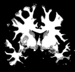
6 BET: brain extraction = skull stripping 1. segmentation goal: automatic segmentation of brain & non-brain tissue (skin, skull, eyeballs, etc.) preparation for: registration/motion correction tissue segmentation masking out non-brain problems of manual segmentation: time, training, reproducibility benefits of BET 5-20 sec, high reproducibility different contrasts T1-/T2-/T2*-w data, robust to bias field (uses local intensity changes) can estimate inner & outer skull & outer scalp surface brain non-brain Smith, SM, Fast robust automated brain extraction, HBM 17(3), Jenkinson, M et al., BET2: MR-based estimation of brain, skull and scalp surfaces, OHBM, 2005.

 spherical surface initialization subdividing")

7 BET: how does it work? 1. segmentation surface = mesh of connected triangles (no folds, not cortex) histogram-based threshold t estimation t-based binarization center of gravity (COG) spherical surface initialization subdividing each triangle expanding the surface vertex locations updated based on local intensities not self-intersecting surface smoothness condition Smith, SM, Fast robust automated brain extraction, HBM 17(3), 2002.

![output [parameters] -f <f>](/docs-images/75/71831840/images/8-1.jpg "-g <g> f brain outline")
![estimate, [0,1], =0.](/docs-images/75/71831840/images/8-2.jpg "5 g brain outline bottom,")
![top, [-1,1], =0 7T Philips](/docs-images/75/71831840/images/8-3.jpg "MPRAGE 1.0 mm 3 default: f=0.")
8 BET: how to use it? 1. segmentation $ bet input output [parameters] -f <f> -g <g> f brain outline estimate, [0,1], =0.5 g brain outline bottom, top, [-1,1], =0 7T Philips MPRAGE 1.0 mm 3 default: f=0.5 g=0 modified: f=0.3 g=0.3
9 BET: how to use it? 1. segmentation $ bet input output [parameters] -c <x y z> -r <r> initial center of gravity [voxels] head radius [mm], initial r/2 -R -Z -F -m -s -o -n -A robust brain center estimation temporary add slices for small z-fov apply to 4D fmri data (-f dilation) save binary brain mask save skull image save brain surface outline skip segmented brain image generate skull & scalp surfaces (BETSURF)
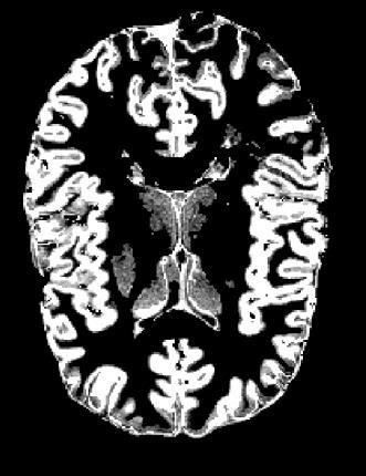
10 FAST: tissue segmentation FMRIB s Automated Segmentation Tool goal: automatic segmentation of WM, GM & CSF input: BET processed images (extracted brain) single image: T1, T2, PD multichannel images pre-aligned (with FLIRT) output: binary tissue masks or probability maps 1. segmentation 1. histogram-based: uses Gaussian mixture model 2. estimates & removes bias field 3. considers voxel s neighborhood robust to noise 4. use of prior tissue probability maps GM CSF WM Zhang, Y et al., Segmentation of brain MR images through a hidden Markov random field model and the expectation-maximization algorithm, IEEE Trans Med Imag, 20(1), 2001.

, P(WM) CSF GM WM WM GM CSF P(CSF) = 0 P(GM) = 400 P(WM) = 9000 GM/WM border http://fsl.")
11 FAST: histogram as a mixture of Gaussians 1. segmentation data intensity histogram model = mixture of Gaussians separation of the peaks segmentation peaks overlap segmentation more difficult for each voxel calculates: P(CSF), P(GM), P(WM) CSF GM WM WM GM CSF P(CSF) = 0 P(GM) = 400 P(WM) = 9000 GM/WM border

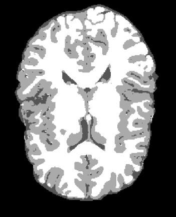
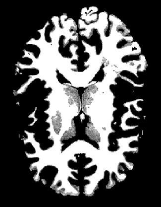
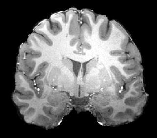
12 FAST: hard segmentation vs probability maps 1. segmentation original image 3 class probability maps segmentation CSF GM WM 7T Siemens MEMPRAGE 0.75 mm 3


13 FAST: bias field correction 1. segmentation low resolution, head motion, noise, blurring & bias field histogram peaks overlap RF inhomogeneity spatial intensity variations = bias field original image estimated bias field corrected image

 + P(neighborhood) controls")

14 FAST: neighborhood 1. segmentation local neighborhood information robust to noise P(class) = P(intensity) + P(neighborhood) controls contribution of neighbors vs intensity, can be set by user =0 =0.1 =0.3 =0.5
![FAST: how to use it? 1. segmentation $ fast [options] input -H -t -S -b -B -N -I -l, =0.](/docs-images/75/71831840/images/15-0.jpg "3 input data type: 1=T1, 2=T2, 3=PD number of input data channels, =1 save estimated bias field save bias-corrected input image no bias field correction number of iteration for bias field removal, =4")
15 FAST: how to use it? 1. segmentation $ fast [options] input -H -t -S -b -B -N -I -l, =0.3 input data type: 1=T1, 2=T2, 3=PD number of input data channels, =1 save estimated bias field save bias-corrected input image no bias field correction number of iteration for bias field removal, =4 bias field smoothing FWHM, =20mm -n -g -nopve number of tissue type classes, =3 save a binary mask for each class skip probability maps


 alternative prior")

16 FAST: use of priors 1. segmentation -a t.mat -P -A p1 p2 p3 -s file use prior probability maps for initialization (requires transformation to standard space) use prior probability maps at all stages (requires a/-a) alternative prior images for tissue classes initial tissue-type means T1-w mean GM WM CSF
17 2. registration


 MNI152 = 152 structural images, averaged after high-dimensional nonlinear")


18 registration 2. registration = transformation of 3D volumes into the same space (coordinate system) 1. transformation 2. cost function 3. interpolation intra-subject: different modalities, contrasts, frames etc. inter-subject: different subjects to common space (population analysis etc.) MNI152 = 152 structural images, averaged after high-dimensional nonlinear registration $FSLDIR/data/standards Jenkinson M & Smith. A global optimisation method for robust affine registration of brain images. Medical Image Analysis, 5(2): , M. Jenkinson, P.R. Bannister, J.M. Brady, and S.M. Smith. Improved optimisation for the robust and accurate linear registration and motion correction of brain images. NeuroImage, 17(2): , 2002.


19 FLIRT: linear transformation FMRIB s Linear Registration Tool intra-subject, same/different modality, DOF = degrees of freedom 2. registration rigid body = 6DOF = 3 rotations + 3 translations affine = 12 DOF = 6DOF + 3 scalings + 3 skews/shears represented by a transformation matrix a 11 a 12 a 13 a 14 a 21 a 22 a 23 a 24 a 31 a 32 a 33 a coordinate vectors multiplied by the matrix define transformed coordinates

 represented by a 3D deformation field =")
 deformation field x-component FLIRT MNI152 FNIRT y-component")

20 FNIRT: non-linear transformation FMRIB s Non-Linear Registration Tool 2. registration inter-subject: same anatomical region in all subjects (atlas with anatomical labels) many many DOFs, same modality, least squares cost function, bias field model (FNIRT) represented by a 3D deformation field = warp, displacement field stored as 3 x 3D volumes (x-, y-, z-components of the vectors) deformation field x-component FLIRT MNI152 FNIRT y-component z-component mm inter-subject variability not very accurate! some individual features lost
21 cost function 2. registration registration = iterative process of finding the best alignment of 3D volumes minimizing cost function = measure of goodness of the alignment beware of local minima! local min global min local min least squares: same modality, same brightness, contrast (includes bias field model) normalized correlation: same modality, different brightness, contrast correlation ratio: any MRI modalities mutual information: any modalities (also CT, PET etc.) normalized mutual information: any modalities (also CT, PET etc.) boundary-based-registration = BBR: intra-subject, EPI to structural


 fast, blocky (pixelized)")
 fast, blurring")

22 Why N How Seminar Martinos Center March 30, 2017 Anna Blazejewska ablazejewska@mgh.harvard.edu interpolation =finding intensity values between grid points 2. registration nearest neighbor (NN) fast, blocky (pixelized) for labels, binary masks tri linear (TL) fast, blurring most common option spline, sinc slower, sharper images (spline not that slow) 7T Siemens GRE EPI 1.5 mm 3

23 FLIRT: how to use it? 2. registration $ flirt -in input ref reference out output -omat matrix -dof df -cost cf -interp it degrees of freedom, =12 cost function, =corratio interpolation method, =trilinear -searchrx min max -nosearch -usesqform -noresample search angles, = all search angles = 0 initialize using sform/qform estimates transformation


24 FLIRT: example 2. registration 7T Siemens GRE EPI 1.5 mm 3 7T Siemens MEMPRAGE 0.75 mm 3 $ flirt -in input ref reference out output -omat matrix -cost mutualinfo -dof 6 interp sinc searchrx -3 3 searchry -3 3 searchrx -3 3


25 FLIRT: mask registration 2. registration applying existing transformation: $ flirt -in input ref reference out output applyxfm init matrix.mat -interp nearestneighbour integer, rounded mask shrinks -interp trilinear followed by thresholding: = 0.5 same size mask as input < 0.5 more PVE, > 0.5 less PVE original NN TL TL vs NN TL 0.5 vs NN TL 0.2 vs NN


26 FLIRT & FNIRT: weighted registration 2. registration cost function weighting for input and/or reference image values range [0,1], for corrupted areas, for important areas artifacts pathologies =1 =0 -refweight <volume> -inweight <volume> use weights for reference volume use weights for input volume

27 MCFLIRT: motion correction 2. registration registration of multiple frames of the same fmri scan $ mcflirt in input out output [options] -refvol -meanvol -cost cf -dof df reference frame, =middle use mean volume as a reference cost function, =normcorr degrees of freedom, =6 before -sinc_final -spline_final final interpolation choice, =trilinear -stats -mats -plots save variance & stdev images save motion parameters in*.par file save transformation matrices (in subdir) after Jenkinson, M, et al., Improved Optimisation for the Robust and Accurate Linear Registration and Motion Correction of Brain Images. NeuroImage, 17(2), , 2002.
28 3. distortion correction


29 EPI geometric distortion 3. distortion correction magnetic susceptibility = χ material s property, quantifies ability to magnetize susceptibility effects T 2 *w contrast B 0 PA brain χ <<1 air χ 0 B +Δφ +Δφ +Δφ φ φ φ Δφ Δφ Δφ dephasing 0 G y distortion AP 1. field map based distortion correction (FUGUE) 2. EPI data acquired with the opposite PE direction (TOPUP)
 need to have/know: field map, phase encoding direction, ESP, TEs ( TE) $ fugue -i epi p phase u output --unwarpdir=dir")
30 FUGUE: how to use it? 3. distortion correction field map B voxel shift map undo distortion (unwarp) need to have/know: field map, phase encoding direction, ESP, TEs ( TE) $ fugue -i epi p phase u output --unwarpdir=dir -dwell=esp --asym=dte -s sigma phase encoding direction, =y (usually AP) echo spacing (ESP, in sequence parameters) TE 2D Gaussian smoothing (field map regularization)! phase data must have the same FOV & resolution as EPI (may require prior resampling)

 manually: resampling to EPI resolution:")
31 FUGUE: how to use it? 3. distortion correction magnitude TE1 magnitude TE2 phase 7T Siemens, 2mm 3 Siemens: GUI/script, magnitude data brain only (BET), output in rad/s $ fsl_prepare_fieldmap SIEMENS phz mag output dte others (or Siemens) manually: resampling to EPI resolution: flirt -applyxfm convert phase to radians & rad/s: fslmaths unwrapping phase: prelude
32 ${FSLDIR}/etc/flirtsch/ TOPUP: distortion correction 3. distortion correction EPI data acquired with opposite phase encoding directions

33 ${FSLDIR}/etc/flirtsch/ TOPUP: distortion correction 3. distortion correction EPI data acquired with opposite phase encoding directions
34 ${FSLDIR}/etc/flirtsch/ TOPUP: how to use it? 3. distortion correction $ topup - imain=input - datain=ap_pa.txt -config=topup.cfg --out=output --fout=fmap - iout=corrected --dfout=deform input: combined frames with opposite phase encoding directions ap_pa.txt: columns 1-3 = phase encoding directions (3 rd one must be 0), column 4 = readout time (if vary between the volumes) config: default one $FSLDIR/etc/flirtsch/b02b0.cnf (data resolution <3.0 mm) corrected: input set of frames otherwise applytopup ap_pa.txt
35 shell scripting REF=${PATH_TO_ALL_MY_DATA}/struct_brain.nii.gz for s in ${PATH_TO_ALL_MY_DATA}/epi*brain.nii.gz; do flirt -in ${s} ref ${REF} dof 6 cost mutualinfo... done; automatic! work by themselves! run multiple subjects/datasets automatically systematically explore different parameters/options run multiple processing streams in parallel reproducibility: each subjects processed the same way replicability: easy to re-run with some modification save (are) documentation of what was run easy to share with others! check the results on each stage of processing & monitor potential crashing..
36 Thank you! Anna Blazejewska
Brain Extraction, Registration & EPI Distortion Correction
 Brain Extraction, Registration & EPI Distortion Correction What use is Registration? Some common uses of registration: Combining across individuals in group studies: including fmri & diffusion Quantifying
Structural Segmentation
 Structural Segmentation FAST tissue-type segmentation FIRST sub-cortical structure segmentation FSL-VBM voxelwise grey-matter density analysis SIENA atrophy analysis FAST FMRIB s Automated Segmentation
Structural Segmentation
 Structural Segmentation FAST tissue-type segmentation FIRST sub-cortical structure segmentation FSL-VBM voxelwise grey-matter density analysis SIENA atrophy analysis FAST FMRIB s Automated Segmentation
Basic principles of MR image analysis. Basic principles of MR image analysis. Basic principles of MR image analysis
 Basic principles of MR image analysis Basic principles of MR image analysis Julien Milles Leiden University Medical Center Terminology of fmri Brain extraction Registration Linear registration Non-linear
Image Registration + Other Stuff
 Image Registration + Other Stuff John Ashburner Pre-processing Overview fmri time-series Motion Correct Anatomical MRI Coregister m11 m 21 m 31 m12 m13 m14 m 22 m 23 m 24 m 32 m 33 m 34 1 Template Estimate
FSL Pre-Processing Pipeline
 The Art and Pitfalls of fmri Preprocessing FSL Pre-Processing Pipeline Mark Jenkinson FMRIB Centre, University of Oxford FSL Pre-Processing Pipeline Standard pre-processing: Task fmri Resting-state fmri
Playing with data from lab
 Playing with data from lab Getting data off the scanner From the Patient Browser, select the folder for the study you want (or within that study, the set of images you want), and then from the Transfer
User s Guide Neuroimage Processing ToolKit (NPTK) Version.1.7 (beta) fmri Registration Software Pipeline for Functional Localization
 Version.1.7 (beta) fmri Registration Software Pipeline for Functional Localization") User s Guide Neuroimage Processing ToolKit (NPTK) Version.1.7 (beta) fmri Registration Software Pipeline for Functional Localization Software Written by Ali Gholipour SIP Lab, UTD, 2005-2007 Revision 1.7
User s Guide Neuroimage Processing ToolKit (NPTK) Version.1.7 (beta) fmri Registration Software Pipeline for Functional Localization Software Written by Ali Gholipour SIP Lab, UTD, 2005-2007 Revision 1.7
Basic fmri Design and Analysis. Preprocessing
 Basic fmri Design and Analysis Preprocessing fmri Preprocessing Slice timing correction Geometric distortion correction Head motion correction Temporal filtering Intensity normalization Spatial filtering
Computational Neuroanatomy
 Computational Neuroanatomy John Ashburner john@fil.ion.ucl.ac.uk Smoothing Motion Correction Between Modality Co-registration Spatial Normalisation Segmentation Morphometry Overview fmri time-series kernel
FSL Pre-Processing Pipeline
 The Art and Pitfalls of fmri Preprocessing FSL Pre-Processing Pipeline Mark Jenkinson FMRIB Centre, University of Oxford FSL Pre-Processing Pipeline Standard pre-processing: Task fmri Resting-state fmri
SPM8 for Basic and Clinical Investigators. Preprocessing. fmri Preprocessing
 SPM8 for Basic and Clinical Investigators Preprocessing fmri Preprocessing Slice timing correction Geometric distortion correction Head motion correction Temporal filtering Intensity normalization Spatial
Functional MRI in Clinical Research and Practice Preprocessing
 Functional MRI in Clinical Research and Practice Preprocessing fmri Preprocessing Slice timing correction Geometric distortion correction Head motion correction Temporal filtering Intensity normalization
Fmri Spatial Processing
 Educational Course: Fmri Spatial Processing Ray Razlighi Jun. 8, 2014 Spatial Processing Spatial Re-alignment Geometric distortion correction Spatial Normalization Smoothing Why, When, How, Which Why is
EPI Data Are Acquired Serially. EPI Data Are Acquired Serially 10/23/2011. Functional Connectivity Preprocessing. fmri Preprocessing
 Functional Connectivity Preprocessing Geometric distortion Head motion Geometric distortion Head motion EPI Data Are Acquired Serially EPI Data Are Acquired Serially descending 1 EPI Data Are Acquired
Automated MR Image Analysis Pipelines
 Automated MR Image Analysis Pipelines Andy Simmons Centre for Neuroimaging Sciences, Kings College London Institute of Psychiatry. NIHR Biomedical Research Centre for Mental Health at IoP & SLAM. Neuroimaging
Functional MRI data preprocessing. Cyril Pernet, PhD
 Functional MRI data preprocessing Cyril Pernet, PhD Data have been acquired, what s s next? time No matter the design, multiple volumes (made from multiple slices) have been acquired in time. Before getting
Diffusion MRI Acquisition. Karla Miller FMRIB Centre, University of Oxford
 Diffusion MRI Acquisition Karla Miller FMRIB Centre, University of Oxford karla@fmrib.ox.ac.uk Diffusion Imaging How is diffusion weighting achieved? How is the image acquired? What are the limitations,
An Introduction To Automatic Tissue Classification Of Brain MRI. Colm Elliott Mar 2014
 An Introduction To Automatic Tissue Classification Of Brain MRI Colm Elliott Mar 2014 Tissue Classification Tissue classification is part of many processing pipelines. We often want to classify each voxel
Introduction to fmri. Pre-processing
 Introduction to fmri Pre-processing Tibor Auer Department of Psychology Research Fellow in MRI Data Types Anatomical data: T 1 -weighted, 3D, 1/subject or session - (ME)MPRAGE/FLASH sequence, undistorted
SPM8 for Basic and Clinical Investigators. Preprocessing
 SPM8 for Basic and Clinical Investigators Preprocessing fmri Preprocessing Slice timing correction Geometric distortion correction Head motion correction Temporal filtering Intensity normalization Spatial
2. Creating Field Maps Using the Field Map GUI (Version 2.0) in SPM5
 in SPM5") 1. Introduction This manual describes how to use the Field Map Toolbox Version 2.0 for creating unwrapped field maps that can be used to do geometric distortion correction of EPI images in SPM5. 1. 1.
1. Introduction This manual describes how to use the Field Map Toolbox Version 2.0 for creating unwrapped field maps that can be used to do geometric distortion correction of EPI images in SPM5. 1. 1.
fmri pre-processing Juergen Dukart
 fmri pre-processing Juergen Dukart Outline Why do we need pre-processing? fmri pre-processing Slice time correction Realignment Unwarping Coregistration Spatial normalisation Smoothing Overview fmri time-series
The organization of the human cerebral cortex estimated by intrinsic functional connectivity
 1 The organization of the human cerebral cortex estimated by intrinsic functional connectivity Journal: Journal of Neurophysiology Author: B. T. Thomas Yeo, et al Link: https://www.ncbi.nlm.nih.gov/pubmed/21653723
2019 FSL Course. Course Handbook February 2019 Dunedin, New Zealand
 2019 FSL Course Course Handbook 18-22 February 2019 Dunedin, New Zealand Organised by: Wellcome Centre for Integrative Neuroimaging University of Oxford Welcome! We are pleased to welcome you to the
White Pixel Artifact. Caused by a noise spike during acquisition Spike in K-space <--> sinusoid in image space
 White Pixel Artifact Caused by a noise spike during acquisition Spike in K-space sinusoid in image space Susceptibility Artifacts Off-resonance artifacts caused by adjacent regions with different
Supplementary methods
 Supplementary methods This section provides additional technical details on the sample, the applied imaging and analysis steps and methods. Structural imaging Trained radiographers placed all participants
User s Guide Neuroimage Processing ToolKit (NPTK) Version 2.0 fmri Registration Software Pipeline for Functional Localization
 Version 2.0 fmri Registration Software Pipeline for Functional Localization") User s Guide Neuroimage Processing ToolKit (NPTK) Version 2.0 fmri Registration Software Pipeline for Functional Localization Software Written by Ali Gholipour SIP Lab, UTD, 2005-2010 Revision 2.0 February
User s Guide Neuroimage Processing ToolKit (NPTK) Version 2.0 fmri Registration Software Pipeline for Functional Localization Software Written by Ali Gholipour SIP Lab, UTD, 2005-2010 Revision 2.0 February
How to create a head model
 How to create a head model This document describes the command line tools: mri2mesh: Central tool to reconstruct a head model from T1w and T2w data dwi2cond: Reconstruct conductivity tensors for brain
Nonrigid Registration using Free-Form Deformations
 Nonrigid Registration using Free-Form Deformations Hongchang Peng April 20th Paper Presented: Rueckert et al., TMI 1999: Nonrigid registration using freeform deformations: Application to breast MR images
Image Processing for fmri John Ashburner. Wellcome Trust Centre for Neuroimaging, 12 Queen Square, London, UK.
 Iage Processing for fmri John Ashburner Wellcoe Trust Centre for Neuroiaging, 12 Queen Square, London, UK. Contents * Preliinaries * Rigid-Body and Affine Transforations * Optiisation and Objective Functions
ASAP_2.0 (Automatic Software for ASL Processing) USER S MANUAL
 USER S MANUAL") ASAP_2.0 (Automatic Software for ASL Processing) USER S MANUAL ASAP was developed as part of the COST Action "Arterial Spin Labelling Initiative in Dementia (AID)" by: Department of Neuroimaging, Institute
ASAP_2.0 (Automatic Software for ASL Processing) USER S MANUAL ASAP was developed as part of the COST Action "Arterial Spin Labelling Initiative in Dementia (AID)" by: Department of Neuroimaging, Institute
1 Introduction Motivation and Aims Functional Imaging Computational Neuroanatomy... 12
 Contents 1 Introduction 10 1.1 Motivation and Aims....... 10 1.1.1 Functional Imaging.... 10 1.1.2 Computational Neuroanatomy... 12 1.2 Overview of Chapters... 14 2 Rigid Body Registration 18 2.1 Introduction.....
Preprocessing II: Between Subjects John Ashburner
 Preprocessing II: Between Subjects John Ashburner Pre-processing Overview Statistics or whatever fmri time-series Anatomical MRI Template Smoothed Estimate Spatial Norm Motion Correct Smooth Coregister
Methods for data preprocessing
 Methods for data preprocessing John Ashburner Wellcome Trust Centre for Neuroimaging, 12 Queen Square, London, UK. Overview Voxel-Based Morphometry Morphometry in general Volumetrics VBM preprocessing
Automatic Registration-Based Segmentation for Neonatal Brains Using ANTs and Atropos
 Automatic Registration-Based Segmentation for Neonatal Brains Using ANTs and Atropos Jue Wu and Brian Avants Penn Image Computing and Science Lab, University of Pennsylvania, Philadelphia, USA Abstract.
BDP: BrainSuite Diffusion Pipeline. Chitresh Bhushan
 BDP: BrainSuite Diffusion Pipeline Chitresh Bhushan Why diffusion MRI? T 2 weighted MPRAGE FA map Fiber track Quantify microstructural tissue characteristics Structural connectivity Connectome Clinical
FMRI Pre-Processing and Model- Based Statistics
 FMRI Pre-Processing and Model- Based Statistics Brief intro to FMRI experiments and analysis FMRI pre-stats image processing Simple Single-Subject Statistics Multi-Level FMRI Analysis Advanced FMRI Analysis
Anatomic parcellation based on DTI data with FSL taking the example of SMA/preSMA
 Groupe de Travail IRMf/MEG 02/2014 Anatomic parcellation based on DTI data with FSL taking the example of SMA/preSMA Magdalena Wutte, Lucile Brun & Boris Burle SMA/preSMA Parcellation - what for? - connectivity
Pre-processing of ASL data T CT
 Wed October 2, 2013 Image Processing Pre-processing: motion correction, denoising, outlier detection Alessandra Bertoldo Pre-processing of ASL data T CT C T C Single TI ASL T T T T C CCC average Pre-processing
Statistical Analysis of Neuroimaging Data. Phebe Kemmer BIOS 516 Sept 24, 2015
 Statistical Analysis of Neuroimaging Data Phebe Kemmer BIOS 516 Sept 24, 2015 Review from last time Structural Imaging modalities MRI, CAT, DTI (diffusion tensor imaging) Functional Imaging modalities
Where are we now? Structural MRI processing and analysis
 Where are we now? Structural MRI processing and analysis Pierre-Louis Bazin bazin@cbs.mpg.de Leipzig, Germany Structural MRI processing: why bother? Just use the standards? SPM FreeSurfer FSL However:
BDP: BrainSuite Diffusion Pipeline. Chitresh Bhushan
 BDP: BrainSuite Diffusion Pipeline Chitresh Bhushan Why diffusion MRI? T 2 weighted MPRAGE FA map Fiber track Quantify microstructural tissue characteristics Structural connectivity Connectome Clinical
ECSE 626 Project Report Multimodality Image Registration by Maximization of Mutual Information
 ECSE 626 Project Report Multimodality Image Registration by Maximization of Mutual Information Emmanuel Piuze McGill University Montreal, Qc, Canada. epiuze@cim.mcgill.ca Abstract In 1997, Maes et al.
Correction of Partial Volume Effects in Arterial Spin Labeling MRI
 Correction of Partial Volume Effects in Arterial Spin Labeling MRI By: Tracy Ssali Supervisors: Dr. Keith St. Lawrence and Udunna Anazodo Medical Biophysics 3970Z Six Week Project April 13 th 2012 Introduction
Overview of fmri Analysis Software. McConnell BIC Open Methods Meetup January 13th 2013
 Overview of fmri Analysis Software McConnell BIC Open Methods Meetup January 13th 2013 FSL Michael Ferreira Outline Introduction MRI scanners and fmri equipment FSL Image formats and conversion FSLView
Math in image processing
 Math in image processing Math in image processing Nyquist theorem Math in image processing Discrete Fourier Transformation Math in image processing Image enhancement: scaling Math in image processing Image
Head motion in diffusion MRI
 Head motion in diffusion MRI Anastasia Yendiki HMS/MGH/MIT Athinoula A. Martinos Center for Biomedical Imaging 11/06/13 Head motion in diffusion MRI 0/33 Diffusion contrast Basic principle of diffusion
Slide 1. Technical Aspects of Quality Control in Magnetic Resonance Imaging. Slide 2. Annual Compliance Testing. of MRI Systems.
 Slide 1 Technical Aspects of Quality Control in Magnetic Resonance Imaging Slide 2 Compliance Testing of MRI Systems, Ph.D. Department of Radiology Henry Ford Hospital, Detroit, MI Slide 3 Compliance Testing
MR IMAGE SEGMENTATION
 MR IMAGE SEGMENTATION Prepared by : Monil Shah What is Segmentation? Partitioning a region or regions of interest in images such that each region corresponds to one or more anatomic structures Classification
RIGID IMAGE REGISTRATION
 RIGID IMAGE REGISTRATION Duygu Tosun-Turgut, Ph.D. Center for Imaging of Neurodegenerative Diseases Department of Radiology and Biomedical Imaging duygu.tosun@ucsf.edu What is registration? Image registration
The simulator can be applied in a number of diverse applications which span both
 Chapter 6 Simulator applications The simulator can be applied in a number of diverse applications which span both MRI and FMRI fields These applications include the simulation and removal of various imaging
SPM Introduction. SPM : Overview. SPM: Preprocessing SPM! SPM: Preprocessing. Scott Peltier. FMRI Laboratory University of Michigan
 SPM Introduction Scott Peltier FMRI Laboratory University of Michigan! Slides adapted from T. Nichols SPM! SPM : Overview Library of MATLAB and C functions Graphical user interface Four main components:
SPM Introduction SPM! Scott Peltier. FMRI Laboratory University of Michigan. Software to perform computation, manipulation and display of imaging data
 SPM Introduction Scott Peltier FMRI Laboratory University of Michigan Slides adapted from T. Nichols SPM! Software to perform computation, manipulation and display of imaging data 1 1 SPM : Overview Library
Spatial Preprocessing
 Spatial Preprocessing Overview of SPM Analysis fmri time-series Design matrix Statistical Parametric Map John Ashburner john@fil.ion.ucl.ac.uk Motion Correction Smoothing General Linear Model Smoothing
Phase Difference Reconstruction. Outline
 Advanced MRI Phase Difference Reconstruction Faik Can MERAL Outline Introduction Quantitative Description Arctangent operation ATAN2 Phased-Array Multiple Coil Data Correction of Predictable Phase Errors
Norbert Schuff VA Medical Center and UCSF
 Norbert Schuff Medical Center and UCSF Norbert.schuff@ucsf.edu Medical Imaging Informatics N.Schuff Course # 170.03 Slide 1/67 Objective Learn the principle segmentation techniques Understand the role
Surface-based Analysis: Inter-subject Registration and Smoothing
 Surface-based Analysis: Inter-subject Registration and Smoothing Outline Exploratory Spatial Analysis Coordinate Systems 3D (Volumetric) 2D (Surface-based) Inter-subject registration Volume-based Surface-based
The Insight Toolkit. Image Registration Algorithms & Frameworks
 The Insight Toolkit Image Registration Algorithms & Frameworks Registration in ITK Image Registration Framework Multi Resolution Registration Framework Components PDE Based Registration FEM Based Registration
Performance Evaluation of the TINA Medical Image Segmentation Algorithm on Brainweb Simulated Images
 Tina Memo No. 2008-003 Internal Memo Performance Evaluation of the TINA Medical Image Segmentation Algorithm on Brainweb Simulated Images P. A. Bromiley Last updated 20 / 12 / 2007 Imaging Science and
Whole Body MRI Intensity Standardization
 Whole Body MRI Intensity Standardization Florian Jäger 1, László Nyúl 1, Bernd Frericks 2, Frank Wacker 2 and Joachim Hornegger 1 1 Institute of Pattern Recognition, University of Erlangen, {jaeger,nyul,hornegger}@informatik.uni-erlangen.de
Lilla Zöllei A.A. Martinos Center, MGH; Boston, MA
 Lilla Zöllei lzollei@nmr.mgh.harvard.edu A.A. Martinos Center, MGH; Boston, MA Bruce Fischl Gheorghe Postelnicu Jean Augustinack Anastasia Yendiki Allison Stevens Kristen Huber Sita Kakonoori + the FreeSurfer
Comparison Study of Clinical 3D MRI Brain Segmentation Evaluation
 Comparison Study of Clinical 3D MRI Brain Segmentation Evaluation Ting Song 1, Elsa D. Angelini 2, Brett D. Mensh 3, Andrew Laine 1 1 Heffner Biomedical Imaging Laboratory Department of Biomedical Engineering,
Histograms. h(r k ) = n k. p(r k )= n k /NM. Histogram: number of times intensity level rk appears in the image
 = n k. p(r k )= n k /NM. Histogram: number of times intensity level rk appears in the image") Histograms h(r k ) = n k Histogram: number of times intensity level rk appears in the image p(r k )= n k /NM normalized histogram also a probability of occurence 1 Histogram of Image Intensities Create
Histograms h(r k ) = n k Histogram: number of times intensity level rk appears in the image p(r k )= n k /NM normalized histogram also a probability of occurence 1 Histogram of Image Intensities Create
A Model-Independent, Multi-Image Approach to MR Inhomogeneity Correction
 Tina Memo No. 2007-003 Published in Proc. MIUA 2007 A Model-Independent, Multi-Image Approach to MR Inhomogeneity Correction P. A. Bromiley and N.A. Thacker Last updated 13 / 4 / 2007 Imaging Science and
Image Registration I
 Image Registration I Comp 254 Spring 2002 Guido Gerig Image Registration: Motivation Motivation for Image Registration Combine images from different modalities (multi-modality registration), e.g. CT&MRI,
This Time. fmri Data analysis
 This Time Reslice example Spatial Normalization Noise in fmri Methods for estimating and correcting for physiologic noise SPM Example Spatial Normalization: Remind ourselves what a typical functional image
Normalization for clinical data
 Normalization for clinical data Christopher Rorden, Leonardo Bonilha, Julius Fridriksson, Benjamin Bender, Hans-Otto Karnath (2012) Agespecific CT and MRI templates for spatial normalization. NeuroImage
Neuroimaging and mathematical modelling Lesson 2: Voxel Based Morphometry
 Neuroimaging and mathematical modelling Lesson 2: Voxel Based Morphometry Nivedita Agarwal, MD Nivedita.agarwal@apss.tn.it Nivedita.agarwal@unitn.it Volume and surface morphometry Brain volume White matter
Sampling-Based Ensemble Segmentation against Inter-operator Variability
 Sampling-Based Ensemble Segmentation against Inter-operator Variability Jing Huo 1, Kazunori Okada, Whitney Pope 1, Matthew Brown 1 1 Center for Computer vision and Imaging Biomarkers, Department of Radiological
Preprocessing I: Within Subject John Ashburner
 Preprocessing I: Within Subject John Ashburner Pre-processing Overview Statistics or whatever fmri tie-series Anatoical MRI Teplate Soothed Estiate Spatial Nor Motion Correct Sooth Coregister 11 21 31
MEDICAL IMAGE COMPUTING (CAP 5937) LECTURE 4: Pre-Processing Medical Images (II)
 LECTURE 4: Pre-Processing Medical Images (II)") SPRING 2016 1 MEDICAL IMAGE COMPUTING (CAP 5937) LECTURE 4: Pre-Processing Medical Images (II) Dr. Ulas Bagci HEC 221, Center for Research in Computer Vision (CRCV), University of Central Florida (UCF),
SPRING 2016 1 MEDICAL IMAGE COMPUTING (CAP 5937) LECTURE 4: Pre-Processing Medical Images (II) Dr. Ulas Bagci HEC 221, Center for Research in Computer Vision (CRCV), University of Central Florida (UCF),
MRI Physics II: Gradients, Imaging
 MRI Physics II: Gradients, Imaging Douglas C., Ph.D. Dept. of Biomedical Engineering University of Michigan, Ann Arbor Magnetic Fields in MRI B 0 The main magnetic field. Always on (0.5-7 T) Magnetizes
Norbert Schuff Professor of Radiology VA Medical Center and UCSF
 Norbert Schuff Professor of Radiology Medical Center and UCSF Norbert.schuff@ucsf.edu 2010, N.Schuff Slide 1/67 Overview Definitions Role of Segmentation Segmentation methods Intensity based Shape based
Motion Correction in fmri by Mapping Slice-to-Volume with Concurrent Field-Inhomogeneity Correction
 Motion Correction in fmri by Mapping Slice-to-Volume with Concurrent Field-Inhomogeneity Correction Desmond T.B. Yeo 1,2, Jeffery A. Fessler 2, and Boklye Kim 1 1 Department of Radiology, University of
Classification of Subject Motion for Improved Reconstruction of Dynamic Magnetic Resonance Imaging
 1 CS 9 Final Project Classification of Subject Motion for Improved Reconstruction of Dynamic Magnetic Resonance Imaging Feiyu Chen Department of Electrical Engineering ABSTRACT Subject motion is a significant
AFNI Preprocessing: Outline, Recommendations, and New(ish) Stuff. Robert W Cox SSCC / NIMH & NINDS / NIH / DHHS / USA / EARTH
 Stuff. Robert W Cox SSCC / NIMH & NINDS / NIH / DHHS / USA / EARTH") AFNI Preprocessing: Outline, Recommendations, and New(ish) Stuff Robert W Cox SSCC / NIMH & NINDS / NIH / DHHS / USA / EARTH HBM 2016 As a work of a US Government official, this presentation is not copyrighted
AFNI Preprocessing: Outline, Recommendations, and New(ish) Stuff Robert W Cox SSCC / NIMH & NINDS / NIH / DHHS / USA / EARTH HBM 2016 As a work of a US Government official, this presentation is not copyrighted
fmri Image Preprocessing
 fmri Image Preprocessing Rick Hoge, Ph.D. Laboratoire de neuroimagerie vasculaire (LINeV) Centre de recherche de l institut universitaire de gériatrie de Montréal, Université de Montréal Outline Motion
Data pre-processing framework in SPM. Bogdan Draganski
 Data pre-processing fraework in SPM Bogdan Draganski Outline Why do we need pre-processing? Overview Structural MRI pre-processing fmri pre-processing Why do we need pre-processing? What do we want? Reason
ADAPTIVE GRAPH CUTS WITH TISSUE PRIORS FOR BRAIN MRI SEGMENTATION
 ADAPTIVE GRAPH CUTS WITH TISSUE PRIORS FOR BRAIN MRI SEGMENTATION Abstract: MIP Project Report Spring 2013 Gaurav Mittal 201232644 This is a detailed report about the course project, which was to implement
Methodological progress in image registration for ventilation estimation, segmentation propagation and multi-modal fusion
 Methodological progress in image registration for ventilation estimation, segmentation propagation and multi-modal fusion Mattias P. Heinrich Julia A. Schnabel, Mark Jenkinson, Sir Michael Brady 2 Clinical
Journal of Articles in Support of The Null Hypothesis
 Data Preprocessing Martin M. Monti, PhD UCLA Psychology NITP 2016 Typical (task-based) fmri analysis sequence Image Pre-processing Single Subject Analysis Group Analysis Journal of Articles in Support
A Spatio-temporal Denoising Approach based on Total Variation Regularization for Arterial Spin Labeling
 A Spatio-temporal Denoising Approach based on Total Variation Regularization for Arterial Spin Labeling Cagdas Ulas 1,2, Stephan Kaczmarz 3, Christine Preibisch 3, Jonathan I Sperl 2, Marion I Menzel 2,
Ensemble registration: Combining groupwise registration and segmentation
 PURWANI, COOTES, TWINING: ENSEMBLE REGISTRATION 1 Ensemble registration: Combining groupwise registration and segmentation Sri Purwani 1,2 sri.purwani@postgrad.manchester.ac.uk Tim Cootes 1 t.cootes@manchester.ac.uk
mritc: A Package for MRI Tissue Classification
 mritc: A Package for MRI Tissue Classification Dai Feng 1 Luke Tierney 2 1 Merck Research Labratories 2 University of Iowa July 2010 Feng & Tierney (Merck & U of Iowa) MRI Tissue Classification July 2010
Registration Techniques
 EMBO Practical Course on Light Sheet Microscopy Junior-Prof. Dr. Olaf Ronneberger Computer Science Department and BIOSS Centre for Biological Signalling Studies University of Freiburg Germany O. Ronneberger,
Chapter 3 Set Redundancy in Magnetic Resonance Brain Images
 16 Chapter 3 Set Redundancy in Magnetic Resonance Brain Images 3.1 MRI (magnetic resonance imaging) MRI is a technique of measuring physical structure within the human anatomy. Our proposed research focuses
Preprocessing of fmri data
 Preprocessing of fmri data Pierre Bellec CRIUGM, DIRO, UdM Flowchart of the NIAK fmri preprocessing pipeline fmri run 1 fmri run N individual datasets CIVET NUC, segmentation, spatial normalization slice
Image Registration. Prof. Dr. Lucas Ferrari de Oliveira UFPR Informatics Department
 Image Registration Prof. Dr. Lucas Ferrari de Oliveira UFPR Informatics Department Introduction Visualize objects inside the human body Advances in CS methods to diagnosis, treatment planning and medical
Function-Structure Integration in FreeSurfer
 Function-Structure Integration in FreeSurfer Outline Function-Structure Integration Function-Structure Registration in FreeSurfer fmri Analysis Preprocessing First-Level Analysis Higher-Level (Group) Analysis
Supplementary Online Content
 Supplementary Online Content Palacios EM, Sala-Llonch R, Junque C, Roig T, Tormos JM, Bargallo N, Vendrella P. Restingstate functional magnetic resonance imaging activity and connectivity and cognitive
Role of Parallel Imaging in High Field Functional MRI
 Role of Parallel Imaging in High Field Functional MRI Douglas C. Noll & Bradley P. Sutton Department of Biomedical Engineering, University of Michigan Supported by NIH Grant DA15410 & The Whitaker Foundation
Non-rigid Image Registration using Electric Current Flow
 Non-rigid Image Registration using Electric Current Flow Shu Liao, Max W. K. Law and Albert C. S. Chung Lo Kwee-Seong Medical Image Analysis Laboratory, Department of Computer Science and Engineering,
MRI image formation 8/3/2016. Disclosure. Outlines. Chen Lin, PhD DABR 3. Indiana University School of Medicine and Indiana University Health
 MRI image formation Indiana University School of Medicine and Indiana University Health Disclosure No conflict of interest for this presentation 2 Outlines Data acquisition Spatial (Slice/Slab) selection
Norbert Schuff Professor of Radiology VA Medical Center and UCSF
 Norbert Schuff Professor of Radiology Medical Center and UCSF Norbert.schuff@ucsf.edu Slide 1/67 Overview Definitions Role of Segmentation Segmentation methods Intensity based Shape based Texture based
Analysis of fmri data within Brainvisa Example with the Saccades database
 Analysis of fmri data within Brainvisa Example with the Saccades database 18/11/2009 Note : All the sentences in italic correspond to informations relative to the specific dataset under study TP participants
EMSegment Tutorial. How to Define and Fine-Tune Automatic Brain Compartment Segmentation and the Detection of White Matter Hyperintensities
 EMSegment Tutorial How to Define and Fine-Tune Automatic Brain Compartment Segmentation and the Detection of White Matter Hyperintensities This documentation serves as a tutorial to learn to customize
Human Connectom Project : The minimal processing Pipeline
 Human Connectom Project : The minimal processing Pipeline Human Connectom Project : The minimal processing Pipeline Van Essen DC, The WU-Minn Human Connectome Project: an overview. Neuroimage. 2013 Marcus
HST.583 Functional Magnetic Resonance Imaging: Data Acquisition and Analysis Fall 2008
 MIT OpenCourseWare http://ocw.mit.edu HST.583 Functional Magnetic Resonance Imaging: Data Acquisition and Analysis Fall 2008 For information about citing these materials or our Terms of Use, visit: http://ocw.mit.edu/terms.
Robust Realignment of fmri Time Series Data
 Robust Realignment of fmri Time Series Data Ben Dodson bjdodson@stanford.edu Olafur Gudmundsson olafurg@stanford.edu December 12, 2008 Abstract FMRI data has become an increasingly popular source for exploring