Association between pathology and texture features of multi parametric MRI of the prostate
|
|
|
- Sharyl Blankenship
- 6 years ago
- Views:
Transcription


1 Association between pathology and texture features of multi parametric MRI of the prostate 1,2 Peter Kuess, 3 D. Nilsson, 1,2 P. Andrzejewski, 2,4 P. Georg, 1 J. Knoth, 5 M. Susani, 3 J. Trygg, 2,6 T. Helbich, 1,2 D. Georg, 7 T. Nyholm 1 Department of Radiation Oncology / Medical University Vienna & AKH Wien 2 Christian Doppler Laboratory for Medical Radiation Research for Radiation Oncology 3 Computational Life Science Cluster (CliC), Department of Chemistry, Umea University 4 EBG MedAustron GmbH, Wiener Neustadt (Austria) 5 Clinical Institute of Pathology, Medical University of Vienna 2,6 Department of biomedical Imaging and Image-guided Therapy, Medical University of Vienna 7 Department of Radiation Sciences, Radiation Physics, Umea University Peter Kuess - ESTRO 35, Turin


2 Acknowledgements Visit Umeå: Tufve Nyholm, David Nilsson, Patrik Brynolfsson The financial support by the Federal Ministry of Science, Research and Economy and the National Foundation for Research, Technology and Development is gratefully acknowledged. 2
3 Motivation Finding a correlation between imaging parameters (textures) derived from mpmri and pathological verified tumor occurrence in the prostate Investigation of orthogonal partial least squares (OPLS) modelling approaches and the predictive power of parameter combinations Long-term goal: Usage of tumor prediction models as assistance during tumor delineation / diagnostic 3
 o DCE: 0s, 79s, 300s (b-d) A B C D b1) o o AUC (e)")
 Slice thickness 3-4")
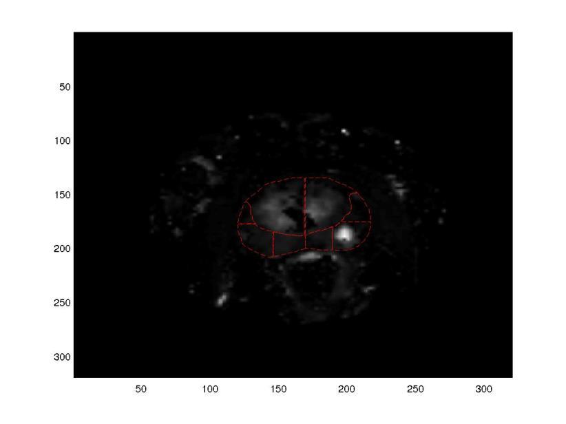
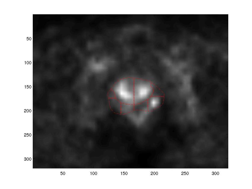
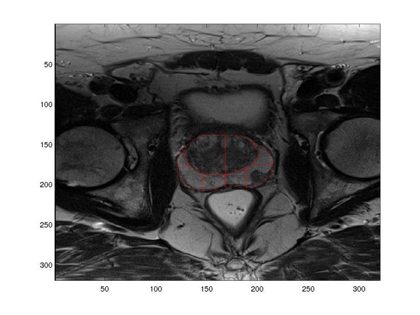
4 Material & Methods: Dataset 25 Patient Data sets T2 (a) DCE (70 timepoints) o DCE: 0s, 79s, 300s (b-d) A B C D b1) o o AUC (e) ktrans (f) E F G H DWI (ADC map measured based on 4 b-values) (g) Pathology information after prostatectomy (h) Slice thickness 3-4 mm 4

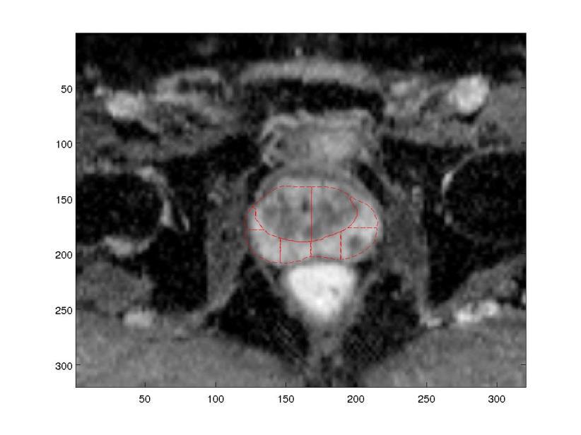
 o 6 substructures in CG and 16 substructures in")
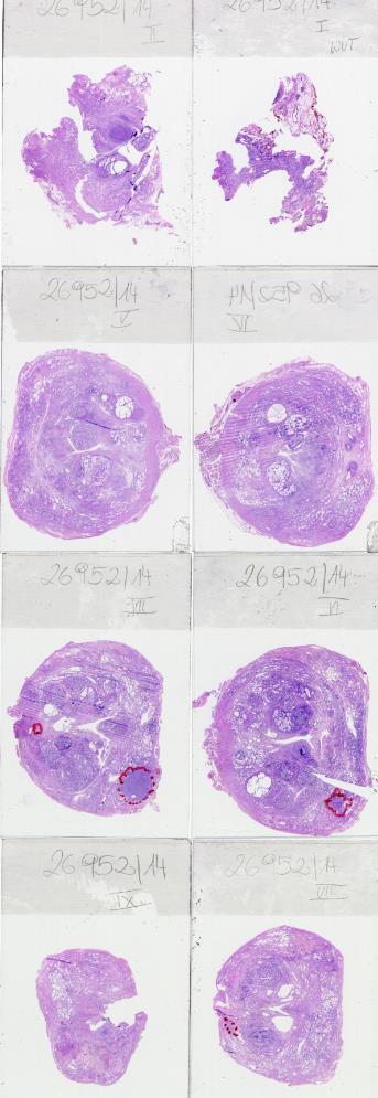
5 Material & Methods: Delineation Registration of pathological slices and MR images is challenging Central Gland and Peripheral Zone were delineated on T2 Based on histological information Tumor was delineated by visual comparison and propagated to all image modalities In addition geometrical substructures (PIRADS) were used and scored in accordance to pathological information (4 distinct scoring levels) o 6 substructures in CG and 16 substructures in PZ 5
6 Material & Methods: Delineation Registration of pathological slices and MR images is challenging Central Gland and Peripheral Zone were delineated on T2 Based on histological information Tumor was delineated by visual comparison and propagated to all image modalities In addition geometrical substructures PIRADS Structure (PIRADS) 10p: were used and scored in accordance to pathological information (4 distinct scoring levels) Score: 0.75 / 1 o 6 substructures in CG and 16 substructures in PZ 6


7 Material & Methods: Delineation In 24/25 cases the situation was more complex 7
8 Material & Methods: Workflow ADC ADC T2 DCE 3 Timepoints Tumor GLCM (BinSize=8,16,32) X Tumor-free ktrans AUC Multivariate Image Analysis PCA OPLS 8

9 Material & Methods: Workflow ADC ADC T2 DCE 3 Timepoints Tumor GLCM (BinSize=8,16,32) X Tumor-free ktrans AUC Multivariate Image Analysis PCA OPLS 11 Histogramm -- based parameters per image modality Peter Kuess - ESTRO 35, Turin 9
10 Evaluation Parameters Textual Parameters Autocorrelation, Cluster Prominence, Cluster Shade, Maximum Probability, Energy, Sum of Squares Variance, Sum Variance, Sum Entropy Gray-level co-occurrence matrixes (GLCM) Bin Size (N=8,16 and 32) 4 Orientations GLCM Histogram based parameters Min, Max, 2%,15%...85%,98%, mean, median, standard deviation, skewness, kurtosis 10
11 Orthogonal partial least squares (OPLS) Modeling OPLS is a multivariate regression technique X data matrix: textures and histogram-based parameters of image modalities X = ഥ x + tp + T o P o + E y = തy + tq + F y: response representing histological information OPLS removes variations in X, T 0 P 0, that is orthogonal to response Quality of Model: R 2 Y: goodnes of model itself [0-1] Q 2 Y: explaining the cross-correlation [0-1] 11
12 Preliminary Results: PCA on PIRADS sturctures Histograms of Score projections of ADC Blue bars = distribution of tumor free PIRADS structures Orange bars = PIRADS structures with tumor occurence 12

13 Preliminary Results: PIRADS using PCA Histograms of Score projections of ADC Blue bars = distribution of tumor free PIRADS structures Orange bars = PIRADS structures with tumor occurence 13
14 Preliminary Resutls: OPLS modeling Imaging Parameters Q2Y R2Y ADC Txt ADC Hist ADC Txt+Hist DCE (79s) Txt DCE (79s) Hist DCE (79s) Txt+Hist T2 Txt T2 Hist T2 Txt+Hist OPLS Model including textual and histogrambased paramters for ADC Blue dots = tumor structures Red dots = tumor free structurs ktrans Txt ktrans Hist ktrans Txt+Hist
15 Q²Y OPLS Modeling ADC T2 DCE (79s) AUC ktrans DCE (0s) DCE (300s) Histogram based parameters Textual Parameters Combined: Hist & Txt 15
16 Q²Y OPLS Modeling ADC T2 DCE (79s) AUC ktrans DCE (0s) DCE (300s) Histogram based parameters Textual Parameters Combined: Hist & Txt 16
17 OPLS Modeling Benefit from using all mpmri image methods available R2Y Q2Y Linear (R2Y) Linear (Q2Y) 17
18 OPLS Modeling Benefit from using all mpmri image methods available R2Y Q2Y Linear (R2Y) Linear (Q2Y) 18
19 Conclusion Textual parameters proved to be an additional supplement to histogram-based parameters in mpmri analysis Tumor prediction using OPLS shows encouraging results Best prediction value obtained were based on ADC PIRADS classification for tumor prediction is promising But structure size is too large Next step: voxel-based GLCM Thanks for your attention! 19
20 Additonal Slides: Peter Kuess - ESTRO 35, Turin 20

21 Background: DIL boosting study Andrzejewski et al 2015 Rad Oncol in press Material and Methods: DILs were defined based on multiparametric magnetic resonance imaging and fused with planning computed tomography images for twelve patients. VMAT, IMPT and HDR-BT treatment plans were created for each patient with the EQD2 α/β dose to the DIL escalated up to Gy, PTV initial D pres = 80.9 Gy (EBRT) and CTV D 90% = 81.9 Gy (HDR-BT). Hard dose constraints were applied to spare the OARs. Treatment plans were evaluated and compared between used techniques in CERR software. Results: Higher boost doses were achieved with IMPT compared to VMAT, keeping major OARs doses at similar level. HDR-BT was superior both in terms of OARs sparing and DIL boosting. 21


22 Outlook: Voxel-based GLCM Local texture around every voxel is analyzed A GLCM is generated for all voxels in image Data is assembled as X and subjected to multivariate methods Score values can be refolded to original image dimensions for visualization Example from glioma data set 22

23 Outlook: Local Binary Patterns Analyzes the texture around a voxel Surrounded voxels are thresholded, which gives 0s and 1s Put together, these form a pattern, which is a binary number The LBP is the numerical number All LBPs from a ROI can be binned in a histogram, so the frequency of each pattern can be accessed The histograms can be subjected to multivariate data analysis 23
24 Multi parametric MR Images ADC T2 Histoscore: Substructure 8p: 0.75 ktrans PET 24
25 Speciificity Sensitivity Sensitivity and Specificity of OPLS % 95.00% 90.00% 85.00% 80.00% 75.00% 70.00% 65.00% 60.00% 55.00% 50.00% % 95.00% 90.00% 85.00% ADC T2 AUC ktrans DCE_early DCE_pre DCE_late Image Modality Histo Text Hist+Txt 80.00% 75.00% 70.00% 65.00% 60.00% 55.00% Histo Text Hist+Txt 50.00% ADC T2 AUC ktrans DCE_early DCE_pre DCE_late Image Modality 25
26 Sensitivity and Specificity of OPLS 95.00% 90.00% 85.00% 80.00% 75.00% 70.00% Sensitivity Specificity 26
27 Textual descriptors All: autocorrelation, Cluster prominence, cluster shade, contrast, correlation, difference entropy, dissimilarity, energy, entropy, homogeneity 1 (as described by Soh et al.), homogeneity 2 (as implemented in MatLab 2014a, Image Processing Toolbox v. 9.0), information measure of correlations 1 and 2, inverse difference moment, normalized inverse difference moment, maximum probability, sum average, sum entropy, sum of squares, variance, sum variance Not included due to size dependency: contrast, correlation, difference entropy, difference variance, dissimilarity, energy, entropy, homogeneity, inverse difference, information measures of correlation 1 and 2. Finally used textual parameters: Autocorrelation, Cluster Prominence, Cluster Shade, Maximum Probability, Energy, Sum of Squares Variance, Sum Variance, Sum Entropy 28


28 Loading Plot Size dependency of textual parameters 29
29 Zonal Segmentation 30
RADIOMICS: potential role in the clinics and challenges
 27 giugno 2018 Dipartimento di Fisica Università degli Studi di Milano RADIOMICS: potential role in the clinics and challenges Dr. Francesca Botta Medical Physicist Istituto Europeo di Oncologia (Milano)
Is deformable image registration a solved problem?
 Is deformable image registration a solved problem? Marcel van Herk On behalf of the imaging group of the RT department of NKI/AVL Amsterdam, the Netherlands DIR 1 Image registration Find translation.deformation
Global Journal of Engineering Science and Research Management
 ADVANCED K-MEANS ALGORITHM FOR BRAIN TUMOR DETECTION USING NAIVE BAYES CLASSIFIER Veena Bai K*, Dr. Niharika Kumar * MTech CSE, Department of Computer Science and Engineering, B.N.M. Institute of Technology,
Package radiomics. March 30, 2018
 Type Package Title 'Radiomic' Image Processing Toolbox Version 0.1.3 Date 2018-03-15 Author Joel Carlson Package radiomics March 30, 2018 Maintainer Joel Carlson Functions to extract
MEDICAL IMAGE ANALYSIS
 SECOND EDITION MEDICAL IMAGE ANALYSIS ATAM P. DHAWAN g, A B IEEE Engineering in Medicine and Biology Society, Sponsor IEEE Press Series in Biomedical Engineering Metin Akay, Series Editor +IEEE IEEE PRESS
Texture Analysis in Quantitative Osteoporosis Assessment
 Texture Analysis in Quantitative Osteoporosis Assessment Characterizing Micro-architecture in High Resolution Peripheral Quantitative Computed Tomography Alexander Valentinitsch 1, Janina Patsch 1, Dirk
BRAIN CANCER CLASSIFICATION USING BACK PROPAGATION NEURAL NETWORK AND PRINCIPLE COMPONENT ANALYSIS Ganesh Ram Nayak 1, Mr.
 International Journal of Technical Research and Applications e-issn:2320-8163, www.ijtra.com Volume 2, Issue 4 (July-Aug 2014), PP. 26-31 BRAIN CANCER CLASSIFICATION USING BACK PROPAGATION NEURAL NETWORK
Dosimetric Analysis Report
 RT-safe 48, Artotinis str 116 33, Athens Greece +30 2107563691 info@rt-safe.com Dosimetric Analysis Report SAMPLE, for demonstration purposes only Date of report: ----------- Date of irradiation: -----------
Sequences Plane Technical characteristics Axial: TR=3425ms, TE=110ms, NSA: 2, Axial (renal hilum-pubis)
") Table 1: PET/CT and MRI protocols PET CT Preparation Patients fasted for 4h before acquisition The blood glucose level had to be less than 7 mmol/l Injection of 5 MBq/kg of 18 F-FDG PET acquisitions were
Table 1: PET/CT and MRI protocols PET CT Preparation Patients fasted for 4h before acquisition The blood glucose level had to be less than 7 mmol/l Injection of 5 MBq/kg of 18 F-FDG PET acquisitions were
ECE 176 Digital Image Processing Handout #14 Pamela Cosman 4/29/05 TEXTURE ANALYSIS
 ECE 176 Digital Image Processing Handout #14 Pamela Cosman 4/29/ TEXTURE ANALYSIS Texture analysis is covered very briefly in Gonzalez and Woods, pages 66 671. This handout is intended to supplement that
Digital Image Processing. Lecture # 15 Image Segmentation & Texture
 Digital Image Processing Lecture # 15 Image Segmentation & Texture 1 Image Segmentation Image Segmentation Group similar components (such as, pixels in an image, image frames in a video) Applications:
CHAPTER 2 TEXTURE CLASSIFICATION METHODS GRAY LEVEL CO-OCCURRENCE MATRIX AND TEXTURE UNIT
 CHAPTER 2 TEXTURE CLASSIFICATION METHODS GRAY LEVEL CO-OCCURRENCE MATRIX AND TEXTURE UNIT 2.1 BRIEF OUTLINE The classification of digital imagery is to extract useful thematic information which is one
SUPPLEMENTARY APPENDIX A: Definition of texture features.
 SUPPLEMETARY APPEDIX A: Definition of texture features. Input volume: Volume of interest V (x, y, z) with isotropic voxel size. The necessity for isotropically resampling V to a given voxel size prior
Classification of Protein Crystallization Imagery
 Classification of Protein Crystallization Imagery Xiaoqing Zhu, Shaohua Sun, Samuel Cheng Stanford University Marshall Bern Palo Alto Research Center September 2004, EMBC 04 Outline Background X-ray crystallography
K-Means Clustering Using Localized Histogram Analysis
 K-Means Clustering Using Localized Histogram Analysis Michael Bryson University of South Carolina, Department of Computer Science Columbia, SC brysonm@cse.sc.edu Abstract. The first step required for many
Tumor Detection in Breast Ultrasound images
 I J C T A, 8(5), 2015, pp. 1881-1885 International Science Press Tumor Detection in Breast Ultrasound images R. Vanithamani* and R. Dhivya** Abstract: Breast ultrasound is becoming a popular screening
Abbie M. Diak, PhD Loyola University Medical Center Dept. of Radiation Oncology
 Abbie M. Diak, PhD Loyola University Medical Center Dept. of Radiation Oncology Outline High Spectral and Spatial Resolution MR Imaging (HiSS) What it is How to do it Ways to use it HiSS for Radiation
Classification of Subject Motion for Improved Reconstruction of Dynamic Magnetic Resonance Imaging
 1 CS 9 Final Project Classification of Subject Motion for Improved Reconstruction of Dynamic Magnetic Resonance Imaging Feiyu Chen Department of Electrical Engineering ABSTRACT Subject motion is a significant
ADVANCING CANCER TREATMENT
 3 ADVANCING CANCER TREATMENT SUPPORTING CLINICS WORLDWIDE RaySearch is advancing cancer treatment through pioneering software. We believe software has un limited potential, and that it is now the driving
8/3/2017. Contour Assessment for Quality Assurance and Data Mining. Objective. Outline. Tom Purdie, PhD, MCCPM
 Contour Assessment for Quality Assurance and Data Mining Tom Purdie, PhD, MCCPM Objective Understand the state-of-the-art in contour assessment for quality assurance including data mining-based techniques
Image Segmentation and Registration
 Image Segmentation and Registration Dr. Christine Tanner (tanner@vision.ee.ethz.ch) Computer Vision Laboratory, ETH Zürich Dr. Verena Kaynig, Machine Learning Laboratory, ETH Zürich Outline Segmentation
Prostate Detection Using Principal Component Analysis
 Prostate Detection Using Principal Component Analysis Aamir Virani (avirani@stanford.edu) CS 229 Machine Learning Stanford University 16 December 2005 Introduction During the past two decades, computed
REAL-TIME ADAPTIVITY IN HEAD-AND-NECK AND LUNG CANCER RADIOTHERAPY IN A GPU ENVIRONMENT
 REAL-TIME ADAPTIVITY IN HEAD-AND-NECK AND LUNG CANCER RADIOTHERAPY IN A GPU ENVIRONMENT Anand P Santhanam Assistant Professor, Department of Radiation Oncology OUTLINE Adaptive radiotherapy for head and
Hybrid Approach for MRI Human Head Scans Classification using HTT based SFTA Texture Feature Extraction Technique
 Volume 118 No. 17 2018, 691-701 ISSN: 1311-8080 (printed version); ISSN: 1314-3395 (on-line version) url: http://www.ijpam.eu ijpam.eu Hybrid Approach for MRI Human Head Scans Classification using HTT
Lucy Phantom MR Grid Evaluation
 Lucy Phantom MR Grid Evaluation Anil Sethi, PhD Loyola University Medical Center, Maywood, IL 60153 November 2015 I. Introduction: The MR distortion grid, used as an insert with Lucy 3D QA phantom, is
Extraction and Features of Tumour from MR brain images
 IOSR Journal of Electronics and Communication Engineering (IOSR-JECE) e-issn: 2278-2834,p- ISSN: 2278-8735.Volume 13, Issue 2, Ver. I (Mar. - Apr. 2018), PP 67-71 www.iosrjournals.org Sai Prasanna M 1,
MPRAD: A Multiparametric Radiomics Framework
 MPRAD: A Multiparametric Radiomics Framework Vishwa S. Parekh 1,3, Michael A. Jacobs* 1,2 1 The Russell H. Morgan Department of Radiology and Radiological Science and 2 Sidney Kimmel Comprehensive Cancer
Tumor Detection and classification of Medical MRI UsingAdvance ROIPropANN Algorithm
 International Journal of Engineering Research and Advanced Technology (IJERAT) DOI:http://dx.doi.org/10.31695/IJERAT.2018.3273 E-ISSN : 2454-6135 Volume.4, Issue 6 June -2018 Tumor Detection and classification
Use of MRI in Radiotherapy: Technical Consideration
 Use of MRI in Radiotherapy: Technical Consideration Yanle Hu, PhD Department of Radiation Oncology, Mayo Clinic Arizona 04/07/2018 2015 MFMER slide-1 Conflict of Interest: None 2015 MFMER slide-2 Objectives
DEEP LEARNING WITH ORTHOGONAL VOLUMETRIC HED SEGMENTATION AND 3D SURFACE RECONSTRUCTION MODEL OF PROSTATE MRI
 DEEP LEARNING WITH ORTHOGONAL VOLUMETRIC HED SEGMENTATION AND 3D SURFACE RECONSTRUCTION MODEL OF PROSTATE MRI Ruida Cheng a, Nathan Lay b, Francesca Mertan c, Baris Turkbey c, Holger R. Roth b, Le Lu b,
ADVANCING CANCER TREATMENT
 The RayPlan treatment planning system makes proven, innovative RayStation technology accessible to clinics that need a cost-effective and streamlined solution. Fast, efficient and straightforward to use,
6 credits. BMSC-GA Practical Magnetic Resonance Imaging II
 BMSC-GA 4428 - Practical Magnetic Resonance Imaging II 6 credits Course director: Ricardo Otazo, PhD Course description: This course is a practical introduction to image reconstruction, image analysis
13 CP Addition of Measurement Report Root Template for Planar and Volumetric ROIs
 13 CP-1386 - Addition of Measurement Report Root Template for Planar and Volumetric ROIs Page 1 1 STATUS Final Text 2 Date of Last Update 2014/11/11 3 Person Assigned David Clunie 4 mailto:dclunie@dclunie.com
13 CP Addition of Measurement Report Root Template for Planar and Volumetric ROIs
 13 CP-1386 - Addition of Measurement Report Root Template for Planar and Volumetric ROIs Page 1 1 STATUS Letter Ballot 2 Date of Last Update 2014/09/08 3 Person Assigned David Clunie 4 mailto:dclunie@dclunie.com
TEXTURE. Plan for today. Segmentation problems. What is segmentation? INF 4300 Digital Image Analysis. Why texture, and what is it?
 INF 43 Digital Image Analysis TEXTURE Plan for today Why texture, and what is it? Statistical descriptors First order Second order Gray level co-occurrence matrices Fritz Albregtsen 8.9.21 Higher order
Coverage based treatment planning to accommodate organ deformable motions and contouring uncertainties for prostate treatment. Huijun Xu, Ph.D.
 Coverage based treatment planning to accommodate organ deformable motions and contouring uncertainties for prostate treatment Huijun Xu, Ph.D. Acknowledgement and Disclosure Dr. Jeffrey Siebers Dr. DJ
A Robust Brain MRI Classification with GLCM Features
 A Robust Brain MRI with GLCM Features Sahar Jafarpour Zahra Sedghi Mehdi Chehel Amirani ABSTRACT Automated and accurate classification of brain MRI is such important that leads us to present a new robust
Segmentation Using a Region Growing Thresholding
 Segmentation Using a Region Growing Thresholding Matei MANCAS 1, Bernard GOSSELIN 1, Benoît MACQ 2 1 Faculté Polytechnique de Mons, Circuit Theory and Signal Processing Laboratory Bâtiment MULTITEL/TCTS
CHAPTER 4 TEXTURE FEATURE EXTRACTION
 83 CHAPTER 4 TEXTURE FEATURE EXTRACTION This chapter deals with various feature extraction technique based on spatial, transform, edge and boundary, color, shape and texture features. A brief introduction
SuRVoS Workbench. Super-Region Volume Segmentation. Imanol Luengo
 SuRVoS Workbench Super-Region Volume Segmentation Imanol Luengo Index - The project - What is SuRVoS - SuRVoS Overview - What can it do - Overview of the internals - Current state & Limitations - Future
Machine Learning for Medical Image Analysis. A. Criminisi
 Machine Learning for Medical Image Analysis A. Criminisi Overview Introduction to machine learning Decision forests Applications in medical image analysis Anatomy localization in CT Scans Spine Detection
Creating a Knowledge Based Model using RapidPlan TM : The Henry Ford Experience
 DVH Estimates Creating a Knowledge Based Model using RapidPlan TM : The Henry Ford Experience Karen Chin Snyder, MS, DABR AAMD Region V Meeting October 4, 2014 Disclosures The Department of Radiation Oncology
UNIVERSITY OF SOUTHAMPTON
 UNIVERSITY OF SOUTHAMPTON PHYS2007W1 SEMESTER 2 EXAMINATION 2014-2015 MEDICAL PHYSICS Duration: 120 MINS (2 hours) This paper contains 10 questions. Answer all questions in Section A and only two questions
Thermographic Image Analysis Method in Detection of Canine Bone Cancer (Osteosarcoma)
") 2012 5th International Congress on Image and Signal Processing (CISP 2012) Thermographic Image Analysis Method in Detection of Canine Bone Cancer (Osteosarcoma) Maryamsadat Amini, Peng Liu and Scott E
2012 5th International Congress on Image and Signal Processing (CISP 2012) Thermographic Image Analysis Method in Detection of Canine Bone Cancer (Osteosarcoma) Maryamsadat Amini, Peng Liu and Scott E
MR-guided radiotherapy: Vision, status and research at the UMC Utrecht. Dipl. Ing. Dr. Markus Glitzner
 MR-guided radiotherapy: Vision, status and research at the UMC Utrecht Dipl. Ing. Dr. Markus Glitzner About myself Training Medizintechnik TU Graz PhD UMC Utrecht Clinical work Software implementation
Detection of Focal Cortical Dysplasia Lesions in MRI using Textural Features
 Detection of Focal Cortical Dysplasia Lesions in MRI using Textural Features Christian Loyek 1, Friedrich G. Woermann 2, Tim W. Nattkemper 1 1 Applied Neuroinformatics, Faculty of Technology, Bielefeld
Ensemble Prostate Tumor Classification in H&E Whole Slide Imaging via Stain Normalization and Cell Density Estimation
 Ensemble Prostate Tumor Classification in H&E Whole Slide Imaging via Stain Normalization and Cell Density Estimation Michaela Weingant 1, Hayley M. Reynolds 2,3, Annette Haworth 2,3, Catherine Mitchell
Help Guide. mm Copyright Mirada Medical Ltd, Mirada Medical RTx 1
 Help Guide mm3237-1.6-1 Copyright Mirada Medical Ltd, 2000-2014. Mirada Medical RTx 1 Contents Help Guide... 1 Contents... 2 Introduction to RTx... 4 Regulatory Statement... 6 Notes... 15 Data Supported...
Automated segmentation methods for liver analysis in oncology applications
 University of Szeged Department of Image Processing and Computer Graphics Automated segmentation methods for liver analysis in oncology applications Ph. D. Thesis László Ruskó Thesis Advisor Dr. Antal
Medical Image Feature, Extraction, Selection And Classification
 Medical Image Feature, Extraction, Selection And Classification 1 M.VASANTHA *, 2 DR.V.SUBBIAH BHARATHI, 3 R.DHAMODHARAN 1. Research Scholar, Mother Teresa Women s University, KodaiKanal, and Asst.Professor,
Computer-Aided Diagnosis in Abdominal and Cardiac Radiology Using Neural Networks
 Computer-Aided Diagnosis in Abdominal and Cardiac Radiology Using Neural Networks Du-Yih Tsai, Masaru Sekiya and Yongbum Lee Department of Radiological Technology, School of Health Sciences, Faculty of
Using Pinnacle 16 Deformable Image registration in a re-treat scenario
 Introduction Using Pinnacle 16 Deformable Image registration in a re-treat scenario This short Hands On exercise will introduce how the Deformable Image Registration (DIR) tools in Pinnacle can be used
TG 132: Use of Image Registration and Fusion in RT
 TG 132: Use of Image Registration and Fusion in RT Kristy K Brock, PhD, DABR, FAAPM Associate Professor Department of Radiation Oncology, University of Michigan Chair, AAPM TG 132: Image Registration and
1 School of Biological Science and Medical Engineering, Beihang University, Beijing, China
 A Radio-genomics Approach for Identifying High Risk Estrogen Receptor-positive Breast Cancers on DCE-MRI: Preliminary Results in Predicting OncotypeDX Risk Scores Tao Wan 1,2, PhD, B. Nicolas Bloch 3,
Medical Imaging Projects
 NSF REU MedIX Summer 2006 Medical Imaging Projects Daniela Stan Raicu, PhD http://facweb.cs.depaul.edu/research draicu@cs.depaul.edu Outline Medical Informatics Imaging Modalities Computed Tomography Medical
TomoTherapy Related Projects. An image guidance alternative on Tomo Low dose MVCT reconstruction Patient Quality Assurance using Sinogram
 TomoTherapy Related Projects An image guidance alternative on Tomo Low dose MVCT reconstruction Patient Quality Assurance using Sinogram Development of A Novel Image Guidance Alternative for Patient Localization
 Grupo Física Médica de Sevilla (CTS-233) Antonio Leal Plaza alplaza@us.es Dept. Fisiología Médica y Biofísica, Fac. Medicina. Univ. Sevilla http://grupos.us.es/medicalphysics/ Overview Introduction to
Walk Through of CERR Capabilities. CERR: Introduction. Outline. Getting CERR: Control Panel. Documentation Support Community
 Walk Through of CERR Capabilities Aditya P. Apte, Ph.D. Department of Medical Physics Memorial Sloan Kettering Cancer Center New York aptea@mskcc.org AAPM 2015, July 15, 2015 CERR: Computational Environment
Image Registration. Prof. Dr. Lucas Ferrari de Oliveira UFPR Informatics Department
 Image Registration Prof. Dr. Lucas Ferrari de Oliveira UFPR Informatics Department Introduction Visualize objects inside the human body Advances in CS methods to diagnosis, treatment planning and medical
Improving the detection of excessive activation of ciliaris muscle by clustering thermal images
 11 th International Conference on Quantitative InfraRed Thermography Improving the detection of excessive activation of ciliaris muscle by clustering thermal images *University of Debrecen, Faculty of
Business Club. Decision Trees
 Business Club Decision Trees Business Club Analytics Team December 2017 Index 1. Motivation- A Case Study 2. The Trees a. What is a decision tree b. Representation 3. Regression v/s Classification 4. Building
Clinical Prospects and Technological Challenges for Multimodality Imaging Applications in Radiotherapy Treatment Planning
 Clinical Prospects and Technological Challenges for Multimodality Imaging Applications in Radiotherapy Treatment Planning Issam El Naqa, PhD Assistant Professor Department of Radiation Oncology Washington
K Means Clustering Using Localized Histogram Analysis and Multiple Assignment. Michael Bryson 4/18/2007
 1 K Means Clustering Using Localized Histogram Analysis and Multiple Assignment Michael Bryson 4/18/2007 2 Outline Introduction Redefining Distance Preliminary Results Multiple Assignment Discussion 3
MRI Brain Image Analysis and Classification for Computer-Assisted Diagnosis
 MRI Brain Image Analysis and Classification for Computer-Assisted Diagnosis MADINA HAMIANE Department of Telecommunication Engineering Ahlia University Manama, Kingdom of Bahrain mhamiane@ahlia.edu.bh
Supplementary Material: Guided Volume Editing based on Histogram Dissimilarity
 Supplementary Material: Guided Volume Editing based on Histogram Dissimilarity A. Karimov, G. Mistelbauer, T. Auzinger and S. Bruckner 2 Institute of Computer Graphics and Algorithms, Vienna University
SVM BASED AUTOMATIC MEDICAL DECISION SUPPORT SYSTEM FOR MEDICAL IMAGE
 SVM BASED AUTOMATIC MEDICAL DECISION SUPPORT SYSTEM FOR MEDICAL IMAGE 1 JOSEPHINE SUTHA.V, 2 Dr.P.LATHA 1 Sardar Raja College Of Engineering, Tirunelveli, India 2 Government College Of Engineering, Tirunelveli,India
iplan RT Image Advanced Contouring Workstation - Driving Physician Collaboration
 iplan RT Image Advanced Contouring Workstation - Driving Physician Collaboration The iplan Contouring Workstation offers unique and innovative capabilities for faster contouring and consistent segmentation
CONTOURING ACCURACY. What Have We Learned? And Where Do We Go From Here? BEN NELMS, PH.D. AUGUST 15, 2016
 CONTOURING ACCURACY What Have We Learned? And Where Do We Go From Here? BEN NELMS, PH.D. AUGUST 15, 2016 FIRST THINGS FIRST Happy Medical Dosimetrist s Week! OUTLINE 1. Objectives 2. The importance of
Interactive Differential Segmentation of the Prostate using Graph-Cuts with a Feature Detector-based Boundary Term
 MOSCHIDIS, GRAHAM: GRAPH-CUTS WITH FEATURE DETECTORS 1 Interactive Differential Segmentation of the Prostate using Graph-Cuts with a Feature Detector-based Boundary Term Emmanouil Moschidis emmanouil.moschidis@postgrad.manchester.ac.uk
An independent component analysis based tool for exploring functional connections in the brain
 An independent component analysis based tool for exploring functional connections in the brain S. M. Rolfe a, L. Finney b, R. F. Tungaraza b, J. Guan b, L.G. Shapiro b, J. F. Brinkely b, A. Poliakov c,
Chapter 3 Set Redundancy in Magnetic Resonance Brain Images
 16 Chapter 3 Set Redundancy in Magnetic Resonance Brain Images 3.1 MRI (magnetic resonance imaging) MRI is a technique of measuring physical structure within the human anatomy. Our proposed research focuses
Radio-morphology: Parametric Shape-Based Features in Radiotherapy
 Radio-morphology: Parametric Shape-Based Features in Radiotherapy by Pranav Lakshminarayanan A thesis submitted to Johns Hopkins University in conformity with the requirements for the degree of Master
Improvement and Evaluation of a Time-of-Flight-based Patient Positioning System
 Improvement and Evaluation of a Time-of-Flight-based Patient Positioning System Simon Placht, Christian Schaller, Michael Balda, André Adelt, Christian Ulrich, Joachim Hornegger Pattern Recognition Lab,
NIH Public Access Author Manuscript Proc IEEE Int Symp Biomed Imaging. Author manuscript; available in PMC 2014 November 15.
 NIH Public Access Author Manuscript Published in final edited form as: Proc IEEE Int Symp Biomed Imaging. 2013 April ; 2013: 748 751. doi:10.1109/isbi.2013.6556583. BRAIN TUMOR SEGMENTATION WITH SYMMETRIC
Hugues Mailleux Medical Physics Department Institut Paoli-Calmettes Marseille France. Sunday 17 July 2016
 Hugues Mailleux Medical Physics Department Institut Paoli-Calmettes Marseille France Sunday 17 July 2016 AGENDA 1. Introduction 2. Material 3. Optimization process 4. Results 5. Comments 6. Conclusion
DETECTING MILD TRAUMATIC BRAIN INJURY USING DYNAMIC LOW LEVEL CONTEXT
 DETECTING MILD TRAUMATIC BRAIN INJURY USING DYNAMIC LOW LEVEL CONTEXT Anthony Bianchi, Bir Bhanu, Virginia Donovan*, and Andre Obenaus*,** Center for Research in Intelligent Systems, University of California,
Feature Extraction and Texture Classification in MRI
 Extraction and Texture Classification in MRI Jayashri Joshi, Mrs.A.C.Phadke. Marathwada Mitra Mandal s College of Engineering, Pune.. Maharashtra Institute of Technology, Pune. kjayashri@rediffmail.com.
Norbert Schuff VA Medical Center and UCSF
 Norbert Schuff Medical Center and UCSF Norbert.schuff@ucsf.edu Medical Imaging Informatics N.Schuff Course # 170.03 Slide 1/67 Objective Learn the principle segmentation techniques Understand the role
Available Online through
 Available Online through www.ijptonline.com ISSN: 0975-766X CODEN: IJPTFI Research Article ANALYSIS OF CT LIVER IMAGES FOR TUMOUR DIAGNOSIS BASED ON CLUSTERING TECHNIQUE AND TEXTURE FEATURES M.Krithika
Histograms. h(r k ) = n k. p(r k )= n k /NM. Histogram: number of times intensity level rk appears in the image
 = n k. p(r k )= n k /NM. Histogram: number of times intensity level rk appears in the image") Histograms h(r k ) = n k Histogram: number of times intensity level rk appears in the image p(r k )= n k /NM normalized histogram also a probability of occurence 1 Histogram of Image Intensities Create
Histograms h(r k ) = n k Histogram: number of times intensity level rk appears in the image p(r k )= n k /NM normalized histogram also a probability of occurence 1 Histogram of Image Intensities Create
VALIDATION OF DIR. Raj Varadhan, PhD, DABMP Minneapolis Radiation Oncology
 VALIDATION OF DIR Raj Varadhan, PhD, DABMP Minneapolis Radiation Oncology Overview Basics: Registration Framework, Theory Discuss Validation techniques Using Synthetic CT data & Phantoms What metrics to
CHAPTER 3 TUMOR DETECTION BASED ON NEURO-FUZZY TECHNIQUE
 32 CHAPTER 3 TUMOR DETECTION BASED ON NEURO-FUZZY TECHNIQUE 3.1 INTRODUCTION In this chapter we present the real time implementation of an artificial neural network based on fuzzy segmentation process
4 CIM&Lab, Universidad Nacional de Colombia, Bogota, Colombia {edromero,
 Segmentation of pelvic structures from planning CT based on a statistical shape model with a multiscale edge detector and geometrical likelihood measures Fabio Martinez 1,2,4, Oscar Acosta 1,2, Gaël Dréan
Modern Medical Image Analysis 8DC00 Exam
 Parts of answers are inside square brackets [... ]. These parts are optional. Answers can be written in Dutch or in English, as you prefer. You can use drawings and diagrams to support your textual answers.
A New GPU-Based Level Set Method for Medical Image Segmentation
 A New GPU-Based Level Set Method for Medical Image Segmentation Wenzhe Xue Research Assistant Radiology Department Mayo Clinic, Scottsdale, AZ Ph.D. Student Biomedical Informatics Arizona State University,
An Integrated Visual Analysis System for Fusing MR Spectroscopy and Multi-Modal Radiology Imaging
 An Integrated Visual Analysis System for Fusing MR Spectroscopy and Multi-Modal Radiology Imaging Miguel Nunes, Benjamin Rowland, Matthias Schlachter, Soléakhéna Ken, Kresimir Matkovic, Anne Laprie and
Comparison Study of Clinical 3D MRI Brain Segmentation Evaluation
 Comparison Study of Clinical 3D MRI Brain Segmentation Evaluation Ting Song 1, Elsa D. Angelini 2, Brett D. Mensh 3, Andrew Laine 1 1 Heffner Biomedical Imaging Laboratory Department of Biomedical Engineering,
Effect of Image Quality Improvement on the Leaf Image Classification Accuracy
 Arun Kumar et al, / (IJCSIT) International Journal of Computer Science and Information Technologies, Vol. 6 (6), 15, 488-4887 Effect of Image Quality Improvement on the Leaf Image Classification Accuracy
Detection of Bone Fracture using Image Processing Methods
 Detection of Bone Fracture using Image Processing Methods E Susmitha, M.Tech Student, Susmithasrinivas3@gmail.com Mr. K. Bhaskar Assistant Professor bhasi.adc@gmail.com MVR college of engineering and Technology
Current state of multi-criteria treatment planning
 Current state of multi-criteria treatment planning June 11, 2010 Fall River NE AAPM meeting David Craft Massachusetts General Hospital Talk outline - Overview of optimization - Multi-criteria optimization
Good Morning! Thank you for joining us
 Good Morning! Thank you for joining us Deformable Registration, Contour Propagation and Dose Mapping: 101 and 201 Marc Kessler, PhD, FAAPM The University of Michigan Conflict of Interest I receive direct
Fractional labelmaps for computing accurate dose volume histograms
 Fractional labelmaps for computing accurate dose volume histograms Kyle Sunderland, Csaba Pinter, Andras Lasso, Gabor Fichtinger Laboratory for Percutaneous Surgery, School of Computing, Queen s University,
Pixels to Voxels: Modeling Visual Representation in the Human Brain
 Pixels to Voxels: Modeling Visual Representation in the Human Brain Authors: Pulkit Agrawal, Dustin Stansbury, Jitendra Malik, Jack L. Gallant Presenters: JunYoung Gwak, Kuan Fang Outlines Background Motivation
SIIM 2017 Scientific Session Analytics & Deep Learning Part 2 Friday, June 2 8:00 am 9:30 am
 SIIM 2017 Scientific Session Analytics & Deep Learning Part 2 Friday, June 2 8:00 am 9:30 am Performance of Deep Convolutional Neural Networks for Classification of Acute Territorial Infarct on Brain MRI:
Image Processing. Image Features
 Image Processing Image Features Preliminaries 2 What are Image Features? Anything. What they are used for? Some statements about image fragments (patches) recognition Search for similar patches matching
Medical Image Analysis
 Computer assisted Image Analysis VT04 29 april 2004 Medical Image Analysis Lecture 10 (part 1) Xavier Tizon Medical Image Processing Medical imaging modalities XRay,, CT Ultrasound MRI PET, SPECT Generic
Elastic Registration of Prostate MR Images Based on State Estimation of Dynamical Systems
 Elastic Registration of Prostate MR Images Based on State Estimation of Dynamical Systems Bahram Marami a,b,c, Suha Ghoul a, Shahin Sirouspour c, David W. Capson d, Sean R. H. Davidson e, John Trachtenberg
Using a research real-time control interface to go beyond dynamic MLC tracking
 in partnership with Using a research real-time control interface to go beyond dynamic MLC tracking Dr. Simeon Nill Joint Department of Physics at The Institute of Cancer Research and the Royal Marsden
Interactive Boundary Detection for Automatic Definition of 2D Opacity Transfer Function
 Interactive Boundary Detection for Automatic Definition of 2D Opacity Transfer Function Martin Rauberger, Heinrich Martin Overhoff Medical Engineering Laboratory, University of Applied Sciences Gelsenkirchen,
Supplementary Material
 Supplementary Material Figure 1S: Scree plot of the 400 dimensional data. The Figure shows the 20 largest eigenvalues of the (normalized) correlation matrix sorted in decreasing order; the insert shows