Advanced MRI Techniques (and Applications)
|
|
|
- Dorcas Stafford
- 6 years ago
- Views:
Transcription
1 Advanced MRI Techniques (and Applications) Jeffry R. Alger, PhD Department of Neurology Ahmanson-Lovelace Brain Mapping Center Brain Research Institute Jonsson Comprehensive Cancer Center University of California, Los Angeles K30 course January 2012
2 (Selected) Advanced MRI Techniques Gradient Echo MRI High resolution anatomic imaging Magnetic Resonance Angiography Echo Planar MRI Diffusion MRI Diffusion-weighted MRI Diffusion Tensor MRI Perfusion MRI (contrast passage) Diffusion and Perfusion MRI for acute stroke assessment
3 Gradient Echo MRI
4 Gradient (Recalled) Echo Imaging More efficient than conventional forms of MRI Makes 3-dimensional MRI practical Time needed for a 3D acquisition is 6-15 min TR is much shorter than in conventional MRI Inherently produces T1-weighted contrast

 3D Volumetric T1-weighted MRI (1 mm isotropic")
5 3-Dimensional Gradient Echo MRI 3-dimensional gradient echo imaging is becoming more practical due to advances in MRI hardware design Conventional Spin Echo Multiple Slice T1- weighted MRI (3 mm slice thickness) 3D Volumetric T1-weighted MRI (1 mm isotropic voxels)


6 Right Lateral Rendering of surface anatomy from MRI Superior Left Lateral
 Quantification of brain tissue")
7 3-Dimensional Gradient Echo Imaging (Thompson PM et al) Quantification of brain tissue composition
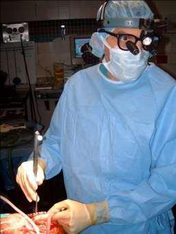

8 Image-guided Neurosurgery
9 Magnetic Resonance Angiography (MRA) Imaging of the blood vessels using MRI Usually arteries Sometimes veins Most MRA techniques use gradient echo MRI Time-of-flight (TOF) MRA Contrast enhanced MRA Phase contrast MRA (not covered in this lecture)


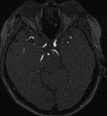
10 TOF-MRA Arteries appear as very hyperintense signal


11 Time-of-Flight MRA 1 mm slice from volume acquisition Volume maximum intensity projection
 with Contrast Agents")
12 Magnetic Resonance Angiography (MRA) with Contrast Agents Magnevist
13 Contrast Enhanced MRA Fast Gradient echo MRI Usually 2D 2-10 sec per time frame Venous injection of MRI contrast agent Image acquisition is timed such that the image is collected when the contrast agent is in the arteries

14 Contrast Enhanced MRA Complete angiographic coverage from arotic arch to Circle of Willis Other peripheral vascular applications
15 Echo Planar Imaging (EPI)
16 Echo Planar Imaging (EPI) Collects an entire slice in one TR (2 4 sec) Greater degree of image distortion slice images per second Facilitates studies that require large numbers of images Typically provides T2- weighted images Typical resolution of 2 mm x 2 mm x 5 mm
17 EPI-based Applications Diffusion imaging Diffusion tensor imaging Perfusion imaging BOLD Functional imaging Uncooperative patients
18 Diffusion MRI
19 Flavors of Diffusion MRI Diffusion Weighted Imaging (DWI) Apparent Diffusion Coefficient Imaging (ADCI) Diffusion Tensor MRI (DTI)

 www.")

20 Robert Brown ( )
21
22 Diffusion-Weighted MRI (DWI) D. Le Bihan. Nature Reviews Neuroscience 2003;4:
23 Diffusion-Weighted MRI (DWI) The degree to which the pulse sequence is sensitive to diffusion is expressed through the b-value or b-factor The signal intensity of diffusion weighted images are inversely related to the b-value directly related to the degree of diffusion restriction (at a particular b-value) directly related to T2
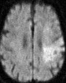
24 MRI of Acute Stroke T2w PDw T1w DWI (b = 1000)


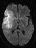
25 MRI of Acute Stroke T2w PDw T1w DWI (b = 1000)
26 Cell Swelling Hypothesis Normal IC EC Intracellular diffusion is slower than extracellular diffusion Ischemic IC EC Swollen intracellular volume Increased volume average DWI Reduced volume average ADC
27 DWI Hyperintensity in Stroke A marker of cytotoxic (cellular) edema seen in acute ischemic stroke
28 Diffusion MRI Typical diffusion-weighted MRIs are also T2-weighted T2 shine through Apparent Diffusion Coefficient (ADC) calculation using a series of diffusion-weighted images provides diffusion rate without the influence of T2

29 Diffusion-Weighted MRI (DWI) D. Le Bihan. Nature Reviews Neuroscience 2003;4:
30 ADC Calculation L/R gradient sensitization S/I gradient sensitization A/P gradient sensitization ln(s b ) Average Slope = - ADC Directionally averaged gradient sensitization b-value

")

31 Apparent Diffusion Coefficient (ADC) Imaging T2w DWI (b = 1000) ADC
32 Apparent Diffusion Coefficient Quantitative measure of diffusion rate Higher values mean less restricted diffusion Lower values mean more restricted diffusion A diffusion speedometer Independent of T2 Numbers that are comparable across subjects
33 Diffusion Tensor Imaging (DTI) Formal evaluation of the directionality of the diffusion Relies on the ability to measure the diffusion speed in any arbitrary direction Possible because of the directional specificity of the gradient hardware
: 750-1000 micron 2 /sec Parallel to white matter bundle: 1200 1500 micron 2 /sec Perpendicular to white matter bundle: 200 400 micron 2 /sec CSF: 3000 micron")
34 White Matter Diffusion Restriction Water diffuses more readily parallel to the fiber bundles than perpendicular to the fiber bundles Typical values of water diffusion coefficient in brain Gray matter or white matter (directionally averaged): micron 2 /sec Parallel to white matter bundle: micron 2 /sec Perpendicular to white matter bundle: micron 2 /sec CSF: 3000 micron 2 /sec
35
36 ADC L/R >> ADC A/P = ADC S/I ADC S/I ADC L/R ADC A/P
37 Diffusion Tensor Imaging Measure the component of the ADC In at least 6 unique anatomic directions In each voxel Determine in each voxel The direction in which the ADC is maximal The value of the directionally averaged ADC
38 Diffusion Tensor Imaging Voxel is likely white matter fiber if there is a great difference in the ADC between directions The direction showing the maximal ADC is parallel to the fiber


39 ICBM Protocol UCLA 1.5 T Sonata 5 acquisitions March 4, 2005 A/P fiber bundles L/R fiber bundles S/I fiber bundles
 color")


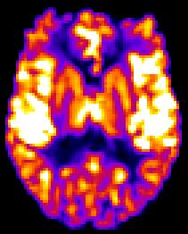
40 b0 fa Trace (ADC) color ICBM Protocol UCLA 1.5 T Sonata 1 acquisition March 4, 2005
41 Parametric DTI Images Trace (or ADC) conveys the average diffusion speed ADC is a quantitative measure that can be compared across patients Fractional Anisotropy (FA) conveys the degree of directional coherence within the voxel FA is an index such that 0 < FA < 1.0 FA is a quantitative measure that can be compared across patients
42
43 DTI Fiber Tractography

44 White Matter Fiber Tracking with DTI Connect the principal direction between voxels with smooth stream lines R. Bammer et al Eur J Radiol 2003;45:

45 White Matter Fiber Tracking with DTI D. Jones Institute of Psychiatry King s College, London

46 Catani Curr Opin Neurol 2006
47 Contrast Bolus Passage Perfusion MRI
48 Perfusion Perfusion is a physiologically-defined parameter Passage of blood through capillaries

49 Field Distortion Around Capillaries Filled with Contrast Agent Gradient echo sequences are sensitive to magnetic field gradients Signal from water molecules outside of the capillaries is reduced due to magnetic field gradients Buxton RB. Introduction to Functional Magnetic Resonance Imaging Principles & Techniques. Cambridge 2002
50 MRI Bolus Passage Perfusion Imaging Design Perfusion imaging is performed using a timed bolus of contrast given during rapid image acquisition Gradient echo echo planar imaging conveys sensitivity to the passage of contrast 2 sec time resolution for each 3D image Image intensity becomes hypointense as contrast passes

51 Perfusion Imaging using Contrast Passage
52 Contrast Bolus Passage Perfusion Imaging Qualitative information Tissue volumes that experience low contrast delivery Tissue volumes experience delayed contrast delivery Quantitative information Fractional Cerebral Blood Volume Cerebral Blood Flow (limited accuracy) Mean Transit Time (limited accuracy)
53 14 14 Bolus max T MAX (sec) TTP 4 (sec) CBF (R 0 ) ml/100 g/min 0 CBF (R max ) ml/100 g/min CBV 0 ml/100 g FHD 0.1 FHK 0.0 FHS 0.0
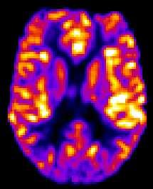
54 DSC-MRI-CBF Images 3T RRUCLAMC-1


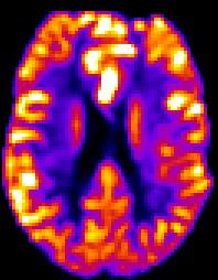
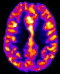
55 100 cc per 100g per min MP-RAGE GRE FLAIR 1000 cc per 100g per min 0 PET-CBF MRI-CBF 0 Normal Same day Slice # 23
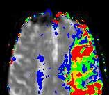
56 Stroke Diffusion and Perfusion MRI The combined use of diffusion and perfusion MRI for acute stroke is based on the concept of the ischemic penumbra The ischemic penumbra is believed to be what can be saved with prompt treatment The combination of diffusion and perfusion MRI offers a means of visualizing the size of the ischemic penumbra
57 Stroke Penumbra Imaging Diffusion and Perfusion MRI Perfusion MRI shows the entire affected territory Diffusion MRI shows the already damaged and unrepairable territory The penumbra is the difference (the mismatch)
58 Diffusion-Perfusion Mismatch Tmax
59 Diffusion-Perfusion Mismatch At Risk Tmax
60 Diffusion-Perfusion Mismatch At Risk Tmax DWI
61 Diffusion-Perfusion Mismatch At Risk Core Tmax DWI
62 Diffusion-Perfusion Mismatch At Risk At Risk Core Tmax DWI
63 Diffusion-Perfusion Mismatch At Risk Salvageable At Risk Core Tmax DWI
64 Stroke Diffusion and Perfusion MRI The combined use of diffusion and perfusion MRI for acute stroke is based on the concept of the ischemic penumbra The ischemic penumbra is believed to be what can be saved with prompt treatment The combination of diffusion and perfusion MRI offers a means of visualizing the size of the ischemic penumbra
65 Advanced MRI Techniques Gradient Echo MRI 3D brain morphometry Magnetic Resonance Angiography Neurosurgical planning Diffusion MRI Diffusivity Imaging Stroke assessment Diffusion Tensor Imaging Visualization of white matter structure Visualization of white matter connections Perfusion MRI (contrast passage) Stroke assessment

66
Quantitative MRI of the Brain: Investigation of Cerebral Gray and White Matter Diseases
 Quantities Measured by MR - Quantitative MRI of the Brain: Investigation of Cerebral Gray and White Matter Diseases Static parameters (influenced by molecular environment): T, T* (transverse relaxation)
MR Advance Techniques. Vascular Imaging. Class III
 MR Advance Techniques Vascular Imaging Class III 1 Vascular Imaging There are several methods that can be used to evaluate the cardiovascular systems with the use of MRI. MRI will aloud to evaluate morphology
HST.583 Functional Magnetic Resonance Imaging: Data Acquisition and Analysis Fall 2008
 MIT OpenCourseWare http://ocw.mit.edu HST.583 Functional Magnetic Resonance Imaging: Data Acquisition and Analysis Fall 2008 For information about citing these materials or our Terms of Use, visit: http://ocw.mit.edu/terms.
Supplementary Figure 1
 Supplementary Figure 1 BOLD and CBV functional maps showing EPI versus line-scanning FLASH fmri. A. Colored BOLD and CBV functional maps are shown in the highlighted window (green frame) of the raw EPI
Diffusion Tensor Imaging and Reading Development
 Diffusion Tensor Imaging and Reading Development Bob Dougherty Stanford Institute for Reading and Learning Reading and Anatomy Every brain is different... Not all brains optimized for highly proficient
Lilla Zöllei A.A. Martinos Center, MGH; Boston, MA
 Lilla Zöllei lzollei@nmr.mgh.harvard.edu A.A. Martinos Center, MGH; Boston, MA Bruce Fischl Gheorghe Postelnicu Jean Augustinack Anastasia Yendiki Allison Stevens Kristen Huber Sita Kakonoori + the FreeSurfer
Diffusion model fitting and tractography: A primer
 Diffusion model fitting and tractography: A primer Anastasia Yendiki HMS/MGH/MIT Athinoula A. Martinos Center for Biomedical Imaging 03/18/10 Why n how Diffusion model fitting and tractography 0/18 Why
XI Conference "Medical Informatics & Technologies" VALIDITY OF MRI BRAIN PERFUSION IMAGING METHOD
 XI Conference "Medical Informatics & Technologies" - 2006 medical imaging, MRI, brain perfusion Bartosz KARCZEWSKI 1, Jacek RUMIŃSKI 1 VALIDITY OF MRI BRAIN PERFUSION IMAGING METHOD Brain perfusion imaging
Dynamic Contrast enhanced MRA
 Dynamic Contrast enhanced MRA Speaker: Yung-Chieh Chang Date : 106.07.22 Department of Radiology, Taichung Veterans General Hospital, Taichung, Taiwan 1 Outline Basic and advanced principles of Diffusion
MRI. When to use What sequences. Outline 2012/09/19. Sequence: Definition. Basic Principles: Step 2. Basic Principles: Step 1. Govind Chavhan, MD
 MRI When to use What sequences Govind Chavhan, MD Assistant Professor and Staff Radiologist The Hospital For Sick Children, Toronto Planning Acquisition Post processing Interpretation Patient history and
Automatic Quantification of DTI Parameters along Fiber Bundles
 Automatic Quantification of DTI Parameters along Fiber Bundles Jan Klein 1, Simon Hermann 1, Olaf Konrad 1, Horst K. Hahn 1, and Heinz-Otto Peitgen 1 1 MeVis Research, 28359 Bremen Email: klein@mevis.de
Evaluation of Local Filter Approaches for Diffusion Tensor based Fiber Tracking
 Evaluation of Local Filter Approaches for Diffusion Tensor based Fiber Tracking D. Merhof 1, M. Buchfelder 2, C. Nimsky 3 1 Visual Computing, University of Konstanz, Konstanz 2 Department of Neurosurgery,
n o r d i c B r a i n E x Tutorial DSC Module
 m a k i n g f u n c t i o n a l M R I e a s y n o r d i c B r a i n E x Tutorial DSC Module Please note that this tutorial is for the latest released nordicbrainex. If you are using an older version please
Automatic Quantification of DTI Parameters along Fiber Bundles
 Automatic Quantification of DTI Parameters along Fiber Bundles Jan Klein, Simon Hermann, Olaf Konrad, Horst K. Hahn, Heinz-Otto Peitgen MeVis Research, 28359 Bremen Email: klein@mevis.de Abstract. We introduce
GE Healthcare CLINICAL GALLERY. Discovery * MR750w 3.0T. This brochure is intended for European healthcare professionals.
 GE Healthcare CLINICAL GALLERY Discovery * MR750w 3.0T This brochure is intended for European healthcare professionals. NEURO PROPELLER delivers high resolution, motion insensitive imaging in all planes.
Fiber Selection from Diffusion Tensor Data based on Boolean Operators
 Fiber Selection from Diffusion Tensor Data based on Boolean Operators D. Merhof 1, G. Greiner 2, M. Buchfelder 3, C. Nimsky 4 1 Visual Computing, University of Konstanz, Konstanz, Germany 2 Computer Graphics
CP Generalize Concepts in Abstract Multi-dimensional Image Model Component Semantics. David Clunie.
 CP-1390 - Generalize Concepts in Abstract Multi-dimensional Image Model Semantics Page 1 STATUS Date of Last Update Person Assigned Submitter Name Submission Date Assigned 2014/06/09 David Clunie mailto:dclunie@dclunie.com
Correction of Partial Volume Effects in Arterial Spin Labeling MRI
 Correction of Partial Volume Effects in Arterial Spin Labeling MRI By: Tracy Ssali Supervisors: Dr. Keith St. Lawrence and Udunna Anazodo Medical Biophysics 3970Z Six Week Project April 13 th 2012 Introduction
MB-EPI PCASL. Release Notes for Version February 2015
 MB-EPI PCASL Release Notes for Version 1.0 20 February 2015 1 Background High-resolution arterial spin labeling (ASL) imaging is highly desirable in both neuroscience research and clinical applications
HST.583 Functional Magnetic Resonance Imaging: Data Acquisition and Analysis Fall 2008
 MIT OpenCourseWare http://ocw.mit.edu HST.583 Functional Magnetic Resonance Imaging: Data Acquisition and Analysis Fall 2008 For information about citing these materials or our Terms of Use, visit: http://ocw.mit.edu/terms.
better images mean better results
 better images mean better results A better way for YOU and YOUR patient brought to you by Advanced Neuro analysis with access to studies wherever you need it Advanced Neuro from Invivo Advancements in
ThE ultimate, INTuITIVE Mr INTErFAcE
 ThE ultimate, INTuITIVE Mr INTErFAcE Empowering you to do more The revolutionary Toshiba M-power user interface takes Mr performance and flexibility to levels higher than ever before. M-power is able to
E. Mark Haacke, PhD. The MRI Institute for Biomedical Research Detroit, Michigan Wayne State University Detroit, Michigan 48201
 E. Mark Haacke, PhD The MRI Institute for Biomedical Research Detroit, Michigan 48202 Wayne State University Detroit, Michigan 48201 Acknowledgements The testing and establishment of these protocols has
TOF-MRA Using Multi-Oblique-Stack Acquisition (MOSA)
") JOURNAL OF MAGNETIC RESONANCE IMAGING 26:432 436 (2007) Technical Note TOF-MRA Using Multi-Oblique-Stack Acquisition (MOSA) Ed X. Wu, PhD, 1,2 * Edward S. Hui, BEng, 1,2 and Jerry S. Cheung, BEng 1,2 Purpose:
JOURNAL OF MAGNETIC RESONANCE IMAGING 26:432 436 (2007) Technical Note TOF-MRA Using Multi-Oblique-Stack Acquisition (MOSA) Ed X. Wu, PhD, 1,2 * Edward S. Hui, BEng, 1,2 and Jerry S. Cheung, BEng 1,2 Purpose:
Functional MRI. Jerry Allison, Ph. D. Medical College of Georgia
 Functional MRI Jerry Allison, Ph. D. Medical College of Georgia BOLD Imaging Technique Blood Oxygen Level Dependent contrast can be used to map brain function Right Hand Motor Task Outline fmri BOLD Contrast
Magnetic Resonance Angiography
 Magnetic Resonance Angiography Course: Advance MRI (BIOE 594) Instructors: Dr Xiaohong Joe Zhou Dr. Shadi Othman By, Nayan Pasad Phase Contrast Angiography By Moran 1982, Bryan et. Al. 1984 and Moran et.
Patient-Motion Analysis in Perfusion Weighted MRI
 Patient-Motion Analysis in Perfusion Weighted MRI Thesis submitted in partial fulfillment of the requirements for the degree of MS by Research in Computer Science and Engineering by ROHIT GAUTAM 200702035
DIFFUSION TENSOR IMAGING ANALYSIS. Using Analyze
 DIFFUSION TENSOR IMAGING ANALYSIS Using Analyze 2 Table of Contents 1. Introduction page 3 2. Loading DTI Data page 4 3. Computing DTI Maps page 5 4. Defining ROIs for Fiber Tracking page 6 5. Visualizing
n o r d i c B r a i n E x Tutorial DTI Module
 m a k i n g f u n c t i o n a l M R I e a s y n o r d i c B r a i n E x Tutorial DTI Module Please note that this tutorial is for the latest released nordicbrainex. If you are using an older version please
NEURO M203 & BIOMED M263 WINTER 2014
 NEURO M203 & BIOMED M263 WINTER 2014 MRI Lab 2: Neuroimaging Connectivity Lab In today s lab we will work with sample diffusion imaging data and the group averaged fmri data collected during your scanning
Advanced Visual Medicine: Techniques for Visual Exploration & Analysis
 Advanced Visual Medicine: Techniques for Visual Exploration & Analysis Interactive Visualization of Multimodal Volume Data for Neurosurgical Planning Felix Ritter, MeVis Research Bremen Multimodal Neurosurgical
Diffusion MRI Acquisition. Karla Miller FMRIB Centre, University of Oxford
 Diffusion MRI Acquisition Karla Miller FMRIB Centre, University of Oxford karla@fmrib.ox.ac.uk Diffusion Imaging How is diffusion weighting achieved? How is the image acquired? What are the limitations,
CHAPTER 9: Magnetic Susceptibility Effects in High Field MRI
 Figure 1. In the brain, the gray matter has substantially more blood vessels and capillaries than white matter. The magnified image on the right displays the rich vasculature in gray matter forming porous,
SPECIFICATIONS FOR A NEW STATE OF ART 16 SLICE ALL PURPOSE C. T. SCANNER
 SPECIFICATIONS FOR A NEW STATE OF ART 16 SLICE ALL PURPOSE C. T. SCANNER A) Scanner Design X-Ray generator and tube: 1. Scanner: Whole body spiral CT scanner (16 slices) of latest technology. 2. X-Ray
CTA HEAD Perfusion AqONE without and with IV Contrast
 CTA HEAD Perfusion AqONE without and with IV Contrast Patient Position Adult Contrast Adult Injection Rate Supine IOML perpendicular to table top. IV: 100 ml with helical head CTA 50 ml without helical
Appendix E1. Supplementary Methods. MR Image Acquisition. MR Image Analysis
 RSNA, 2015 10.1148/radiol.2015150532 Appendix E1 Supplementary Methods MR Image Acquisition By using a 1.5-T system (Avanto, Siemens Medical, Erlangen, Germany) under a program of regular maintenance (no
New Technology Allows Multiple Image Contrasts in a Single Scan
 These images were acquired with an investigational device. PD T2 T2 FLAIR T1 MAP T1 FLAIR PSIR T1 New Technology Allows Multiple Image Contrasts in a Single Scan MR exams can be time consuming. A typical
Module 5: Dynamic Imaging and Phase Sharing. (true-fisp, TRICKS, CAPR, DISTAL, DISCO, HYPR) Review. Improving Temporal Resolution.
 Review. Improving Temporal Resolution.") MRES 7005 - Fast Imaging Techniques Module 5: Dynamic Imaging and Phase Sharing (true-fisp, TRICKS, CAPR, DISTAL, DISCO, HYPR) Review Improving Temporal Resolution True-FISP (I) True-FISP (II) Keyhole
MRES 7005 - Fast Imaging Techniques Module 5: Dynamic Imaging and Phase Sharing (true-fisp, TRICKS, CAPR, DISTAL, DISCO, HYPR) Review Improving Temporal Resolution True-FISP (I) True-FISP (II) Keyhole
BrainSuite. presented at the UCLA/NITP Advanced Neuroimaging Summer Program 29 July 2014
 BrainSuite presented at the UCLA/NITP Advanced Neuroimaging Summer Program 29 July 2014 David Shattuck Ahmanson-Lovelace Brain Mapping Center Department of Neurology David Geffen School of Medicine at
NeuroQLab A Software Assistant for Neurosurgical Planning and Quantitative Image Analysis
 NeuroQLab A Software Assistant for Neurosurgical Planning and Quantitative Image Analysis Florian Weiler 1, Jan Rexilius 2, Jan Klein 1, Horst K. Hahn 1 1 Fraunhofer MEVIS, Universitätsallee 29, 28359
An Automatic Algorithm for Quality Assurance of MRI Scanners using a DWI Phantom
 An Automatic Algorithm for Quality Assurance of MRI Scanners using a DWI Phantom Aalborg University Biomedical Engineering and informatics School of Medicine and Health Master s Thesis By Andreas Ormstrup
Image Acquisition Systems
 Image Acquisition Systems Goals and Terminology Conventional Radiography Axial Tomography Computer Axial Tomography (CAT) Magnetic Resonance Imaging (MRI) PET, SPECT Ultrasound Microscopy Imaging ITCS
Diffusion-MRI processing for group analysis
 Diffusion-MRI processing for group analysis Felix Renard IRMaGe: Inserm US 17 / CNRS UMS 3552 University Hospital of Grenoble - France 25/09/2015 felixrenard@gmail.com 1 Diffusion-MRI processing for group
Supplementary methods
 Supplementary methods This section provides additional technical details on the sample, the applied imaging and analysis steps and methods. Structural imaging Trained radiographers placed all participants
UNIVERSITY OF SOUTHAMPTON
 UNIVERSITY OF SOUTHAMPTON PHYS2007W1 SEMESTER 2 EXAMINATION 2014-2015 MEDICAL PHYSICS Duration: 120 MINS (2 hours) This paper contains 10 questions. Answer all questions in Section A and only two questions
A validation of the biexponential model in diffusion MRI signal attenuation using diffusion Monte Carlo simulator
 A validation of the biexponential model in diffusion MRI signal attenuation using diffusion Monte Carlo simulator Poster No.: C-0331 Congress: ECR 2014 Type: Scientific Exhibit Authors: D. Nishigake, S.
ASAP_2.0 (Automatic Software for ASL Processing) USER S MANUAL
 USER S MANUAL") ASAP_2.0 (Automatic Software for ASL Processing) USER S MANUAL ASAP was developed as part of the COST Action "Arterial Spin Labelling Initiative in Dementia (AID)" by: Department of Neuroimaging, Institute
ASAP_2.0 (Automatic Software for ASL Processing) USER S MANUAL ASAP was developed as part of the COST Action "Arterial Spin Labelling Initiative in Dementia (AID)" by: Department of Neuroimaging, Institute
Network connectivity via inference over curvature-regularizing line graphs
 Network connectivity via inference over curvature-regularizing line graphs Asian Conference on Computer Vision Maxwell D. Collins 1,2, Vikas Singh 2,1, Andrew L. Alexander 3 1 Department of Computer Sciences
Functional analysis with DTI and diffusion-neurography of cranial nerves
 Functional analysis with DTI and diffusion-neurography of cranial nerves Poster No.: C-1942 Congress: ECR 2013 Type: Educational Exhibit Authors: J. P. Martínez Barbero, T. Martín Noguerol, A. Luna Alcalá;
Complex Fiber Visualization
 Annales Mathematicae et Informaticae 34 (2007) pp. 103 109 http://www.ektf.hu/tanszek/matematika/ami Complex Fiber Visualization Henrietta Tomán a, Róbert Tornai b, Marianna Zichar c a Department of Computer
MRI Physics II: Gradients, Imaging
 MRI Physics II: Gradients, Imaging Douglas C., Ph.D. Dept. of Biomedical Engineering University of Michigan, Ann Arbor Magnetic Fields in MRI B 0 The main magnetic field. Always on (0.5-7 T) Magnetizes
Magnetic Resonance Imaging Velocity. Information. Joe Lee. April 4, 2000
 Locating Arteriovenous Malformations using Magnetic Resonance Imaging Velocity Information Joe Lee April 4, 2000 1 Introduction An arteriovenous malformation (AVM) is a congenital vascular defect where
A Novel Contrast for DTI Visualization for Thalamus Delineation
 A Novel Contrast for DTI Visualization for Thalamus Delineation Xian Fan a, Meredith Thompson a,b, John A. Bogovic a, Pierre-Louis Bazin c, Jerry L. Prince a,c a Johns Hopkins University, Baltimore, MD,
Page 1 of 9. Protocol: adult_other_adni3basichumanprotocol25x_ _ _1. 3 Plane Localizer. 3 Plane Localizer PATIENT POSITION
 3 Localizer FOV 26.0 Slice Thickness 5.0 Slice Spacing 0.0 Freq 256 Phase 128 3-PLANE 3 Localizer Unswap Phase Correction Gradient Echo Imaging Options Seq, Fast Recon All Images 3 Localizer Pause / SCIC
A Ray-based Approach for Boundary Estimation of Fiber Bundles Derived from Diffusion Tensor Imaging
 A Ray-based Approach for Boundary Estimation of Fiber Bundles Derived from Diffusion Tensor Imaging M. H. A. Bauer 1,3, S. Barbieri 2, J. Klein 2, J. Egger 1,3, D. Kuhnt 1, B. Freisleben 3, H.-K. Hahn
Politecnico di Torino. Porto Institutional Repository
 Politecnico di Torino Porto Institutional Repository [Proceeding] Motion artifact correction in ASL images: automated procedure an improved Original Citation: Di Cataldo S., Ficarra E., Acquaviva A., Macii
MR IMAGE SEGMENTATION
 MR IMAGE SEGMENTATION Prepared by : Monil Shah What is Segmentation? Partitioning a region or regions of interest in images such that each region corresponds to one or more anatomic structures Classification
Fiber Selection from Diffusion Tensor Data based on Boolean Operators
 Fiber Selection from Diffusion Tensor Data based on Boolean Operators D. Merhofl, G. Greiner 2, M. Buchfelder 3, C. Nimsky4 1 Visual Computing, University of Konstanz, Konstanz, Germany 2 Computer Graphics
Body Diffusion MRI: Basics and Beyond
 What is Diffusion? The process of Brownian motion Body Diffusion MRI: Basics and Beyond Xiaodong Zhong, PhD Senior R&D Expert, MR R&D Collaborations, Siemens Healthcare Adjunct Assistant Professor, Radiologyand
Parameter estimation in arterial spin labeling MRI: comparing the four phase model and the buxton model with fourier transform
 Original Article Parameter estimation in arterial spin labeling MRI: comparing the four phase model and the buxton model with fourier transform Jim Ji 1, Vincent Pham 1, Xiao-Ping Zhu 2, Ka-loh Li 2 1
Cocozza S., et al. : ALTERATIONS OF FUNCTIONAL CONNECTIVITY OF THE MOTOR CORTEX IN FABRY'S DISEASE: AN RS-FMRI STUDY
 ALTERATIONS OF FUNCTIONAL CONNECTIVITY OF THE MOTOR CORTEX IN FABRY'S DISEASE: AN RS-FMRI STUDY SUPPLEMENTARY MATERIALS Sirio Cocozza, MD 1*, Antonio Pisani, MD, PhD 2, Gaia Olivo, MD 1, Francesco Saccà,
Magnetic Resonance Imaging of Perfusion *
 MAGNETIC RESONANCE IN MEDICINE 14,283-292 ( 1990) Magnetic Resonance Imaging of Perfusion * D. LE BIHAN Diagnostic Radiology Department, The Warren Grant Magnuson Clinical Center, Building 10, Room IC660,
UCLA Advanced Neuroimaging Summer Program David W. Shattuck, PhD
 BrainSuite UCLA Advanced Neuroimaging Summer Program Presented 18 July 2013 David W. Shattuck, PhD Associate Professor Department of Neurology David Geffen School of Medicine at UCLA http://www.loni.ucla.edu/~shattuck/
Original Research. Anne M. Smith, PhD,* Cécile B. Grandin, MD, Thierry Duprez, MD, Frédéric Mataigne, MD, and Guy Cosnard, PhD
 JOURNAL OF MAGNETIC RESONANCE IMAGING 12:400 410 (2000) Original Research Whole Brain Quantitative CBF, CBV, and MTT Measurements Using MRI Bolus Tracking: Implementation and Application to Data Acquired
The VesselGlyph: Focus & Context Visualization in CT-Angiography
 The VesselGlyph: Focus & Context Visualization in CT-Angiography Matúš Straka M. Šrámek, A. La Cruz E. Gröller, D. Fleischmann Contents Motivation:» Why again a new visualization method for vessel data?
SEGMENTATION OF STROKE REGIONS FROM DWI AND ADC SEQUENCES USING A MODIFIED WATERSHED METHOD
 SEGMENTATION OF STROKE REGIONS FROM DWI AND ADC SEQUENCES USING A MODIFIED WATERSHED METHOD Ravi S. 1, A.M. Khan 2 1 Research Student, Dept. of Electronics, Mangalore University, Mangalagangotri, India
FROM IMAGE RECONSTRUCTION TO CONNECTIVITY ANALYSIS: A JOURNEY THROUGH THE BRAIN'S WIRING. Francesca Pizzorni Ferrarese
 FROM IMAGE RECONSTRUCTION TO CONNECTIVITY ANALYSIS: A JOURNEY THROUGH THE BRAIN'S WIRING Francesca Pizzorni Ferrarese Pipeline overview WM and GM Segmentation Registration Data reconstruction Tractography
HST.583 Functional Magnetic Resonance Imaging: Data Acquisition and Analysis Fall 2008
 MIT OpenCourseWare http://ocw.mit.edu HST.583 Functional Magnetic Resonance Imaging: Data Acquisition and Analysis Fall 2008 For information about citing these materials or our Terms of Use, visit: http://ocw.mit.edu/terms.
Automated Method for Generating the Arterial Input Function on Perfusion-Weighted MR Imaging: Validation in Patients with Stroke
 AJNR Am J Neuroradiol 26:1479 1486, June/July 2005 Automated Method for Generating the Arterial Input Function on Perfusion-Weighted MR Imaging: Validation in Patients with Stroke Michael Mlynash, Irina
Unsupervised Non Linear Dimensionality Reduction Machine Learning methods applied to Multiparametric MRI in cerebral ischemia: Preliminary Results
 Unsupervised Non Linear Dimensionality Reduction Machine Learning methods applied to Multiparametric MRI in cerebral ischemia: Preliminary Results Vishwa S. Parekh 1,3, Jeremy R. Jacobs 1, Michael A. Jacobs
Acquisition Methods for fmri at 7 Tesla
 Acquisition Methods for fmri at 7 Tesla Peter J. Koopmans, Nuffield department for clinical neurosciences, University of Oxford, UK fmri at 7 Tesla is mostly used in the context of high spatial resolution
Integrating spatially resolved 3D MALDI imaging mass spectrometry with in vivo MRI
 Integrating spatially resolved 3D MALDI imaging mass spectrometry with in vivo MRI Tuhin K Sinha, Sheerin Khatib-Shahidi, Thomas E Yankeelov, Khubaib Mapara, Moneeb Ehtesham, D Shannon Cornett, Benoit
Quantitative Analysis of White Matter Fiber Properties along Geodesic Paths
 Quantitative Analysis of White Matter Fiber Properties along Geodesic Paths Pierre Fillard 1,4, John Gilmore 2, Joseph Piven 2, Weili Lin 3, and Guido Gerig 1,2 1 Department of Computer Science, 2 Department
MITK-DI. A new Diffusion Imaging Component for MITK. Klaus Fritzsche, Hans-Peter Meinzer
 MITK-DI A new Diffusion Imaging Component for MITK Klaus Fritzsche, Hans-Peter Meinzer Division of Medical and Biological Informatics, DKFZ Heidelberg k.fritzsche@dkfz-heidelberg.de Abstract. Diffusion-MRI
An Introduction to Neuro-Imaging for Neuroscience (PhD Program) Dr M A Oghabian `
 Dr M A Oghabian `") An Introduction to Neuro-Imaging for Neuroscience (PhD Program) Dr M A Oghabian ` www.oghabian.net تصويربرداري بر مبناي نوع اطالعات 1( تصويربرداري اطالعات اناتوميك 2( تصويربرداري اطالعات فيزيولوزيك 3(
An Introduction to Neuro-Imaging for Neuroscience (PhD Program) Dr M A Oghabian ` www.oghabian.net تصويربرداري بر مبناي نوع اطالعات 1( تصويربرداري اطالعات اناتوميك 2( تصويربرداري اطالعات فيزيولوزيك 3(
Playing with data from lab
 Playing with data from lab Getting data off the scanner From the Patient Browser, select the folder for the study you want (or within that study, the set of images you want), and then from the Transfer
11/18/ CPT Preauthorization Groupings Effective January 1, Computerized Tomography (CT) Abdomen 6. CPT Description SEGR CT01
 Abdomen 6. CPT Description SEGR CT01") Computerized Tomography (CT) 6 & 101 5 Upper Extremity 11 Lower Extremity 12 Head 3 Orbit 1 Sinus 2 Neck 4 7 Cervical Spine 8 Thoracic Spine 9 Lumbar Spine 10 Colon 13 CPT Description SEGR 74150 74160
Computerized Tomography (CT) 6 & 101 5 Upper Extremity 11 Lower Extremity 12 Head 3 Orbit 1 Sinus 2 Neck 4 7 Cervical Spine 8 Thoracic Spine 9 Lumbar Spine 10 Colon 13 CPT Description SEGR 74150 74160
A Framework to Assist Acute Stroke Diagnosis
 A Framework to Assist Acute Stroke Diagnosis A.Bardera, I.Boada, M.Feixas, S.Pedraza and J.Rodríguez Institut d Informàtica i Aplicacions, Universitat de Girona, 17071-Girona, Spain Hospital Universitari
Pre-processing of ASL data T CT
 Wed October 2, 2013 Image Processing Pre-processing: motion correction, denoising, outlier detection Alessandra Bertoldo Pre-processing of ASL data T CT C T C Single TI ASL T T T T C CCC average Pre-processing
Southwest Conference on Integrated Mathematical Methods in Medical Imaging. Conference Speaker Abstracts
 Saturday, February 6, 2010 College of Design, North Building, Room 60 Justin Romberg, Georgia Tech Presenting at 9:15 AM FFTs on Spirals and Dynamic Updating for L1 Minimization We will discuss two algorithms
Clinical Importance. Aortic Stenosis. Aortic Regurgitation. Ultrasound vs. MRI. Carotid Artery Stenosis
 Clinical Importance Rapid cardiovascular flow quantitation using sliceselective Fourier velocity encoding with spiral readouts Valve disease affects 10% of patients with heart disease in the U.S. Most
Generating Fiber Crossing Phantoms Out of Experimental DWIs
 Generating Fiber Crossing Phantoms Out of Experimental DWIs Matthan Caan 1,2, Anne Willem de Vries 2, Ganesh Khedoe 2,ErikAkkerman 1, Lucas van Vliet 2, Kees Grimbergen 1, and Frans Vos 1,2 1 Department
Basic fmri Design and Analysis. Preprocessing
 Basic fmri Design and Analysis Preprocessing fmri Preprocessing Slice timing correction Geometric distortion correction Head motion correction Temporal filtering Intensity normalization Spatial filtering
Super-Resolution Reconstruction of Diffusion-Weighted Images from Distortion Compensated Orthogonal Anisotropic Acquisitions.
 Super-Resolution Reconstruction of Diffusion-Weighted Images from Distortion Compensated Orthogonal Anisotropic Acquisitions. Benoit Scherrer Ali Gholipour Simon K. Warfield Children s Hospital Boston,
Outline: Contrast-enhanced MRA
 Outline: Contrast-enhanced MRA Background Technique Clinical Indications Future Directions Disclosures: GE Health Care: Research support Consultant: Bracco, Bayer The Basics During rapid IV infusion, Gadolinium
White Pixel Artifact. Caused by a noise spike during acquisition Spike in K-space <--> sinusoid in image space
 White Pixel Artifact Caused by a noise spike during acquisition Spike in K-space sinusoid in image space Susceptibility Artifacts Off-resonance artifacts caused by adjacent regions with different
Measuring baseline whole-brain perfusion on GE 3.0T using arterial spin labeling (ASL) MRI
 MRI") Measuring baseline whole-brain perfusion on GE 3.0T using arterial spin labeling (ASL) MRI Revision date: 09/15/2008 Overview This document describes the procedure for measuring baseline whole-brain perfusion
Measuring baseline whole-brain perfusion on GE 3.0T using arterial spin labeling (ASL) MRI Revision date: 09/15/2008 Overview This document describes the procedure for measuring baseline whole-brain perfusion
Methods for data preprocessing
 Methods for data preprocessing John Ashburner Wellcome Trust Centre for Neuroimaging, 12 Queen Square, London, UK. Overview Voxel-Based Morphometry Morphometry in general Volumetrics VBM preprocessing
NIH Public Access Author Manuscript Med Image Comput Comput Assist Interv. Author manuscript; available in PMC 2009 December 4.
 NIH Public Access Author Manuscript Med Image Comput Comput Assist Interv. Author manuscript; available in PMC 2009 December 4. Published in final edited form as: Med Image Comput Comput Assist Interv.
Digital Imaging and Communications in Medicine (DICOM) Supplement 189: Parametrical Blending Presentation State Storage
 Supplement 189: Parametrical Blending Presentation State Storage") 5 Digital Imaging and Communications in edicine (DICO) Supplement 189: Parametrical Blending Presentation State Storage 10 15 Prepared by: DICO Standards Committee, Working Group 16 (R sub group Functional
5 Digital Imaging and Communications in edicine (DICO) Supplement 189: Parametrical Blending Presentation State Storage 10 15 Prepared by: DICO Standards Committee, Working Group 16 (R sub group Functional
Saturn User Manual. Rubén Cárdenes. 29th January 2010 Image Processing Laboratory, University of Valladolid. Abstract
 Saturn User Manual Rubén Cárdenes 29th January 2010 Image Processing Laboratory, University of Valladolid Abstract Saturn is a software package for DTI processing and visualization, provided with a graphic
The Effects of Inter-slice Gap Thickness on Parametric Methods for Diffusion Tensor Imaging Abstract
 The Effects of Inter-slice Gap Thickness on Parametric Methods for Diffusion Tensor Imaging Beth Hutchinson; Neuroscience Training program, Statistics 592 Abstract Although high-resolution, whole-brain
syngo.mr Neuro 3D: Your All-In-One Post Processing, Visualization and Reporting Engine for BOLD Functional and Diffusion Tensor MR Imaging Datasets
 syngo.mr Neuro 3D: Your All-In-One Post Processing, Visualization and Reporting Engine for BOLD Functional and Diffusion Tensor MR Imaging Datasets Julien Gervais; Lisa Chuah Siemens Healthcare, Magnetic
Estimation of Extracellular Volume from Regularized Multi-shell Diffusion MRI
 Estimation of Extracellular Volume from Regularized Multi-shell Diffusion MRI The Harvard community has made this article openly available. Please share how this access benefits you. Your story matters
High Resolution Multi-modal in vivo Imaging Platform
 High Resolution Multi-modal in vivo Imaging Platform The world s only customizable imaging platform combining ultra high frequency ultrasound and photoacoustics Experience the next generation of in vivo
Diffusion Imaging Models 1: from DTI to HARDI models
 Diffusion Imaging Models 1: from DTI to HARDI models Flavio Dell Acqua, PhD. www.natbrainlab.com flavio.dellacqua@kcl.ac.uk @flaviodellacqua Diffusion Tensor Imaging (DTI) z λ 1 λ 2 The profile of the
Automatic Determination of Arterial Input Function for Dynamic Contrast Enhanced MRI in Tumor Assessment
 Automatic Determination of Arterial Input Function for Dynamic Contrast Enhanced MRI in Tumor Assessment Jeremy Chen, Jianhua Yao, and David Thomasson Diagnostic Radiology Department, Clinical Center,
A NEURAL NETWORK BASED IMAGING SYSTEM FOR fmri ANALYSIS IMPLEMENTING WAVELET METHOD
 6th WSEAS International Conference on CIRCUITS, SYSTEMS, ELECTRONICS,CONTROL & SIGNAL PROCESSING, Cairo, Egypt, Dec 29-31, 2007 454 A NEURAL NETWORK BASED IMAGING SYSTEM FOR fmri ANALYSIS IMPLEMENTING
MITK-DI. A new Diffusion Imaging Component for MITK. Klaus Fritzsche, Hans-Peter Meinzer
 MITK-DI A new Diffusion Imaging Component for MITK Klaus Fritzsche, Hans-Peter Meinzer Division of Medical and Biological Informatics, DKFZ Heidelberg k.fritzsche@dkfz-heidelberg.de Abstract. Diffusion-MRI
Medical Imaging Image Quality, Diffusion MRI, Functional MRI
 G16.4426/EL5823/BE6203 Medical Imaging Image Quality, Diffusion MRI, Functional MRI Riccardo Lattanzi, Ph.D. Assistant Professor Department of Radiology, NYU School of Medicine Department of Electrical