Sairam Geethanath, Ph.D. Medical Imaging Research Centre Dayananda Sagar Institutions, Bangalore
|
|
|
- Abigail Tyler
- 6 years ago
- Views:
Transcription


1 Sairam Geethanath, Ph.D. Medical Imaging Research Centre Dayananda Sagar Institutions, Bangalore
2 Contrast SNR MRI Speed Data provided by Baek
3 Number of non-zero coefficients in a data vector Importance due to conservation of energy Sinusoidal signal for 3 hours in time domain or frequency domain? Move towards time-frequency transforms

4 CS: what is it all about? Matlab demo Steps ahead on CS Resources on CS
5 Childlike question on compression Acceleration technique involving both acquisition and reconstruction paradigms Technically challenging, pragmatically feasible and clinically valuable

6 2D FFT Good data quality but takes a long time! Hence, may not be suitable for certain imaging protocols. Limits spatial and temporal resolutions Higher spatial resolution aids in morphological analysis of tumors breast DCE-MRI Temporal resolution is important for accurate pharmacokinetic analysis. Several approaches like keyhole, parallel imaging and other fast sequences have been used. 200 X 2D IFFT Data provided by Baek


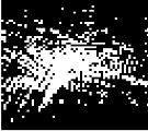
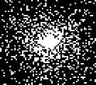
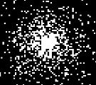
7 X 2D IFFT Uniform Sampling 120 X 2D IFFT Incoherent Sampling


![[1] David L. Donoho, IEEE Transactions on Information theory, Vol.52, no.](/docs-images/75/72888969/images/8-2.jpg "4, April 2006 [2] Candes, E.J. et al.")
8 Wavelet Transform Complete data reconstruction Most objects in nature are approximately sparse in a transformed domain. Utilize above concept to obtain very few measurements and yet reconstruct with high fidelity [1,2] X Only 33% of complete data Data provided by Baek [1] David L. Donoho, IEEE Transactions on Information theory, Vol.52, no. 4, April 2006 [2] Candes, E.J. et al., IEEE Transactions on Information theory, Vol.52, no.2, Feb. 2006


9 Generate a 2D phantom Cartesian undersampling of data Obtain undersampled data and zfwdc recon Choice of ROI if required for diagnostic evaluation purposes Recon params, post L-curve optimization Nonlinear conjugate gradient iterative reconstruction Comparative quality

10 Point spread function analyses 1. Incoherence 2. Design of this sampling mask
11 K-space trajectories with 2 constraints: 1. Slew rate 2. Smoothness of k-space coverage
12 Every MRI method: Angiography DWI/DTI/SWI/DCE-MRI/ASL fmri/mrsi/cmr. Because MRI is inherently a slow acquisition process, mostly dictated by the physics of acquisition Magnetic Resonance Fingerprinting
13 1. Rapid 1 H MR metabolic imaging 2. Accelerated DCE-MRI 3. Swifter Sweep Imaging with Fourier Transform (SWIFT) MRI
14 It has been well established that magnetic resonance imaging (MRI) provides critical information about cancer [3]. Magnetic resonance spectroscopic imaging (MRSI) furthers this capability by providing information about the presence of certain metabolites which are known to be important prognostic markers of cancer [4] (stroke, AD, energy metabolism, TCA cycle). MRSI provides information about the spatial distribution of these metabolites, hence enabling metabolic imaging. [3] Huk WJ et al., Neurosurgical Review 7(4) 1984; [4] Preul MC et al., Nat. Med. 2(3) 1996;

15 Increased choline level Reduced N-Acetylaspartate (NAA) level Reduced creatine level CANCER NORMAL [5] [5] H Kugel et al., Radiology 183 June 1992
16 Long acquisition times for MRSI A typical MRSI protocol (32 X 32 X 512) takes ~ 20 minutes Difficult to maintain anatomical posture for long time Increases patient discomfort, likelihood of early termination of study Discourages routine clinical use of this powerful MRI technique To increase throughput (decreased scanner time, technician time) Reduction of acquisition time is usually accomplished by under sampling measured data (k-space). Limitations of Shannon-Nyquist criterion. Compressed sensing provides a framework to achieve sub-nyquist sampling rates with good data fidelity.
 Philips 3.")
17 MRSI data Scanner TR(ms) TE(ms) # Averages Grid Size FOV (mm 3 ) Brain - normal (N=6) Siemens 3.0T Trio Tim x 16 x x 100 x 15 Brain cancer (N=2) Philips 3.0T Achieva x 21 x x 22 x x 210 x x 220 x 15 Prostate cancer (N=2) Philips 3.0T Achieva x 10 x x 12 x x 50 x x 51 x 26 Brain - normal (N=6) Brain - cancer (N=2) Prostate -cancer (N=2)
18 Minimal data processing done using jmrui [7] FID Apodization Gaussian (~3Hz) Removal of water peak using HLSVD Phase correction To allow correct integration of the real part of the spectra QUEST based quantitation. [8] To generate specific metabolite maps. [7] A. Naressi, et al., Computers in Biology and Medicine, vol. 31, [8] H. Ratiney, et al., Magnetic Resonance Materials in Physics Biology and Medicine, vol. 16, 2004.
19 1X 5X Cho Cr NAA
20 1X Normal Brain cancer Cancer Cho NAA Cho Cr Cho Cr 2 Cr 2 Cr NAA Cr Prostate cancer Normal Cancer Cit Cho + Cr Cit 2X 5X 10X

21 Brain - Normal Metabolite maps Brain - cancer Prostate - cancer
22 Mean SD of pooled data for each data type 2 tailed paired t-test Ratio: CNI for brain data and (Cho + cr)/cit for prostate data Excluded voxels with denominator value of 0 in 1X case For CS cases, if the denominator had a value of 0, the ratio was set to 0 P value less than 0.05 was chosen as a significant difference (* p <0.05) Brain (Normal) Brain (Cancer) Prostate (Cancer) NAA (a.u.) Cr (a.u.) Cho (a.u.) Cit (a.u.) Ratio 1X X X X * * * * 1X X X X * * 1X X X X * *

23 RMSE 1 N N i 1 ( i i '') 2 N = total number of elements of the MRSI data; Θ, Θ = the data reconstructed from full k-space and undersampled k-space respectively.
24 Application of compressed sensing on 1 H MRSI has been performed for the first time It has been demonstrated that compressed sensing based reconstruction can be successfully applied on 1 H MRSI in vivo human brain (normal and cancer), prostate cancer data and in vitro, computer generated phantom data sets Our results indicate a potential to reduce MRSI acquisition times by 75% thus significantly reducing the time spent by the patient in the MR scanner for spectroscopic studies Current and future work involves the implementation of compressed sensing based pulse sequences on preclinical and clinical scanners Other groups in the world are working on this demonstration now!
25 Rapid 1 H MR metabolic imaging Accelerated DCE-MRI Swifter Sweep Imaging with Fourier Transform (SWIFT) MRI
. 2005 *Model implemented by Dr. Vikram Kodibagkar in MATLAB [10]")
26 C(t) = f(δr 1 (t)) T1 weighted images for baseline T1 shortening contrast agent Tissue perfusion, microvascular density and extravascular -extracellular volume -- tumor staging, monitor treatment response [10] Yankeelov TE, et. al MRI;23(4) *Model implemented by Dr. Vikram Kodibagkar in MATLAB [10]
![[11] Vanvaals JJ et. al. JMRI; 3(4) 1993 [12] Jim J et.](/docs-images/75/72888969/images/27-0.jpg "al. IEEE TMI 2008 [13] Lustig M et. al.")
 S pre (ω) y diff Data was normalized to a")
 = L pre (ω) + H")
 = FI diff y diff 2 + λ LI WI diff 1 +λ TV")
27 [11] Vanvaals JJ et. al. JMRI; 3(4) 1993 [12] Jim J et. al. IEEE TMI 2008 [13] Lustig M et. al. MRM;58(6) 2007 I post-contrast I pre-contrast I diff S post (ω) S pre (ω) y diff Data was normalized to a range of 0 to 1 before retrospective reconstruction Keyhole for DCE CS for DCE S pre (ω) = L pre (ω) + H pre (ω) (1a) S post (ω) = L post (ω) + H pre (ω) (1b) Є( I diff ) = FI diff y diff 2 + λ LI WI diff 1 +λ TV (I diff ) (2) [11] [12,13]
28 5 DCE-MRI breast cancer data sets consisting of 64 frames (4 precontrast images and 60 post-contrast images) were used for retrospective reconstructions. The contrast agent used was Omniscan (intravenously administered through the tail vein at a dose of 0.1 mmol/kg). Reconstructions based on 2 approaches: keyhole and compressed sensing, were performed as function of masks and acceleration factors were performed. These reconstructions were quantified by the root mean square error metric defined below N RMSE 1 N i 1 ( i i '') 2


29 Masks Recon Original Keyhole Keylines Keythresh CS_Gauss CS_Glines CS_Thresh 2X 3X 4X 5X 2X 3X 4X 5X


30 Keyhole CS Keylines CS_Gauss Keyhole CS_Glines Keythresh CS_Thresh Starts at frame 1 Starts at frame 6 (post-contrast)


31 K trans Ve Original Keyhole Keylines Keythresh CS_Gauss CS_Glines CS_Thresh 2X 3X 4X 5X 2X 3X 4X 5X


32 ROI Intensity ROI Intensity ROI Intensity T1w precontrast T1w postcontrast Muscle T2w Overlay Well perfused region Original Keyhole Keylines Keythresh Gauss Glines Gthresh Frame # Poorly perfused region Original Keyhole Keylines Keythresh Gauss Glines Gthresh Frame # Original Keyhole Keylines Keythresh Gauss Glines Gthresh Frame #


33 Muscle Well perfused Poorly perfused
34 It has been shown here and previously that DCE MRI can be reliably accelerated through methods like compressed sensing and keyhole reconstructions to obtain increased spatial and/or temporal resolution. CS based masks Gauss and Gthresh provide better performance when compared to Glines mask, which out do the keyhole masks as observed by the RMSE graphs. Keyhole based masks keyhole mask performs relatively poorer when compared to keythresh and keylines masks Acceleration factors the values of RMSE increases with acceleration as expected (not shown); the CS masks show a RMSE of less than even at an acceleration factor of 5 while keyhole masks result in a RMSE of less than 0.1
35 Rapid 1 H MR metabolic imaging Accelerated DCE-MRI Swifter Sweep Imaging with Fourier Transform (SWIFT) MRI
![Sweep imaging with Fourier transformation [14] Time](/docs-images/75/72888969/images/36-0.jpg "domain signals are acquired during a swept radiofrequency")


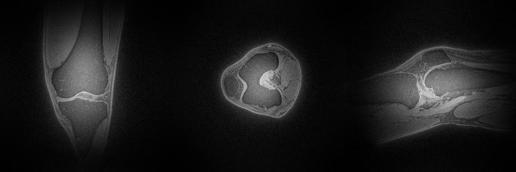
36 Sweep imaging with Fourier transformation [14] Time domain signals are acquired during a swept radiofrequency excitation in a time shared way This results in a significantly negligible echo time. Insensitive to motion, restricted dynamic range, low gradient noise Bovine tibia [14] D.Idiyatullin et al., JMR, 181, [14] GRE SWIFT Photograph
37 Full k-space recon was performed using gridding. The volume was restricted to a range of [0,1] by normalizing it to the highest absolute value. Prospective implementation is straight forward due to the nature of k-space trajectory. Acceleration of 5.33 X was achieved directly proportional to time saved MR data is sparse in the total variation domain. Since the data in this case is 3D, a 3D total variation norm is most apt. Reconstruction involves minimization of the convex functional given below. This is accomplished by a custom implementation of non-linear conjugate gradient algorithm. Є(m) = F u m y 2 +λ TV TV(m) where m is the desired MRI volume, F u is the Fourier transform operator, TV is the 3D total variation operator,. 2 is the L2 norm operator, λ TV is the regularization parameter for the TV term respectively, and Є is the value of the cost function.
38 The initial estimate of the volume is given by the zero-filled case with density compensation (zfwdc). This produces artifacts which are incoherent as can be seen in the zfwdc images. A total of 8 iterations were used and the recon was performed in 4 mins. NRMSE given by RMSE/ range of input; i.e. 1; hence NRMSE = RMSE calculated as given below N 1 2 RMSE ( i i '') N i 1 N = total number of elements of the MRI volume; Θ, Θ = the data reconstructed from full k- space and undersampled k-space respectively.

39 Original Zero filled with density compensation 5.33 X

 0.")
40 Intensity (au) Intensity (au) Intensity (au) Original Pixel number Zero filled with density compensation (Zfwdc) Pixel Number 0.6 5X Pixel Number
41 Original 5X Scan time ~ 8 min Estimated scan time ~1.6 min

42 Original Zfwdc 5 X
43 Review on CS MRI Critical reviews in biomedical engineering Miki Lustig, UC Berkley John M Pauly, Stanford

44 25+ member team ( ) Impact factor > 15 for Considered world experts in CS Work on CS has been showcased in the American Society of Neuroradiologists 2013 annual conference Several groups worldwide are working on our idea including Oxford and Yale
45

46 Human Scan Scanning Started from: Number of volunteers scanned till : 20
47 Mr. Rajesh Harsh, Mr. Ravindran Nair, Mr. T.S. Datta, Mr. R.S.Verma MIRC students Knowledge partners for MRI India Consortium: AIIMS, Harvard, NYU, Minnesota, Auburn ASU, KCL/ICL, Wipro-GE Healthcare Scientists/Participants Management of DSCE
48
G Practical Magnetic Resonance Imaging II Sackler Institute of Biomedical Sciences New York University School of Medicine. Compressed Sensing
 G16.4428 Practical Magnetic Resonance Imaging II Sackler Institute of Biomedical Sciences New York University School of Medicine Compressed Sensing Ricardo Otazo, PhD ricardo.otazo@nyumc.org Compressed
Compressed Sensing for Rapid MR Imaging
 Compressed Sensing for Rapid Imaging Michael Lustig1, Juan Santos1, David Donoho2 and John Pauly1 1 Electrical Engineering Department, Stanford University 2 Statistics Department, Stanford University rapid
Sparse sampling in MRI: From basic theory to clinical application. R. Marc Lebel, PhD Department of Electrical Engineering Department of Radiology
 Sparse sampling in MRI: From basic theory to clinical application R. Marc Lebel, PhD Department of Electrical Engineering Department of Radiology Objective Provide an intuitive overview of compressed sensing
A Novel Iterative Thresholding Algorithm for Compressed Sensing Reconstruction of Quantitative MRI Parameters from Insufficient Data
 A Novel Iterative Thresholding Algorithm for Compressed Sensing Reconstruction of Quantitative MRI Parameters from Insufficient Data Alexey Samsonov, Julia Velikina Departments of Radiology and Medical
Accelerated MRI Techniques: Basics of Parallel Imaging and Compressed Sensing
 Accelerated MRI Techniques: Basics of Parallel Imaging and Compressed Sensing Peng Hu, Ph.D. Associate Professor Department of Radiological Sciences PengHu@mednet.ucla.edu 310-267-6838 MRI... MRI has low
Compressed Sensing Reconstructions for Dynamic Contrast Enhanced MRI
 1 Compressed Sensing Reconstructions for Dynamic Contrast Enhanced MRI Kevin T. Looby klooby@stanford.edu ABSTRACT The temporal resolution necessary for dynamic contrast enhanced (DCE) magnetic resonance
Information about presenter
 Information about presenter 2013-now Engineer R&D ithera Medical GmbH 2011-2013 M.Sc. in Biomedical Computing (TU München) Thesis title: A General Reconstruction Framework for Constrained Optimisation
Compressed Sensing Algorithm for Real-Time Doppler Ultrasound Image Reconstruction
 Mathematical Modelling and Applications 2017; 2(6): 75-80 http://www.sciencepublishinggroup.com/j/mma doi: 10.11648/j.mma.20170206.14 ISSN: 2575-1786 (Print); ISSN: 2575-1794 (Online) Compressed Sensing
Abbie M. Diak, PhD Loyola University Medical Center Dept. of Radiation Oncology
 Abbie M. Diak, PhD Loyola University Medical Center Dept. of Radiation Oncology Outline High Spectral and Spatial Resolution MR Imaging (HiSS) What it is How to do it Ways to use it HiSS for Radiation
Enhao Gong, PhD Candidate, Electrical Engineering, Stanford University Dr. John Pauly, Professor in Electrical Engineering, Stanford University Dr.
 Enhao Gong, PhD Candidate, Electrical Engineering, Stanford University Dr. John Pauly, Professor in Electrical Engineering, Stanford University Dr. Greg Zaharchuk, Associate Professor in Radiology, Stanford
Spiral keyhole imaging for MR fingerprinting
 Spiral keyhole imaging for MR fingerprinting Guido Buonincontri 1, Laura Biagi 1,2, Pedro A Gómez 3,4, Rolf F Schulte 4, Michela Tosetti 1,2 1 IMAGO7 Research Center, Pisa, Italy 2 IRCCS Stella Maris,
Improved Spatial Localization in 3D MRSI with a Sequence Combining PSF-Choice, EPSI and a Resolution Enhancement Algorithm
 Improved Spatial Localization in 3D MRSI with a Sequence Combining PSF-Choice, EPSI and a Resolution Enhancement Algorithm L.P. Panych 1,3, B. Madore 1,3, W.S. Hoge 1,3, R.V. Mulkern 2,3 1 Brigham and
Optimal Sampling Geometries for TV-Norm Reconstruction of fmri Data
 Optimal Sampling Geometries for TV-Norm Reconstruction of fmri Data Oliver M. Jeromin, Student Member, IEEE, Vince D. Calhoun, Senior Member, IEEE, and Marios S. Pattichis, Senior Member, IEEE Abstract
Single Breath-hold Abdominal T 1 Mapping using 3-D Cartesian Sampling and Spatiotemporally Constrained Reconstruction
 Single Breath-hold Abdominal T 1 Mapping using 3-D Cartesian Sampling and Spatiotemporally Constrained Reconstruction Felix Lugauer 1,3, Jens Wetzl 1, Christoph Forman 2, Manuel Schneider 1, Berthold Kiefer
Redundancy Encoding for Fast Dynamic MR Imaging using Structured Sparsity
 Redundancy Encoding for Fast Dynamic MR Imaging using Structured Sparsity Vimal Singh and Ahmed H. Tewfik Electrical and Computer Engineering Dept., The University of Texas at Austin, USA Abstract. For
29 th NATIONAL RADIO SCIENCE CONFERENCE (NRSC 2012) April 10 12, 2012, Faculty of Engineering/Cairo University, Egypt
 April 10 12, 2012, Faculty of Engineering/Cairo University, Egypt") K1. High Performance Compressed Sensing MRI Image Reconstruction Ahmed Abdel Salam, Fadwa Fawzy, Norhan Shaker, Yasser M.Kadah Biomedical Engineering, Cairo University, Cairo, Egypt, ymk@k-space.org Computer
K1. High Performance Compressed Sensing MRI Image Reconstruction Ahmed Abdel Salam, Fadwa Fawzy, Norhan Shaker, Yasser M.Kadah Biomedical Engineering, Cairo University, Cairo, Egypt, ymk@k-space.org Computer
TITLE: Regularized Reconstruction of Dynamic Contrast-Enhanced MR Images for Evaluation of Breast Lesions
 AD Award Number: W81XWH-08-1-0273 TITLE: Regularized Reconstruction of Dynamic Contrast-Enhanced MR Images for Evaluation of Breast Lesions PRINCIPAL INVESTIGATOR: Kimberly A. Khalsa CONTRACTING ORGANIZATION:
NIH Public Access Author Manuscript Med Phys. Author manuscript; available in PMC 2009 March 13.
 NIH Public Access Author Manuscript Published in final edited form as: Med Phys. 2008 February ; 35(2): 660 663. Prior image constrained compressed sensing (PICCS): A method to accurately reconstruct dynamic
Compressive Sensing Could Accelerate 1 H MR Metabolic Imaging in the Clinic 1
 Note: This copy is for your personal non-commercial use only. To order presentation-ready copies for distribution to your colleagues or clients, contact us at www.rsna.org/rsnarights. Sairam Geethanath,
MRI reconstruction from partial k-space data by iterative stationary wavelet transform thresholding
 MRI reconstruction from partial k-space data by iterative stationary wavelet transform thresholding Mohammad H. Kayvanrad 1,2, Charles A. McKenzie 1,2,3, Terry M. Peters 1,2,3 1 Robarts Research Institute,
6 credits. BMSC-GA Practical Magnetic Resonance Imaging II
 BMSC-GA 4428 - Practical Magnetic Resonance Imaging II 6 credits Course director: Ricardo Otazo, PhD Course description: This course is a practical introduction to image reconstruction, image analysis
Classification of Subject Motion for Improved Reconstruction of Dynamic Magnetic Resonance Imaging
 1 CS 9 Final Project Classification of Subject Motion for Improved Reconstruction of Dynamic Magnetic Resonance Imaging Feiyu Chen Department of Electrical Engineering ABSTRACT Subject motion is a significant
Combination of Parallel Imaging and Compressed Sensing for high acceleration factor at 7T
 Combination of Parallel Imaging and Compressed Sensing for high acceleration factor at 7T DEDALE Workshop Nice Loubna EL GUEDDARI (NeuroSPin) Joint work with: Carole LAZARUS, Alexandre VIGNAUD and Philippe
Fast Imaging Trajectories: Non-Cartesian Sampling (1)
") Fast Imaging Trajectories: Non-Cartesian Sampling (1) M229 Advanced Topics in MRI Holden H. Wu, Ph.D. 2018.05.03 Department of Radiological Sciences David Geffen School of Medicine at UCLA Class Business
Fast Imaging Trajectories: Non-Cartesian Sampling (1) M229 Advanced Topics in MRI Holden H. Wu, Ph.D. 2018.05.03 Department of Radiological Sciences David Geffen School of Medicine at UCLA Class Business
RADIOMICS: potential role in the clinics and challenges
 27 giugno 2018 Dipartimento di Fisica Università degli Studi di Milano RADIOMICS: potential role in the clinics and challenges Dr. Francesca Botta Medical Physicist Istituto Europeo di Oncologia (Milano)
HST.583 Functional Magnetic Resonance Imaging: Data Acquisition and Analysis Fall 2008
 MIT OpenCourseWare http://ocw.mit.edu HST.583 Functional Magnetic Resonance Imaging: Data Acquisition and Analysis Fall 2008 For information about citing these materials or our Terms of Use, visit: http://ocw.mit.edu/terms.
(a Scrhon5 R2iwd b. P)jc%z 5. ivcr3. 1. I. ZOms Xn,s. 1E IDrAS boms. EE225E/BIOE265 Spring 2013 Principles of MRI. Assignment 8 Solutions
jc%z 5. ivcr3. 1. I. ZOms Xn,s. 1E IDrAS boms. EE225E/BIOE265 Spring 2013 Principles of MRI. Assignment 8 Solutions") EE225E/BIOE265 Spring 2013 Principles of MRI Miki Lustig Assignment 8 Solutions 1. Nishimura 7.1 P)jc%z 5 ivcr3. 1. I Due Wednesday April 10th, 2013 (a Scrhon5 R2iwd b 0 ZOms Xn,s r cx > qs 4-4 8ni6 4
EE225E/BIOE265 Spring 2013 Principles of MRI Miki Lustig Assignment 8 Solutions 1. Nishimura 7.1 P)jc%z 5 ivcr3. 1. I Due Wednesday April 10th, 2013 (a Scrhon5 R2iwd b 0 ZOms Xn,s r cx > qs 4-4 8ni6 4
Slide 1. Technical Aspects of Quality Control in Magnetic Resonance Imaging. Slide 2. Annual Compliance Testing. of MRI Systems.
 Slide 1 Technical Aspects of Quality Control in Magnetic Resonance Imaging Slide 2 Compliance Testing of MRI Systems, Ph.D. Department of Radiology Henry Ford Hospital, Detroit, MI Slide 3 Compliance Testing
High dynamic range magnetic resonance flow imaging in the abdomen
 High dynamic range magnetic resonance flow imaging in the abdomen Christopher M. Sandino EE 367 Project Proposal 1 Motivation Time-resolved, volumetric phase-contrast magnetic resonance imaging (also known
Constrained Reconstruction of Sparse Cardiac MR DTI Data
 Constrained Reconstruction of Sparse Cardiac MR DTI Data Ganesh Adluru 1,3, Edward Hsu, and Edward V.R. DiBella,3 1 Electrical and Computer Engineering department, 50 S. Central Campus Dr., MEB, University
Spread Spectrum Using Chirp Modulated RF Pulses for Incoherent Sampling Compressive Sensing MRI
 Spread Spectrum Using Chirp Modulated RF Pulses for Incoherent Sampling Compressive Sensing MRI Sulaiman A. AL Hasani Department of ECSE, Monash University, Melbourne, Australia Email: sulaiman.alhasani@monash.edu
arxiv: v2 [physics.med-ph] 22 Jul 2014
![arxiv: v2 [physics.med-ph] 22 Jul 2014](/thumbs/83/87658058.jpg "arxiv: v2 [physics.med-ph] 22 Jul 2014") Multichannel Compressive Sensing MRI Using Noiselet Encoding arxiv:1407.5536v2 [physics.med-ph] 22 Jul 2014 Kamlesh Pawar 1,2,3, Gary Egan 4, and Jingxin Zhang 1,5,* 1 Department of Electrical and Computer
Multichannel Compressive Sensing MRI Using Noiselet Encoding arxiv:1407.5536v2 [physics.med-ph] 22 Jul 2014 Kamlesh Pawar 1,2,3, Gary Egan 4, and Jingxin Zhang 1,5,* 1 Department of Electrical and Computer
An Iterative Approach for Reconstruction of Arbitrary Sparsely Sampled Magnetic Resonance Images
 An Iterative Approach for Reconstruction of Arbitrary Sparsely Sampled Magnetic Resonance Images Hamed Pirsiavash¹, Mohammad Soleymani², Gholam-Ali Hossein-Zadeh³ ¹Department of electrical engineering,
Deviceless respiratory motion correction in PET imaging exploring the potential of novel data driven strategies
 g Deviceless respiratory motion correction in PET imaging exploring the potential of novel data driven strategies Presented by Adam Kesner, Ph.D., DABR Assistant Professor, Division of Radiological Sciences,
Collaborative Sparsity and Compressive MRI
 Modeling and Computation Seminar February 14, 2013 Table of Contents 1 T2 Estimation 2 Undersampling in MRI 3 Compressed Sensing 4 Model-Based Approach 5 From L1 to L0 6 Spatially Adaptive Sparsity MRI
Nuts & Bolts of Advanced Imaging. Image Reconstruction Parallel Imaging
 Nuts & Bolts of Advanced Imaging Image Reconstruction Parallel Imaging Michael S. Hansen, PhD Magnetic Resonance Technology Program National Institutes of Health, NHLBI Declaration of Financial Interests
CT NOISE POWER SPECTRUM FOR FILTERED BACKPROJECTION AND ITERATIVE RECONSTRUCTION
 CT NOISE POWER SPECTRUM FOR FILTERED BACKPROJECTION AND ITERATIVE RECONSTRUCTION Frank Dong, PhD, DABR Diagnostic Physicist, Imaging Institute Cleveland Clinic Foundation and Associate Professor of Radiology
Magnetic Resonance Angiography
 Magnetic Resonance Angiography Course: Advance MRI (BIOE 594) Instructors: Dr Xiaohong Joe Zhou Dr. Shadi Othman By, Nayan Pasad Phase Contrast Angiography By Moran 1982, Bryan et. Al. 1984 and Moran et.
Image reconstruction using compressed sensing for individual and collective coil methods.
 Biomedical Research 2016; Special Issue: S287-S292 ISSN 0970-938X www.biomedres.info Image reconstruction using compressed sensing for individual and collective coil methods. Mahmood Qureshi *, Muhammad
MIDAS Signal Calibration
 Contents MIDAS Signal Calibration A. Maudsley, 2004-2007 Introduction... 1 The Calibration Phantom... 2 Calibration Method... 4 Additional Considerations... 11 Acknowledgements... 12 Introduction The MR
Outline: Contrast-enhanced MRA
 Outline: Contrast-enhanced MRA Background Technique Clinical Indications Future Directions Disclosures: GE Health Care: Research support Consultant: Bracco, Bayer The Basics During rapid IV infusion, Gadolinium
Module 5: Dynamic Imaging and Phase Sharing. (true-fisp, TRICKS, CAPR, DISTAL, DISCO, HYPR) Review. Improving Temporal Resolution.
 Review. Improving Temporal Resolution.") MRES 7005 - Fast Imaging Techniques Module 5: Dynamic Imaging and Phase Sharing (true-fisp, TRICKS, CAPR, DISTAL, DISCO, HYPR) Review Improving Temporal Resolution True-FISP (I) True-FISP (II) Keyhole
MRES 7005 - Fast Imaging Techniques Module 5: Dynamic Imaging and Phase Sharing (true-fisp, TRICKS, CAPR, DISTAL, DISCO, HYPR) Review Improving Temporal Resolution True-FISP (I) True-FISP (II) Keyhole
Functional MRI in Clinical Research and Practice Preprocessing
 Functional MRI in Clinical Research and Practice Preprocessing fmri Preprocessing Slice timing correction Geometric distortion correction Head motion correction Temporal filtering Intensity normalization
EE123 Digital Signal Processing
 EE123 Digital Signal Processing Lecture 24 Compressed Sensing III M. Lustig, EECS UC Berkeley RADIOS https://inst.eecs.berkeley.edu/~ee123/ sp15/radio.html Interfaces and radios on Wednesday -- please
Role of Parallel Imaging in High Field Functional MRI
 Role of Parallel Imaging in High Field Functional MRI Douglas C. Noll & Bradley P. Sutton Department of Biomedical Engineering, University of Michigan Supported by NIH Grant DA15410 & The Whitaker Foundation
Basic fmri Design and Analysis. Preprocessing
 Basic fmri Design and Analysis Preprocessing fmri Preprocessing Slice timing correction Geometric distortion correction Head motion correction Temporal filtering Intensity normalization Spatial filtering
Weighted-CS for reconstruction of highly under-sampled dynamic MRI sequences
 Weighted- for reconstruction of highly under-sampled dynamic MRI sequences Dornoosh Zonoobi and Ashraf A. Kassim Dept. Electrical and Computer Engineering National University of Singapore, Singapore E-mail:
K11. Modified Hybrid Median Filter for Image Denoising
 April 10 12, 2012, Faculty of Engineering/Cairo University, Egypt K11. Modified Hybrid Median Filter for Image Denoising Zeinab A.Mustafa, Banazier A. Abrahim and Yasser M. Kadah Biomedical Engineering
Lab Location: MRI, B2, Cardinal Carter Wing, St. Michael s Hospital, 30 Bond Street
 Lab Location: MRI, B2, Cardinal Carter Wing, St. Michael s Hospital, 30 Bond Street MRI is located in the sub basement of CC wing. From Queen or Victoria, follow the baby blue arrows and ride the CC south
Steen Moeller Center for Magnetic Resonance research University of Minnesota
 Steen Moeller Center for Magnetic Resonance research University of Minnesota moeller@cmrr.umn.edu Lot of material is from a talk by Douglas C. Noll Department of Biomedical Engineering Functional MRI Laboratory
Supplementary methods
 Supplementary methods This section provides additional technical details on the sample, the applied imaging and analysis steps and methods. Structural imaging Trained radiographers placed all participants
PERFORMANCE ANALYSIS BETWEEN TWO SPARSITY-CONSTRAINED MRI METHODS: HIGHLY CONSTRAINED BACKPROJECTION (HYPR) AND
 AND") PERFORMANCE ANALYSIS BETWEEN TWO SPARSITY-CONSTRAINED MRI METHODS: HIGHLY CONSTRAINED BACKPROJECTION (HYPR) AND COMPRESSED SENSING (CS) FOR DYNAMIC IMAGING A Thesis by NIBAL ARZOUNI Submitted to the Office
PERFORMANCE ANALYSIS BETWEEN TWO SPARSITY-CONSTRAINED MRI METHODS: HIGHLY CONSTRAINED BACKPROJECTION (HYPR) AND COMPRESSED SENSING (CS) FOR DYNAMIC IMAGING A Thesis by NIBAL ARZOUNI Submitted to the Office
SPM8 for Basic and Clinical Investigators. Preprocessing. fmri Preprocessing
 SPM8 for Basic and Clinical Investigators Preprocessing fmri Preprocessing Slice timing correction Geometric distortion correction Head motion correction Temporal filtering Intensity normalization Spatial
SIVIC GUI Tutorial. HMTRC Workshop - March 23-24, 2017
 SIVIC GUI Tutorial HMTRC Workshop - March 23-24, 2017 Department of Radiology and Biomedical Imaging, UCSF Supported by NIBIB P41EB013598 Goal: The purpose of this tutorial is to introduce you to the SIVIC
Enhanced DICOM MR for spectroscopy, structural and functional imaging
 The Medicine Behind the Image Enhanced DICOM MR for spectroscopy, structural and functional imaging Dr. David A. Clunie, MB.,BS., FRACR Chief Technology Officer RadPharm, Inc. Acknowledgments Mark Day,
K-Space Trajectories and Spiral Scan
 K-Space and Spiral Scan Presented by: Novena Rangwala nrangw2@uic.edu 1 Outline K-space Gridding Reconstruction Features of Spiral Sampling Pulse Sequences Mathematical Basis of Spiral Scanning Variations
Motion Artifacts and Suppression in MRI At a Glance
 Motion Artifacts and Suppression in MRI At a Glance Xiaodong Zhong, PhD MR R&D Collaborations Siemens Healthcare MRI Motion Artifacts and Suppression At a Glance Outline Background Physics Common Motion
3/27/2012 WHY SPECT / CT? SPECT / CT Basic Principles. Advantages of SPECT. Advantages of CT. Dr John C. Dickson, Principal Physicist UCLH
 3/27/212 Advantages of SPECT SPECT / CT Basic Principles Dr John C. Dickson, Principal Physicist UCLH Institute of Nuclear Medicine, University College London Hospitals and University College London john.dickson@uclh.nhs.uk
Automatic Determination of Arterial Input Function for Dynamic Contrast Enhanced MRI in Tumor Assessment
 Automatic Determination of Arterial Input Function for Dynamic Contrast Enhanced MRI in Tumor Assessment Jeremy Chen, Jianhua Yao, and David Thomasson Diagnostic Radiology Department, Clinical Center,
Institute of Cardiovascular Science, UCL Centre for Cardiovascular Imaging, London, United Kingdom, 2
 Grzegorz Tomasz Kowalik 1, Jennifer Anne Steeden 1, Bejal Pandya 1, David Atkinson 2, Andrew Taylor 1, and Vivek Muthurangu 1 1 Institute of Cardiovascular Science, UCL Centre for Cardiovascular Imaging,
Evaluations of k-space Trajectories for Fast MR Imaging for project of the course EE591, Fall 2004
 Evaluations of k-space Trajectories for Fast MR Imaging for project of the course EE591, Fall 24 1 Alec Chi-Wah Wong Department of Electrical Engineering University of Southern California 374 McClintock
FOREWORD TO THE SPECIAL ISSUE ON MOTION DETECTION AND COMPENSATION
 Philips J. Res. 51 (1998) 197-201 FOREWORD TO THE SPECIAL ISSUE ON MOTION DETECTION AND COMPENSATION This special issue of Philips Journalof Research includes a number of papers presented at a Philips
EPI Data Are Acquired Serially. EPI Data Are Acquired Serially 10/23/2011. Functional Connectivity Preprocessing. fmri Preprocessing
 Functional Connectivity Preprocessing Geometric distortion Head motion Geometric distortion Head motion EPI Data Are Acquired Serially EPI Data Are Acquired Serially descending 1 EPI Data Are Acquired
SPM8 for Basic and Clinical Investigators. Preprocessing
 SPM8 for Basic and Clinical Investigators Preprocessing fmri Preprocessing Slice timing correction Geometric distortion correction Head motion correction Temporal filtering Intensity normalization Spatial
Sparse MRI: The Application of Compressed Sensing for Rapid MR Imaging
 Magnetic Resonance in Medicine 58:1182 1195 (2007) Sparse MRI: The Application of Compressed Sensing for Rapid MR Imaging Michael Lustig, 1 David Donoho, 2 and John M. Pauly 1 The sparsity which is implicit
Rat 2D EPSI Dual Band Variable Flip Angle 13 C Dynamic Spectroscopy
 Rat 2D EPSI Dual Band Variable Flip Angle 13 C Dynamic Spectroscopy In this example you will load a dynamic MRS animal data set acquired on a GE 3T scanner. This data was acquired with an EPSI sequence
CP Generalize Concepts in Abstract Multi-dimensional Image Model Component Semantics. David Clunie.
 CP-1390 - Generalize Concepts in Abstract Multi-dimensional Image Model Semantics Page 1 STATUS Date of Last Update Person Assigned Submitter Name Submission Date Assigned 2014/06/09 David Clunie mailto:dclunie@dclunie.com
Accelerated 4D flow MRI
 Accelerated 4D flow MRI Comparing SENSE, k-t PCA and Compressed Sensing Tomas Kaandorp 10356169 Patient comfort can be increased by accelerating an MRI scanner to reduce scan time. 4D flow MRI scans can
Separate Magnitude and Phase Regularization via Compressed Sensing
 IEEE TRANSACTIONS ON MEDICAL IMAGING, VOL. 31, NO. 9, SEPTEMBER 2012 1713 Separate Magnitude and Phase Regularization via Compressed Sensing Feng Zhao*, Douglas C. Noll, Senior Member, IEEE, Jon-Fredrik
Iterative CT Reconstruction Using Curvelet-Based Regularization
 Iterative CT Reconstruction Using Curvelet-Based Regularization Haibo Wu 1,2, Andreas Maier 1, Joachim Hornegger 1,2 1 Pattern Recognition Lab (LME), Department of Computer Science, 2 Graduate School in
Correction of Partial Volume Effects in Arterial Spin Labeling MRI
 Correction of Partial Volume Effects in Arterial Spin Labeling MRI By: Tracy Ssali Supervisors: Dr. Keith St. Lawrence and Udunna Anazodo Medical Biophysics 3970Z Six Week Project April 13 th 2012 Introduction
Acknowledgments and financial disclosure
 AAPM 2012 Annual Meeting Digital breast tomosynthesis: basic understanding of physics principles James T. Dobbins III, Ph.D., FAAPM Director, Medical Physics Graduate Program Ravin Advanced Imaging Laboratories
Efficient MR Image Reconstruction for Compressed MR Imaging
 Efficient MR Image Reconstruction for Compressed MR Imaging Junzhou Huang, Shaoting Zhang, and Dimitris Metaxas Division of Computer and Information Sciences, Rutgers University, NJ, USA 08854 Abstract.
Breast MRI Accreditation Program Clinical Image Quality Guide
 Breast MRI Accreditation Program Clinical Image Quality Guide Introduction This document provides guidance on breast MRI clinical image quality and describes the criteria used by the ACR Breast MRI Accreditation
Chapter 3 Set Redundancy in Magnetic Resonance Brain Images
 16 Chapter 3 Set Redundancy in Magnetic Resonance Brain Images 3.1 MRI (magnetic resonance imaging) MRI is a technique of measuring physical structure within the human anatomy. Our proposed research focuses
M R I Physics Course
 M R I Physics Course Multichannel Technology & Parallel Imaging Nathan Yanasak, Ph.D. Jerry Allison Ph.D. Tom Lavin, B.S. Department of Radiology Medical College of Georgia References: 1) The Physics of
MEDICAL IMAGE COMPUTING (CAP 5937) LECTURE 4: Pre-Processing Medical Images (II)
 LECTURE 4: Pre-Processing Medical Images (II)") SPRING 2016 1 MEDICAL IMAGE COMPUTING (CAP 5937) LECTURE 4: Pre-Processing Medical Images (II) Dr. Ulas Bagci HEC 221, Center for Research in Computer Vision (CRCV), University of Central Florida (UCF),
SPRING 2016 1 MEDICAL IMAGE COMPUTING (CAP 5937) LECTURE 4: Pre-Processing Medical Images (II) Dr. Ulas Bagci HEC 221, Center for Research in Computer Vision (CRCV), University of Central Florida (UCF),
Introduction to Topics in Machine Learning
 Introduction to Topics in Machine Learning Namrata Vaswani Department of Electrical and Computer Engineering Iowa State University Namrata Vaswani 1/ 27 Compressed Sensing / Sparse Recovery: Given y :=
Advanced Imaging Trajectories
 Advanced Imaging Trajectories Cartesian EPI Spiral Radial Projection 1 Radial and Projection Imaging Sample spokes Radial out : from k=0 to kmax Projection: from -kmax to kmax Trajectory design considerations
8/2/2016. Measures the degradation/distortion of the acquired image (relative to an ideal image) using a quantitative figure-of-merit
 using a quantitative figure-of-merit") Ke Li Assistant Professor Department of Medical Physics and Department of Radiology School of Medicine and Public Health, University of Wisconsin-Madison This work is partially supported by an NIH Grant
Ke Li Assistant Professor Department of Medical Physics and Department of Radiology School of Medicine and Public Health, University of Wisconsin-Madison This work is partially supported by an NIH Grant
Diffusion MRI Acquisition. Karla Miller FMRIB Centre, University of Oxford
 Diffusion MRI Acquisition Karla Miller FMRIB Centre, University of Oxford karla@fmrib.ox.ac.uk Diffusion Imaging How is diffusion weighting achieved? How is the image acquired? What are the limitations,
The Benefit of Tree Sparsity in Accelerated MRI
 The Benefit of Tree Sparsity in Accelerated MRI Chen Chen and Junzhou Huang Department of Computer Science and Engineering, The University of Texas at Arlington, TX, USA 76019 Abstract. The wavelet coefficients
Sparse Spectral Deconvolution Algorithm for Noncartesian MR Spectroscopic Imaging
 SPECTROSCOPIC METHODOLOGY Note Magnetic Resonance in Medicine 71:469 476 (2014) Sparse Spectral Deconvolution Algorithm for Noncartesian MR Spectroscopic Imaging Sampada Bhave, 1 Ramin Eslami, 2 and Mathews
Sampling, Ordering, Interleaving
 Sampling, Ordering, Interleaving Sampling patterns and PSFs View ordering Modulation due to transients Temporal modulations Slice interleaving Sequential, Odd/even, bit-reversed Arbitrary Other considerations:
ComputerLab: compressive sensing and application to MRI
 Compressive Sensing, 207-8 ComputerLab: compressive sensing and application to MRI Aline Roumy This computer lab addresses the implementation and analysis of reconstruction algorithms for compressive sensing.
Sparse Reconstruction / Compressive Sensing
 Sparse Reconstruction / Compressive Sensing Namrata Vaswani Department of Electrical and Computer Engineering Iowa State University Namrata Vaswani Sparse Reconstruction / Compressive Sensing 1/ 20 The
Dynamic Contrast enhanced MRA
 Dynamic Contrast enhanced MRA Speaker: Yung-Chieh Chang Date : 106.07.22 Department of Radiology, Taichung Veterans General Hospital, Taichung, Taiwan 1 Outline Basic and advanced principles of Diffusion
MIDAS FAQ. 1) Supported MR Systems and SI Acquisition Protocols. 2) Recommended Computer Systems
 Supported MR Systems and SI Acquisition Protocols. 2) Recommended Computer Systems") MIDAS FAQ Contents: 1) Supported MR Systems and SI Acquisition Protocols... 1 2) Recommended Computer Systems... 1 3) MIDAS Error Messages... 2 4) Miscellaneous Questions and Artifacts... 4 a) Why does
MIDAS FAQ Contents: 1) Supported MR Systems and SI Acquisition Protocols... 1 2) Recommended Computer Systems... 1 3) MIDAS Error Messages... 2 4) Miscellaneous Questions and Artifacts... 4 a) Why does
Respiratory Motion Compensation for Simultaneous PET/MR Based on Strongly Undersampled Radial MR Data
 Respiratory Motion Compensation for Simultaneous PET/MR Based on Strongly Undersampled Radial MR Data Christopher M Rank 1, Thorsten Heußer 1, Andreas Wetscherek 1, and Marc Kachelrieß 1 1 German Cancer
BME I5000: Biomedical Imaging
 1 Lucas Parra, CCNY BME I5000: Biomedical Imaging Lecture 4 Computed Tomography Lucas C. Parra, parra@ccny.cuny.edu some slides inspired by lecture notes of Andreas H. Hilscher at Columbia University.
Analysis of Functional MRI Timeseries Data Using Signal Processing Techniques
 Analysis of Functional MRI Timeseries Data Using Signal Processing Techniques Sea Chen Department of Biomedical Engineering Advisors: Dr. Charles A. Bouman and Dr. Mark J. Lowe S. Chen Final Exam October
Supplementary Figure 1
 Supplementary Figure 1 BOLD and CBV functional maps showing EPI versus line-scanning FLASH fmri. A. Colored BOLD and CBV functional maps are shown in the highlighted window (green frame) of the raw EPI
Initial Experience of Applying TWIST Dixon with Flexible View Sharing in Breast DCE-MRI
 Initial Experience of Applying TWIST Dixon with Flexible View Sharing in Breast DCE-MRI Yuan Le PhD 1, Hal D. Kipfer MD 1, Dominik M. Nickel PhD 2, Randall Kroeker PhD 2, Brian Dale PhD 2, Stephanie P.
A Spatio-temporal Denoising Approach based on Total Variation Regularization for Arterial Spin Labeling
 A Spatio-temporal Denoising Approach based on Total Variation Regularization for Arterial Spin Labeling Cagdas Ulas 1,2, Stephan Kaczmarz 3, Christine Preibisch 3, Jonathan I Sperl 2, Marion I Menzel 2,
Acknowledgments. High Performance Cone-Beam CT of Acute Traumatic Brain Injury
 A. Sisniega et al. (presented at RSNA 214) High Performance Cone-Beam CT of Acute Traumatic Brain Injury A. Sisniega 1 W. Zbijewski 1, H. Dang 1, J. Xu 1 J. W. Stayman 1, J. Yorkston 2, N. Aygun 3 V. Koliatsos
Optimizing Flip Angle Selection in Breast MRI for Accurate Extraction and Visualization of T1 Tissue Relaxation Time
 Optimizing Flip Angle Selection in Breast MRI for Accurate Extraction and Visualization of T1 Tissue Relaxation Time GEORGIOS KETSETZIS AND MICHAEL BRADY Medical Vision Laboratory Department of Engineering
MRI image formation 8/3/2016. Disclosure. Outlines. Chen Lin, PhD DABR 3. Indiana University School of Medicine and Indiana University Health
 MRI image formation Indiana University School of Medicine and Indiana University Health Disclosure No conflict of interest for this presentation 2 Outlines Data acquisition Spatial (Slice/Slab) selection
Exploration and Study of MultiVolume Image Data using 3D Slicer
 Exploration and Study of MultiVolume Image Data using 3D Slicer Meysam Torabi and Andriy Fedorov torabi@bwh.harvard.edu, fedorov@bwh.harvard.edu Surgical Navigation and Robotics Lab and Surgical Planning
Fmri Spatial Processing
 Educational Course: Fmri Spatial Processing Ray Razlighi Jun. 8, 2014 Spatial Processing Spatial Re-alignment Geometric distortion correction Spatial Normalization Smoothing Why, When, How, Which Why is
Clinical Importance. Aortic Stenosis. Aortic Regurgitation. Ultrasound vs. MRI. Carotid Artery Stenosis
 Clinical Importance Rapid cardiovascular flow quantitation using sliceselective Fourier velocity encoding with spiral readouts Valve disease affects 10% of patients with heart disease in the U.S. Most