Human Connectom Project : The minimal processing Pipeline
|
|
|
- Clyde Phillips
- 6 years ago
- Views:
Transcription
1 Human Connectom Project : The minimal processing Pipeline
2 Human Connectom Project : The minimal processing Pipeline Van Essen DC, The WU-Minn Human Connectome Project: an overview. Neuroimage Marcus DS,... Human Connectome Project informatics: quality control, database services, and data visualization. Neuroimage Glasser MF,... The minimal preprocessing pipelines for the Human Connectome Project. Neuroimage Sotiropoulos... Advances in diffusion MRI acquisition and processing in the Human Connectome Project. Neuroimage Smith SM,... Resting-state fmri in the Human Connectome Project. Neuroimage. Uğurbil K, Xu J,... Pushing spatial and temporal resolution for functional and diffusion MRI in the Human Connectome Project. Neuroimage Larson-Prior LJ,... Adding dynamics to the Human Connectome Project with MEG. Neuroimage. connectome 300mT McNab JA,... The Human Connectome Project and beyond: initial applications of 300 mt/m gradients. Neuroimage Setsompop K,... Pushing the limits of in vivo diffusion MRI for the Human Connectome Project. Neuroimage Marcus DS,... Van Essen DC. Informatics and data mining tools and strategies for the human connectome project. Front Neuroinform Jun
3 HCP overview Objective : characterize brain connectivity and function and their variability in healthy adults years of preparation 3 year of scanning 1200 Subjects (22-35 years) : a population of adult twins and their non-twin siblings The state of art of acquisition Share the data and the process (first release march 2013) more than 100 investigators and technical staff from ten institutions in the consortium Scan : New 3T scanner (skyra) + new acquisition sequence Multi Band EPI customize 3T skyra (gradient 100mT/m) (commercial version 80mT/m) Scan : ~ 3H30 per subjects in 4 or 5 session Structural : 40 mn : 0.7 iso + calibration scans (B0 B1+ B1-) resting states fmri : 1H : 2 mm iso, TR 0.7 s ( 2x2 sesion RL / LR) task- fmri : 1 H : 7 task working memory, reward processing, motor processing language, social cognition, relational processing and emotion processing session difusion MRI : 1H : 1.25 mm iso. 18 B0 3*90 DWI with Bvalue of repeated twice RL/LR same acquisition at 7T for subset of 200 subjects MEEG/EEG task and resting states for a subset of 100 subject Behaviour Genetic Head motion monitoring with a camera. cardiac and respiratory signals associated with each scan

4 Acquisition parameters optimisation A new scanner 3T with Hig gradient
5 Multi Band SNR no SNR loss due to reduced data collection (a small SNR penalty associated with slice aliasing) BUT T1 relaxation effects from the shorter achievable Trs avec IPAT large signal fluctuations (amplify by diffusion encoding)
6 Acquisitions Details Structural T1 w (mprage) / T2w (sapce) at 0.7 iso optimisation of contrast parameters : FA, TI and TE B0 field maps / B1+ B1- (for readout distortion and bias correction) at 2mm iso B1- : receive field from a FLASH 32 head coil versus body coil B1+ : transmit field from flip-angle imaging (Yarnykh,2007) FA 50 TR1/TR2 20/120ms fmri multiband factor of 8, 2 mm iso, TR 0.7 s FA=52 no PF. TE 33ms matrix 104*90*72 for each session a Spin echo field map : 2 spin echo EPI with reversed phase encoding directions (2*3 images : 60s) A single band reference scan (for a better gray/white contrast) Always 2 sesion RL / LR Diffusion multiband factor of 3, 1.25 mm iso. 18 B0 3*90 DWI with Bvalue of repeated twice RL/LR. Mono polar diffusion-enconding sheme. Direction-resversed images for each diffusion direction (whole sphere) B1- acquisition twice LR / RL Multiband acceleration how much acceleration could be achieved before significant slicecross-talk occurred, or undesirable artefacts arose in the multiband reconstruction due to interactions with head motion (up to 8) We determined that distortion-corrected L R and R L datasets are, in general, anatomically well aligned with each other, even in regions of different dropout it is only the dropout that differs. Q sampling strategie
7 A short TR the basic single-voxel timeseries detection power is minimally affected by reduction in TR (at least in the range ~0.5-3 s), as the simple statistical gain from the increased number of data points is balanced by the loss in signal level (as TR falls below T1). Advantage : the denser temporal sampling of physiological confounds; the importance of temporal degrees-of-freedom for many analysis techniques (such as high-dimensional ICA) richer temporal characterisation of resting-state fluctuations.

8 Spatial resolution For DWI

9 For Tractography
 caused by the dominant draining vein contribution at this field strength (2mm at 7T) but A more specific cortical signal : decrease cross-sulcal and")
10 Spatial resolution for fmri : choice of spatial resolution SNR but also the point spread function of the BOLD effect at 3 T (Parkes et al., 2005), which was measured to be ~3.5 mm at full-width-at-half-maximum (FWHM) caused by the dominant draining vein contribution at this field strength (2mm at 7T) but A more specific cortical signal : decrease cross-sulcal and cross-gyral timeseries correlation.

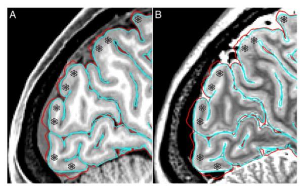
11 Spatial Specificity of correlation map

12 Sulcus specificity of Resting-state components
 Header : contains the grayordinates space definition (l 1000=vertext 2152; line 91000 = 3.9-1.")
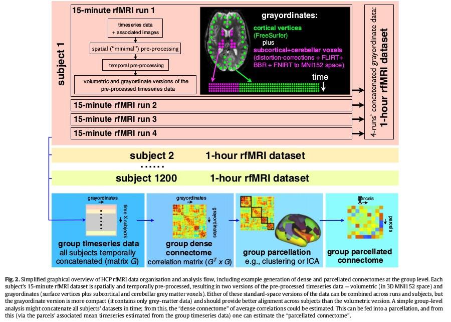
13 Pre-processing The cifti file format & grayordinates CIFTI format : Grayordinates : Combinations of cortical surface and subcortical volumetric parcels dense timeseries : spatial grayordinate x time dense Connectome : seed points x spatial map of the connectivity dense scalar file (ica component z-map ) Header : contains the grayordinates space definition (l 1000=vertext 2152; line = normalisation resampling the sets of voxels in individually defined subcortical parcels to a standard set of voxels of the atlas parcel. surface-constrained smoothing ( adapted to cortex folding) subcortical parcel-constrained smoothing ( no partial volume of surrounding structures) save space (60 to 80%) cifti nifti or nifti + gifti


14 The minimal preprocessing pipelines * Native Volume space (rigidly aligned to MNI aces)
 brain extraction with the template mask (after a non linear")
15 PreFreeSurfer Pipeline Undistorted native structural volume space (siemens correction not for all sequences, but use the Siemens gradient coefficient file) align T1 T2 B1 bias readout distortion is different for T1 and T2 crop with fsl robustfov tool ACPC alignnment space (for visualisation and to remove rotation in the nifti sform) brain extraction with the template mask (after a non linear normalisation to MNI) more robust than BET readout distortion (very small same as EPI distortion) field map is register to T1 and T2 separatly. Boundary-based registration (BBR) cost function. Bias correction from T1*T2 : perserve gray matter inhomogeneity in T1 and T2 (different myelin content Fishl 2004) final output with spline interpolation (less blurring)
16

17 FreeSurefer Pipeline segment the volume (ribon and CIFTI subcortical parcels) white and pial cortical surface definition standard surfaces registration (to fsaverage) addon : Initial Mask 1 mm to 0.7 mm 0.7 T1 intensity normalized and white matter surface is adjusted gray matter surface is adjusted with T1 and T2 volume : T1w intensity normalisation with 4 sigmas (defautl is 3) do not exclude lightly myelinated gray matter. Then T2w is used to erode the pial surface T1w is smoothed with a ribbon constrained approach (sigma 5mm) and pial surface is done again (with 2 sigmas)
18
19

20 improve pial surface

21 Extra Bias Field correction. Differenc with the Group average myelin maps (smooth of 14 mm) substracted to the result.
22
 and low-resolution (32k) meshes.")
23 PostFreeSurfer Pipeline Make all nifti and gifti files for viewing wmparce 3 dilate 2 erosion white matter mask for diffusion single spline interpolation to make T1 and T2 in MNI. registration to Conte69 surface template downsampling surfaces bidirectional resampling between high-resolution (164k) and low-resolution (32k) meshes. myelin maps Conte69 : population-average surfaces which have correspondence between the left and right hemispheres VanEssen 2012
 normalizes intensity to a 4D mean (10 000) apply the final mask Same correction of the gradient-nonlinearity-induced distortion (all images) realign on the single band reference image 12 parameters")
24 fmrivolume pipeline remove spatial distortions realing volumes register to structural reduces the bias filed (from T1 estimation of different session!) normalizes intensity to a 4D mean (10 000) apply the final mask Same correction of the gradient-nonlinearity-induced distortion (all images) realign on the single band reference image 12 parameters mvt (mvt+derived) demeaned and detrended for nuisance regression
 (wheighted according to the partial volume inside the ribbon ) exclusion of")
 = voxel near the edge or with blood vessel.")
25 fmrisurface pipeline bring the timeseries to CIFTI space 2 mm FWHM smooth (surface and parcel constrained smoothing) (wheighted according to the partial volume inside the ribbon ) exclusion of noisy voxel (0.5 std above the 5mm sigma gaussian neighborhood) = voxel near the edge or with blood vessel. Gyral bias in high coefficient of variation is eliminated surface smoothin with vertex aera correction

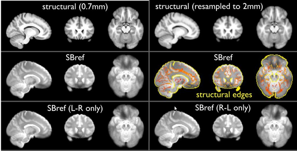
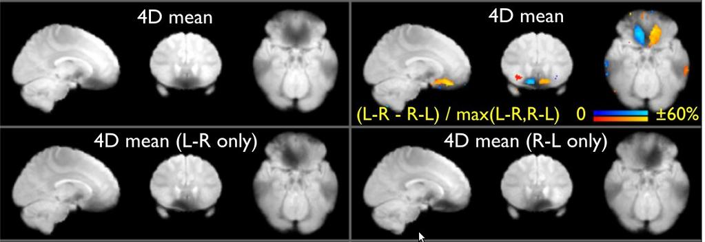
26 SB Ref / MB, same dropout same distortion dropout, which are different in L R and R L

27 distortion-corrected SBRef images after alignment to the structural images green : white-grey boundary from T1w. there is no obvious residual distortion that is different between these two images
28
29
30

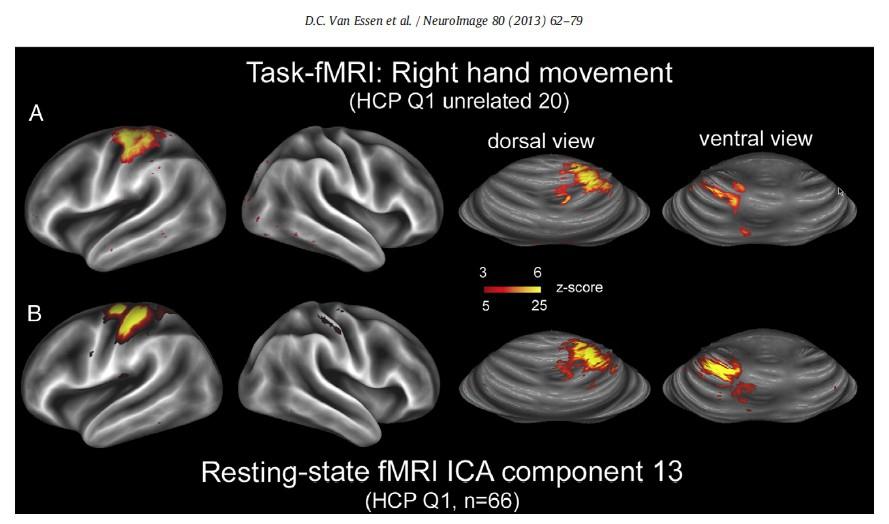
31 Consistent activation in the majority of the global contrasts, BUT some focused contrasts did not show consistent individual subject level activation. No orbital frontal activation in at least 50% of individual subjects in the emotion processing task; no striatal activation in at least 50% of individual subjects in the comparison of reward versus baseline for the gambling task (SNR in subcortical regions); no activation in parietal regions in the tools compared to other stimulus types contrast.

32 Improvment of surface analysis
 eddy-current-induced distortions + subject motion (eddy) corrects for gradient-nolinearities registers to")
33 Diffusion Pipeline normalizes b0 intensity across runs removes EPI distortions : top up with extra spin echo EPI (RL LR) eddy-current-induced distortions + subject motion (eddy) corrects for gradient-nolinearities registers to structural

34 Gradient non-linearity for diffusion encoding

35 Advance processing Diffusion Qsampling Strategie
36
37 rfmri post-processing : FIX Data is provided from spatial ( minimal ) pre-processing, in both volumetric and grayordinate forms. Weak highpass temporal filtering (>2000s FWHM) is applied to both forms, achieving slow drift removal. MELODIC ICA is applied to volumetric data; artefact components are identified using FIX. Artefact and motion-related timecourses are regressed out of both volumetric and grayordinate data. Optionally (and depending on further investigations), possibly also some combination of: further motion cleanup/scrubbing; further removal of physiological confounds based on physiological monitoring data; removal of globally-related signals. (mean total signal or mean gray signal)
38
39 Results
40
FSL Pre-Processing Pipeline
 The Art and Pitfalls of fmri Preprocessing FSL Pre-Processing Pipeline Mark Jenkinson FMRIB Centre, University of Oxford FSL Pre-Processing Pipeline Standard pre-processing: Task fmri Resting-state fmri
FSL Pre-Processing Pipeline
 The Art and Pitfalls of fmri Preprocessing FSL Pre-Processing Pipeline Mark Jenkinson FMRIB Centre, University of Oxford FSL Pre-Processing Pipeline Standard pre-processing: Task fmri Resting-state fmri
SPM8 for Basic and Clinical Investigators. Preprocessing. fmri Preprocessing
 SPM8 for Basic and Clinical Investigators Preprocessing fmri Preprocessing Slice timing correction Geometric distortion correction Head motion correction Temporal filtering Intensity normalization Spatial
Functional MRI data preprocessing. Cyril Pernet, PhD
 Functional MRI data preprocessing Cyril Pernet, PhD Data have been acquired, what s s next? time No matter the design, multiple volumes (made from multiple slices) have been acquired in time. Before getting
Basic fmri Design and Analysis. Preprocessing
 Basic fmri Design and Analysis Preprocessing fmri Preprocessing Slice timing correction Geometric distortion correction Head motion correction Temporal filtering Intensity normalization Spatial filtering
SUPPLEMENTARY INFORMATION
 doi:10.1038/nature18933 Supplementary Methods For A Multi-modal Parcellation of Human Cerebral Cortex Matthew F. Glasser 1, Timothy S. Coalson 1 *, Emma C. Robinson 2,3 *, Carl D. Hacker 4 *, John Harwell
FMRI Pre-Processing and Model- Based Statistics
 FMRI Pre-Processing and Model- Based Statistics Brief intro to FMRI experiments and analysis FMRI pre-stats image processing Simple Single-Subject Statistics Multi-Level FMRI Analysis Advanced FMRI Analysis
Functional MRI in Clinical Research and Practice Preprocessing
 Functional MRI in Clinical Research and Practice Preprocessing fmri Preprocessing Slice timing correction Geometric distortion correction Head motion correction Temporal filtering Intensity normalization
Fmri Spatial Processing
 Educational Course: Fmri Spatial Processing Ray Razlighi Jun. 8, 2014 Spatial Processing Spatial Re-alignment Geometric distortion correction Spatial Normalization Smoothing Why, When, How, Which Why is
EPI Data Are Acquired Serially. EPI Data Are Acquired Serially 10/23/2011. Functional Connectivity Preprocessing. fmri Preprocessing
 Functional Connectivity Preprocessing Geometric distortion Head motion Geometric distortion Head motion EPI Data Are Acquired Serially EPI Data Are Acquired Serially descending 1 EPI Data Are Acquired
SPM8 for Basic and Clinical Investigators. Preprocessing
 SPM8 for Basic and Clinical Investigators Preprocessing fmri Preprocessing Slice timing correction Geometric distortion correction Head motion correction Temporal filtering Intensity normalization Spatial
Introduction to fmri. Pre-processing
 Introduction to fmri Pre-processing Tibor Auer Department of Psychology Research Fellow in MRI Data Types Anatomical data: T 1 -weighted, 3D, 1/subject or session - (ME)MPRAGE/FLASH sequence, undistorted
The organization of the human cerebral cortex estimated by intrinsic functional connectivity
 1 The organization of the human cerebral cortex estimated by intrinsic functional connectivity Journal: Journal of Neurophysiology Author: B. T. Thomas Yeo, et al Link: https://www.ncbi.nlm.nih.gov/pubmed/21653723
Diffusion MRI Acquisition. Karla Miller FMRIB Centre, University of Oxford
 Diffusion MRI Acquisition Karla Miller FMRIB Centre, University of Oxford karla@fmrib.ox.ac.uk Diffusion Imaging How is diffusion weighting achieved? How is the image acquired? What are the limitations,
Analysis of Functional MRI Timeseries Data Using Signal Processing Techniques
 Analysis of Functional MRI Timeseries Data Using Signal Processing Techniques Sea Chen Department of Biomedical Engineering Advisors: Dr. Charles A. Bouman and Dr. Mark J. Lowe S. Chen Final Exam October
NeuroImage 80 (2013) Contents lists available at SciVerse ScienceDirect. NeuroImage. journal homepage:
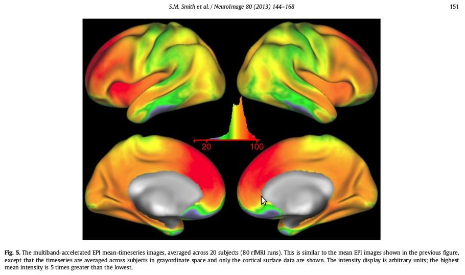
 Contents lists available at SciVerse ScienceDirect. NeuroImage. journal homepage:") NeuroImage 80 (2013) 144 168 Contents lists available at SciVerse ScienceDirect NeuroImage journal homepage: www.elsevier.com/locate/ynimg Resting-state fmri in the Human Connectome Project Stephen M.
NeuroImage 80 (2013) 144 168 Contents lists available at SciVerse ScienceDirect NeuroImage journal homepage: www.elsevier.com/locate/ynimg Resting-state fmri in the Human Connectome Project Stephen M.
The WU-Minn HCP Open Access Initial Data Release: User Guide. Appendix 2 File Names and Directory Structure for Minimally Pre-Processed Data
 The WU-Minn HCP Open Access Initial Data Release: User Guide Appendix 2 File Names and Directory Structure for Minimally Pre-Processed Data Version 2 release: 21 December 2012 Table of Contents File Names
Brain Extraction, Registration & EPI Distortion Correction
 Brain Extraction, Registration & EPI Distortion Correction What use is Registration? Some common uses of registration: Combining across individuals in group studies: including fmri & diffusion Quantifying
Supplementary methods
 Supplementary methods This section provides additional technical details on the sample, the applied imaging and analysis steps and methods. Structural imaging Trained radiographers placed all participants
White Pixel Artifact. Caused by a noise spike during acquisition Spike in K-space <--> sinusoid in image space
 White Pixel Artifact Caused by a noise spike during acquisition Spike in K-space sinusoid in image space Susceptibility Artifacts Off-resonance artifacts caused by adjacent regions with different
Statistical Analysis of Neuroimaging Data. Phebe Kemmer BIOS 516 Sept 24, 2015
 Statistical Analysis of Neuroimaging Data Phebe Kemmer BIOS 516 Sept 24, 2015 Review from last time Structural Imaging modalities MRI, CAT, DTI (diffusion tensor imaging) Functional Imaging modalities
Lilla Zöllei A.A. Martinos Center, MGH; Boston, MA
 Lilla Zöllei lzollei@nmr.mgh.harvard.edu A.A. Martinos Center, MGH; Boston, MA Bruce Fischl Gheorghe Postelnicu Jean Augustinack Anastasia Yendiki Allison Stevens Kristen Huber Sita Kakonoori + the FreeSurfer
Journal of Articles in Support of The Null Hypothesis
 Data Preprocessing Martin M. Monti, PhD UCLA Psychology NITP 2016 Typical (task-based) fmri analysis sequence Image Pre-processing Single Subject Analysis Group Analysis Journal of Articles in Support
This Time. fmri Data analysis
 This Time Reslice example Spatial Normalization Noise in fmri Methods for estimating and correcting for physiologic noise SPM Example Spatial Normalization: Remind ourselves what a typical functional image
Surface-based Analysis: Inter-subject Registration and Smoothing
 Surface-based Analysis: Inter-subject Registration and Smoothing Outline Exploratory Spatial Analysis Coordinate Systems 3D (Volumetric) 2D (Surface-based) Inter-subject registration Volume-based Surface-based
Basic principles of MR image analysis. Basic principles of MR image analysis. Basic principles of MR image analysis
 Basic principles of MR image analysis Basic principles of MR image analysis Julien Milles Leiden University Medical Center Terminology of fmri Brain extraction Registration Linear registration Non-linear
HCP1200 July 2017 release of high-level rfmri connectivity analyses
 HCP1200 July 2017 release of high-level rfmri connectivity analyses Summary Resting-state fmri (rfmri) data from the 2017 HCP data release was processed, yielding the following outputs: 1. Group-PCA group-average
Head motion in diffusion MRI
 Head motion in diffusion MRI Anastasia Yendiki HMS/MGH/MIT Athinoula A. Martinos Center for Biomedical Imaging 11/06/13 Head motion in diffusion MRI 0/33 Diffusion contrast Basic principle of diffusion
fmri Image Preprocessing
 fmri Image Preprocessing Rick Hoge, Ph.D. Laboratoire de neuroimagerie vasculaire (LINeV) Centre de recherche de l institut universitaire de gériatrie de Montréal, Université de Montréal Outline Motion
Preprocessing of fmri data
 Preprocessing of fmri data Pierre Bellec CRIUGM, DIRO, UdM Flowchart of the NIAK fmri preprocessing pipeline fmri run 1 fmri run N individual datasets CIVET NUC, segmentation, spatial normalization slice
Cocozza S., et al. : ALTERATIONS OF FUNCTIONAL CONNECTIVITY OF THE MOTOR CORTEX IN FABRY'S DISEASE: AN RS-FMRI STUDY
 ALTERATIONS OF FUNCTIONAL CONNECTIVITY OF THE MOTOR CORTEX IN FABRY'S DISEASE: AN RS-FMRI STUDY SUPPLEMENTARY MATERIALS Sirio Cocozza, MD 1*, Antonio Pisani, MD, PhD 2, Gaia Olivo, MD 1, Francesco Saccà,
UNC 4D Infant Cortical Surface Atlases, from Neonate to 6 Years of Age
 UNC 4D Infant Cortical Surface Atlases, from Neonate to 6 Years of Age Version 1.0 UNC 4D infant cortical surface atlases from neonate to 6 years of age contain 11 time points, including 1, 3, 6, 9, 12,
HST.583 Functional Magnetic Resonance Imaging: Data Acquisition and Analysis Fall 2008
 MIT OpenCourseWare http://ocw.mit.edu HST.583 Functional Magnetic Resonance Imaging: Data Acquisition and Analysis Fall 2008 For information about citing these materials or our Terms of Use, visit: http://ocw.mit.edu/terms.
Playing with data from lab
 Playing with data from lab Getting data off the scanner From the Patient Browser, select the folder for the study you want (or within that study, the set of images you want), and then from the Transfer
Image Registration + Other Stuff
 Image Registration + Other Stuff John Ashburner Pre-processing Overview fmri time-series Motion Correct Anatomical MRI Coregister m11 m 21 m 31 m12 m13 m14 m 22 m 23 m 24 m 32 m 33 m 34 1 Template Estimate
Supplementary Figure 1
 Supplementary Figure 1 BOLD and CBV functional maps showing EPI versus line-scanning FLASH fmri. A. Colored BOLD and CBV functional maps are shown in the highlighted window (green frame) of the raw EPI
Acquisition Methods for fmri at 7 Tesla
 Acquisition Methods for fmri at 7 Tesla Peter J. Koopmans, Nuffield department for clinical neurosciences, University of Oxford, UK fmri at 7 Tesla is mostly used in the context of high spatial resolution
Computational Neuroanatomy
 Computational Neuroanatomy John Ashburner john@fil.ion.ucl.ac.uk Smoothing Motion Correction Between Modality Co-registration Spatial Normalisation Segmentation Morphometry Overview fmri time-series kernel
Analysis of fmri data within Brainvisa Example with the Saccades database
 Analysis of fmri data within Brainvisa Example with the Saccades database 18/11/2009 Note : All the sentences in italic correspond to informations relative to the specific dataset under study TP participants
FEAT 3 - Advanced FMRI Analysis
 FEAT 3 - Advanced FMRI Analysis Pipeline overview Advanced preprocessing steps Motion artefact correction Physiological noise correction Demeaning EVs Advanced designs: Parametric designs and F- tests
HST.583 Functional Magnetic Resonance Imaging: Data Acquisition and Analysis Fall 2006
 MIT OpenCourseWare http://ocw.mit.edu HST.583 Functional Magnetic Resonance Imaging: Data Acquisition and Analysis Fall 2006 For information about citing these materials or our Terms of Use, visit: http://ocw.mit.edu/terms.
Introduction to MRI data processing with FSL. Anna Blazejewska
 Introduction to MRI data processing with FSL Anna Blazejewska FSL = FMRIB Software Library FMRIB = Functional Magnetic Resonance Imaging of the Brain @ Oxford since 2000, last stable FSL 5.0, free! for
NEURO M203 & BIOMED M263 WINTER 2014
 NEURO M203 & BIOMED M263 WINTER 2014 MRI Lab 2: Neuroimaging Connectivity Lab In today s lab we will work with sample diffusion imaging data and the group averaged fmri data collected during your scanning
FROM IMAGE RECONSTRUCTION TO CONNECTIVITY ANALYSIS: A JOURNEY THROUGH THE BRAIN'S WIRING. Francesca Pizzorni Ferrarese
 FROM IMAGE RECONSTRUCTION TO CONNECTIVITY ANALYSIS: A JOURNEY THROUGH THE BRAIN'S WIRING Francesca Pizzorni Ferrarese Pipeline overview WM and GM Segmentation Registration Data reconstruction Tractography
BDP: BrainSuite Diffusion Pipeline. Chitresh Bhushan
 BDP: BrainSuite Diffusion Pipeline Chitresh Bhushan Why diffusion MRI? T 2 weighted MPRAGE FA map Fiber track Quantify microstructural tissue characteristics Structural connectivity Connectome Clinical
CHAPTER 9: Magnetic Susceptibility Effects in High Field MRI
 Figure 1. In the brain, the gray matter has substantially more blood vessels and capillaries than white matter. The magnified image on the right displays the rich vasculature in gray matter forming porous,
User s Guide Neuroimage Processing ToolKit (NPTK) Version.1.7 (beta) fmri Registration Software Pipeline for Functional Localization
 Version.1.7 (beta) fmri Registration Software Pipeline for Functional Localization") User s Guide Neuroimage Processing ToolKit (NPTK) Version.1.7 (beta) fmri Registration Software Pipeline for Functional Localization Software Written by Ali Gholipour SIP Lab, UTD, 2005-2007 Revision 1.7
User s Guide Neuroimage Processing ToolKit (NPTK) Version.1.7 (beta) fmri Registration Software Pipeline for Functional Localization Software Written by Ali Gholipour SIP Lab, UTD, 2005-2007 Revision 1.7
BDP: BrainSuite Diffusion Pipeline. Chitresh Bhushan
 BDP: BrainSuite Diffusion Pipeline Chitresh Bhushan Why diffusion MRI? T 2 weighted MPRAGE FA map Fiber track Quantify microstructural tissue characteristics Structural connectivity Connectome Clinical
AFNI Preprocessing: Outline, Recommendations, and New(ish) Stuff. Robert W Cox SSCC / NIMH & NINDS / NIH / DHHS / USA / EARTH
 Stuff. Robert W Cox SSCC / NIMH & NINDS / NIH / DHHS / USA / EARTH") AFNI Preprocessing: Outline, Recommendations, and New(ish) Stuff Robert W Cox SSCC / NIMH & NINDS / NIH / DHHS / USA / EARTH HBM 2016 As a work of a US Government official, this presentation is not copyrighted
AFNI Preprocessing: Outline, Recommendations, and New(ish) Stuff Robert W Cox SSCC / NIMH & NINDS / NIH / DHHS / USA / EARTH HBM 2016 As a work of a US Government official, this presentation is not copyrighted
Supplementary Information. Task-induced brain state manipulation improves prediction of individual traits. Greene et al.
 Supplementary Information Task-induced brain state manipulation improves prediction of individual traits Greene et al. Supplementary Note 1 Analyses of effects of gf measurement technique PNC CPM results
Preprocessing II: Between Subjects John Ashburner
 Preprocessing II: Between Subjects John Ashburner Pre-processing Overview Statistics or whatever fmri time-series Anatomical MRI Template Smoothed Estimate Spatial Norm Motion Correct Smooth Coregister
fmri pre-processing Juergen Dukart
 fmri pre-processing Juergen Dukart Outline Why do we need pre-processing? fmri pre-processing Slice time correction Realignment Unwarping Coregistration Spatial normalisation Smoothing Overview fmri time-series
SPM Introduction. SPM : Overview. SPM: Preprocessing SPM! SPM: Preprocessing. Scott Peltier. FMRI Laboratory University of Michigan
 SPM Introduction Scott Peltier FMRI Laboratory University of Michigan! Slides adapted from T. Nichols SPM! SPM : Overview Library of MATLAB and C functions Graphical user interface Four main components:
Structural Segmentation
 Structural Segmentation FAST tissue-type segmentation FIRST sub-cortical structure segmentation FSL-VBM voxelwise grey-matter density analysis SIENA atrophy analysis FAST FMRIB s Automated Segmentation
SPM Introduction SPM! Scott Peltier. FMRI Laboratory University of Michigan. Software to perform computation, manipulation and display of imaging data
 SPM Introduction Scott Peltier FMRI Laboratory University of Michigan Slides adapted from T. Nichols SPM! Software to perform computation, manipulation and display of imaging data 1 1 SPM : Overview Library
Structural Segmentation
 Structural Segmentation FAST tissue-type segmentation FIRST sub-cortical structure segmentation FSL-VBM voxelwise grey-matter density analysis SIENA atrophy analysis FAST FMRIB s Automated Segmentation
Role of Parallel Imaging in High Field Functional MRI
 Role of Parallel Imaging in High Field Functional MRI Douglas C. Noll & Bradley P. Sutton Department of Biomedical Engineering, University of Michigan Supported by NIH Grant DA15410 & The Whitaker Foundation
Methods for data preprocessing
 Methods for data preprocessing John Ashburner Wellcome Trust Centre for Neuroimaging, 12 Queen Square, London, UK. Overview Voxel-Based Morphometry Morphometry in general Volumetrics VBM preprocessing
Release Notes. Multi-Band EPI C2P. Release HCP_v1 10 February 2014
 Release Notes Multi-Band EPI C2P Release HCP_v1 10 February 2014 Installation 1. Restart the system (reboot host and MRIR) 2. Extract the.zip file to a temporary directory 3. Run the installer.bat file
COBRE Scan Information
 COBRE Scan Information Below is more information on the directory structure for the COBRE imaging data. Also below are the imaging parameters for each series. Directory structure: var/www/html/dropbox/1139_anonymized/human:
Neuroimaging and mathematical modelling Lesson 2: Voxel Based Morphometry
 Neuroimaging and mathematical modelling Lesson 2: Voxel Based Morphometry Nivedita Agarwal, MD Nivedita.agarwal@apss.tn.it Nivedita.agarwal@unitn.it Volume and surface morphometry Brain volume White matter
NA-MIC National Alliance for Medical Image Computing fmri Data Analysis
 NA-MIC fmri Data Analysis Sonia Pujol, Ph.D. Wendy Plesniak, Ph.D. Randy Gollub, M.D., Ph.D. Acknowledgments NIH U54EB005149 Neuroimage Analysis Center NIH P41RR013218 FIRST Biomedical Informatics Research
A Spatio-temporal Denoising Approach based on Total Variation Regularization for Arterial Spin Labeling
 A Spatio-temporal Denoising Approach based on Total Variation Regularization for Arterial Spin Labeling Cagdas Ulas 1,2, Stephan Kaczmarz 3, Christine Preibisch 3, Jonathan I Sperl 2, Marion I Menzel 2,
HST.583 Functional Magnetic Resonance Imaging: Data Acquisition and Analysis Fall 2008
 MIT OpenCourseWare http://ocw.mit.edu HST.583 Functional Magnetic Resonance Imaging: Data Acquisition and Analysis Fall 2008 For information about citing these materials or our Terms of Use, visit: http://ocw.mit.edu/terms.
MB-EPI PCASL. Release Notes for Version February 2015
 MB-EPI PCASL Release Notes for Version 1.0 20 February 2015 1 Background High-resolution arterial spin labeling (ASL) imaging is highly desirable in both neuroscience research and clinical applications
GLM for fmri data analysis Lab Exercise 1
 GLM for fmri data analysis Lab Exercise 1 March 15, 2013 Medical Image Processing Lab Medical Image Processing Lab GLM for fmri data analysis Outline 1 Getting Started 2 AUDIO 1 st level Preprocessing
Multimodal Imaging Brain Connectivity Analysis (MIBCA)
") Multimodal Imaging Brain Connectivity Analysis (MIBCA) Andre Santos Ribeiro, Luis Miguel Lacerda, Hugo Ferreira April 23, 2015 Abstract In recent years, connectivity studies using neuroimaging data have
Multimodal Imaging Brain Connectivity Analysis (MIBCA) Andre Santos Ribeiro, Luis Miguel Lacerda, Hugo Ferreira April 23, 2015 Abstract In recent years, connectivity studies using neuroimaging data have
Basic Introduction to Data Analysis. Block Design Demonstration. Robert Savoy
 Basic Introduction to Data Analysis Block Design Demonstration Robert Savoy Sample Block Design Experiment Demonstration Use of Visual and Motor Task Separability of Responses Combined Visual and Motor
Pre-processing of ASL data T CT
 Wed October 2, 2013 Image Processing Pre-processing: motion correction, denoising, outlier detection Alessandra Bertoldo Pre-processing of ASL data T CT C T C Single TI ASL T T T T C CCC average Pre-processing
Module 4. K-Space Symmetry. Review. K-Space Review. K-Space Symmetry. Partial or Fractional Echo. Half or Partial Fourier HASTE
 MRES 7005 - Fast Imaging Techniques Module 4 K-Space Symmetry Review K-Space Review K-Space Symmetry Partial or Fractional Echo Half or Partial Fourier HASTE Conditions for successful reconstruction Interpolation
Function-Structure Integration in FreeSurfer
 Function-Structure Integration in FreeSurfer Outline Function-Structure Integration Function-Structure Registration in FreeSurfer fmri Analysis Preprocessing First-Level Analysis Higher-Level (Group) Analysis
INDEPENDENT COMPONENT ANALYSIS APPLIED TO fmri DATA: A GENERATIVE MODEL FOR VALIDATING RESULTS
 INDEPENDENT COMPONENT ANALYSIS APPLIED TO fmri DATA: A GENERATIVE MODEL FOR VALIDATING RESULTS V. Calhoun 1,2, T. Adali, 2 and G. Pearlson 1 1 Johns Hopkins University Division of Psychiatric Neuro-Imaging,
SIEMENS MAGNETOM TrioTim syngo MR B17
 \\USER\KNARRGROUP\MultiBand\LavretskyMultiBand\trufi localizer 3-plane TA: 5.1 s PAT: Voxel size: 1.2 1.2 5. Rel. SNR: 1.00 SIEMENS: trufi Load to stamp Slice group 1 Slices 1 Dist. factor 20 % Phase enc.
Lab Location: MRI, B2, Cardinal Carter Wing, St. Michael s Hospital, 30 Bond Street
 Lab Location: MRI, B2, Cardinal Carter Wing, St. Michael s Hospital, 30 Bond Street MRI is located in the sub basement of CC wing. From Queen or Victoria, follow the baby blue arrows and ride the CC south
Last Time. This Time. Thru-plane dephasing: worse at long TE. Local susceptibility gradients: thru-plane dephasing
 Motion Correction Last Time Mutual Information Optimiation Decoupling Translation & Rotation Interpolation SPM Example (Least Squares & MI) A Simple Derivation This Time Reslice example SPM Example : Remind
Correction of Partial Volume Effects in Arterial Spin Labeling MRI
 Correction of Partial Volume Effects in Arterial Spin Labeling MRI By: Tracy Ssali Supervisors: Dr. Keith St. Lawrence and Udunna Anazodo Medical Biophysics 3970Z Six Week Project April 13 th 2012 Introduction
ASAP_2.0 (Automatic Software for ASL Processing) USER S MANUAL
 USER S MANUAL") ASAP_2.0 (Automatic Software for ASL Processing) USER S MANUAL ASAP was developed as part of the COST Action "Arterial Spin Labelling Initiative in Dementia (AID)" by: Department of Neuroimaging, Institute
ASAP_2.0 (Automatic Software for ASL Processing) USER S MANUAL ASAP was developed as part of the COST Action "Arterial Spin Labelling Initiative in Dementia (AID)" by: Department of Neuroimaging, Institute
Single Breath-hold Abdominal T 1 Mapping using 3-D Cartesian Sampling and Spatiotemporally Constrained Reconstruction
 Single Breath-hold Abdominal T 1 Mapping using 3-D Cartesian Sampling and Spatiotemporally Constrained Reconstruction Felix Lugauer 1,3, Jens Wetzl 1, Christoph Forman 2, Manuel Schneider 1, Berthold Kiefer
CIVET 2.0 (marching cubes), An Automated Pipeline For Structural Human MRI
, An Automated Pipeline For Structural Human MRI") CIVET 2.0 (marching cubes), An Automated Pipeline For Structural Human MRI Lindsay B. Lewis, Ph.D. OMM March 17, 2014 SurfStat Outline Who is behind CIVET? What is CIVET? What s new in CIVET 2.0? Preparing
CIVET 2.0 (marching cubes), An Automated Pipeline For Structural Human MRI Lindsay B. Lewis, Ph.D. OMM March 17, 2014 SurfStat Outline Who is behind CIVET? What is CIVET? What s new in CIVET 2.0? Preparing
Functional MRI. Jerry Allison, Ph. D. Medical College of Georgia
 Functional MRI Jerry Allison, Ph. D. Medical College of Georgia BOLD Imaging Technique Blood Oxygen Level Dependent contrast can be used to map brain function Right Hand Motor Task Outline fmri BOLD Contrast
CS 229 Final Project Report Learning to Decode Cognitive States of Rat using Functional Magnetic Resonance Imaging Time Series
 CS 229 Final Project Report Learning to Decode Cognitive States of Rat using Functional Magnetic Resonance Imaging Time Series Jingyuan Chen //Department of Electrical Engineering, cjy2010@stanford.edu//
The simulator can be applied in a number of diverse applications which span both
 Chapter 6 Simulator applications The simulator can be applied in a number of diverse applications which span both MRI and FMRI fields These applications include the simulation and removal of various imaging
Tutorial BOLD Module
 m a k i n g f u n c t i o n a l M R I e a s y n o r d i c B r a i n E x Tutorial BOLD Module Please note that this tutorial is for the latest released nordicbrainex. If you are using an older version please
HST.583 Functional Magnetic Resonance Imaging: Data Acquisition and Analysis Fall 2006
 MIT OpenCourseWare http://ocw.mit.edu HST.583 Functional Magnetic Resonance Imaging: Data Acquisition and Analysis Fall 2006 For information about citing these materials or our Terms of Use, visit: http://ocw.mit.edu/terms.
SPATIO-TEMPORAL DATA ANALYSIS WITH NON-LINEAR FILTERS: BRAIN MAPPING WITH fmri DATA
 Image Anal Stereol 2000;19:189-194 Original Research Paper SPATIO-TEMPORAL DATA ANALYSIS WITH NON-LINEAR FILTERS: BRAIN MAPPING WITH fmri DATA KARSTEN RODENACKER 1, KLAUS HAHN 1, GERHARD WINKLER 1 AND
A Model-Independent, Multi-Image Approach to MR Inhomogeneity Correction
 Tina Memo No. 2007-003 Published in Proc. MIUA 2007 A Model-Independent, Multi-Image Approach to MR Inhomogeneity Correction P. A. Bromiley and N.A. Thacker Last updated 13 / 4 / 2007 Imaging Science and
How to create a head model
 How to create a head model This document describes the command line tools: mri2mesh: Central tool to reconstruct a head model from T1w and T2w data dwi2cond: Reconstruct conductivity tensors for brain
Slide 1. Technical Aspects of Quality Control in Magnetic Resonance Imaging. Slide 2. Annual Compliance Testing. of MRI Systems.
 Slide 1 Technical Aspects of Quality Control in Magnetic Resonance Imaging Slide 2 Compliance Testing of MRI Systems, Ph.D. Department of Radiology Henry Ford Hospital, Detroit, MI Slide 3 Compliance Testing
I.e. Sex differences in child appetitive traits and Eating in the Absence of Hunger:
 Supplementary Materials I. Evidence of sex differences on eating behavior in children I.e. Sex differences in child appetitive traits and Eating in the Absence of Hunger: Table 2. Parent Report for Child
syngo.mr Neuro 3D: Your All-In-One Post Processing, Visualization and Reporting Engine for BOLD Functional and Diffusion Tensor MR Imaging Datasets
 syngo.mr Neuro 3D: Your All-In-One Post Processing, Visualization and Reporting Engine for BOLD Functional and Diffusion Tensor MR Imaging Datasets Julien Gervais; Lisa Chuah Siemens Healthcare, Magnetic
Attention modulates spatial priority maps in human occipital, parietal, and frontal cortex
 Attention modulates spatial priority maps in human occipital, parietal, and frontal cortex Thomas C. Sprague 1 and John T. Serences 1,2 1 Neuroscience Graduate Program, University of California San Diego
Functional Network Organization of the Human Brain
 Neuron, Volume 72 Supplemental Information Functional Network Organization of the Human Brain Jonathan D. Power, Alexander L. Cohen, Steven M. Nelson, Gagan S. Wig, Kelly Anne Barnes, Jessica A. Church,
Normalization for clinical data
 Normalization for clinical data Christopher Rorden, Leonardo Bonilha, Julius Fridriksson, Benjamin Bender, Hans-Otto Karnath (2012) Agespecific CT and MRI templates for spatial normalization. NeuroImage
arxiv: v2 [q-bio.qm] 16 Oct 2017
![arxiv: v2 [q-bio.qm] 16 Oct 2017](/thumbs/72/66710022.jpg "arxiv: v2 [q-bio.qm] 16 Oct 2017") Gulban this is page 1 The relation between color spaces and compositional data analysis demonstrated with magnetic resonance image processing applications O.F. Gulban Maastricht University, Maastricht,
Gulban this is page 1 The relation between color spaces and compositional data analysis demonstrated with magnetic resonance image processing applications O.F. Gulban Maastricht University, Maastricht,
Image Processing for fmri John Ashburner. Wellcome Trust Centre for Neuroimaging, 12 Queen Square, London, UK.
 Iage Processing for fmri John Ashburner Wellcoe Trust Centre for Neuroiaging, 12 Queen Square, London, UK. Contents * Preliinaries * Rigid-Body and Affine Transforations * Optiisation and Objective Functions
QIBA PET Amyloid BC March 11, Agenda
 QIBA PET Amyloid BC March 11, 2016 - Agenda 1. QIBA Round 6 Funding a. Deadlines b. What projects can be funded, what cannot c. Discussion of projects Mechanical phantom and DRO Paul & John? Any Profile
Diffusion-MRI processing for group analysis
 Diffusion-MRI processing for group analysis Felix Renard IRMaGe: Inserm US 17 / CNRS UMS 3552 University Hospital of Grenoble - France 25/09/2015 felixrenard@gmail.com 1 Diffusion-MRI processing for group
An Introduction To Automatic Tissue Classification Of Brain MRI. Colm Elliott Mar 2014
 An Introduction To Automatic Tissue Classification Of Brain MRI Colm Elliott Mar 2014 Tissue Classification Tissue classification is part of many processing pipelines. We often want to classify each voxel
Artifact detection and repair in fmri
 Artifact detection and repair in fmri Paul K. Mazaika, Ph.D. Center for Interdisciplinary Brain Sciences Research (CIBSR) Division of Interdisciplinary Behavioral Sciences Stanford University School of
This exercise uses one anatomical data set (ANAT1) and two functional data sets (FUNC1 and FUNC2).
 and two functional data sets (FUNC1 and FUNC2).") Exploring Brain Anatomy This week s exercises will let you explore the anatomical organization of the brain to learn some of its basic properties, as well as the location of different structures. The human
Exploring Brain Anatomy This week s exercises will let you explore the anatomical organization of the brain to learn some of its basic properties, as well as the location of different structures. The human