Image Registration + Other Stuff
|
|
|
- Bernard Hunter
- 6 years ago
- Views:
Transcription

.")
1 Image Registration + Other Stuff John Ashburner Pre-processing Overview fmri time-series Motion Correct Anatomical MRI Coregister m11 m 21 m 31 m12 m13 m14 m 22 m 23 m 24 m 32 m 33 m 34 1 Template Estimate Spatial Norm Deformation Statistics or whatever Smoothed Smooth Spatially normalised Contents * Preliminaries * Smooth * Rigid-Body and Affine Transformations * Optimisation and Objective Functions * Transformations and Interpolation * Intra-Subject Registration * Inter-Subject Registration * VBM Smooth Smoothing is done by convolution. Each voxel after smoothing effectively becomes the result of applying a weighted region of interest (ROI). Before convolution Convolved with a circle Convolved with a Gaussian
 images Transformation - i.e. Re-sample according to the")
2 Image Registration Registration - i.e. Optimise the parameters that describe a spatial transformation between the source and reference (template) images Transformation - i.e. Re-sample according to the determined transformation parameters 2D Affine Transforms * Translations by t x and t y * x 1 = x + t x * y 1 = y + t y * Rotation around the origin by Θ radians * x 1 = cos(θ) x + sin(θ) y * y 1 = -sin(θ) x + cos(θ) y * Zooms by s x and s y * x 1 = s x x * y 1 = s y y *Shear *x 1 = x + h y *y 1 = y 2D Affine Transforms * Translations by t x and t y * x 1 = 1 x + y + t x * y 1 = x + 1 y + t y * Rotation around the origin by Θ radians * x 1 = cos(θ) x + sin(θ) y + * y 1 = -sin(θ) x + cos(θ) y + * Zooms by s x and s y : * x 1 = s x x + y + * y 1 = x + s y y + *Shear *x 1 = 1 x + h y + *y 1 = x + 1 y + 3D Rigid-body Transformations * A 3D rigid body transform is defined by: * 3 translations - in X, Y & Z directions * 3 rotations - about X, Y & Z axes * The order of the operations matters Translations X trans 1 Y trans Zt rans 1 cos Φ sin Φ sin Φ cos Φ Pitch about x axis cos Θ sin Θ 1 1 sin Θ cos Θ Roll about y axis cos Ω sin Ω 1 sin Ω cos Ω 1 Yaw about z axis 1
 to some world coordinate system. e.g.")

3 Voxel-to-world Transforms * Affine transform associated with each image * Maps from voxels (x=1..n x, y=1..n y, z=1..n z ) to some world coordinate system. e.g., * Scanner co-ordinates - images from DICOM toolbox * T&T/MNI coordinates - spatially normalised * Registering image B (source) to image A (target) will update B s voxel-to-world mapping * Mapping from voxels in A to voxels in B is by * A-to-world using M A, then world-to-b using M B -1 Left- and Right-handed Coordinate Systems * NIfTI format files are stored in either a left- or right-handed system * Indicated in the header * Talairach & Tournoux uses a right-handed system * Mapping between them sometimes requires a flip * Affine transform has a negative determinant * M B -1 M A Optimisation * Image registration is done by optimisation. * Optimisation involves finding some best parameters according to an objective function, which is either minimised or maximised * The objective function is often related to a probability based on some model Objective function Most probable solution (global optimum) Local optimum Local optimum Objective Functions * Intra-modal * Mean squared difference (minimise) * Normalised cross correlation (maximise) * Entropy of difference (minimise) * Inter-modal (or intra-modal) * Mutual information (maximise) * Normalised mutual information (maximise) * Entropy correlation coefficient (maximise) * AIR cost function (minimise) Value of parameter


4 Simple Interpolation * Nearest neighbour * Take the value of the closest voxel * Tri-linear * Just a weighted average of the neighbouring voxels * f 5 = f 1 x 2 + f 2 x 1 * f 6 = f 3 x 2 + f 4 x 1 * f 7 = f 5 y 2 + f 6 y 1 B-spline Interpolation A continuous function is represented by a linear combination of basis functions B-splines are piecewise polynomials 2D B-spline basis functions of degrees, 1, 2 and 3 Nearest neighbour and trilinear interpolation are the same as B-spline interpolation with degrees and 1. Contents * Preliminaries * Intra-Subject Registration * Realign * Mean-squared difference objective function * Residual artifacts and distortion correction * Coregister * Inter-Subject Registration * VBM Mean-squared Difference * Minimising mean-squared difference works for intramodal registration (realignment) * Simple relationship between intensities in one image, versus those in the other * Assumes normally distributed differences


5 Residual Errors from aligned fmri * Re-sampling can introduce interpolation errors * especially tri-linear interpolation * Gaps between slices can cause aliasing artefacts * Slices are not acquired simultaneously * rapid movements not accounted for by rigid body model * Image artefacts may not move according to a rigid body model * image distortion * image dropout * Nyquist ghost Movement by Distortion Interaction of fmri *Subject disrupts B field, rendering it inhomogeneous * distortions in phase-encode direction *Subject moves during EPI time series *Distortions vary with subject orientation *shape varies * Functions of the estimated motion parameters can be modelled as confounds in subsequent analyses Movement by distortion interaction Correcting for distortion changes using Unwarp Estimate reference from mean of all scans. Estimate movement parameters. Estimate new distortion fields for each image: estimate rate of change of field with respect to the current estimate of movement parameters in pitch and roll. Unwarp time series. Δϕ +Δθ B ϕ B θ Andersson et al, 21


 log 2 [P(a,b)/( P(a)")
![P(b) )] * Related to entropy: MI = -H(a,b) + H(a) + H(b) Contents *](/docs-images/76/73422761/images/6-4.jpg "Preliminaries * Intra-Subject Registration * Inter-Subject")
")
6 Contents * Preliminaries * Intra-Subject Registration * Realign * Coregister * Mutual Information objective function * Inter-Subject Registration Inter-modal registration Match images from same subject but different modalities: anatomical localisation of single subject activations achieve more precise spatial normalisation of functional image using anatomical image. Mutual Information * Used for between-modality registration * Derived from joint histograms * MI= ab P(a,b) log 2 [P(a,b)/( P(a) P(b) )] * Related to entropy: MI = -H(a,b) + H(a) + H(b) Contents * Preliminaries * Intra-Subject Registration * Inter-Subject Registration * Normalise * Affine Registration * Nonlinear Registration * Regularisation * Segment * DARTEL * VBM * Where H(a) = - a P(a) log 2 P(a) and H(a,b) = - a P(a,b) log 2 P(a,b)
 T2 T1 Transm T1 35 EPI PD PET PD T2 SS Template Images Canonical images Spatial")


7 Spatial Normalisation - Procedure * Minimise mean squared difference from template image(s) T2 T1 Transm T1 35 EPI PD PET PD T2 SS Template Images Canonical images Spatial normalisation can be weighted so that non-brain voxels do not influence the result. Similar weighting masks can be used for normalising lesioned brains. Affine registration Non-linear registration Spatial Normalisation - Templates Spatial Normalisation - Affine * The first part is a 12 parameter affine transform * 3 translations * 3 rotations * 3 zooms * 3 shears * Fits overall shape and size * Algorithm simultaneously minimises * Mean-squared difference between template and source image * Squared distance between parameters and their expected values (regularisation) Spatial Normalisation - Non-linear Deformations consist of a linear combination of smooth basis functions These are the lowest frequencies of a 3D discrete cosine transform (DCT) Algorithm simultaneously minimises * Mean squared difference between template and source image * Squared distance between parameters and their known expectation
 Affine registration. (χ 2 = 472.1) Non-linear registration without regularisation. (χ 2 = 287.")
 * Segmentation in SPM5 also estimates a spatial transformation")
 * Bias Correction Component * Warping (Non-linear Registration) Component * Classification")
8 Spatial Normalisation - Overfitting Without regularisation, the non-linear spatial normalisation can introduce unnecessary warps. Target image Non-linear registration using regularisation. (χ 2 = 32.7) Affine registration. (χ 2 = 472.1) Non-linear registration without regularisation. (χ 2 = 287.3) Contents * Preliminaries * Intra-Subject Registration * Inter-Subject Registration * Normalise * Segment * Gaussian mixture model * Intensity non-uniformity correction * Deformed tissue probability maps * DARTEL * VBM Segmentation Mixture of Gaussians (MOG) * Segmentation in SPM5 also estimates a spatial transformation that can be used for spatially normalising images. * It uses a generative model, which involves: * Mixture of Gaussians (MOG) * Bias Correction Component * Warping (Non-linear Registration) Component * Classification is based on a Mixture of Gaussians model (MOG), which represents the intensity probability density by a number of Gaussian distributions. Frequency Image Intensity


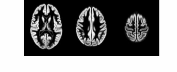
9 Belonging Probabilities Belonging probabilities are assigned by normalising to one. Non-Gaussian Intensity Distributions * Multiple Gaussians per tissue class allow non-gaussian intensity distributions to be modelled. * E.g. accounting for partial volume effects Modelling a Bias Field * A bias field is modelled as a linear combination of basis functions. Tissue Probability Maps * Tissue probability maps (TPMs) are used instead of the proportion of voxels in each Gaussian as the prior. Corrupted image Bias Field Corrected image ICBM Tissue Probabilistic Atlases. These tissue probability maps are kindly provided by the International Consortium for Brain Mapping, John C. Mazziotta and Arthur W. Toga.

 E = log ρi( β) K i= 1 k = 1 j= 1 γ jbij( α) 1 2πσ 2 k ( ρi( β) yi μk ) exp 2 2σk 2 Steepest Descent Start Spatially")

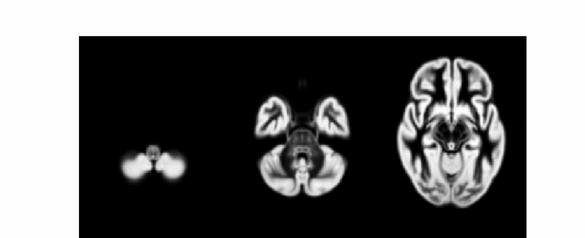
10 Deforming the Tissue Probability Maps * Tissue probability images are deformed so that they can be overlaid on top of the image to segment. Optimisation * The best parameters are those that minimise this objective function. * Optimisation involves finding them. * Begin with starting estimates, and repeatedly change them so that the objective function decreases each time. I K γ kbik( α) E = log ρi( β) K i= 1 k = 1 j= 1 γ jbij( α) 1 2πσ 2 k ( ρi( β) yi μk ) exp 2 2σk 2 Steepest Descent Start Spatially normalised BrainWeb phantoms (T1, T2 and PD) Optimum Tissue probability maps of GM and WM Alternate between optimising different groups of parameters Cocosco, Kollokian, Kwan & Evans. BrainWeb: Online Interface to a 3D MRI Simulated Brain Database. NeuroImage 5(4):S425 (1997)


11 Extensions for SPM8 * Additional tissue classes * Grey matter, white matter, CSF, skull, scalp. * Multi-channel Segmentation * More detailed nonlinear registration * More robust initial affine registration * Extra tissue class maps can be generated Image Intensity Distributions (T1-weighted MRI) Belonging Probabilities
12 Small deformation approximation


13 Forward and backward transforms Registration objective function * Simultaneously minimize the sum of: * Likelihood component * Drives the matching of the images. * Multinomial assumption * Prior component * A measure of deformation roughness * Regularises the registration. * ½u T Hu * A balance between the two terms.


14 Likelihood Model * Current DARTEL model is multinomial for matching tissue class images. * Template represents probability of obtaining different tissues at each point. log p(t μ,ϕ) = Σ j Σ k t jk log(μ k (ϕ j )) t μ ϕ individual GM, WM and background template GM, WM and background deformation Prior Model Simultaneous registration of GM to GM and WM to WM Subject 1 Grey matter White matter Subject 2 Grey matter White matter Grey matter White matter Template Grey matter White matter Grey matter White matter Subject 4 Subject 3 Template Iteratively generated from 471 subjects Began with rigidly aligned tissue probability maps Used an inverse consistent formulation Initial Average After a few iterations Final template


15 Grey matter average of 452 subjects affine Grey matter average of 471 subjects Initial GM images Warped GM images
16
17


18 Contents * Preliminaries * Intra-Subject Registration * Inter-Subject Registration * VBM Volumetry Spatial Normalisation T1-Weighted MRI Grey Matter Original Registered Template All subjects data are aligned with a common reference compare like with like
.")


19 Preserving Tissue Volumes Smoothing Each voxel after smoothing effectively becomes the result of applying a weighted region of interest (ROI). Before convolution Convolved with a circle Convolved with a Gaussian Warped without correction Warped with correction Jacobian Determinants Tissue class images are warped such that the volumes of tissue are preserved VBM Pre-processing * Use Segment button for characterising intensity distributions of tissue classes. * DARTEL import, to generate rigidly aligned grey and white matter maps. * DARTEL warping to generate modulated warped grey matter. * Smooth. * Statistics. New Segment Generate low resolution GM and WM images for each subject ( DARTEL imported ). Generate full resolution GM map for each subject.
20 Run DARTEL (create Templates) Simultaneously align DARTEL imported GM and WM for all subjects. Generates templates and parameterisations of relative shapes. Normalise to MNI Space Use shape parameterisations to generate smoothed Jacobian scaled and spatially normalised GM images for each subject. Possible Explanations for VBM Findings Mis-classify Mis-register Folding Thinning Thickening Mis-register Mis-classify References * Friston et al. Spatial registration and normalisation of images. Human Brain Mapping 3: (1995). * Collignon et al. Automated multi-modality image registration based on information theory. IPMI 95 pp (1995). * Ashburner et al. Incorporating prior knowledge into image registration. NeuroImage 6: (1997). * Ashburner & Friston. Nonlinear spatial normalisation using basis functions. Human Brain Mapping 7: (1999). * Thévenaz et al. Interpolation revisited. IEEE Trans. Med. Imaging 19: (2). * Andersson et al. Modeling geometric deformations in EPI time series. Neuroimage 13: (21). * Ashburner & Friston. Unified Segmentation. NeuroImage 26: (25). * Ashburner. A Fast Diffeomorphic Image Registration Algorithm. NeuroImage 38: (27).
Preprocessing II: Between Subjects John Ashburner
 Preprocessing II: Between Subjects John Ashburner Pre-processing Overview Statistics or whatever fmri time-series Anatomical MRI Template Smoothed Estimate Spatial Norm Motion Correct Smooth Coregister
Spatial Preprocessing
 Spatial Preprocessing Overview of SPM Analysis fmri time-series Design matrix Statistical Parametric Map John Ashburner john@fil.ion.ucl.ac.uk Motion Correction Smoothing General Linear Model Smoothing
Image Processing for fmri John Ashburner. Wellcome Trust Centre for Neuroimaging, 12 Queen Square, London, UK.
 Iage Processing for fmri John Ashburner Wellcoe Trust Centre for Neuroiaging, 12 Queen Square, London, UK. Contents * Preliinaries * Rigid-Body and Affine Transforations * Optiisation and Objective Functions
Preprocessing I: Within Subject John Ashburner
 Preprocessing I: Within Subject John Ashburner Pre-processing Overview Statistics or whatever fmri tie-series Anatoical MRI Teplate Soothed Estiate Spatial Nor Motion Correct Sooth Coregister 11 21 31
Computational Neuroanatomy
 Computational Neuroanatomy John Ashburner john@fil.ion.ucl.ac.uk Smoothing Motion Correction Between Modality Co-registration Spatial Normalisation Segmentation Morphometry Overview fmri time-series kernel
Methods for data preprocessing
 Methods for data preprocessing John Ashburner Wellcome Trust Centre for Neuroimaging, 12 Queen Square, London, UK. Overview Voxel-Based Morphometry Morphometry in general Volumetrics VBM preprocessing
fmri pre-processing Juergen Dukart
 fmri pre-processing Juergen Dukart Outline Why do we need pre-processing? fmri pre-processing Slice time correction Realignment Unwarping Coregistration Spatial normalisation Smoothing Overview fmri time-series
Neuroimaging and mathematical modelling Lesson 2: Voxel Based Morphometry
 Neuroimaging and mathematical modelling Lesson 2: Voxel Based Morphometry Nivedita Agarwal, MD Nivedita.agarwal@apss.tn.it Nivedita.agarwal@unitn.it Volume and surface morphometry Brain volume White matter
Functional MRI data preprocessing. Cyril Pernet, PhD
 Functional MRI data preprocessing Cyril Pernet, PhD Data have been acquired, what s s next? time No matter the design, multiple volumes (made from multiple slices) have been acquired in time. Before getting
1 Introduction Motivation and Aims Functional Imaging Computational Neuroanatomy... 12
 Contents 1 Introduction 10 1.1 Motivation and Aims....... 10 1.1.1 Functional Imaging.... 10 1.1.2 Computational Neuroanatomy... 12 1.2 Overview of Chapters... 14 2 Rigid Body Registration 18 2.1 Introduction.....
Introduction to fmri. Pre-processing
 Introduction to fmri Pre-processing Tibor Auer Department of Psychology Research Fellow in MRI Data Types Anatomical data: T 1 -weighted, 3D, 1/subject or session - (ME)MPRAGE/FLASH sequence, undistorted
Basic fmri Design and Analysis. Preprocessing
 Basic fmri Design and Analysis Preprocessing fmri Preprocessing Slice timing correction Geometric distortion correction Head motion correction Temporal filtering Intensity normalization Spatial filtering
SPM8 for Basic and Clinical Investigators. Preprocessing. fmri Preprocessing
 SPM8 for Basic and Clinical Investigators Preprocessing fmri Preprocessing Slice timing correction Geometric distortion correction Head motion correction Temporal filtering Intensity normalization Spatial
Functional MRI in Clinical Research and Practice Preprocessing
 Functional MRI in Clinical Research and Practice Preprocessing fmri Preprocessing Slice timing correction Geometric distortion correction Head motion correction Temporal filtering Intensity normalization
EPI Data Are Acquired Serially. EPI Data Are Acquired Serially 10/23/2011. Functional Connectivity Preprocessing. fmri Preprocessing
 Functional Connectivity Preprocessing Geometric distortion Head motion Geometric distortion Head motion EPI Data Are Acquired Serially EPI Data Are Acquired Serially descending 1 EPI Data Are Acquired
Data pre-processing framework in SPM. Bogdan Draganski
 Data pre-processing fraework in SPM Bogdan Draganski Outline Why do we need pre-processing? Overview Structural MRI pre-processing fmri pre-processing Why do we need pre-processing? What do we want? Reason
FIL SPM Course Spatial Preprocessing
 Registration, noralisation, segentation, VBM Matthan Caan, BIC/CSCA suer school 2012 Neuroiage, 2012 slides copied/adjusted fro: FIL SPM Course Spatial Preprocessing 1. smri 2. DTI 3. fmri By John Ashburner
SPM8 for Basic and Clinical Investigators. Preprocessing
 SPM8 for Basic and Clinical Investigators Preprocessing fmri Preprocessing Slice timing correction Geometric distortion correction Head motion correction Temporal filtering Intensity normalization Spatial
Basic principles of MR image analysis. Basic principles of MR image analysis. Basic principles of MR image analysis
 Basic principles of MR image analysis Basic principles of MR image analysis Julien Milles Leiden University Medical Center Terminology of fmri Brain extraction Registration Linear registration Non-linear
Zurich SPM Course Voxel-Based Morphometry. Ged Ridgway (Oxford & UCL) With thanks to John Ashburner and the FIL Methods Group
 With thanks to John Ashburner and the FIL Methods Group") Zurich SPM Course 2015 Voxel-Based Morphometry Ged Ridgway (Oxford & UCL) With thanks to John Ashburner and the FIL Methods Group Examples applications of VBM Many scientifically or clinically interesting
Zurich SPM Course 2015 Voxel-Based Morphometry Ged Ridgway (Oxford & UCL) With thanks to John Ashburner and the FIL Methods Group Examples applications of VBM Many scientifically or clinically interesting
Supplementary methods
 Supplementary methods This section provides additional technical details on the sample, the applied imaging and analysis steps and methods. Structural imaging Trained radiographers placed all participants
An Introduction To Automatic Tissue Classification Of Brain MRI. Colm Elliott Mar 2014
 An Introduction To Automatic Tissue Classification Of Brain MRI Colm Elliott Mar 2014 Tissue Classification Tissue classification is part of many processing pipelines. We often want to classify each voxel
Structural Segmentation
 Structural Segmentation FAST tissue-type segmentation FIRST sub-cortical structure segmentation FSL-VBM voxelwise grey-matter density analysis SIENA atrophy analysis FAST FMRIB s Automated Segmentation
Structural Segmentation
 Structural Segmentation FAST tissue-type segmentation FIRST sub-cortical structure segmentation FSL-VBM voxelwise grey-matter density analysis SIENA atrophy analysis FAST FMRIB s Automated Segmentation
Brain Extraction, Registration & EPI Distortion Correction
 Brain Extraction, Registration & EPI Distortion Correction What use is Registration? Some common uses of registration: Combining across individuals in group studies: including fmri & diffusion Quantifying
Fmri Spatial Processing
 Educational Course: Fmri Spatial Processing Ray Razlighi Jun. 8, 2014 Spatial Processing Spatial Re-alignment Geometric distortion correction Spatial Normalization Smoothing Why, When, How, Which Why is
Voxel-Based Morphometry & DARTEL. Ged Ridgway, London With thanks to John Ashburner and the FIL Methods Group
 Zurich SPM Course 2012 Voxel-Based Morphometry & DARTEL Ged Ridgway, London With thanks to John Ashburner and the FIL Methods Group Aims of computational neuroanatomy * Many interesting and clinically
Last Time. This Time. Thru-plane dephasing: worse at long TE. Local susceptibility gradients: thru-plane dephasing
 Motion Correction Last Time Mutual Information Optimiation Decoupling Translation & Rotation Interpolation SPM Example (Least Squares & MI) A Simple Derivation This Time Reslice example SPM Example : Remind
A Model-Independent, Multi-Image Approach to MR Inhomogeneity Correction
 Tina Memo No. 2007-003 Published in Proc. MIUA 2007 A Model-Independent, Multi-Image Approach to MR Inhomogeneity Correction P. A. Bromiley and N.A. Thacker Last updated 13 / 4 / 2007 Imaging Science and
UBC SPM Course Voxel-Based Morphometry & DARTEL. Ged Ridgway
 UBC SPM Course 2010 Voxel-Based Morphometry & DARTEL Ged Ridgway Aims of computational neuroanatomy * Many interesting and clinically important questions might relate to the shape or local size of regions
Nonrigid Registration using Free-Form Deformations
 Nonrigid Registration using Free-Form Deformations Hongchang Peng April 20th Paper Presented: Rueckert et al., TMI 1999: Nonrigid registration using freeform deformations: Application to breast MR images
Neuroimage Processing
 Neuroimage Processing Instructor: Moo K. Chung mkchung@wisc.edu Lecture 2-3. General Linear Models (GLM) Voxel-based Morphometry (VBM) September 11, 2009 What is GLM The general linear model (GLM) is a
White Pixel Artifact. Caused by a noise spike during acquisition Spike in K-space <--> sinusoid in image space
 White Pixel Artifact Caused by a noise spike during acquisition Spike in K-space sinusoid in image space Susceptibility Artifacts Off-resonance artifacts caused by adjacent regions with different
腦部結構影像 標準化 組織分割 體素型態 本週課程內容. Analysis Softwares. A Course of MRI
 本週課程內容 腦部結構影像 A Course of MRI 盧家鋒助理教授國立陽明大學物理治療暨輔助科技學系 alvin4016@ym.edu.tw 腦部結構影像 空間標準化 (Spatial normalization) 均勻度校正 (Bias correction) 組織分割 (Segmentation) 體素形態學分析 (Voxel-based morphometry, VBM) 影像平滑化
This Time. fmri Data analysis
 This Time Reslice example Spatial Normalization Noise in fmri Methods for estimating and correcting for physiologic noise SPM Example Spatial Normalization: Remind ourselves what a typical functional image
MR IMAGE SEGMENTATION
 MR IMAGE SEGMENTATION Prepared by : Monil Shah What is Segmentation? Partitioning a region or regions of interest in images such that each region corresponds to one or more anatomic structures Classification
RIGID IMAGE REGISTRATION
 RIGID IMAGE REGISTRATION Duygu Tosun-Turgut, Ph.D. Center for Imaging of Neurodegenerative Diseases Department of Radiology and Biomedical Imaging duygu.tosun@ucsf.edu What is registration? Image registration
Image Registration I
 Image Registration I Comp 254 Spring 2002 Guido Gerig Image Registration: Motivation Motivation for Image Registration Combine images from different modalities (multi-modality registration), e.g. CT&MRI,
Performance Evaluation of the TINA Medical Image Segmentation Algorithm on Brainweb Simulated Images
 Tina Memo No. 2008-003 Internal Memo Performance Evaluation of the TINA Medical Image Segmentation Algorithm on Brainweb Simulated Images P. A. Bromiley Last updated 20 / 12 / 2007 Imaging Science and
Where are we now? Structural MRI processing and analysis
 Where are we now? Structural MRI processing and analysis Pierre-Louis Bazin bazin@cbs.mpg.de Leipzig, Germany Structural MRI processing: why bother? Just use the standards? SPM FreeSurfer FSL However:
Automatic Registration-Based Segmentation for Neonatal Brains Using ANTs and Atropos
 Automatic Registration-Based Segmentation for Neonatal Brains Using ANTs and Atropos Jue Wu and Brian Avants Penn Image Computing and Science Lab, University of Pennsylvania, Philadelphia, USA Abstract.
VBM Tutorial. 1 Getting Started. John Ashburner. March 12, 2015
 VBM Tutorial John Ashburner March 12, 2015 1 Getting Started The data provided are a selection of T1-weighted scans from the freely available IXI dataset 1. The overall plan will be to Start up SPM. Check
2. Creating Field Maps Using the Field Map GUI (Version 2.0) in SPM5
 in SPM5") 1. Introduction This manual describes how to use the Field Map Toolbox Version 2.0 for creating unwrapped field maps that can be used to do geometric distortion correction of EPI images in SPM5. 1. 1.
1. Introduction This manual describes how to use the Field Map Toolbox Version 2.0 for creating unwrapped field maps that can be used to do geometric distortion correction of EPI images in SPM5. 1. 1.
Introduction to Medical Image Registration
 Introduction to Medical Image Registration Sailesh Conjeti Computer Aided Medical Procedures (CAMP), Technische Universität München, Germany sailesh.conjeti@tum.de Partially adapted from slides by: 1.
Normalization for clinical data
 Normalization for clinical data Christopher Rorden, Leonardo Bonilha, Julius Fridriksson, Benjamin Bender, Hans-Otto Karnath (2012) Agespecific CT and MRI templates for spatial normalization. NeuroImage
Spatial Normalization using Basis Functions
 Chapter 3 Spatial Normalization using Basis Functions John Ashburner & Karl J. Friston The Wellcome Dept. of Imaging Neuroscience, 12 Queen Square, London WC1N 3BG, UK. Contents 3.1 Introduction.................................
Introduction to MRI data processing with FSL. Anna Blazejewska
 Introduction to MRI data processing with FSL Anna Blazejewska FSL = FMRIB Software Library FMRIB = Functional Magnetic Resonance Imaging of the Brain @ Oxford since 2000, last stable FSL 5.0, free! for
SPM Introduction. SPM : Overview. SPM: Preprocessing SPM! SPM: Preprocessing. Scott Peltier. FMRI Laboratory University of Michigan
 SPM Introduction Scott Peltier FMRI Laboratory University of Michigan! Slides adapted from T. Nichols SPM! SPM : Overview Library of MATLAB and C functions Graphical user interface Four main components:
SPM Introduction SPM! Scott Peltier. FMRI Laboratory University of Michigan. Software to perform computation, manipulation and display of imaging data
 SPM Introduction Scott Peltier FMRI Laboratory University of Michigan Slides adapted from T. Nichols SPM! Software to perform computation, manipulation and display of imaging data 1 1 SPM : Overview Library
Structural MRI analysis
 Structural MRI analysis volumetry and voxel-based morphometry cortical thickness measurements structural covariance network mapping Boris Bernhardt, PhD Department of Social Neuroscience, MPI-CBS bernhardt@cbs.mpg.de
CHAPTER 2. Morphometry on rodent brains. A.E.H. Scheenstra J. Dijkstra L. van der Weerd
 CHAPTER 2 Morphometry on rodent brains A.E.H. Scheenstra J. Dijkstra L. van der Weerd This chapter was adapted from: Volumetry and other quantitative measurements to assess the rodent brain, In vivo NMR
Methodological progress in image registration for ventilation estimation, segmentation propagation and multi-modal fusion
 Methodological progress in image registration for ventilation estimation, segmentation propagation and multi-modal fusion Mattias P. Heinrich Julia A. Schnabel, Mark Jenkinson, Sir Michael Brady 2 Clinical
Automated MR Image Analysis Pipelines
 Automated MR Image Analysis Pipelines Andy Simmons Centre for Neuroimaging Sciences, Kings College London Institute of Psychiatry. NIHR Biomedical Research Centre for Mental Health at IoP & SLAM. Neuroimaging
Image Registration. Prof. Dr. Lucas Ferrari de Oliveira UFPR Informatics Department
 Image Registration Prof. Dr. Lucas Ferrari de Oliveira UFPR Informatics Department Introduction Visualize objects inside the human body Advances in CS methods to diagnosis, treatment planning and medical
GLM for fmri data analysis Lab Exercise 1
 GLM for fmri data analysis Lab Exercise 1 March 15, 2013 Medical Image Processing Lab Medical Image Processing Lab GLM for fmri data analysis Outline 1 Getting Started 2 AUDIO 1 st level Preprocessing
Math in image processing
 Math in image processing Math in image processing Nyquist theorem Math in image processing Discrete Fourier Transformation Math in image processing Image enhancement: scaling Math in image processing Image
Cocozza S., et al. : ALTERATIONS OF FUNCTIONAL CONNECTIVITY OF THE MOTOR CORTEX IN FABRY'S DISEASE: AN RS-FMRI STUDY
 ALTERATIONS OF FUNCTIONAL CONNECTIVITY OF THE MOTOR CORTEX IN FABRY'S DISEASE: AN RS-FMRI STUDY SUPPLEMENTARY MATERIALS Sirio Cocozza, MD 1*, Antonio Pisani, MD, PhD 2, Gaia Olivo, MD 1, Francesco Saccà,
Statistical Analysis of Neuroimaging Data. Phebe Kemmer BIOS 516 Sept 24, 2015
 Statistical Analysis of Neuroimaging Data Phebe Kemmer BIOS 516 Sept 24, 2015 Review from last time Structural Imaging modalities MRI, CAT, DTI (diffusion tensor imaging) Functional Imaging modalities
Analysis of fmri data within Brainvisa Example with the Saccades database
 Analysis of fmri data within Brainvisa Example with the Saccades database 18/11/2009 Note : All the sentences in italic correspond to informations relative to the specific dataset under study TP participants
Signal, Noise and Preprocessing*
 Translational Neuroodeling Unit SNR & Preproc Teporal Spatial General Realign Coreg Noralise Sooth Signal, Noise and Preprocessing* Methods and Models for fmri Analysis Septeber 25 th, 2015 Lars Kasper,
Journal of Articles in Support of The Null Hypothesis
 Data Preprocessing Martin M. Monti, PhD UCLA Psychology NITP 2016 Typical (task-based) fmri analysis sequence Image Pre-processing Single Subject Analysis Group Analysis Journal of Articles in Support
Spatial Normalization of Brain Images with Focal Lesions Using Cost Function Masking
 NeuroImage 14, 486 500 (2001) doi:10.1006/nimg.2001.0845, available online at http://www.idealibrary.com on Spatial Normalization of Brain Images with Focal Lesions Using Cost Function Masking Matthew
ADAPTIVE GRAPH CUTS WITH TISSUE PRIORS FOR BRAIN MRI SEGMENTATION
 ADAPTIVE GRAPH CUTS WITH TISSUE PRIORS FOR BRAIN MRI SEGMENTATION Abstract: MIP Project Report Spring 2013 Gaurav Mittal 201232644 This is a detailed report about the course project, which was to implement
Automatic Generation of Training Data for Brain Tissue Classification from MRI
 MICCAI-2002 1 Automatic Generation of Training Data for Brain Tissue Classification from MRI Chris A. Cocosco, Alex P. Zijdenbos, and Alan C. Evans McConnell Brain Imaging Centre, Montreal Neurological
HST.582J / 6.555J / J Biomedical Signal and Image Processing Spring 2007
 MIT OpenCourseWare http://ocw.mit.edu HST.582J / 6.555J / 16.456J Biomedical Signal and Image Processing Spring 2007 For information about citing these materials or our Terms of Use, visit: http://ocw.mit.edu/terms.
Norbert Schuff VA Medical Center and UCSF
 Norbert Schuff Medical Center and UCSF Norbert.schuff@ucsf.edu Medical Imaging Informatics N.Schuff Course # 170.03 Slide 1/67 Objective Learn the principle segmentation techniques Understand the role
Whole Body MRI Intensity Standardization
 Whole Body MRI Intensity Standardization Florian Jäger 1, László Nyúl 1, Bernd Frericks 2, Frank Wacker 2 and Joachim Hornegger 1 1 Institute of Pattern Recognition, University of Erlangen, {jaeger,nyul,hornegger}@informatik.uni-erlangen.de
Computational anatomy with the SPM software
 Available online at www.sciencedirect.com Magnetic Resonance Imaging 27 (2009) 1163 1174 Computational anatomy with the SPM software John Ashburner Wellcome Trust Centre for Neuroimaging, WC1N 3BG London,
Preprocessing of fmri data
 Preprocessing of fmri data Pierre Bellec CRIUGM, DIRO, UdM Flowchart of the NIAK fmri preprocessing pipeline fmri run 1 fmri run N individual datasets CIVET NUC, segmentation, spatial normalization slice
Noise and Artifacts in FMRI
 Noise and Artifacts in FMRI Instructor: Luis Hernandez-Garcia, Ph.D. Associate Research Professor FMRI Laboratory, Biomedical Engineering FMRI analysis - synopsis of what you ll do next week 1. Formulate
Automatic Generation of Training Data for Brain Tissue Classification from MRI
 Automatic Generation of Training Data for Brain Tissue Classification from MRI Chris A. COCOSCO, Alex P. ZIJDENBOS, and Alan C. EVANS http://www.bic.mni.mcgill.ca/users/crisco/ McConnell Brain Imaging
Non-rigid Image Registration
 Overview Non-rigid Image Registration Introduction to image registration - he goal of image registration - Motivation for medical image registration - Classification of image registration - Nonrigid registration
Manual image registration in BrainVoyager QX Table of Contents
 Manual image registration in BrainVoyager QX Table of Contents Manual image registration in BrainVoyager QX......1 Performing manual alignment for functional to anatomical images......2 Step 1: preparation......2
OnDemand3D Fusion Technology
 CYBERMED INC., ONDEMAND3D TECHNOLOGY INC. OnDemand3D Fusion Technology White Paper December 2009 USA Republic of Korea www.ondemand3d.com Introduction OnDemand3D TM Fusion is registration technology to
Spatial normalization of injured brains for neuroimaging research: An illustrative introduction of available options
 Spatial normalization of injured brains for neuroimaging research: An illustrative introduction of available options Junghoon Kim, PhD, Brian Avants, PhD, Sunil Patel, MS, and John Whyte, MD, PhD 1 Recent
MEDICAL IMAGE COMPUTING (CAP 5937) LECTURE 4: Pre-Processing Medical Images (II)
 LECTURE 4: Pre-Processing Medical Images (II)") SPRING 2016 1 MEDICAL IMAGE COMPUTING (CAP 5937) LECTURE 4: Pre-Processing Medical Images (II) Dr. Ulas Bagci HEC 221, Center for Research in Computer Vision (CRCV), University of Central Florida (UCF),
SPRING 2016 1 MEDICAL IMAGE COMPUTING (CAP 5937) LECTURE 4: Pre-Processing Medical Images (II) Dr. Ulas Bagci HEC 221, Center for Research in Computer Vision (CRCV), University of Central Florida (UCF),
FSL Pre-Processing Pipeline
 The Art and Pitfalls of fmri Preprocessing FSL Pre-Processing Pipeline Mark Jenkinson FMRIB Centre, University of Oxford FSL Pre-Processing Pipeline Standard pre-processing: Task fmri Resting-state fmri
Overview of Proposed TG-132 Recommendations
 Overview of Proposed TG-132 Recommendations Kristy K Brock, Ph.D., DABR Associate Professor Department of Radiation Oncology, University of Michigan Chair, AAPM TG 132: Image Registration and Fusion Conflict
Evaluation of multiple voxel-based morphometry approaches and applications in the analysis of white matter changes in temporal lobe epilepsy
 Evaluation of multiple voxel-based morphometry approaches and applications in the analysis of white matter changes in temporal lobe epilepsy Wenjing Li a, Huiguang He a, Jingjing Lu b, Bin Lv a, Meng Li
A Design Toolbox to Generate Complex Phantoms for the Evaluation of Medical Image Processing Algorithms
 A Design Toolbox to Generate Complex Phantoms for the Evaluation of Medical Image Processing Algorithms Omar Hamo, Georg Nelles, Gudrun Wagenknecht Central Institute for Electronics, Research Center Juelich,
Automatic segmentation of the cortical grey and white matter in MRI using a Region Growing approach based on anatomical knowledge
 Automatic segmentation of the cortical grey and white matter in MRI using a Region Growing approach based on anatomical knowledge Christian Wasserthal 1, Karin Engel 1, Karsten Rink 1 und André Brechmann
Medical Image Synthesis Methods and Applications
 MR Intensity Scale is Arbitrary This causes problems in most postprocessing methods Inconsistency or algorithm failure 11/5/2015 2 Joint Histogram 1.5 T GE SPGR 3 T Philips MPRAGE 11/5/2015 3 Problem With
Spline-Based Probabilistic Model for Anatomical Landmark Detection
 Spline-Based Probabilistic Model for Anatomical Landmark Detection Camille Izard 1,2, Bruno Jedynak 1,2, and Craig E.L. Stark 3 1 Laboratoire Paul Painlevé, Université des Sciences et Technologies de Lille,
ECSE 626 Project Report Multimodality Image Registration by Maximization of Mutual Information
 ECSE 626 Project Report Multimodality Image Registration by Maximization of Mutual Information Emmanuel Piuze McGill University Montreal, Qc, Canada. epiuze@cim.mcgill.ca Abstract In 1997, Maes et al.
Learning-based Neuroimage Registration
 Learning-based Neuroimage Registration Leonid Teverovskiy and Yanxi Liu 1 October 2004 CMU-CALD-04-108, CMU-RI-TR-04-59 School of Computer Science Carnegie Mellon University Pittsburgh, PA 15213 Abstract
Registration by continuous optimisation. Stefan Klein Erasmus MC, the Netherlands Biomedical Imaging Group Rotterdam (BIGR)
") Registration by continuous optimisation Stefan Klein Erasmus MC, the Netherlands Biomedical Imaging Group Rotterdam (BIGR) Registration = optimisation C t x t y 1 Registration = optimisation C t x t y
Registration by continuous optimisation Stefan Klein Erasmus MC, the Netherlands Biomedical Imaging Group Rotterdam (BIGR) Registration = optimisation C t x t y 1 Registration = optimisation C t x t y
User s Guide Neuroimage Processing ToolKit (NPTK) Version.1.7 (beta) fmri Registration Software Pipeline for Functional Localization
 Version.1.7 (beta) fmri Registration Software Pipeline for Functional Localization") User s Guide Neuroimage Processing ToolKit (NPTK) Version.1.7 (beta) fmri Registration Software Pipeline for Functional Localization Software Written by Ali Gholipour SIP Lab, UTD, 2005-2007 Revision 1.7
User s Guide Neuroimage Processing ToolKit (NPTK) Version.1.7 (beta) fmri Registration Software Pipeline for Functional Localization Software Written by Ali Gholipour SIP Lab, UTD, 2005-2007 Revision 1.7
Multiple Model Estimation : The EM Algorithm & Applications
 Multiple Model Estimation : The EM Algorithm & Applications Princeton University COS 429 Lecture Dec. 4, 2008 Harpreet S. Sawhney hsawhney@sarnoff.com Plan IBR / Rendering applications of motion / pose
Data mining for neuroimaging data. John Ashburner
 Data mining for neuroimaging data John Ashburner MODELLING The Scientific Process MacKay, David JC. Bayesian interpolation. Neural computation 4, no. 3 (1992): 415-447. Model Selection Search for the best
The simulator can be applied in a number of diverse applications which span both
 Chapter 6 Simulator applications The simulator can be applied in a number of diverse applications which span both MRI and FMRI fields These applications include the simulation and removal of various imaging
ANALYSIS OF FUNCTIONAL MAGNETIC RESONANCE IMAGING DATA USING SPM99: VOXEL-BASED MORPHOMETRY DONNA ROSE ADDIS
 Donna Rose Addis, TWRI, May 2004 1 ANALYSIS OF FUNCTIONAL MAGNETIC RESONANCE IMAGING DATA USING SPM99: VOXEL-BASED MORPHOMETRY DONNA ROSE ADDIS DEPT. OF PSYCHOLOGY, UNIVERSITY OF TORONTO TORONTO WESTERN
Image Warping. Srikumar Ramalingam School of Computing University of Utah. [Slides borrowed from Ross Whitaker] 1
![Image Warping. Srikumar Ramalingam School of Computing University of Utah. [Slides borrowed from Ross Whitaker] 1](/thumbs/78/77189591.jpg "Image Warping. Srikumar Ramalingam School of Computing University of Utah. [Slides borrowed from Ross Whitaker] 1") Image Warping Srikumar Ramalingam School of Computing University of Utah [Slides borrowed from Ross Whitaker] 1 Geom Trans: Distortion From Optics Barrel Distortion Pincushion Distortion Straight lines
Image Warping Srikumar Ramalingam School of Computing University of Utah [Slides borrowed from Ross Whitaker] 1 Geom Trans: Distortion From Optics Barrel Distortion Pincushion Distortion Straight lines
Introduction to Neuroimaging Janaina Mourao-Miranda
 Introduction to Neuroimaging Janaina Mourao-Miranda Neuroimaging techniques have changed the way neuroscientists address questions about functional anatomy, especially in relation to behavior and clinical
Robust Realignment of fmri Time Series Data
 Robust Realignment of fmri Time Series Data Ben Dodson bjdodson@stanford.edu Olafur Gudmundsson olafurg@stanford.edu December 12, 2008 Abstract FMRI data has become an increasingly popular source for exploring
MRI Segmentation. MRI Bootcamp, 14 th of January J. Miguel Valverde
 MRI Segmentation MRI Bootcamp, 14 th of January 2019 Segmentation Segmentation Information Segmentation Algorithms Approach Types of Information Local 4 45 100 110 80 50 76 42 27 186 177 120 167 111 56
Good Morning! Thank you for joining us
 Good Morning! Thank you for joining us Deformable Registration, Contour Propagation and Dose Mapping: 101 and 201 Marc Kessler, PhD, FAAPM The University of Michigan Conflict of Interest I receive direct
for Images A Bayesian Deformation Model
 Statistics in Imaging Workshop July 8, 2004 A Bayesian Deformation Model for Images Sining Chen Postdoctoral Fellow Biostatistics Division, Dept. of Oncology, School of Medicine Outline 1. Introducing
Norbert Schuff Professor of Radiology VA Medical Center and UCSF
 Norbert Schuff Professor of Radiology Medical Center and UCSF Norbert.schuff@ucsf.edu 2010, N.Schuff Slide 1/67 Overview Definitions Role of Segmentation Segmentation methods Intensity based Shape based
l ealgorithms for Image Registration
 FAIR: exib Image Registration l F l ealgorithms for Jan Modersitzki Computing And Software, McMaster University 1280 Main Street West, Hamilton On, L8S 4K1, Canada modersit@cas.mcmaster.ca August 13, 2008
Detecting Changes In Non-Isotropic Images
 Detecting Changes In Non-Isotropic Images K.J. Worsley 1, M. Andermann 1, T. Koulis 1, D. MacDonald, 2 and A.C. Evans 2 August 4, 1999 1 Department of Mathematics and Statistics, 2 Montreal Neurological