UBC SPM Course Voxel-Based Morphometry & DARTEL. Ged Ridgway
|
|
|
- Shona Hubbard
- 6 years ago
- Views:
Transcription
1 UBC SPM Course 2010 Voxel-Based Morphometry & DARTEL Ged Ridgway
2 Aims of computational neuroanatomy * Many interesting and clinically important questions might relate to the shape or local size of regions of the brain * For example, whether (and where) local patterns of brain morphometry help to:? Distinguish schizophrenics from healthy controls? Understand plasticity, e.g. when learning new skills? Explain the changes seen in development and aging? Differentiate degenerative disease from healthy aging? Evaluate subjects on drug treatments versus placebo


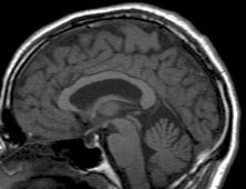
3 Alzheimer s Disease example Baseline Image Standard clinical MRI 1.5T T1 SPGR 1x1x1.5mm voxels Repeat image 12 month follow-up rigidly registered Subtraction image
4 Alzheimer s Disease example * Some changes are apparent in this patient... * Some might be noise or misregistration * Perhaps confounding biological effects like hydration changes * But some might genuinely reflect underlying AD pathology... * If we acquired more than two time-points, we could rule out some of the potential confounds * Would changes generalise from the patient to the disease? * Many morphological questions are not longitudinal, e.g. IQ, sex * It is appealing to try a second-level SPM analysis of structural data variation over subjects * E.g. AD vs. healthy, male vs. female or a correlation with IQ
5 SPM for group fmri Group-wise statistics fmri time-series Preprocessing Stat. modelling Results query Contrast spm T Image fmri time-series Preprocessing Stat. modelling Results query Contrast Image fmri time-series Preprocessing Stat. modelling Results query Contrast Image


6 SPM for structural MRI High-res T1 MRI? Group-wise statistics? High-res T1 MRI? High-res T1 MRI?
7 The need for tissue segmentation * High-resolution MRI reveals fine structural detail in the brain, but not all of it reliable or interesting * Noise, intensity-inhomogeneity, vasculature, * MR Intensity is usually not quantitatively meaningful (in the same way that e.g. CT is) * fmri time-series allow signal changes to be analysed statistically, compared to baseline or global values * Regional volumes of the three main tissue types: gray matter, white matter and CSF, are well-defined and potentially very interesting * Other aspects (and other sequences) can also be of interest
8 Voxel-Based Morphometry * In essence VBM is Statistical Parametric Mapping of regional segmented tissue density or volume * The exact interpretation of gray matter density or volume is complicated, and depends on the preprocessing steps used * It is not interpretable as neuronal packing density or other cytoarchitectonic tissue properties * The hope is that changes in these microscopic properties may lead to macro- or mesoscopic VBM-detectable differences
9 A brief history of VBM * A Voxel-Based Method for the Statistical Analysis of Gray and White Matter Density Wright, McGuire, Poline, Travere, Murrary, Frith, Frackowiak and Friston. NeuroImage 2(4), 1995 (!) * Rigid reorientation (by eye), semi-automatic scalp editing and segmentation, 8mm smoothing, SPM statistics, global covars. * Voxel-Based Morphometry The Methods. Ashburner and Friston. NeuroImage 11(6 pt.1), 2000 * Non-linear spatial normalisation, automatic segmentation * Thorough consideration of assumptions and confounds
10 A brief history of VBM * A Voxel-Based Morphometric Study of Ageing Good, Johnsrude, Ashburner, Henson and Friston. NeuroImage 14(1), 2001 * Optimised GM-normalisation ( a half-baked procedure ) * Unified Segmentation. Ashburner and Friston. NeuroImage 26(3), 2005 * Principled generative model for segmentation using deformable priors * A Fast Diffeomorphic Image Registration Algorithm. Ashburner. Neuroimage 38(1), 2007 * Large deformation normalisation to average shape templates *
11 VBM overview * Unified segmentation and spatial normalisation * More flexible groupwise normalisation using DARTEL * [Optional] modulation with Jacobian determinant * Optional computation of tissue totals/globals * Gaussian smoothing * Voxel-wise statistical analysis


12 VBM in pictures Segment Normalise


13 VBM in pictures Segment Normalise Modulate (?) Smooth

 Smooth")
14 VBM in pictures Segment Normalise Modulate (?) Smooth Voxel-wise statistics


")
15 VBM in pictures Segment Normalise Modulate (?) Smooth Voxel-wise statistics
16 VBM Subtleties * Whether to modulate * How much to smooth * Interpreting results * Adjusting for total GM or Intracranial Volume * Limitations of linear correlation * Statistical validity
17 Modulation Native 1 1 intensity = tissue density * Multiplication of the warped (normalised) tissue intensities so that their regional or global volume is preserved * Can detect differences in completely registered areas * Otherwise, we preserve concentrations, and are detecting mesoscopic effects that remain after approximate registration has removed the macroscopic effects * Flexible (not necessarily perfect ) registration may not leave any such differences Unmodulated Modulated 2/3 1/3 1/3 2/3
18 Modulation tutorial X = x 2 X = dx/dx = 2x X (2.5) = 5 Available from
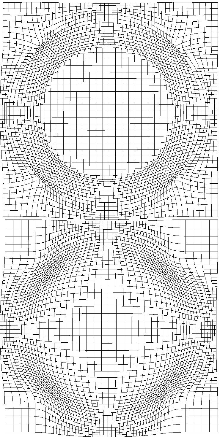
19 Modulation tutorial Square area = (p+q)(r+s) = pr+ps+qr+qs Red area = Square cyan magenta green = pr+ps+qr+qs 2qr qs pr = ps qr
20 VBM Subtleties * Whether to modulate * How much to smooth * Interpreting results * Adjusting for total GM or Intracranial Volume * Limitations of linear correlation * Statistical validity
21 Smoothing * The analysis will be most sensitive to effects that match the shape and size of the kernel * The data will be more Gaussian and closer to a continuous random field for larger kernels * Results will be rough and noise-like if too little smoothing is used * Too much will lead to distributed, indistinct blobs


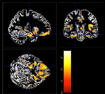
22 Smoothing * Between 7 and 14mm is probably reasonable * (DARTEL s greater precision allows less smoothing) * The results below show two fairly extreme choices, 5mm on the left, and 16mm, right


23 Interpreting findings Mis-classify Mis-register Folding Thickening Thinning Mis-register Mis-classify
24 Globals for VBM * Shape is really a multivariate concept * Dependencies among volumes in different regions * SPM is mass univariate * Combining voxel-wise information with global integrated tissue volume provides a compromise * Using either ANCOVA or proportional scaling (ii) is globally thicker, but locally thinner than (i) either of these effects may be of interest to us. Fig. from: Voxel-based morphometry of the human brain Mechelli, Price, Friston and Ashburner. Current Medical Imaging Reviews 1(2), 2005.
25 Total Intracranial Volume (TIV/ICV) * Global integrated tissue volume may be correlated with interesting regional effects * Correcting for globals in this case may overly reduce sensitivity to local differences * Total intracranial volume integrates GM, WM and CSF, or attempts to measure the skull-volume directly * Not sensitive to global reduction of GM+WM (cancelled out by CSF expansion skull is fixed!) * Correcting for TIV in VBM statistics may give more powerful and/or more interpretable results * See also Pell et al (2009) doi: /j.neuroimage

26 Nonlinearity Caution may be needed when interpreting linear relationships between grey matter concentrations and some covariate of interest. Circles of uniformly increasing area. Smoothed Plot of intensity at circle centres versus area
27 VBM s statistical validity * Residuals are not normally distributed * Little impact on uncorrected statistics for experiments comparing reasonably sized groups * Probably invalid for experiments that compare single subjects or tiny patient groups with a larger control group * Mitigate with large amounts of smoothing * Or use nonparametric tests that make fewer assumptions, e.g. permutation testing with SnPM
28 VBM s statistical validity * Correction for multiple comparisons * RFT correction based on peak heights should be fine * Correction using cluster extents is problematic * SPM usually assumes that the smoothness of the residuals is spatially stationary * VBM residuals have spatially varying smoothness * Bigger blobs expected in smoother regions * Cluster-based correction accounting for nonstationary smoothness is under development * See also Satoru Hayasaka s nonstationarity toolbox
29 VBM s statistical validity * False discovery rate * Less conservative than FWE * Popular in morphometric work * (almost universal for cortical thickness in FreeSurfer) * Recently questioned * Topological FDR (for clusters and peaks) * See SPM8 release notes and Justin s papers * *
30 Longitudinal VBM * The simplest method for longitudinal VBM is to use cross-sectional preprocessing, but longitudinal statistical analyses * Standard preprocessing not optimal, but unbiased * Non-longitudinal statistics would be severely biased * (Estimates of standard errors would be too small) * Simplest longitudinal statistical analysis: two-stage summary statistic approach (common in fmri) * Within subject longitudinal differences or beta estimates from linear regressions against time
31 Longitudinal VBM variations * Intra-subject registration over time is much more accurate than inter-subject normalisation * Different approaches suggested to capitalise * A simple approach is to apply one set of normalisation parameters (e.g. Estimated from baseline images) to both baseline and repeat(s) * Draganski et al (2004) Nature 427: * Voxel Compression mapping separates expansion and contraction before smoothing * Scahill et al (2002) PNAS 99:
32 Longitudinal VBM variations * Can also multiply longitudinal volume change with baseline or average grey matter density * Chételat et al (2005) NeuroImage 27: * Kipps et al (2005) JNNP 76:650 * Hobbs et al (2009) doi: /jnnp * Note that use of baseline (or repeat) instead of average might lead to bias * Thomas et al (2009) doi: /j.neuroimage * Unfortunately, the explanations in this reference relating to interpolation differences are not quite right... there are several open questions here...
33 Spatial normalisation with DARTEL * VBM is crucially dependent on registration performance * The limited flexibility of DCT normalisation has been criticised * Inverse transformations are useful, but not always well-defined * More flexible registration requires careful modelling and regularisation (prior belief about reasonable warping) * MNI/ICBM templates/priors are not universally representative * The DARTEL toolbox combines several methodological advances to address these limitations
34 Mathematical advances in registration * Large deformation concept * Regularise velocity not displacement * (syrup instead of elastic) * Leads to concept of geodesic * Provides a metric for distance between shapes * Geodesic or Riemannian average = mean shape * If velocity assumed constant computation is fast * Ashburner (2007) NeuroImage 38: * DARTEL toolbox in SPM8 * Currently initialised from unified seg_sn.mat files
35 Motivation for using DARTEL * Recent papers comparing different approaches have favoured more flexible methods * DARTEL usually outperforms DCT normalisation * Also comparable to the best algorithms from other software packages (though note that DARTEL and others have many tunable parameters...) * Klein et al. (2009) is a particularly thorough comparison, using expert segmentations * Results summarised in the next slide

36 Part of Fig. 1 in Klein et al. Part of Fig. 5 in Klein et al.
37 Spatial normalisation with DARTEL * VBM is crucially dependent on registration performance * The limited flexibility of DCT normalisation has been criticised * Inverse transformations are useful, but not always well-defined * More flexible registration requires careful modelling and regularisation (prior belief about reasonable warping) * MNI/ICBM templates/priors are not universally representative * The DARTEL toolbox combines several methodological advances to address these limitations
 (x) = x 1 * φ (1) (x) = u(φ (t) (x))dt t=0 * Scaling and squaring is used to generate deformations * Inverse simply")
38 DARTEL * Parameterising the deformation * u is a flow field to be estimated * 3 (x,y,z) DF per 1.5mm cubic voxel * 10^6 DF vs. 10^3 DCT bases * φ (0) (x) = x 1 * φ (1) (x) = u(φ (t) (x))dt t=0 * Scaling and squaring is used to generate deformations * Inverse simply integrates -u

39 Fig.5 in DARTEL paper
40 Registration objective function * Likelihood component * Drives the matching of the images. * Multinomial assumption * Prior component * A measure of deformation roughness * Regularises the registration * ½u T Hu * Need to choose H and a balance between the two terms
41 Likelihood Model * Current DARTEL model is multinomial for matching tissue class images. * Template represents probability of obtaining different tissues at each point. * log p(t µ,ϕ) = Σ j Σ k t jk log(µ k (ϕ j )) t individual GM, WM and background µ template GM, WM and background ϕ deformation

42 Prior Models
43 A word of caution * Different models have different parameterisations and will therefore give different findings * Shape models (image registration models) are no exception * Need to have a good model to reliably report details about differences among parameters * Not always easy to determine good/best model * Bayesian model comparison not yet feasible for Dartel * Classification or prediction are useful; work in progress...

44 Example geodesic shape average Average on Riemannian manifold Uses average flow field Linear Average (Not on Riemannian manifold)
45 Simultaneous registration of GM to GM and WM to WM, for a group of subjects Subject 1 Grey matter White matter Grey matter White matter Subject 3 Grey matter White matter Subject 2 Grey matter White matter Template Grey matter White matter Subject 4
 Average of mwc1 using segment/dct")
46 DARTEL average template evolution Template 1 Rigid average (Template_0) Average of mwc1 using segment/dct Template 6

47
48
49 Summary * VBM performs voxel-wise statistical analysis on smoothed (modulated) normalised tissue segments * SPM8 performs segmentation and spatial normalisation in a unified generative model * Based on Gaussian mixture modelling, with DCT-warped spatial priors, and multiplicative bias field * The new segment toolbox includes non-brain priors and more flexible/precise warping of them * Subsequent (currently non-unified) use of DARTEL improves normalisation for VBM * And perhaps also fmri...

 MNI")
50 Preprocessing overview fmri time-series Anatomical MRI TPMs Input Output Segmentation Transformation (seg_sn.mat) Kernel REALIGN COREG SEGMENT NORM WRITE SMOOTH Motion corrected Mean functional (Headers changed) MNI Space ANALYSIS

51 Preprocessing with Dartel fmri time-series Anatomical MRI TPMs... DARTEL CREATE TEMPLATE REALIGN COREG SEGMENT DARTEL NORM 2 MNI & SMOOTH Motion corrected Mean functional (Headers changed) ANALYSIS
52 Mathematical advances in computational anatomy * VBM is well-suited to find focal volumetric differences * Assumes independence among voxels * Not very biologically plausible * But shows differences that are easy to interpret * Some anatomical differences can not be localised * Need multivariate models * Differences in terms of proportions among measurements * Where would the difference between male and female faces be localised?
53 Mathematical advances in computational anatomy * In theory, assumptions about structural covariance among brain regions are more biologically plausible * Form influenced by spatio-temporal modes of gene expression * Empirical evidence, e.g. * Mechelli, Friston, Frackowiak & Price. Structural covariance in the human cortex. Journal of Neuroscience 25: (2005) * Recent introductory review: * Ashburner & Klöppel. Multivariate models of inter-subject anatomical variability. NeuroImage, In press.
54 Conclusion * VBM uses the machinery of SPM to localise patterns in regional volumetric variation * Use of globals as covariates is a step towards multivariate modelling of volume and shape * More advanced approaches typically benefit from the same preprocessing methods * New segmentation and DARTEL close to state of the art * Though possibly little or no smoothing * Elegant mathematics related to transformations (diffeomorphism group with Riemannian metric) * VBM easier interpretation complementary role
55 Key references for VBM * Ashburner & Friston. Unified Segmentation. NeuroImage 26: (2005). * Mechelli et al. Voxel-based morphometry of the human brain Current Medical Imaging Reviews 1(2) (2005). * Ashburner. A Fast Diffeomorphic Image Registration Algorithm. NeuroImage 38: (2007). * Ashburner & Friston. Computing average shaped tissue probability templates. NeuroImage 45(2): (2009).
56 References for more advanced computational anatomy * Ashburner, Hutton, Frackowiak, Johnsrude, Price & Friston. Identifying global anatomical differences: deformation-based morphometry. Human Brain Mapping 6(5-6): , * Bishop. Pattern recognition and machine learning * Younes, Arrate & Miller. Evolutions equations in computational anatomy. NeuroImage 45(1):S40-S50, * Ashburner & Klöppel. Multivariate models of intersubject anatomical variability. NeuroImage, In press.
57 EXTRA MATERIAL
 * This can be removed automatically using simple morphological filtering operations * Erosion * Conditional dilation")
58 Segmentation clean-up * Results may contain some non-brain tissue (dura, scalp, etc.) * This can be removed automatically using simple morphological filtering operations * Erosion * Conditional dilation Lower segmentations have been cleaned up

59 The new segmentation toolbox * An extended work-in-progress algorithm * Multi-spectral * New TPMs including different tissues * Reduces problems in non-brain tissue * New more flexible warping of TPMs * More precise and more sharp/contrasty results

60 New Segmentation TPMs Segment button New Seg Toolbox
61 New Segmentation registration Segment button * 9*10*9 * 3 = 2430 New Seg Toolbox * 59*70*59 * 3 =

62 New Segmentation results Segment button New Seg Toolbox
63 Limitations of the current model * Assumes that the brain consists of only the tissues modelled by the TPMs * No spatial knowledge of lesions (stroke, tumours, etc) * Prior probability model is based on relatively young and healthy brains * Less appropriate for subjects outside this population * Needs reasonable quality images to work with * No severe artefacts * Good separation of intensities * Good initial alignment with TPMs...
64 Possible future extensions * Deeper Bayesian philosophy * E.g. priors over means and variances * Marginalisation of nuisance variables * Model comparison, e.g. for numbers of Gaussians * Groupwise model (enormous!) * Combination with DARTEL (see later) * More tissue priors e.g. deep grey, meninges, etc. * Imaging physics * See Fischl et al. (2004), as cited in A&F (2005) introduction
Voxel-Based Morphometry & DARTEL. Ged Ridgway, London With thanks to John Ashburner and the FIL Methods Group
 Zurich SPM Course 2012 Voxel-Based Morphometry & DARTEL Ged Ridgway, London With thanks to John Ashburner and the FIL Methods Group Aims of computational neuroanatomy * Many interesting and clinically
Zurich SPM Course Voxel-Based Morphometry. Ged Ridgway (Oxford & UCL) With thanks to John Ashburner and the FIL Methods Group
 With thanks to John Ashburner and the FIL Methods Group") Zurich SPM Course 2015 Voxel-Based Morphometry Ged Ridgway (Oxford & UCL) With thanks to John Ashburner and the FIL Methods Group Examples applications of VBM Many scientifically or clinically interesting
Zurich SPM Course 2015 Voxel-Based Morphometry Ged Ridgway (Oxford & UCL) With thanks to John Ashburner and the FIL Methods Group Examples applications of VBM Many scientifically or clinically interesting
Methods for data preprocessing
 Methods for data preprocessing John Ashburner Wellcome Trust Centre for Neuroimaging, 12 Queen Square, London, UK. Overview Voxel-Based Morphometry Morphometry in general Volumetrics VBM preprocessing
Neuroimaging and mathematical modelling Lesson 2: Voxel Based Morphometry
 Neuroimaging and mathematical modelling Lesson 2: Voxel Based Morphometry Nivedita Agarwal, MD Nivedita.agarwal@apss.tn.it Nivedita.agarwal@unitn.it Volume and surface morphometry Brain volume White matter
Preprocessing II: Between Subjects John Ashburner
 Preprocessing II: Between Subjects John Ashburner Pre-processing Overview Statistics or whatever fmri time-series Anatomical MRI Template Smoothed Estimate Spatial Norm Motion Correct Smooth Coregister
Image Registration + Other Stuff
 Image Registration + Other Stuff John Ashburner Pre-processing Overview fmri time-series Motion Correct Anatomical MRI Coregister m11 m 21 m 31 m12 m13 m14 m 22 m 23 m 24 m 32 m 33 m 34 1 Template Estimate
Computational Neuroanatomy
 Computational Neuroanatomy John Ashburner john@fil.ion.ucl.ac.uk Smoothing Motion Correction Between Modality Co-registration Spatial Normalisation Segmentation Morphometry Overview fmri time-series kernel
Structural Segmentation
 Structural Segmentation FAST tissue-type segmentation FIRST sub-cortical structure segmentation FSL-VBM voxelwise grey-matter density analysis SIENA atrophy analysis FAST FMRIB s Automated Segmentation
Structural Segmentation
 Structural Segmentation FAST tissue-type segmentation FIRST sub-cortical structure segmentation FSL-VBM voxelwise grey-matter density analysis SIENA atrophy analysis FAST FMRIB s Automated Segmentation
Supplementary methods
 Supplementary methods This section provides additional technical details on the sample, the applied imaging and analysis steps and methods. Structural imaging Trained radiographers placed all participants
CHAPTER 2. Morphometry on rodent brains. A.E.H. Scheenstra J. Dijkstra L. van der Weerd
 CHAPTER 2 Morphometry on rodent brains A.E.H. Scheenstra J. Dijkstra L. van der Weerd This chapter was adapted from: Volumetry and other quantitative measurements to assess the rodent brain, In vivo NMR
Structural MRI analysis
 Structural MRI analysis volumetry and voxel-based morphometry cortical thickness measurements structural covariance network mapping Boris Bernhardt, PhD Department of Social Neuroscience, MPI-CBS bernhardt@cbs.mpg.de
1 Introduction Motivation and Aims Functional Imaging Computational Neuroanatomy... 12
 Contents 1 Introduction 10 1.1 Motivation and Aims....... 10 1.1.1 Functional Imaging.... 10 1.1.2 Computational Neuroanatomy... 12 1.2 Overview of Chapters... 14 2 Rigid Body Registration 18 2.1 Introduction.....
Evaluation of multiple voxel-based morphometry approaches and applications in the analysis of white matter changes in temporal lobe epilepsy
 Evaluation of multiple voxel-based morphometry approaches and applications in the analysis of white matter changes in temporal lobe epilepsy Wenjing Li a, Huiguang He a, Jingjing Lu b, Bin Lv a, Meng Li
An Introduction To Automatic Tissue Classification Of Brain MRI. Colm Elliott Mar 2014
 An Introduction To Automatic Tissue Classification Of Brain MRI Colm Elliott Mar 2014 Tissue Classification Tissue classification is part of many processing pipelines. We often want to classify each voxel
The Anatomical Equivalence Class Formulation and its Application to Shape-based Computational Neuroanatomy
 The Anatomical Equivalence Class Formulation and its Application to Shape-based Computational Neuroanatomy Sokratis K. Makrogiannis, PhD From post-doctoral research at SBIA lab, Department of Radiology,
Computational anatomy with the SPM software
 Available online at www.sciencedirect.com Magnetic Resonance Imaging 27 (2009) 1163 1174 Computational anatomy with the SPM software John Ashburner Wellcome Trust Centre for Neuroimaging, WC1N 3BG London,
Introduction to fmri. Pre-processing
 Introduction to fmri Pre-processing Tibor Auer Department of Psychology Research Fellow in MRI Data Types Anatomical data: T 1 -weighted, 3D, 1/subject or session - (ME)MPRAGE/FLASH sequence, undistorted
Form follows func-on. Which one of them can fly? And why? Why would anyone bother about brain structure, when we can do func5onal imaging?
 Why would anyone bother about brain structure, when we can do func5onal imaging? Form follows func-on Which one of them can fly? And why? h;p://animals.na5onalgeographic.com Why would anyone bother about
VBM Tutorial. 1 Getting Started. John Ashburner. March 12, 2015
 VBM Tutorial John Ashburner March 12, 2015 1 Getting Started The data provided are a selection of T1-weighted scans from the freely available IXI dataset 1. The overall plan will be to Start up SPM. Check
Functional MRI data preprocessing. Cyril Pernet, PhD
 Functional MRI data preprocessing Cyril Pernet, PhD Data have been acquired, what s s next? time No matter the design, multiple volumes (made from multiple slices) have been acquired in time. Before getting
Data mining for neuroimaging data. John Ashburner
 Data mining for neuroimaging data John Ashburner MODELLING The Scientific Process MacKay, David JC. Bayesian interpolation. Neural computation 4, no. 3 (1992): 415-447. Model Selection Search for the best
Neuroimage Processing
 Neuroimage Processing Instructor: Moo K. Chung mkchung@wisc.edu Lecture 2-3. General Linear Models (GLM) Voxel-based Morphometry (VBM) September 11, 2009 What is GLM The general linear model (GLM) is a
Quantitative MRI of the Brain: Investigation of Cerebral Gray and White Matter Diseases
 Quantities Measured by MR - Quantitative MRI of the Brain: Investigation of Cerebral Gray and White Matter Diseases Static parameters (influenced by molecular environment): T, T* (transverse relaxation)
Functional MRI in Clinical Research and Practice Preprocessing
 Functional MRI in Clinical Research and Practice Preprocessing fmri Preprocessing Slice timing correction Geometric distortion correction Head motion correction Temporal filtering Intensity normalization
Basic fmri Design and Analysis. Preprocessing
 Basic fmri Design and Analysis Preprocessing fmri Preprocessing Slice timing correction Geometric distortion correction Head motion correction Temporal filtering Intensity normalization Spatial filtering
MULTIVARIATE ANALYSES WITH fmri DATA
 MULTIVARIATE ANALYSES WITH fmri DATA Sudhir Shankar Raman Translational Neuromodeling Unit (TNU) Institute for Biomedical Engineering University of Zurich & ETH Zurich Motivation Modelling Concepts Learning
SPM8 for Basic and Clinical Investigators. Preprocessing. fmri Preprocessing
 SPM8 for Basic and Clinical Investigators Preprocessing fmri Preprocessing Slice timing correction Geometric distortion correction Head motion correction Temporal filtering Intensity normalization Spatial
Fmri Spatial Processing
 Educational Course: Fmri Spatial Processing Ray Razlighi Jun. 8, 2014 Spatial Processing Spatial Re-alignment Geometric distortion correction Spatial Normalization Smoothing Why, When, How, Which Why is
Appendix E1. Supplementary Methods. MR Image Acquisition. MR Image Analysis
 RSNA, 2015 10.1148/radiol.2015150532 Appendix E1 Supplementary Methods MR Image Acquisition By using a 1.5-T system (Avanto, Siemens Medical, Erlangen, Germany) under a program of regular maintenance (no
Introductory Concepts for Voxel-Based Statistical Analysis
 Introductory Concepts for Voxel-Based Statistical Analysis John Kornak University of California, San Francisco Department of Radiology and Biomedical Imaging Department of Epidemiology and Biostatistics
Linear Models in Medical Imaging. John Kornak MI square February 22, 2011
 Linear Models in Medical Imaging John Kornak MI square February 22, 2011 Acknowledgement / Disclaimer Many of the slides in this lecture have been adapted from slides available in talks available on the
Automatic Registration-Based Segmentation for Neonatal Brains Using ANTs and Atropos
 Automatic Registration-Based Segmentation for Neonatal Brains Using ANTs and Atropos Jue Wu and Brian Avants Penn Image Computing and Science Lab, University of Pennsylvania, Philadelphia, USA Abstract.
Normalization for clinical data
 Normalization for clinical data Christopher Rorden, Leonardo Bonilha, Julius Fridriksson, Benjamin Bender, Hans-Otto Karnath (2012) Agespecific CT and MRI templates for spatial normalization. NeuroImage
Chapter 21 Structural MRI: Morphometry
 Chapter 21 Structural MRI: Morphometry Christian Gaser Abstract Human brains are characterised by considerable intersubject anatomical variability, which is of interest in both clinical practice and research.
Linear Models in Medical Imaging. John Kornak MI square February 19, 2013
 Linear Models in Medical Imaging John Kornak MI square February 19, 2013 Acknowledgement / Disclaimer Many of the slides in this lecture have been adapted from slides available in talks available on the
Introduction to Neuroimaging Janaina Mourao-Miranda
 Introduction to Neuroimaging Janaina Mourao-Miranda Neuroimaging techniques have changed the way neuroscientists address questions about functional anatomy, especially in relation to behavior and clinical
Correction for multiple comparisons. Cyril Pernet, PhD SBIRC/SINAPSE University of Edinburgh
 Correction for multiple comparisons Cyril Pernet, PhD SBIRC/SINAPSE University of Edinburgh Overview Multiple comparisons correction procedures Levels of inferences (set, cluster, voxel) Circularity issues
Cocozza S., et al. : ALTERATIONS OF FUNCTIONAL CONNECTIVITY OF THE MOTOR CORTEX IN FABRY'S DISEASE: AN RS-FMRI STUDY
 ALTERATIONS OF FUNCTIONAL CONNECTIVITY OF THE MOTOR CORTEX IN FABRY'S DISEASE: AN RS-FMRI STUDY SUPPLEMENTARY MATERIALS Sirio Cocozza, MD 1*, Antonio Pisani, MD, PhD 2, Gaia Olivo, MD 1, Francesco Saccà,
A Model-Independent, Multi-Image Approach to MR Inhomogeneity Correction
 Tina Memo No. 2007-003 Published in Proc. MIUA 2007 A Model-Independent, Multi-Image Approach to MR Inhomogeneity Correction P. A. Bromiley and N.A. Thacker Last updated 13 / 4 / 2007 Imaging Science and
The organization of the human cerebral cortex estimated by intrinsic functional connectivity
 1 The organization of the human cerebral cortex estimated by intrinsic functional connectivity Journal: Journal of Neurophysiology Author: B. T. Thomas Yeo, et al Link: https://www.ncbi.nlm.nih.gov/pubmed/21653723
Automated MR Image Analysis Pipelines
 Automated MR Image Analysis Pipelines Andy Simmons Centre for Neuroimaging Sciences, Kings College London Institute of Psychiatry. NIHR Biomedical Research Centre for Mental Health at IoP & SLAM. Neuroimaging
Where are we now? Structural MRI processing and analysis
 Where are we now? Structural MRI processing and analysis Pierre-Louis Bazin bazin@cbs.mpg.de Leipzig, Germany Structural MRI processing: why bother? Just use the standards? SPM FreeSurfer FSL However:
腦部結構影像 標準化 組織分割 體素型態 本週課程內容. Analysis Softwares. A Course of MRI
 本週課程內容 腦部結構影像 A Course of MRI 盧家鋒助理教授國立陽明大學物理治療暨輔助科技學系 alvin4016@ym.edu.tw 腦部結構影像 空間標準化 (Spatial normalization) 均勻度校正 (Bias correction) 組織分割 (Segmentation) 體素形態學分析 (Voxel-based morphometry, VBM) 影像平滑化
SPM8 for Basic and Clinical Investigators. Preprocessing
 SPM8 for Basic and Clinical Investigators Preprocessing fmri Preprocessing Slice timing correction Geometric distortion correction Head motion correction Temporal filtering Intensity normalization Spatial
EPI Data Are Acquired Serially. EPI Data Are Acquired Serially 10/23/2011. Functional Connectivity Preprocessing. fmri Preprocessing
 Functional Connectivity Preprocessing Geometric distortion Head motion Geometric distortion Head motion EPI Data Are Acquired Serially EPI Data Are Acquired Serially descending 1 EPI Data Are Acquired
Data pre-processing framework in SPM. Bogdan Draganski
 Data pre-processing fraework in SPM Bogdan Draganski Outline Why do we need pre-processing? Overview Structural MRI pre-processing fmri pre-processing Why do we need pre-processing? What do we want? Reason
Medical Image Analysis
 Medical Image Analysis Instructor: Moo K. Chung mchung@stat.wisc.edu Lecture 10. Multiple Comparisons March 06, 2007 This lecture will show you how to construct P-value maps fmri Multiple Comparisons 4-Dimensional
Basic Introduction to Data Analysis. Block Design Demonstration. Robert Savoy
 Basic Introduction to Data Analysis Block Design Demonstration Robert Savoy Sample Block Design Experiment Demonstration Use of Visual and Motor Task Separability of Responses Combined Visual and Motor
MultiVariate Bayesian (MVB) decoding of brain images
 decoding of brain images") MultiVariate Bayesian (MVB) decoding of brain images Alexa Morcom Edinburgh SPM course 2015 With thanks to J. Daunizeau, K. Brodersen for slides stimulus behaviour encoding of sensorial or cognitive state?
MultiVariate Bayesian (MVB) decoding of brain images Alexa Morcom Edinburgh SPM course 2015 With thanks to J. Daunizeau, K. Brodersen for slides stimulus behaviour encoding of sensorial or cognitive state?
MR IMAGE SEGMENTATION
 MR IMAGE SEGMENTATION Prepared by : Monil Shah What is Segmentation? Partitioning a region or regions of interest in images such that each region corresponds to one or more anatomic structures Classification
7/15/2016 ARE YOUR ANALYSES TOO WHY IS YOUR ANALYSIS PARAMETRIC? PARAMETRIC? That s not Normal!
 ARE YOUR ANALYSES TOO PARAMETRIC? That s not Normal! Martin M Monti http://montilab.psych.ucla.edu WHY IS YOUR ANALYSIS PARAMETRIC? i. Optimal power (defined as the probability to detect a real difference)
An Intensity Consistent Approach to the Cross Sectional Analysis of Deformation Tensor Derived Maps of Brain Shape
 An Intensity Consistent Approach to the Cross Sectional Analysis of Deformation Tensor Derived Maps of Brain Shape C. Studholme, V. Cardenas, A. Maudsley, and M. Weiner U.C.S.F., Dept of Radiology, VAMC
Automatic Generation of Training Data for Brain Tissue Classification from MRI
 Automatic Generation of Training Data for Brain Tissue Classification from MRI Chris A. COCOSCO, Alex P. ZIJDENBOS, and Alan C. EVANS http://www.bic.mni.mcgill.ca/users/crisco/ McConnell Brain Imaging
Shape-based Diffeomorphic Registration on Hippocampal Surfaces Using Beltrami Holomorphic Flow
 Shape-based Diffeomorphic Registration on Hippocampal Surfaces Using Beltrami Holomorphic Flow Abstract. Finding meaningful 1-1 correspondences between hippocampal (HP) surfaces is an important but difficult
GLIRT: Groupwise and Longitudinal Image Registration Toolbox
 Software Release (1.0.1) Last updated: March. 30, 2011. GLIRT: Groupwise and Longitudinal Image Registration Toolbox Guorong Wu 1, Qian Wang 1,2, Hongjun Jia 1, and Dinggang Shen 1 1 Image Display, Enhancement,
Ensemble registration: Combining groupwise registration and segmentation
 PURWANI, COOTES, TWINING: ENSEMBLE REGISTRATION 1 Ensemble registration: Combining groupwise registration and segmentation Sri Purwani 1,2 sri.purwani@postgrad.manchester.ac.uk Tim Cootes 1 t.cootes@manchester.ac.uk
Spatial normalization of injured brains for neuroimaging research: An illustrative introduction of available options
 Spatial normalization of injured brains for neuroimaging research: An illustrative introduction of available options Junghoon Kim, PhD, Brian Avants, PhD, Sunil Patel, MS, and John Whyte, MD, PhD 1 Recent
Measuring longitudinal brain changes in humans and small animal models. Christos Davatzikos
 Measuring longitudinal brain changes in humans and small animal models Christos Davatzikos Section of Biomedical Image Analysis University of Pennsylvania (Radiology) http://www.rad.upenn.edu/sbia Computational
Linear Models in Medical Imaging. John Kornak MI square February 23, 2010
 Linear Models in Medical Imaging John Kornak MI square February 23, 2010 Acknowledgement / Disclaimer Many of the slides in this lecture have been adapted from slides available in talks available on the
Bayesian Inference in fmri Will Penny
 Bayesian Inference in fmri Will Penny Bayesian Approaches in Neuroscience Karolinska Institutet, Stockholm February 2016 Overview Posterior Probability Maps Hemodynamic Response Functions Population
Correction of Partial Volume Effects in Arterial Spin Labeling MRI
 Correction of Partial Volume Effects in Arterial Spin Labeling MRI By: Tracy Ssali Supervisors: Dr. Keith St. Lawrence and Udunna Anazodo Medical Biophysics 3970Z Six Week Project April 13 th 2012 Introduction
Detecting Changes In Non-Isotropic Images
 Detecting Changes In Non-Isotropic Images K.J. Worsley 1, M. Andermann 1, T. Koulis 1, D. MacDonald, 2 and A.C. Evans 2 August 4, 1999 1 Department of Mathematics and Statistics, 2 Montreal Neurological
Nonrigid Registration using Free-Form Deformations
 Nonrigid Registration using Free-Form Deformations Hongchang Peng April 20th Paper Presented: Rueckert et al., TMI 1999: Nonrigid registration using freeform deformations: Application to breast MR images
Multiple comparisons problem and solutions
 Multiple comparisons problem and solutions James M. Kilner http://sites.google.com/site/kilnerlab/home What is the multiple comparisons problem How can it be avoided Ways to correct for the multiple comparisons
Methodological progress in image registration for ventilation estimation, segmentation propagation and multi-modal fusion
 Methodological progress in image registration for ventilation estimation, segmentation propagation and multi-modal fusion Mattias P. Heinrich Julia A. Schnabel, Mark Jenkinson, Sir Michael Brady 2 Clinical
Medicale Image Analysis
 Medicale Image Analysis Registration Validation Prof. Dr. Philippe Cattin MIAC, University of Basel Prof. Dr. Philippe Cattin: Registration Validation Contents 1 Validation 1.1 Validation of Registration
MARS: Multiple Atlases Robust Segmentation
 Software Release (1.0.1) Last updated: April 30, 2014. MARS: Multiple Atlases Robust Segmentation Guorong Wu, Minjeong Kim, Gerard Sanroma, and Dinggang Shen {grwu, mjkim, gerard_sanroma, dgshen}@med.unc.edu
Extending the GLM. Outline. Mixed effects motivation Evaluating mixed effects methods Three methods. Conclusions. Overview
 Extending the GLM So far, we have considered the GLM for one run in one subject The same logic can be applied to multiple runs and multiple subjects GLM Stats For any given region, we can evaluate the
SPM Introduction. SPM : Overview. SPM: Preprocessing SPM! SPM: Preprocessing. Scott Peltier. FMRI Laboratory University of Michigan
 SPM Introduction Scott Peltier FMRI Laboratory University of Michigan! Slides adapted from T. Nichols SPM! SPM : Overview Library of MATLAB and C functions Graphical user interface Four main components:
Automatic segmentation of the cortical grey and white matter in MRI using a Region Growing approach based on anatomical knowledge
 Automatic segmentation of the cortical grey and white matter in MRI using a Region Growing approach based on anatomical knowledge Christian Wasserthal 1, Karin Engel 1, Karsten Rink 1 und André Brechmann
SPM Introduction SPM! Scott Peltier. FMRI Laboratory University of Michigan. Software to perform computation, manipulation and display of imaging data
 SPM Introduction Scott Peltier FMRI Laboratory University of Michigan Slides adapted from T. Nichols SPM! Software to perform computation, manipulation and display of imaging data 1 1 SPM : Overview Library
Norbert Schuff VA Medical Center and UCSF
 Norbert Schuff Medical Center and UCSF Norbert.schuff@ucsf.edu Medical Imaging Informatics N.Schuff Course # 170.03 Slide 1/67 Objective Learn the principle segmentation techniques Understand the role
Multiple Testing and Thresholding
 Multiple Testing and Thresholding UCLA Advanced NeuroImaging Summer School, 2007 Thanks for the slides Tom Nichols! Overview Multiple Testing Problem Which of my 100,000 voxels are active? Two methods
Machine Learning for Medical Image Analysis. A. Criminisi
 Machine Learning for Medical Image Analysis A. Criminisi Overview Introduction to machine learning Decision forests Applications in medical image analysis Anatomy localization in CT Scans Spine Detection
Surface-based Analysis: Inter-subject Registration and Smoothing
 Surface-based Analysis: Inter-subject Registration and Smoothing Outline Exploratory Spatial Analysis Coordinate Systems 3D (Volumetric) 2D (Surface-based) Inter-subject registration Volume-based Surface-based
New and best-practice approaches to thresholding
 New and best-practice approaches to thresholding Thomas Nichols, Ph.D. Department of Statistics & Warwick Manufacturing Group University of Warwick FIL SPM Course 17 May, 2012 1 Overview Why threshold?
ADAPTIVE GRAPH CUTS WITH TISSUE PRIORS FOR BRAIN MRI SEGMENTATION
 ADAPTIVE GRAPH CUTS WITH TISSUE PRIORS FOR BRAIN MRI SEGMENTATION Abstract: MIP Project Report Spring 2013 Gaurav Mittal 201232644 This is a detailed report about the course project, which was to implement
Statistical Modeling of Neuroimaging Data: Targeting Activation, Task-Related Connectivity, and Prediction
 Statistical Modeling of Neuroimaging Data: Targeting Activation, Task-Related Connectivity, and Prediction F. DuBois Bowman Department of Biostatistics and Bioinformatics Emory University, Atlanta, GA,
Whole Body MRI Intensity Standardization
 Whole Body MRI Intensity Standardization Florian Jäger 1, László Nyúl 1, Bernd Frericks 2, Frank Wacker 2 and Joachim Hornegger 1 1 Institute of Pattern Recognition, University of Erlangen, {jaeger,nyul,hornegger}@informatik.uni-erlangen.de
White Matter Lesion Segmentation (WMLS) Manual
 Manual") White Matter Lesion Segmentation (WMLS) Manual 1. Introduction White matter lesions (WMLs) are brain abnormalities that appear in different brain diseases, such as multiple sclerosis (MS), head injury,
White Matter Lesion Segmentation (WMLS) Manual 1. Introduction White matter lesions (WMLs) are brain abnormalities that appear in different brain diseases, such as multiple sclerosis (MS), head injury,
Spatio-Temporal Registration of Biomedical Images by Computational Methods
 Spatio-Temporal Registration of Biomedical Images by Computational Methods Francisco P. M. Oliveira, João Manuel R. S. Tavares tavares@fe.up.pt, www.fe.up.pt/~tavares Outline 1. Introduction 2. Spatial
Spatial Preprocessing
 Spatial Preprocessing Overview of SPM Analysis fmri time-series Design matrix Statistical Parametric Map John Ashburner john@fil.ion.ucl.ac.uk Motion Correction Smoothing General Linear Model Smoothing
Ischemic Stroke Lesion Segmentation Proceedings 5th October 2015 Munich, Germany
 0111010001110001101000100101010111100111011100100011011101110101101012 Ischemic Stroke Lesion Segmentation www.isles-challenge.org Proceedings 5th October 2015 Munich, Germany Preface Stroke is the second
Basic principles of MR image analysis. Basic principles of MR image analysis. Basic principles of MR image analysis
 Basic principles of MR image analysis Basic principles of MR image analysis Julien Milles Leiden University Medical Center Terminology of fmri Brain extraction Registration Linear registration Non-linear
Brain Extraction, Registration & EPI Distortion Correction
 Brain Extraction, Registration & EPI Distortion Correction What use is Registration? Some common uses of registration: Combining across individuals in group studies: including fmri & diffusion Quantifying
fmri pre-processing Juergen Dukart
 fmri pre-processing Juergen Dukart Outline Why do we need pre-processing? fmri pre-processing Slice time correction Realignment Unwarping Coregistration Spatial normalisation Smoothing Overview fmri time-series
IS MRI Based Structure a Mediator for Lead s Effect on Cognitive Function?
 IS MRI Based Structure a Mediator for Lead s Effect on Cognitive Function? Brian Caffo, Sining Chen, Brian Schwartz Department of Biostatistics and Environmental Health Sciences Johns Hopkins University
ASAP_2.0 (Automatic Software for ASL Processing) USER S MANUAL
 USER S MANUAL") ASAP_2.0 (Automatic Software for ASL Processing) USER S MANUAL ASAP was developed as part of the COST Action "Arterial Spin Labelling Initiative in Dementia (AID)" by: Department of Neuroimaging, Institute
ASAP_2.0 (Automatic Software for ASL Processing) USER S MANUAL ASAP was developed as part of the COST Action "Arterial Spin Labelling Initiative in Dementia (AID)" by: Department of Neuroimaging, Institute
Manifold Learning: Applications in Neuroimaging
 Your own logo here Manifold Learning: Applications in Neuroimaging Robin Wolz 23/09/2011 Overview Manifold learning for Atlas Propagation Multi-atlas segmentation Challenges LEAP Manifold learning for
Learning-based Neuroimage Registration
 Learning-based Neuroimage Registration Leonid Teverovskiy and Yanxi Liu 1 October 2004 CMU-CALD-04-108, CMU-RI-TR-04-59 School of Computer Science Carnegie Mellon University Pittsburgh, PA 15213 Abstract
Applications of Elastic Functional and Shape Data Analysis
 Applications of Elastic Functional and Shape Data Analysis Quick Outline: 1. Functional Data 2. Shapes of Curves 3. Shapes of Surfaces BAYESIAN REGISTRATION MODEL Bayesian Model + Riemannian Geometry +
Multiple Testing and Thresholding
 Multiple Testing and Thresholding NITP, 2009 Thanks for the slides Tom Nichols! Overview Multiple Testing Problem Which of my 100,000 voxels are active? Two methods for controlling false positives Familywise
Automatic Generation of Training Data for Brain Tissue Classification from MRI
 MICCAI-2002 1 Automatic Generation of Training Data for Brain Tissue Classification from MRI Chris A. Cocosco, Alex P. Zijdenbos, and Alan C. Evans McConnell Brain Imaging Centre, Montreal Neurological
Deriving statistical significance maps for SVM based image classification and group comparisons
 Deriving statistical significance maps for SVM based image classification and group comparisons Bilwaj Gaonkar, Christos Davatzikos Section for Biomedical Image Analysis, University of Pennsylvania, Philadelphia,
Statistical Analysis of Neuroimaging Data. Phebe Kemmer BIOS 516 Sept 24, 2015
 Statistical Analysis of Neuroimaging Data Phebe Kemmer BIOS 516 Sept 24, 2015 Review from last time Structural Imaging modalities MRI, CAT, DTI (diffusion tensor imaging) Functional Imaging modalities
Voxel-MARS and CMARS: Methods for Early Detection of Alzheimer s Disease by Classification of Structural Brain MRI
 Voxel-MARS and CMARS: Methods for Early Detection of Alzheimer s Disease by Classification of Structural Brain MRI Gerhard-Wilhelm Weber, Poznan University of Technology Alper Çevik, Middle East Technical
Image Segmentation. Ross Whitaker SCI Institute, School of Computing University of Utah
 Image Segmentation Ross Whitaker SCI Institute, School of Computing University of Utah What is Segmentation? Partitioning images/volumes into meaningful pieces Partitioning problem Labels Isolating a specific
Image Processing for fmri John Ashburner. Wellcome Trust Centre for Neuroimaging, 12 Queen Square, London, UK.
 Iage Processing for fmri John Ashburner Wellcoe Trust Centre for Neuroiaging, 12 Queen Square, London, UK. Contents * Preliinaries * Rigid-Body and Affine Transforations * Optiisation and Objective Functions
mritc: A Package for MRI Tissue Classification
 mritc: A Package for MRI Tissue Classification Dai Feng 1 Luke Tierney 2 1 Merck Research Labratories 2 University of Iowa July 2010 Feng & Tierney (Merck & U of Iowa) MRI Tissue Classification July 2010