Mohammad Baharvandy & Sina Fazelpour
|
|
|
- Melissa Atkinson
- 6 years ago
- Views:
Transcription

1 Mohammad Baharvandy & Sina Fazelpour
2 Ultrasound Basics Data acquisition in 3D Reconstruction of 2D images 3D Ultrasound Modeling Medical applications 4D Ultrasound 2
. Dr. J.J. Wild English surgeon and J.")
3 Ultrasound consist of sound waves of frequencies above the range of human hearing (15 Hz to 20 KHz). Frequencies between 2 to 10 MHz is used in medical diagnosis ultrasound. the original idea comes from RADAR systems Naval doctor G. Ludwig reported the first measurements of sound speed in body( ave1540m/s). Dr. J.J. Wild English surgeon and J. Reid an electrical engineer attempted to use 15 MHZ radar to detect cancer in stomach. During 1960 s s several companies developed ultrasound systems suitable for Imaging fetus. R. Solender of Siemens designed the first real-time mechanical ultrasound scanner in Dr. I McDonald and DR. T.G. Brown developed the first commercially successful diagnostic Ultrasound imaging system. 3


4 Transducer: piezoelectric crystals in electrical current produce sound waves. (transmission) Sound waves hits crystals and produce electric current. (reception) 4
5 C is assumed to be 1540 m/s in human tissue. If we use an electrical circuit model Z (sound impedance) is analogous to R (resistance) and P (pressure wave) is analogous to V (potential difference), which makes I (intensity) to have similar behavior to power. 5

6 Advantages: Flexibility Real-Time imaging Non-ionizing radiation No known bioeffects Portable Relative low cost Disadvantages: Resolution Interpretation of results 6

7 First experiments occurred over 20 years ago, with CT scan. Research for 3D ultrasound started around the same time. Real-time frame rates, make 3D ultrasound reconstruction much harder. 7


8 Data Acquisition Image Recording Reconstruction Display 8
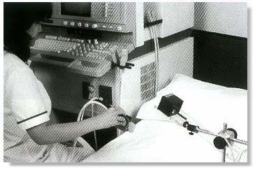
9 Free-hand Acquisition Acoustic Articulated arm Electromagnetic positioner Mechanical Localizers Linear scanning Fan scanning Rotational scanning 2D Transducers arrays Movements of probe should be fast and precise. Position and orientation of probe must be known all the time. 9
10 -Acoustic- Most common method in free-hand acquisition. Position and angle are obtained from three sound-emitting devices mounted on the transducer and is detected by three microphones above the patient. -Electromagnetic- Position and orientation are detected by magnetic field sensor. Continuous of ultrasound transducers using 100 Hz field measurements. -Acoustic- -Articulated Arm- Transducer on a mechanical arm with movable joint. Less movement, more precision. -Electromagnetic- 10
11 11


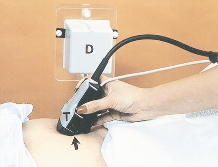
12 Mechanical probe moves in a precise manner. Use the knowledge about anatomy of body. Small, expensive localizers or large localizers than can be assembled on 2D device. Take slices in predefine intervals. Linear distance angle Linear scanning Fan Scanning Rotational Scanning 12


13 Linear scanning Fan scanning Free-hand offers more flexibility where mechanical localizers are more precise in most cases. 13
14 No need to move the probe. Generates 3D data in real-time. (4D) Mostly in research process, because of constraints such as: Large number of transducer in a small area. Higher probability of false detection. 14
15 3D image reconstruction refers to the generation of a 3-D 3 representation of the examined structures from the acquired set of 2D images. There are two distinct ways to implement the reconstruction process: First, 3-dimensional surface model,, Segmenting series of 2D images to extract the desired features before the 3D image is reconstructed. Second, Acquiring series of 2D images to build a 3D voxel based Cartesian volume. 15
16 Segmenting Approach: The boundaries in between tissues should be outlined either manually or automatically. Accurate segmentation is required which is a particularly difficult problem. 3D surface model is developed from the boundary descriptions using different techniques. Provides images with increased contrast between segmented structures which increases the image artifacts. This method reduces the amount of information allowing for efficient 3D rendering. 16
17 Voxel Based Approach: No information is lost during the 3D reconstruction. Allows a variety of rendering techniques such as: - Texture mapping - Ray-casting Requires no user intervention and is easily automated. However this approach results in large data files 17
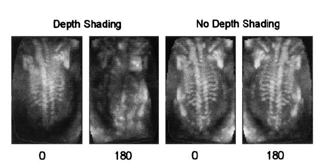
18 Plays an important and at times, dominant role in verifying information to the operator. Four classes of displaying 3D images: Surface-based Multi-planar Combined surface and multi-planar rendering Volume based 18
19 This technique is based on visualization of surfaces of structures or organs A segmentation or classification step is required which can be done either automatically by an algorithm analysis or manually by a skilled operator There are two basic methods available for viewing: - Wire-frames - Surface Rendering 19


20 A Wire-frame image of heart Surface rendered image of fetus It is not an ultrasound image Though! 20
21 Requires that a 3D voxel- based image be first reconstructed Two common techniques are used to view the image: - Using computer user-interface tools to select planes from the volume for viewing as reformatted 2D images. - Multi-planar visualization with texture mapping (the 3D image appears as a polyhedron). 21

22 An example of Texture mapping: Three-dimensional color Doppler US image of the carotid artery shows the sharply jagged irregularity of the vessel wall and the color pattern caused by slight variations in the beat-to to-beat movement of the artery. The image was acquired with cardiac gating, which improves the quality of the 3D image but increases total imaging time. 22

23 Presents to the viewer a display of entire 3D image after it has been projected onto a 2D plane. Density-Weighted The most common approach which is the ray-casting techniques. Maximum intensity projection Another common approach to display only the maximum intensity voxels along each ray 23
 in (1 α(k) is opacity value c(k) ) is shade value P(r) is opacity and color")
24 Cout = Cin (1-α(k))(k)) + c(k). ).α(k) in (1 α(k) is opacity value c(k) ) is shade value P(r) is opacity and color of pixel r, k is the kth voxel along the rth ray 24
25 Subtle surface features can be enhanced by using gradient Subtle can be enhanced by using gradient information from the volume to highlight interfaces. (by using some s type of gradient shading) One method to get the gradient is to use Mark-Hildreth operator which is the convolution of a Gaussian with a Laplacian: 25
26 Exponential depth shading is a useful technique to enhance the perspective of depth in a volume rendered image In this approach the density of voxel is a function of voxel depth in the volume which is reduced by an amount based on an exponential function: β(k) : attenuation factor at kth location along rth ray 26
27 27

28 Fetal Imaging: -3D US has shown most promise in obstetric imaging - Reported to make anomalies easier to recognize - Better understanding of fetal abnormalities *A study of 204 patients with anomalies: 62%:3D more advantageous than 2D 36%: both equal 2%: 3D disadvantageous * Study was performed by Merz et al. 28

29 Images of embryos, demonstrating the degree of detail that can be shown using 3D Ultrasound techniques Images courtesy of Drs. Benoit and Bonilla- Musolles 29

30 Gynecology: One of the most important applications is characterization of uterine anomalies - Viewing transverse plane through uterus More Accurate measurements of endometrial volume Useful in evaluating infertility patients C-plane view of uterus 30

31 Prostate Imaging: 3D US gives the possibility of more accurate repeated measurement than 2D which can be very useful when accurate volume assessment is needed for dosimetry planning or for estimating prostate- specific antigen levels. 3D US image shows hypoechoic tumor invading the seminal vesicle 31
32 There several other applications of 3D US such as: Cardiology Musculoskeletal Breast imaging Biopsy related imaging Dermatology Surgical applications 32
33 4DUS is the main goal of research in ultrasound field. Why is it more difficult to get a 4D ultrasound? Limits in acquisition methods Limits in hardware and software computational devices Limits in present image displaying techniques. Very helpful in diagnosis of dynamic organs, such as heart or fetus. PLAY THE MOVIE! And we are done THANK YOU! 33
34 Fenster,, Aaron, B. Downey, Donal. 3-D D Ultrasound Imaging: A Review, IEEE ENGINEERING IN MEDICINE AND BIOLOGY MAGAZINE /1996. Lees, William, Ultrasound Imaging in Three and Four Dimensions, Seminars in Ultrasound, CT, and MRI, Vol 22, No 1 (February), 2001; pp Nelson, Thomas R., Elvins,, T. Todd, Visualization of 3D Ultrasound Data. IEEE Computer Graphics & Application Nelson, Thomas R., Pretorius,, Dolores H., THREE-DIMENSIONAL ULTRASOUND IMAGING, Ultrasound in Med. & Biol., Vol.24, No 9, pp , Welch, Jacqueline, Johnson, Jeremy A., Bax,, Michael R., Badr, Rana, Shahidi, Ramin, A A Real-Time Freehand 3D Ultrasound System For Image-Guided Surgery, 2000 IEEE Ultrasonics Symposium. Harvey, Christopher A., Pilcher,, James A., Eckersley,, Robert J., Blomley,, Martin J K., Cosgrove, David O. Advances in Ultrasound,, 2002 The Royal College of Radiologists. intro.html#lt4 ultrasound.net/history-3d.html 34
Lecture 6: Medical imaging and image-guided interventions
 ME 328: Medical Robotics Winter 2019 Lecture 6: Medical imaging and image-guided interventions Allison Okamura Stanford University Updates Assignment 3 Due this Thursday, Jan. 31 Note that this assignment
Chapter 32 3-D and 4-D imaging in Obstetrics and Gynecology
 Objectives Define common terms related to 3D/4D ultrasound Chapter 32 3-D and 4-D imaging in Obstetrics and Gynecology Bridgette Lunsford Describe how 3D and 4D imaging differs from 2D ultrasound Identify
Projection-Based Needle Segmentation in 3D Ultrasound Images
 Projection-Based Needle Segmentation in 3D Ultrasound Images Mingyue Ding and Aaron Fenster Imaging Research Laboratories, Robarts Research Institute, 100 Perth Drive, London, ON, Canada, N6A 5K8 ^PGLQJDIHQVWHU`#LPDJLQJUREDUWVFD
IMPROVEMENT OF CONSPICUITY BY FUSION OF PULSE-ECHO DATA
 IMPROVEMENT OF CONSPICUITY BY FUSION OF PULSE-ECHO DATA S. M. Song 1, H. Jang 1, J. Kwon 1, J. Sung 2, H. Ahn 2, J. Lee 2 and S. Jang 2 1 Seoul National Universit Seoul, Korea; 2 Sae-An Engineering Corporation,
Medical Images Analysis and Processing
 Medical Images Analysis and Processing - 25642 Emad Course Introduction Course Information: Type: Graduated Credits: 3 Prerequisites: Digital Image Processing Course Introduction Reference(s): Insight
Shadow casting. What is the problem? Cone Beam Computed Tomography THE OBJECTIVES OF DIAGNOSTIC IMAGING IDEAL DIAGNOSTIC IMAGING STUDY LIMITATIONS
 Cone Beam Computed Tomography THE OBJECTIVES OF DIAGNOSTIC IMAGING Reveal pathology Reveal the anatomic truth Steven R. Singer, DDS srs2@columbia.edu IDEAL DIAGNOSTIC IMAGING STUDY Provides desired diagnostic
Medical Image Analysis
 Computer assisted Image Analysis VT04 29 april 2004 Medical Image Analysis Lecture 10 (part 1) Xavier Tizon Medical Image Processing Medical imaging modalities XRay,, CT Ultrasound MRI PET, SPECT Generic
Medical Image Processing: Image Reconstruction and 3D Renderings
 Medical Image Processing: Image Reconstruction and 3D Renderings 김보형 서울대학교컴퓨터공학부 Computer Graphics and Image Processing Lab. 2011. 3. 23 1 Computer Graphics & Image Processing Computer Graphics : Create,
Image Acquisition Systems
 Image Acquisition Systems Goals and Terminology Conventional Radiography Axial Tomography Computer Axial Tomography (CAT) Magnetic Resonance Imaging (MRI) PET, SPECT Ultrasound Microscopy Imaging ITCS
Ultrasound To Go. MySono U5
 Ultrasound To Go MySono U5 Ultrasound To Go With the introduction of the MySono U5, Samsung Medison brings you a fully featured ultrasound imaging system to go. Delivering exceptional image quality and
Certificate in Clinician Performed Ultrasound (CCPU)
") Certificate in Clinician Performed Ultrasound (CCPU) Syllabus Physics Tutorial Physics Tutorial Purpose: Training: Assessments: This unit is designed to cover the theoretical and practical curriculum for
Certificate in Clinician Performed Ultrasound (CCPU) Syllabus Physics Tutorial Physics Tutorial Purpose: Training: Assessments: This unit is designed to cover the theoretical and practical curriculum for
Digital Image Processing
 Digital Image Processing SPECIAL TOPICS CT IMAGES Hamid R. Rabiee Fall 2015 What is an image? 2 Are images only about visual concepts? We ve already seen that there are other kinds of image. In this lecture
LOGIQ. V2 Ultrasound. Part of LOGIQ Vision Series. Imagination at work LOGIQ is a trademark of General Electric Company.
 TM LOGIQ V2 Ultrasound Part of LOGIQ Vision Series Imagination at work The brilliance of color. The simplicity of GE. Now you can add the advanced capabilities of color Doppler to patient care with the
GPU Ultrasound Simulation and Volume Reconstruction
 GPU Ultrasound Simulation and Volume Reconstruction Athanasios Karamalis 1,2 Supervisor: Nassir Navab1 Advisor: Oliver Kutter1, Wolfgang Wein2 1Computer Aided Medical Procedures (CAMP), Technische Universität
Computer-Aided Diagnosis in Abdominal and Cardiac Radiology Using Neural Networks
 Computer-Aided Diagnosis in Abdominal and Cardiac Radiology Using Neural Networks Du-Yih Tsai, Masaru Sekiya and Yongbum Lee Department of Radiological Technology, School of Health Sciences, Faculty of
Introduction to Medical Image Processing
 Introduction to Medical Image Processing Δ Essential environments of a medical imaging system Subject Image Analysis Energy Imaging System Images Image Processing Feature Images Image processing may be
CHAPTER 2 MEDICAL IMAGING WITH NON-IONIZING RADIATION
 CHAPTER 2 MEDICAL IMAGING WITH NON-IONIZING RADIATION 1 Ultrasound Imaging 1.1 Ultrasound Production and Detection Ultrasound is frequency vibration. To produce and detect ultrasound, we use crystals which
Range Sensors (time of flight) (1)
 (1)") Range Sensors (time of flight) (1) Large range distance measurement -> called range sensors Range information: key element for localization and environment modeling Ultrasonic sensors, infra-red sensors
Range Sensors (time of flight) (1) Large range distance measurement -> called range sensors Range information: key element for localization and environment modeling Ultrasonic sensors, infra-red sensors
PII S (98)00043-X
00043-X") PII S0301-5629(98)00043-X Ultrasound in Med. & Biol., Vol. 24, No. 9, pp. 1243 1270, 1998 Copyright 1998 World Federation for Ultrasound in Medicine & Biology Printed in the USA. All rights reserved 0301-5629/98/$
PII S0301-5629(98)00043-X Ultrasound in Med. & Biol., Vol. 24, No. 9, pp. 1243 1270, 1998 Copyright 1998 World Federation for Ultrasound in Medicine & Biology Printed in the USA. All rights reserved 0301-5629/98/$
CT Basics Principles of Spiral CT Dose. Always Thinking Ahead.
 1 CT Basics Principles of Spiral CT Dose 2 Who invented CT? 1963 - Alan Cormack developed a mathematical method of reconstructing images from x-ray projections Sir Godfrey Hounsfield worked for the Central
Ch. 4 Physical Principles of CT
 Ch. 4 Physical Principles of CT CLRS 408: Intro to CT Department of Radiation Sciences Review: Why CT? Solution for radiography/tomography limitations Superimposition of structures Distinguishing between
Advanced Image Reconstruction Methods for Photoacoustic Tomography
 Advanced Image Reconstruction Methods for Photoacoustic Tomography Mark A. Anastasio, Kun Wang, and Robert Schoonover Department of Biomedical Engineering Washington University in St. Louis 1 Outline Photoacoustic/thermoacoustic
Clinical Importance. Aortic Stenosis. Aortic Regurgitation. Ultrasound vs. MRI. Carotid Artery Stenosis
 Clinical Importance Rapid cardiovascular flow quantitation using sliceselective Fourier velocity encoding with spiral readouts Valve disease affects 10% of patients with heart disease in the U.S. Most
Ultrasound To Go. The MySono U5 -
 Ultrasound To Go The MySono U5 - Ultrasound To Go With the introduction of the MySono U5, MEDISON brings you a fully featured ultrasound imaging system to go. Delivering exceptional image quality and featuring
UNIVERSITY OF SOUTHAMPTON
 UNIVERSITY OF SOUTHAMPTON PHYS2007W1 SEMESTER 2 EXAMINATION 2014-2015 MEDICAL PHYSICS Duration: 120 MINS (2 hours) This paper contains 10 questions. Answer all questions in Section A and only two questions
Computed tomography - outline
 Computed tomography - outline Computed Tomography Systems Jørgen Arendt Jensen and Mikael Jensen (DTU Nutech) October 6, 216 Center for Fast Ultrasound Imaging, Build 349 Department of Electrical Engineering
Refraction Corrected Transmission Ultrasound Computed Tomography for Application in Breast Imaging
 Refraction Corrected Transmission Ultrasound Computed Tomography for Application in Breast Imaging Joint Research With Trond Varslot Marcel Jackowski Shengying Li and Klaus Mueller Ultrasound Detection
10/5/09 1. d = 2. Range Sensors (time of flight) (2) Ultrasonic Sensor (time of flight, sound) (1) Ultrasonic Sensor (time of flight, sound) (2) 4.1.
 (2) Ultrasonic Sensor (time of flight, sound) (1) Ultrasonic Sensor (time of flight, sound) (2) 4.1.") Range Sensors (time of flight) (1) Range Sensors (time of flight) (2) arge range distance measurement -> called range sensors Range information: key element for localization and environment modeling Ultrasonic
Range Sensors (time of flight) (1) Range Sensors (time of flight) (2) arge range distance measurement -> called range sensors Range information: key element for localization and environment modeling Ultrasonic
The Supreme 3D/4D Ultrasound. The Supreme 3D/4D Ultrasound. CT-V20-ICM
 The Supreme 3D/4D Ultrasound The Supreme 3D/4D Ultrasound www.medison.com info@medison.com CT-V20-ICM-06.20.2008 The Supreme 3D/4D Ultrasound Since launching the first commercially available 3D ultrasound
INDUSTRIAL SYSTEM DEVELOPMENT FOR VOLUMETRIC INTEGRITY
 INDUSTRIAL SYSTEM DEVELOPMENT FOR VOLUMETRIC INTEGRITY VERIFICATION AND ANALYSIS M. L. Hsiao and J. W. Eberhard CR&D General Electric Company Schenectady, NY 12301 J. B. Ross Aircraft Engine - QTC General
MEDICAL IMAGE ANALYSIS
 SECOND EDITION MEDICAL IMAGE ANALYSIS ATAM P. DHAWAN g, A B IEEE Engineering in Medicine and Biology Society, Sponsor IEEE Press Series in Biomedical Engineering Metin Akay, Series Editor +IEEE IEEE PRESS
VEVO 1100 Sole Source Specifications
 VEVO 1100 Sole Source Specifications Table of Contents 1.SYSTEM... 2 1-1 Licensing for the User... 2 1-2 Safety... 2 1-3 Portability... 2 2.TRANSDUCERS... 2 2-1 MicroScan transducers... 2 3. IMAGING STATION...
Prostate Detection Using Principal Component Analysis
 Prostate Detection Using Principal Component Analysis Aamir Virani (avirani@stanford.edu) CS 229 Machine Learning Stanford University 16 December 2005 Introduction During the past two decades, computed
Computational Medical Imaging Analysis
 Computational Medical Imaging Analysis Chapter 2: Image Acquisition Systems Jun Zhang Laboratory for Computational Medical Imaging & Data Analysis Department of Computer Science University of Kentucky
Focus on your needs. Ultrasound system HS60 SAMSUNG MEDISON CO., LTD. CT-HS60 V1.0-OB-FT EN
 Samsung Medison, an affiliate of Samsung Electronics, is a global medical company founded in 1985. With a mission to bring health and well-being to people's lives, the company manufactures diagnostic ultrasound
Mindray. Hand-carried Diagnostic Ultrasound System. Expanding the Envelope of Performance and Flexibility
 M7 Mindray M7 Hand-carried Diagnostic Ultrasound System Expanding the Envelope of Performance and Flexibility M7 Diagnostic Ultrasound System Equipped for Quality Radiology Cardiology Outpatient Emergency
3/27/2012 WHY SPECT / CT? SPECT / CT Basic Principles. Advantages of SPECT. Advantages of CT. Dr John C. Dickson, Principal Physicist UCLH
 3/27/212 Advantages of SPECT SPECT / CT Basic Principles Dr John C. Dickson, Principal Physicist UCLH Institute of Nuclear Medicine, University College London Hospitals and University College London john.dickson@uclh.nhs.uk
Direct Scan-Lines Mapping for Ultrasound 3D image Reconstruction
 Abstract 3D Ultrasound has recently been a routine imaging modality for medical diagnosing and in other bioscience fields.compared to the conventional method where D images are used to represent a 3D anatomy,
A Study of Medical Image Analysis System
 Indian Journal of Science and Technology, Vol 8(25), DOI: 10.17485/ijst/2015/v8i25/80492, October 2015 ISSN (Print) : 0974-6846 ISSN (Online) : 0974-5645 A Study of Medical Image Analysis System Kim Tae-Eun
Ultrasound. Q-Station software. Streamlined workflow solutions. Philips Q-Station ultrasound workspace software
 Ultrasound Q-Station software Streamlined workflow solutions Philips Q-Station ultrasound workspace software Managing your off-cart workf low Everyone is being asked to do more with fewer resources it
A method for interactive manipulation and animation of volumetric data
 A method for interactive manipulation and animation of volumetric data Yves Jean, Larry F. Hodges Graphics, Visualization and Usability Center College of Computing Georgia Institute of Technology Atlanta,
Index. aliasing artifacts and noise in CT images, 200 measurement of projection data, nondiffracting
 Index Algebraic equations solution by Kaczmarz method, 278 Algebraic reconstruction techniques, 283-84 sequential, 289, 293 simultaneous, 285-92 Algebraic techniques reconstruction algorithms, 275-96 Algorithms
GE Healthcare. Vivid 7 Dimension 08 Cardiovascular ultrasound system
 GE Healthcare Vivid 7 Dimension 08 Cardiovascular ultrasound system ltra Definition. Technology. Performance. Start with a system that s proven its worth in LV quantification and 4D imaging. Then add even
Backward-Warping Ultrasound Reconstruction for Improving Diagnostic Value and Registration
 Backward-Warping Ultrasound Reconstruction for Improving Diagnostic Value and Registration Wolfgang Wein 1, Fabian Pache 1,BarbaraRöper 2, and Nassir Navab 1 1 Chair for Computer Aided Medical Procedures
FINDING THE TRUE EDGE IN CTA
 FINDING THE TRUE EDGE IN CTA by: John A. Rumberger, PhD, MD, FACC Your patient has chest pain. The Cardiac CT Angiography shows plaque in the LAD. You adjust the viewing window trying to evaluate the stenosis
Recent advances in aerospace inspection with ultrasonic phased arrays
 Recent advances in aerospace inspection with ultrasonic phased arrays David Lines Chief Engineer, Diagnostic Sonar Ltd., UK AeroNDT SEMINAR, Aerospace Testing Expo2007 27 th -29 th March 2007, Munich Content
MEDICAL IMAGING 2nd Part Computed Tomography
 MEDICAL IMAGING 2nd Part Computed Tomography Introduction 2 In the last 30 years X-ray Computed Tomography development produced a great change in the role of diagnostic imaging in medicine. In convetional
APLIO JUST GOT EVEN BETTER INTRODUCING THE INNOVATION 2016 UPGRADE
 APLIO JUST GOT EVEN BETTER INTRODUCING THE INNOVATION 2016 UPGRADE INNOVATION 2016 AT A GLANCE More clinical detail New settings in the Precision technology package provide enhanced border delineation
Phantom-based evaluation of a semi-automatic segmentation algorithm for cerebral vascular structures in 3D ultrasound angiography (3D USA)
") Phantom-based evaluation of a semi-automatic segmentation algorithm for cerebral vascular structures in 3D ultrasound angiography (3D USA) C. Chalopin¹, K. Krissian², A. Müns 3, F. Arlt 3, J. Meixensberger³,
Phantom-based evaluation of a semi-automatic segmentation algorithm for cerebral vascular structures in 3D ultrasound angiography (3D USA) C. Chalopin¹, K. Krissian², A. Müns 3, F. Arlt 3, J. Meixensberger³,
Multipass GPU Surface Rendering in 4D Ultrasound
 2012 Cairo International Biomedical Engineering Conference (CIBEC) Cairo, Egypt, December 20-21, 2012 Multipass GPU Surface Rendering in 4D Ultrasound Ahmed F. Elnokrashy 1,2, Marwan Hassan 1, Tamer Hosny
Limitations of Projection Radiography. Stereoscopic Breast Imaging. Limitations of Projection Radiography. 3-D Breast Imaging Methods
 Stereoscopic Breast Imaging Andrew D. A. Maidment, Ph.D. Chief, Physics Section Department of Radiology University of Pennsylvania Limitations of Projection Radiography Mammography is a projection imaging
Real-time self-calibration of a tracked augmented reality display
 Real-time self-calibration of a tracked augmented reality display Zachary Baum, Andras Lasso, Tamas Ungi, Gabor Fichtinger Laboratory for Percutaneous Surgery, Queen s University, Kingston, Canada ABSTRACT
Compactly SAMSUNG powerful ULTRASOUND H60
 Samsung Medison is a global leading medical devices company. Founded in 1985, the company now sells cutting-edge medical devices including diagnostic ultrasound, digital X-ray and blood analyzer around
Volume Illumination. Visualisation Lecture 11. Taku Komura. Institute for Perception, Action & Behaviour School of Informatics
 Volume Illumination Visualisation Lecture 11 Taku Komura Institute for Perception, Action & Behaviour School of Informatics Taku Komura Volume Illumination & Vector Vis. 1 Previously : Volume Rendering
A Navigation System for Minimally Invasive Abdominal Intervention Surgery Robot
 A Navigation System for Minimally Invasive Abdominal Intervention Surgery Robot Weiming ZHAI, Yannan ZHAO and Peifa JIA State Key Laboratory of Intelligent Technology and Systems Tsinghua National Laboratory
Computational Medical Imaging Analysis Chapter 4: Image Visualization
 Computational Medical Imaging Analysis Chapter 4: Image Visualization Jun Zhang Laboratory for Computational Medical Imaging & Data Analysis Department of Computer Science University of Kentucky Lexington,
C-mode Real Time Tomographic Reflection for a Matrix Array Ultrasound Sonic Flashlight
 C-mode Real Time Tomographic Reflection for a Matrix Array Ultrasound Sonic Flashlight George Stetten 1,2,3, Aaron Cois 1,2,, Wilson Chang 1,2,3, Damion Shelton 2, Robert Tamburo 1,2, John Castellucci
A Survey of Volumetric Visualization Techniques for Medical Images
 International Journal of Research Studies in Computer Science and Engineering (IJRSCSE) Volume 2, Issue 4, April 2015, PP 34-39 ISSN 2349-4840 (Print) & ISSN 2349-4859 (Online) www.arcjournals.org A Survey
Tomographic Reconstruction
 Tomographic Reconstruction 3D Image Processing Torsten Möller Reading Gonzales + Woods, Chapter 5.11 2 Overview Physics History Reconstruction basic idea Radon transform Fourier-Slice theorem (Parallel-beam)
US 1.
 US 1 Sample image: Normal pancreas seen on sonogram. Looking up from abdomen toward the head of the patient. The liver is in front of the pancreas. A vein draining the spleen is behind the pancreas http://www.radiologyinfo.org/photocat/photos.cfm?image=abdo-us-pancr.jpg&&subcategory=abdomen&&stop=9
Beginner s Guide to Vevo 2100
 PN 12090 Beginner s Guide to Vevo 2100 December 2008 This Beginner s Guide to Vevo 2100 provides a quick overview of the Vevo 2100 imaging modes and provides key tips to allow users to start a study, acquire
GE Healthcare. Agile Ultrasound. The Next Revolution in Ultrasound Imaging
 Agile Ultrasound The Next Revolution in Ultrasound Imaging Abstract Diagnostic use of ultrasound has greatly expanded over the past couple of decades because it offers many advantages as an imaging modality.
A THREE-DIMENSIONAL PHASED ARRAY ULTRASONIC TESTING TECHNIQUE
 A THREE-DIMENSIONAL PHASED ARRAY ULTRASONIC TESTING TECHNIQUE So KITAZAWA, Naoyuki KONO, Atsushi BABA and Yuji ADACHI HITACHI, Ltd., Japan Mitsuru ODAKURA HITACHI-GE Nuclear Energy, Ltd., Japan Introduction
Mech. Engineering, Comp. Science, and Rad. Oncology Departments. Schools of Engineering and Medicine, Bio-X Program, Stanford University
 Mech. Engineering, Comp. Science, and Rad. Oncology Departments Schools of Engineering and Medicine, Bio-X Program, Stanford University 1 Conflict of Interest Nothing to disclose 2 Imaging During Beam
Leading the New Standards
 Samsung Medison is a global leading medical devices company. Founded in 1985, the company now sells cutting-edge medical devices including diagnostic ultrasound, digital X-ray and blood analyzer, in 110
Image Thickness Correction for Navigation with 3D Intra-cardiac Ultrasound Catheter
 Image Thickness Correction for Navigation with 3D Intra-cardiac Ultrasound Catheter Hua Zhong 1, Takeo Kanade 1,andDavidSchwartzman 2 1 Computer Science Department, Carnegie Mellon University, USA 2 University
Volume Illumination and Segmentation
 Volume Illumination and Segmentation Computer Animation and Visualisation Lecture 13 Institute for Perception, Action & Behaviour School of Informatics Overview Volume illumination Segmentation Volume
Volume visualization. Volume visualization. Volume visualization methods. Sources of volume visualization. Sources of volume visualization
 Volume visualization Volume visualization Volumes are special cases of scalar data: regular 3D grids of scalars, typically interpreted as density values. Each data value is assumed to describe a cubic
Towards an Estimation of Acoustic Impedance from Multiple Ultrasound Images
 Towards an Estimation of Acoustic Impedance from Multiple Ultrasound Images Christian Wachinger 1, Ramtin Shams 2, Nassir Navab 1 1 Computer Aided Medical Procedures (CAMP), Technische Universität München
Scalar Data. Visualization Torsten Möller. Weiskopf/Machiraju/Möller
 Scalar Data Visualization Torsten Möller Weiskopf/Machiraju/Möller Overview Basic strategies Function plots and height fields Isolines Color coding Volume visualization (overview) Classification Segmentation
Implementing the probe beam deflection technique for acoustic sensing in photoacoustic and ultrasound imaging
 Implementing the probe beam deflection technique for acoustic sensing in photoacoustic and ultrasound imaging Ronald A. Barnes Jr. The University of Texas at San Antonio This work is a collaboration between
Visualisation : Lecture 1. So what is visualisation? Visualisation
 So what is visualisation? UG4 / M.Sc. Course 2006 toby.breckon@ed.ac.uk Computer Vision Lab. Institute for Perception, Action & Behaviour Introducing 1 Application of interactive 3D computer graphics to
Computed tomography (Item No.: P )
") Computed tomography (Item No.: P2550100) Curricular Relevance Area of Expertise: Biology Education Level: University Topic: Modern Imaging Methods Subtopic: X-ray Imaging Experiment: Computed tomography
Computed tomography (Item No.: P2550100) Curricular Relevance Area of Expertise: Biology Education Level: University Topic: Modern Imaging Methods Subtopic: X-ray Imaging Experiment: Computed tomography
Ch 22 Inspection Technologies
 Ch 22 Inspection Technologies Sections: 1. Inspection Metrology 2. Contact vs. Noncontact Inspection Techniques 3. Conventional Measuring and Gaging Techniques 4. Coordinate Measuring Machines 5. Surface
Tissue Doppler gated (TDOG) dynamic three-dimensional ultrasound imaging of the fetal heart
 dynamic three-dimensional ultrasound imaging of the fetal heart") Ultrasound Obstet Gynecol 2004; 24: 192 198 Published online in Wiley InterScience (www.interscience.wiley.com). DOI: 10.1002/uog.1094 Tissue Doppler gated (TDOG) dynamic three-dimensional ultrasound imaging
Ultrasound Obstet Gynecol 2004; 24: 192 198 Published online in Wiley InterScience (www.interscience.wiley.com). DOI: 10.1002/uog.1094 Tissue Doppler gated (TDOG) dynamic three-dimensional ultrasound imaging
Classification of Subject Motion for Improved Reconstruction of Dynamic Magnetic Resonance Imaging
 1 CS 9 Final Project Classification of Subject Motion for Improved Reconstruction of Dynamic Magnetic Resonance Imaging Feiyu Chen Department of Electrical Engineering ABSTRACT Subject motion is a significant
Medical Image Registration by Maximization of Mutual Information
 Medical Image Registration by Maximization of Mutual Information EE 591 Introduction to Information Theory Instructor Dr. Donald Adjeroh Submitted by Senthil.P.Ramamurthy Damodaraswamy, Umamaheswari Introduction
Volume Rendering. Computer Animation and Visualisation Lecture 9. Taku Komura. Institute for Perception, Action & Behaviour School of Informatics
 Volume Rendering Computer Animation and Visualisation Lecture 9 Taku Komura Institute for Perception, Action & Behaviour School of Informatics Volume Rendering 1 Volume Data Usually, a data uniformly distributed
Motion Compensation from Short-Scan Data in Cardiac CT
 Motion Compensation from Short-Scan Data in Cardiac CT Juliane Hahn 1,2, Thomas Allmendinger 1, Herbert Bruder 1, and Marc Kachelrieß 2 1 Siemens Healthcare GmbH, Forchheim, Germany 2 German Cancer Research
Acknowledgments and financial disclosure
 AAPM 2012 Annual Meeting Digital breast tomosynthesis: basic understanding of physics principles James T. Dobbins III, Ph.D., FAAPM Director, Medical Physics Graduate Program Ravin Advanced Imaging Laboratories
A New Approach to Ultrasound Guided Radio-Frequency Needle Placement
 A New Approach to Ultrasound Guided Radio-Frequency Needle Placement Claudio Alcérreca 1,2, Jakob Vogel 2, Marco Feuerstein 2, and Nassir Navab 2 1 Image Analysis and Visualization Lab., CCADET UNAM, 04510
RADIOMICS: potential role in the clinics and challenges
 27 giugno 2018 Dipartimento di Fisica Università degli Studi di Milano RADIOMICS: potential role in the clinics and challenges Dr. Francesca Botta Medical Physicist Istituto Europeo di Oncologia (Milano)
Scalar Data. CMPT 467/767 Visualization Torsten Möller. Weiskopf/Machiraju/Möller
 Scalar Data CMPT 467/767 Visualization Torsten Möller Weiskopf/Machiraju/Möller Overview Basic strategies Function plots and height fields Isolines Color coding Volume visualization (overview) Classification
RADIOLOGY AND DIAGNOSTIC IMAGING
 Day 2 part 2 RADIOLOGY AND DIAGNOSTIC IMAGING Dr hab. Zbigniew Serafin, MD, PhD serafin@cm.umk.pl 2 3 4 5 CT technique CT technique 6 CT system Kanal K: RSNA/AAPM web module: CT Systems & CT Image Quality
BMVC 1996 doi: /c.10.44
 Correcting Motion-Induced Registration Errors in 3-D Ultrasound Images Robert Rohling and Andrew Gee University of Cambridge Department of Engineering Cambridge, England, CB2 1PZ frnr20,ahgg@eng.cam.ac.uk
NEW METHODOLOGIES FOR PROCESSING MEDICAL IMAGES. Ahmed Fathy Mosaad Elnokrashy
 NEW METHODOLOGIES FOR PROCESSING MEDICAL IMAGES By Ahmed Fathy Mosaad Elnokrashy A Thesis Submitted to the Faculty of Engineering at Cairo University in Partial Fulfillment of the Requirements for the
Visualization. Images are used to aid in understanding of data. Height Fields and Contours Scalar Fields Volume Rendering Vector Fields [chapter 26]
![Visualization. Images are used to aid in understanding of data. Height Fields and Contours Scalar Fields Volume Rendering Vector Fields [chapter 26]](/thumbs/74/70771954.jpg "Visualization. Images are used to aid in understanding of data. Height Fields and Contours Scalar Fields Volume Rendering Vector Fields [chapter 26]") Visualization Images are used to aid in understanding of data Height Fields and Contours Scalar Fields Volume Rendering Vector Fields [chapter 26] Tumor SCI, Utah Scientific Visualization Visualize large
Visualization Images are used to aid in understanding of data Height Fields and Contours Scalar Fields Volume Rendering Vector Fields [chapter 26] Tumor SCI, Utah Scientific Visualization Visualize large
MR-Guided Mixed Reality for Breast Conserving Surgical Planning
 MR-Guided Mixed Reality for Breast Conserving Surgical Planning Suba Srinivasan (subashini7@gmail.com) March 30 th 2017 Mentors: Prof. Brian A. Hargreaves, Prof. Bruce L. Daniel MEDICINE MRI Guided Mixed
INTRODUCTION TO MEDICAL IMAGING- 3D LOCALIZATION LAB MANUAL 1. Modifications for P551 Fall 2013 Medical Physics Laboratory
 INTRODUCTION TO MEDICAL IMAGING- 3D LOCALIZATION LAB MANUAL 1 Modifications for P551 Fall 2013 Medical Physics Laboratory Introduction Following the introductory lab 0, this lab exercise the student through
First CT Scanner. How it Works. Contemporary CT. Before and After CT. Computer Tomography: How It Works. Medical Imaging and Pattern Recognition
 Computer Tomography: How t Works Medical maging and Pattern Recognition Lecture 7 Computed Tomography Oleh Tretiak Only one plane is illuminated. Source-subject motion provides added information. 2 How
By choosing to view this document, you agree to all provisions of the copyright laws protecting it.
 Copyright 2009 IEEE. Reprinted from 31 st Annual International Conference of the IEEE Engineering in Medicine and Biology Society, 2009. EMBC 2009. Sept. 2009. This material is posted here with permission
HIGH-PERFORMANCE TOMOGRAPHIC IMAGING AND APPLICATIONS
 HIGH-PERFORMANCE TOMOGRAPHIC IMAGING AND APPLICATIONS Hua Lee and Yuan-Fang Wang Department of Electrical and Computer Engineering University of California, Santa Barbara ABSTRACT Tomographic imaging systems
Data Visualization (CIS/DSC 468)
") Data Visualization (CIS/DSC 46) Volume Rendering Dr. David Koop Visualizing Volume (3D) Data 2D visualization slice images (or multi-planar reformating MPR) Indirect 3D visualization isosurfaces (or surface-shaded
Data Visualization (CIS/DSC 46) Volume Rendering Dr. David Koop Visualizing Volume (3D) Data 2D visualization slice images (or multi-planar reformating MPR) Indirect 3D visualization isosurfaces (or surface-shaded
Data Fusion Virtual Surgery Medical Virtual Reality Team. Endo-Robot. Database Functional. Database
 2017 29 6 16 GITI 3D From 3D to 4D imaging Data Fusion Virtual Surgery Medical Virtual Reality Team Morphological Database Functional Database Endo-Robot High Dimensional Database Team Tele-surgery Robotic
TUMOR DETECTION IN MRI IMAGES
 TUMOR DETECTION IN MRI IMAGES Prof. Pravin P. Adivarekar, 2 Priyanka P. Khatate, 3 Punam N. Pawar Prof. Pravin P. Adivarekar, 2 Priyanka P. Khatate, 3 Punam N. Pawar Asst. Professor, 2,3 BE Student,,2,3
3D Modeling of Objects Using Laser Scanning
 1 3D Modeling of Objects Using Laser Scanning D. Jaya Deepu, LPU University, Punjab, India Email: Jaideepudadi@gmail.com Abstract: In the last few decades, constructing accurate three-dimensional models
Biophysical Techniques (BPHS 4090/PHYS 5800)
") Biophysical Techniques (BPHS 4090/PHYS 5800) Instructors: Prof. Christopher Bergevin (cberge@yorku.ca) Schedule: MWF 1:30-2:30 (CB 122) Website: http://www.yorku.ca/cberge/4090w2017.html York University
Biophysical Techniques (BPHS 4090/PHYS 5800) Instructors: Prof. Christopher Bergevin (cberge@yorku.ca) Schedule: MWF 1:30-2:30 (CB 122) Website: http://www.yorku.ca/cberge/4090w2017.html York University
FIELD PARADIGM FOR 3D MEDICAL IMAGING: Safer, More Accurate, and Faster SPECT/PET, MRI, and MEG
 FIELD PARADIGM FOR 3D MEDICAL IMAGING: Safer, More Accurate, and Faster SPECT/PET, MRI, and MEG July 1, 2011 First, do no harm. --Medical Ethics (Hippocrates) Dr. Murali Subbarao, Ph. D. murali@fieldparadigm.com,
VALIDATION OF THE SIMULATION SOFTWARE CIVA UT IN SEPARATED TRANSMIT/RECEIVE CONFIGURATIONS
 VALIDATION OF THE SIMULATION SOFTWARE CIVA UT IN SEPARATED TRANSMIT/RECEIVE CONFIGURATIONS Fabrice FOUCHER 1, Sébastien LONNE 1, Gwénaël TOULLELAN 2, Steve MAHAUT 2, Sylvain CHATILLON 2, Erica SCHUMACHER
SONOS 7500/5500 Using 3-Dimensional and BiPlane Imaging And Other System Changes for Software Revision D.1
 SONOS 7500/5500 Using 3-Dimensional and BiPlane Imaging And Other System Changes for Software Revision D.1 User s Guide Using 3-Dimensional and BiPlane Imaging And Other System Changes for Software Revision