IS MRI Based Structure a Mediator for Lead s Effect on Cognitive Function?
|
|
|
- Shon O’Brien’
- 6 years ago
- Views:
Transcription
1 IS MRI Based Structure a Mediator for Lead s Effect on Cognitive Function? Brian Caffo, Sining Chen, Brian Schwartz Department of Biostatistics and Environmental Health Sciences Johns Hopkins University
2 Cumulative lead dose Peak tibia lead Brain structure / architecture MRI RAVENS Cognitive function Cognitive domain summary scores
3 Around 500 male organo-lead workers Peak tibia lead measured by extrapolating from recent lead levels using a compartmental model Cognitive function measured by a battery of psychological tests collapsed into six domain scores 1. VM - Visual memory 2. VC - Visuo construction 3. VML - Verbal memory and learning 4. EF - Executive function 5. EHC - Eye-hand coordination 6. PSP - Processing speed
4 Variable Age (years) Year enrolled (n) Domain scores VC VML VM EF EHC PSP Brain volume(cm 3 ) Total Grey White Summary Mean(sd, range) (7.93, 34.70, 78.30) Mean (sd, range) (1.03, -4.51, 1.73) 0.26 (0.80, -2.40, 1.87) 0.15 (0.92, -2.40, 2.19) (0.76, -3.76, 1.64) (0.88, -4.93, 1.61) (0.77, -4.40, 2.07) Mean (sd, range) (105.10, , ) (60.05, , ) (59.03, , )

5 MRI Nuclear magnetic resonance Uniform magnetic field Protons align in the direction of the magnetic field RF pulse knocks some protons out of alignment Protons snap back into place emitting RF pulse Emitted RF differs by tissue type Weighting shows contrast of different types

6 Regions of interest analysis Processed brains are registered with a labeled template Subject-specific volume summaries are calculated for anatomical regions of interest ROI summaries analyzed using standard methods
7 Voxel Based Morphometry MRIs Skull stripping registration segmentation Data representation Spatial smoothing Voxel by voxel regression thresholding Cluster level hypothesis testing Peak value hypothesis testing Bonferoni or FDR correction
8 RAVENS maps Images are skull stripped, segmented and registered to a common template Intensity in a voxel the image represents volume (in mm 3 ) of the original images Original intensity lost Using VBM we investigate cross-sectional volume changes

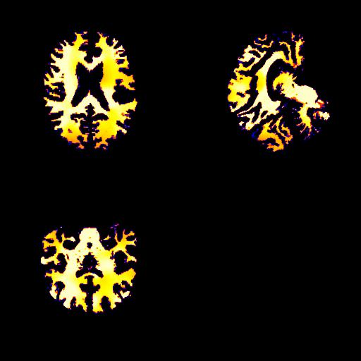
9 Ravens maps Grey White
10 Image representation Images are stored in Analyze format Binary image.img file Header.hdr file Header file contains (among other things) Voxel dimensions Array dimensions Image center Same header file for all images Images need to be rotated by 90 degree pitch to be in standard format
11 More on the images Images are registered to a template, but the template is rotated from the standard SPM template Imperfect registration implies some benefit from smoothing Images were smoothed using a 10mm FWHM Gaussian kernel smoother in SPM Multiplying the value in a voxel by.94x.94x1.5 / 1000 converts it to mm 3
12 Conversion to rda files The (around) 600 smoothed images are represented 256x256x256 arrays These images were processed into 256 separate rda files each containing a 256x256x600 array and the associated subject indices This representation allows for easy parallel processing of the VBM analysis
13 Cumulative lead dose Peak tibia lead Brain structure / architecture MRI RAVENS Cognitive function Cognitive domain summary scores
14 Voxel-level model NBT = Voxel + Age + VI + Height + Ed + Db + Smk + Alc Proportional versus absolute? TBV? Outliers with abnormalities?
15
16
17
18 Domain Expected Anatomical Site ROI VBM Visuoconstruction (ROCF, BD) frontal-parietaloccipital frontal WM frontal-parietaloccipital GM parietal WM Verbal Memory (RAVLT, Serial Digit) Temporalparietal L>R, hippocampus, prefrontal, ACC, thalamus, precuneas Visual Memory (ROCF, symbol digit) Bilateral hippocampus, R>L; Total WM Parietal GM, WM, temporal WM, corpus callosum, cerebellum, insula Occipital, inferior parietal GM, WM, corpus callosum Executive Function (Purdue Assembly, Stroop C-A, Trails B) Pre-frontal, ACC, DLPFC; Trails B medial temporal Occipital GM, frontal WM, occipital WM, insula, corpus callosum frontal-parietaloccipital GM, parietal, ACC, WM Eye-Hand Coordination (Trails A, Purdue) WM hyperintensities, Total WM, frontal GM, WM, parietal GM, temporal GM,WM, occipital WM, cerebellum, insula, amygdale, medial Parietal WM, GM Processing Speed (Reaction Time) DLPFC, ventral pre-motor Frontal GM, WM, parietal GM, occipital WM, medial, cingulate, insula, hippocampus Occipital GM, parietal-occiptial WM
19 General strategy Apply VBM regressing PTL or Domain on volume Threshold the resulting T statistic map to create a lead based mask Ideally represents those areas most associated with lead or domain across subjects Apply the mask to each subject and sum the resulting voxels creating subject-specific ROIs Raises concerns over multiplicity
20 Cumulative lead dose Peak tibia lead Brain structure / architecture MRI RAVENS Cognitive function Cognitive domain summary scores

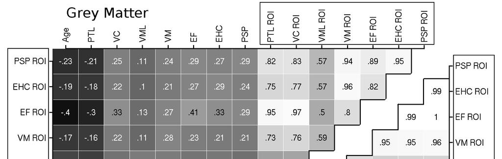
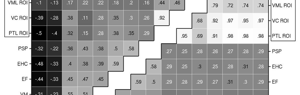
21 Grey matter glass brain images
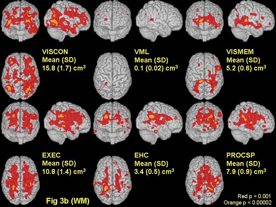
22 White matter glass brain images
23
24 Permutation analysis Investigate the impact of multiplicity and using the same subjects to both create the mask and evaluate significance Permute subject labels, perform the VBM analysis again, threshold the t-map, apply the mask to each subject Now each subjects ROI value represents only chance associations
25
26 Structural equation modeling Domain = a + b PTL + c Volume + OC + error Volume = d + e PTL + OC + error Direct effect of PTL on Domain = b Indirect effect of PTL on Domain = ec Total effect of PTL on Domain = b + ec Proportion direct = b / (b + ec) Estimated by the sem package in R
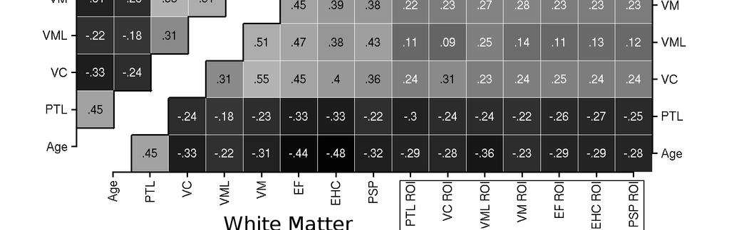
27 Proportion of direct effect roi size matter VC VML VM EXEC EHC PSP lead Grey domain Grey lead Grey domain Grey lead White domain White lead White domain White
Evaluation of multiple voxel-based morphometry approaches and applications in the analysis of white matter changes in temporal lobe epilepsy
 Evaluation of multiple voxel-based morphometry approaches and applications in the analysis of white matter changes in temporal lobe epilepsy Wenjing Li a, Huiguang He a, Jingjing Lu b, Bin Lv a, Meng Li
Neuroimaging and mathematical modelling Lesson 2: Voxel Based Morphometry
 Neuroimaging and mathematical modelling Lesson 2: Voxel Based Morphometry Nivedita Agarwal, MD Nivedita.agarwal@apss.tn.it Nivedita.agarwal@unitn.it Volume and surface morphometry Brain volume White matter
Figure 1. Comparison of the frequency of centrality values for central London and our region of Soho studied. The comparison shows that Soho falls
 A B C Figure 1. Comparison of the frequency of centrality values for central London and our region of Soho studied. The comparison shows that Soho falls well within the distribution for London s streets.
Methods for data preprocessing
 Methods for data preprocessing John Ashburner Wellcome Trust Centre for Neuroimaging, 12 Queen Square, London, UK. Overview Voxel-Based Morphometry Morphometry in general Volumetrics VBM preprocessing
Neuroimage Processing
 Neuroimage Processing Instructor: Moo K. Chung mkchung@wisc.edu Lecture 2-3. General Linear Models (GLM) Voxel-based Morphometry (VBM) September 11, 2009 What is GLM The general linear model (GLM) is a
ASAP_2.0 (Automatic Software for ASL Processing) USER S MANUAL
 USER S MANUAL") ASAP_2.0 (Automatic Software for ASL Processing) USER S MANUAL ASAP was developed as part of the COST Action "Arterial Spin Labelling Initiative in Dementia (AID)" by: Department of Neuroimaging, Institute
ASAP_2.0 (Automatic Software for ASL Processing) USER S MANUAL ASAP was developed as part of the COST Action "Arterial Spin Labelling Initiative in Dementia (AID)" by: Department of Neuroimaging, Institute
Appendix E1. Supplementary Methods. MR Image Acquisition. MR Image Analysis
 RSNA, 2015 10.1148/radiol.2015150532 Appendix E1 Supplementary Methods MR Image Acquisition By using a 1.5-T system (Avanto, Siemens Medical, Erlangen, Germany) under a program of regular maintenance (no
ANALYSIS OF FUNCTIONAL MAGNETIC RESONANCE IMAGING DATA USING SPM99: VOXEL-BASED MORPHOMETRY DONNA ROSE ADDIS
 Donna Rose Addis, TWRI, May 2004 1 ANALYSIS OF FUNCTIONAL MAGNETIC RESONANCE IMAGING DATA USING SPM99: VOXEL-BASED MORPHOMETRY DONNA ROSE ADDIS DEPT. OF PSYCHOLOGY, UNIVERSITY OF TORONTO TORONTO WESTERN
Preprocessing II: Between Subjects John Ashburner
 Preprocessing II: Between Subjects John Ashburner Pre-processing Overview Statistics or whatever fmri time-series Anatomical MRI Template Smoothed Estimate Spatial Norm Motion Correct Smooth Coregister
Supplementary Information. Task-induced brain state manipulation improves prediction of individual traits. Greene et al.
 Supplementary Information Task-induced brain state manipulation improves prediction of individual traits Greene et al. Supplementary Note 1 Analyses of effects of gf measurement technique PNC CPM results
The organization of the human cerebral cortex estimated by intrinsic functional connectivity
 1 The organization of the human cerebral cortex estimated by intrinsic functional connectivity Journal: Journal of Neurophysiology Author: B. T. Thomas Yeo, et al Link: https://www.ncbi.nlm.nih.gov/pubmed/21653723
Introduction to fmri. Pre-processing
 Introduction to fmri Pre-processing Tibor Auer Department of Psychology Research Fellow in MRI Data Types Anatomical data: T 1 -weighted, 3D, 1/subject or session - (ME)MPRAGE/FLASH sequence, undistorted
Supplementary methods
 Supplementary methods This section provides additional technical details on the sample, the applied imaging and analysis steps and methods. Structural imaging Trained radiographers placed all participants
Math in image processing
 Math in image processing Math in image processing Nyquist theorem Math in image processing Discrete Fourier Transformation Math in image processing Image enhancement: scaling Math in image processing Image
CHAPTER 2. Morphometry on rodent brains. A.E.H. Scheenstra J. Dijkstra L. van der Weerd
 CHAPTER 2 Morphometry on rodent brains A.E.H. Scheenstra J. Dijkstra L. van der Weerd This chapter was adapted from: Volumetry and other quantitative measurements to assess the rodent brain, In vivo NMR
Supplementary Online Content
 Supplementary Online Content Palacios EM, Sala-Llonch R, Junque C, Roig T, Tormos JM, Bargallo N, Vendrella P. Restingstate functional magnetic resonance imaging activity and connectivity and cognitive
An Introduction To Automatic Tissue Classification Of Brain MRI. Colm Elliott Mar 2014
 An Introduction To Automatic Tissue Classification Of Brain MRI Colm Elliott Mar 2014 Tissue Classification Tissue classification is part of many processing pipelines. We often want to classify each voxel
Fmri Spatial Processing
 Educational Course: Fmri Spatial Processing Ray Razlighi Jun. 8, 2014 Spatial Processing Spatial Re-alignment Geometric distortion correction Spatial Normalization Smoothing Why, When, How, Which Why is
Computational Neuroanatomy
 Computational Neuroanatomy John Ashburner john@fil.ion.ucl.ac.uk Smoothing Motion Correction Between Modality Co-registration Spatial Normalisation Segmentation Morphometry Overview fmri time-series kernel
17th Annual Meeting of the Organization for Human Brain Mapping (HBM)
") 17th Annual Meeting of the Organization for Human Brain Mapping (HBM) Regionally constrained voxel-based network of left hippocampus in left medial temporal lobe epilepsy Submission No: 2740 Authors: Jarang
17th Annual Meeting of the Organization for Human Brain Mapping (HBM) Regionally constrained voxel-based network of left hippocampus in left medial temporal lobe epilepsy Submission No: 2740 Authors: Jarang
This exercise uses one anatomical data set (ANAT1) and two functional data sets (FUNC1 and FUNC2).
 and two functional data sets (FUNC1 and FUNC2).") Exploring Brain Anatomy This week s exercises will let you explore the anatomical organization of the brain to learn some of its basic properties, as well as the location of different structures. The human
Exploring Brain Anatomy This week s exercises will let you explore the anatomical organization of the brain to learn some of its basic properties, as well as the location of different structures. The human
Correction for multiple comparisons. Cyril Pernet, PhD SBIRC/SINAPSE University of Edinburgh
 Correction for multiple comparisons Cyril Pernet, PhD SBIRC/SINAPSE University of Edinburgh Overview Multiple comparisons correction procedures Levels of inferences (set, cluster, voxel) Circularity issues
Automated MR Image Analysis Pipelines
 Automated MR Image Analysis Pipelines Andy Simmons Centre for Neuroimaging Sciences, Kings College London Institute of Psychiatry. NIHR Biomedical Research Centre for Mental Health at IoP & SLAM. Neuroimaging
Voxel-Based Morphometry & DARTEL. Ged Ridgway, London With thanks to John Ashburner and the FIL Methods Group
 Zurich SPM Course 2012 Voxel-Based Morphometry & DARTEL Ged Ridgway, London With thanks to John Ashburner and the FIL Methods Group Aims of computational neuroanatomy * Many interesting and clinically
Basic fmri Design and Analysis. Preprocessing
 Basic fmri Design and Analysis Preprocessing fmri Preprocessing Slice timing correction Geometric distortion correction Head motion correction Temporal filtering Intensity normalization Spatial filtering
Structural MRI analysis
 Structural MRI analysis volumetry and voxel-based morphometry cortical thickness measurements structural covariance network mapping Boris Bernhardt, PhD Department of Social Neuroscience, MPI-CBS bernhardt@cbs.mpg.de
Multiple comparisons problem and solutions
 Multiple comparisons problem and solutions James M. Kilner http://sites.google.com/site/kilnerlab/home What is the multiple comparisons problem How can it be avoided Ways to correct for the multiple comparisons
Introduction to Neuroimaging Janaina Mourao-Miranda
 Introduction to Neuroimaging Janaina Mourao-Miranda Neuroimaging techniques have changed the way neuroscientists address questions about functional anatomy, especially in relation to behavior and clinical
SPM8 for Basic and Clinical Investigators. Preprocessing. fmri Preprocessing
 SPM8 for Basic and Clinical Investigators Preprocessing fmri Preprocessing Slice timing correction Geometric distortion correction Head motion correction Temporal filtering Intensity normalization Spatial
Functional MRI data preprocessing. Cyril Pernet, PhD
 Functional MRI data preprocessing Cyril Pernet, PhD Data have been acquired, what s s next? time No matter the design, multiple volumes (made from multiple slices) have been acquired in time. Before getting
Quantitative MRI of the Brain: Investigation of Cerebral Gray and White Matter Diseases
 Quantities Measured by MR - Quantitative MRI of the Brain: Investigation of Cerebral Gray and White Matter Diseases Static parameters (influenced by molecular environment): T, T* (transverse relaxation)
Structural Segmentation
 Structural Segmentation FAST tissue-type segmentation FIRST sub-cortical structure segmentation FSL-VBM voxelwise grey-matter density analysis SIENA atrophy analysis FAST FMRIB s Automated Segmentation
Functional MRI in Clinical Research and Practice Preprocessing
 Functional MRI in Clinical Research and Practice Preprocessing fmri Preprocessing Slice timing correction Geometric distortion correction Head motion correction Temporal filtering Intensity normalization
Introductory Concepts for Voxel-Based Statistical Analysis
 Introductory Concepts for Voxel-Based Statistical Analysis John Kornak University of California, San Francisco Department of Radiology and Biomedical Imaging Department of Epidemiology and Biostatistics
Given a network composed of a set of nodes and edges, shortest path length, dij, is the
 SUPPLEMENTARY METHODS Efficiency Given a network composed of a set of nodes and edges, shortest path length, dij, is the minimum number of edges between two nodes i and j, and the efficiency, eij, between
Structural Segmentation
 Structural Segmentation FAST tissue-type segmentation FIRST sub-cortical structure segmentation FSL-VBM voxelwise grey-matter density analysis SIENA atrophy analysis FAST FMRIB s Automated Segmentation
Functional Connectivity in the Thalamus and Hippocampus Studied with Functional MR Imaging
 AJNR Am J Neuroradiol 21:1397 1401, September 2000 Functional Connectivity in the Thalamus and Hippocampus Studied with Functional MR Imaging Thor Stein, Chad Moritz, Michelle Quigley, Dietmar Cordes,
SPM8 for Basic and Clinical Investigators. Preprocessing
 SPM8 for Basic and Clinical Investigators Preprocessing fmri Preprocessing Slice timing correction Geometric distortion correction Head motion correction Temporal filtering Intensity normalization Spatial
Surface-based Analysis: Inter-subject Registration and Smoothing
 Surface-based Analysis: Inter-subject Registration and Smoothing Outline Exploratory Spatial Analysis Coordinate Systems 3D (Volumetric) 2D (Surface-based) Inter-subject registration Volume-based Surface-based
I.e. Sex differences in child appetitive traits and Eating in the Absence of Hunger:
 Supplementary Materials I. Evidence of sex differences on eating behavior in children I.e. Sex differences in child appetitive traits and Eating in the Absence of Hunger: Table 2. Parent Report for Child
Automatic Generation of Training Data for Brain Tissue Classification from MRI
 Automatic Generation of Training Data for Brain Tissue Classification from MRI Chris A. COCOSCO, Alex P. ZIJDENBOS, and Alan C. EVANS http://www.bic.mni.mcgill.ca/users/crisco/ McConnell Brain Imaging
Correction of Partial Volume Effects in Arterial Spin Labeling MRI
 Correction of Partial Volume Effects in Arterial Spin Labeling MRI By: Tracy Ssali Supervisors: Dr. Keith St. Lawrence and Udunna Anazodo Medical Biophysics 3970Z Six Week Project April 13 th 2012 Introduction
QUANTITATION OF THE PREMATURE INFANT BRAIN VOLUME FROM MR IMAGES USING WATERSHED TRANSFORM AND BAYESIAN SEGMENTATION
 QUANTITATION OF THE PREMATURE INFANT BRAIN VOLUME FROM MR IMAGES USING WATERSHED TRANSFORM AND BAYESIAN SEGMENTATION Merisaari Harri 1;2, Teräs Mika 2, Alhoniemi Esa 1, Parkkola Riitta 2;3, Nevalainen
Supplemental Material
 Supplemental Material GABA deficiency in NF1: a multimodal [ 11 C]-Flumazenil and spectroscopy study Inês R. Violante, Miguel Patricio, Inês Bernardino, José Rebola, Antero J. Abrunhosa, Nuno Ferreira,
HST.583 Functional Magnetic Resonance Imaging: Data Acquisition and Analysis Fall 2008
 MIT OpenCourseWare http://ocw.mit.edu HST.583 Functional Magnetic Resonance Imaging: Data Acquisition and Analysis Fall 2008 For information about citing these materials or our Terms of Use, visit: http://ocw.mit.edu/terms.
Processing math: 100% Intensity Normalization
 Intensity Normalization Overall Pipeline 2/21 Intensity normalization Conventional MRI intensites (T1-w, T2-w, PD, FLAIR) are acquired in arbitrary units Images are not comparable across scanners, subjects,
MRI Segmentation MIDAS, 2007, 2010
 MRI Segmentation MIDAS, 2007, 2010 Lawrence O. Hall, Dmitry Goldgof, Yuhua Gu, Prodip Hore Dept. of Computer Science & Engineering University of South Florida CONTENTS: 1. Introduction... 1 2. Installing
Knowledge-Based Segmentation of Brain MRI Scans Using the Insight Toolkit
 Knowledge-Based Segmentation of Brain MRI Scans Using the Insight Toolkit John Melonakos 1, Ramsey Al-Hakim 1, James Fallon 2 and Allen Tannenbaum 1 1 Georgia Institute of Technology, Atlanta GA 30332,
Automatic Registration-Based Segmentation for Neonatal Brains Using ANTs and Atropos
 Automatic Registration-Based Segmentation for Neonatal Brains Using ANTs and Atropos Jue Wu and Brian Avants Penn Image Computing and Science Lab, University of Pennsylvania, Philadelphia, USA Abstract.
FROM IMAGE RECONSTRUCTION TO CONNECTIVITY ANALYSIS: A JOURNEY THROUGH THE BRAIN'S WIRING. Francesca Pizzorni Ferrarese
 FROM IMAGE RECONSTRUCTION TO CONNECTIVITY ANALYSIS: A JOURNEY THROUGH THE BRAIN'S WIRING Francesca Pizzorni Ferrarese Pipeline overview WM and GM Segmentation Registration Data reconstruction Tractography
Norbert Schuff VA Medical Center and UCSF
 Norbert Schuff Medical Center and UCSF Norbert.schuff@ucsf.edu Medical Imaging Informatics N.Schuff Course # 170.03 Slide 1/67 Objective Learn the principle segmentation techniques Understand the role
Image Registration + Other Stuff
 Image Registration + Other Stuff John Ashburner Pre-processing Overview fmri time-series Motion Correct Anatomical MRI Coregister m11 m 21 m 31 m12 m13 m14 m 22 m 23 m 24 m 32 m 33 m 34 1 Template Estimate
PRANA. Project Review and Analysis. March (Pre-release and still under development!)
") PRANA Project Review and Analysis March 2010- (Pre-release and still under development!) A.A. Maudsley Contents: PRANA... 1 1. Introduction... 2 1.1. Examples... 2 2. Subject/Study Selection and Filter...
PRANA Project Review and Analysis March 2010- (Pre-release and still under development!) A.A. Maudsley Contents: PRANA... 1 1. Introduction... 2 1.1. Examples... 2 2. Subject/Study Selection and Filter...
Effect of age and dementia on topology of brain functional networks. Paul McCarthy, Luba Benuskova, Liz Franz University of Otago, New Zealand
 Effect of age and dementia on topology of brain functional networks Paul McCarthy, Luba Benuskova, Liz Franz University of Otago, New Zealand 1 Structural changes in aging brain Age-related changes in
Cerebral Cortical Thickness Measurements
 Tina Memo No. -7 Published in Medical Image Analysis http://dx.doi.org/./j.media.8.. Cerebral Cortical Thickness Measurements M.L.J. Scott and N.A. Thacker Last updated / / Imaging Science and Biomedical
Zurich SPM Course Voxel-Based Morphometry. Ged Ridgway (Oxford & UCL) With thanks to John Ashburner and the FIL Methods Group
 With thanks to John Ashburner and the FIL Methods Group") Zurich SPM Course 2015 Voxel-Based Morphometry Ged Ridgway (Oxford & UCL) With thanks to John Ashburner and the FIL Methods Group Examples applications of VBM Many scientifically or clinically interesting
Zurich SPM Course 2015 Voxel-Based Morphometry Ged Ridgway (Oxford & UCL) With thanks to John Ashburner and the FIL Methods Group Examples applications of VBM Many scientifically or clinically interesting
Normalization for clinical data
 Normalization for clinical data Christopher Rorden, Leonardo Bonilha, Julius Fridriksson, Benjamin Bender, Hans-Otto Karnath (2012) Agespecific CT and MRI templates for spatial normalization. NeuroImage
Optimally-Discriminative Voxel-Based Analysis
 Optimally-Discriminative Voxel-Based Analysis Tianhao Zhang and Christos Davatzikos Section of Biomedical Image Analysis, Department of Radiology, University of Pennsylvania, Philadelphia, PA 19104, USA
Attention modulates spatial priority maps in human occipital, parietal, and frontal cortex
 Attention modulates spatial priority maps in human occipital, parietal, and frontal cortex Thomas C. Sprague 1 and John T. Serences 1,2 1 Neuroscience Graduate Program, University of California San Diego
HST.583 Functional Magnetic Resonance Imaging: Data Acquisition and Analysis Fall 2006
 MIT OpenCourseWare http://ocw.mit.edu HST.583 Functional Magnetic Resonance Imaging: Data Acquisition and Analysis Fall 2006 For information about citing these materials or our Terms of Use, visit: http://ocw.mit.edu/terms.
Cocozza S., et al. : ALTERATIONS OF FUNCTIONAL CONNECTIVITY OF THE MOTOR CORTEX IN FABRY'S DISEASE: AN RS-FMRI STUDY
 ALTERATIONS OF FUNCTIONAL CONNECTIVITY OF THE MOTOR CORTEX IN FABRY'S DISEASE: AN RS-FMRI STUDY SUPPLEMENTARY MATERIALS Sirio Cocozza, MD 1*, Antonio Pisani, MD, PhD 2, Gaia Olivo, MD 1, Francesco Saccà,
A Model-Independent, Multi-Image Approach to MR Inhomogeneity Correction
 Tina Memo No. 2007-003 Published in Proc. MIUA 2007 A Model-Independent, Multi-Image Approach to MR Inhomogeneity Correction P. A. Bromiley and N.A. Thacker Last updated 13 / 4 / 2007 Imaging Science and
EPI Data Are Acquired Serially. EPI Data Are Acquired Serially 10/23/2011. Functional Connectivity Preprocessing. fmri Preprocessing
 Functional Connectivity Preprocessing Geometric distortion Head motion Geometric distortion Head motion EPI Data Are Acquired Serially EPI Data Are Acquired Serially descending 1 EPI Data Are Acquired
Medical Image Analysis
 Medical Image Analysis Instructor: Moo K. Chung mchung@stat.wisc.edu Lecture 10. Multiple Comparisons March 06, 2007 This lecture will show you how to construct P-value maps fmri Multiple Comparisons 4-Dimensional
Measuring longitudinal brain changes in humans and small animal models. Christos Davatzikos
 Measuring longitudinal brain changes in humans and small animal models Christos Davatzikos Section of Biomedical Image Analysis University of Pennsylvania (Radiology) http://www.rad.upenn.edu/sbia Computational
Advanced Visual Medicine: Techniques for Visual Exploration & Analysis
 Advanced Visual Medicine: Techniques for Visual Exploration & Analysis Interactive Visualization of Multimodal Volume Data for Neurosurgical Planning Felix Ritter, MeVis Research Bremen Multimodal Neurosurgical
EMSegmenter Tutorial (Advanced Mode)
") EMSegmenter Tutorial (Advanced Mode) Dominique Belhachemi Section of Biomedical Image Analysis Department of Radiology University of Pennsylvania 1/65 Overview The goal of this tutorial is to apply the
EMSegmenter Tutorial (Advanced Mode) Dominique Belhachemi Section of Biomedical Image Analysis Department of Radiology University of Pennsylvania 1/65 Overview The goal of this tutorial is to apply the
Group (Level 2) fmri Data Analysis - Lab 4
 fmri Data Analysis - Lab 4") Group (Level 2) fmri Data Analysis - Lab 4 Index Goals of this Lab Before Getting Started The Chosen Ten Checking Data Quality Create a Mean Anatomical of the Group Group Analysis: One-Sample T-Test Examine
Group (Level 2) fmri Data Analysis - Lab 4 Index Goals of this Lab Before Getting Started The Chosen Ten Checking Data Quality Create a Mean Anatomical of the Group Group Analysis: One-Sample T-Test Examine
An Automated 3D Algorithm for Neo-cortical Thickness Measurement
 An Automated 3D Algorithm for Neo-cortical Thickness Measurement S. Srivastava, F. Maes, D. Vandermeulen, P. Dupont, W. Van Paesschen, and P. Suetens Katholieke Universiteit Leuven, Faculties of Medicine
MULTI-RESOLUTION STATISTICAL ANALYSIS ON GRAPH STRUCTURED DATA IN NEUROIMAGING
 MULTI-RESOLUTION STATISTICAL ANALYSIS ON GRAPH STRUCTURED DATA IN NEUROIMAGING, Vikas Singh, Moo Chung, Nagesh Adluru, Barbara B. Bendlin, Sterling C. Johnson University of Wisconsin Madison Apr. 19, 2015
Impact of acquisition protocols and processing streams on tissue segmentation of T1 weighted MR images
 www.elsevier.com/locate/ynimg NeuroImage 29 (2006) 185 202 Impact of acquisition protocols and processing streams on tissue segmentation of T1 weighted MR images Kristi A. Clark, a, * Roger P. Woods, a
Chapter 3 Set Redundancy in Magnetic Resonance Brain Images
 16 Chapter 3 Set Redundancy in Magnetic Resonance Brain Images 3.1 MRI (magnetic resonance imaging) MRI is a technique of measuring physical structure within the human anatomy. Our proposed research focuses
Knowledge-Based Segmentation of Brain MRI Scans Using the Insight Toolkit
 Knowledge-Based Segmentation of Brain MRI Scans Using the Insight Toolkit John Melonakos 1, Ramsey Al-Hakim 1, James Fallon 2 and Allen Tannenbaum 1 1 Georgia Institute of Technology, Atlanta GA 30332,
ARTICLE IN PRESS YNIMG-03802; No. of pages: 19; 4C: 6, 8, 9, 10, 11, 12, 13, 14, 15, 16
 YNIMG-03802; No. of pages: 19; 4C: 6, 8, 9, 10, 11, 12, 13, 14, 15, 16 DTD 5 www.elsevier.com/locate/ynimg NeuroImage xx (2006) xxx xxx Tract-based spatial statistics: Voxelwise analysis of multi-subject
Robust Realignment of fmri Time Series Data
 Robust Realignment of fmri Time Series Data Ben Dodson bjdodson@stanford.edu Olafur Gudmundsson olafurg@stanford.edu December 12, 2008 Abstract FMRI data has become an increasingly popular source for exploring
Spatio-Temporal Registration of Biomedical Images by Computational Methods
 Spatio-Temporal Registration of Biomedical Images by Computational Methods Francisco P. M. Oliveira, João Manuel R. S. Tavares tavares@fe.up.pt, www.fe.up.pt/~tavares Outline 1. Introduction 2. Spatial
QIBA PET Amyloid BC March 11, Agenda
 QIBA PET Amyloid BC March 11, 2016 - Agenda 1. QIBA Round 6 Funding a. Deadlines b. What projects can be funded, what cannot c. Discussion of projects Mechanical phantom and DRO Paul & John? Any Profile
An ensemble of classiers guided by the AAL brain atlas for Alzheimer's disease detection
 An ensemble of classiers guided by the AAL brain atlas for Alzheimer's disease detection Alexandre Savio, Manuel Graña Grupo de Inteligencia Computacional (GIC), Universidad del País Vasco (UPV/EHU), San
Elastically Deforming a Three-Dimensional Atlas to Match Anatomical Brain Images
 University of Pennsylvania ScholarlyCommons Technical Reports (CIS) Department of Computer & Information Science May 1993 Elastically Deforming a Three-Dimensional Atlas to Match Anatomical Brain Images
Artifact detection and repair in fmri
 Artifact detection and repair in fmri Paul K. Mazaika, Ph.D. Center for Interdisciplinary Brain Sciences Research (CIBSR) Division of Interdisciplinary Behavioral Sciences Stanford University School of
MR IMAGE SEGMENTATION
 MR IMAGE SEGMENTATION Prepared by : Monil Shah What is Segmentation? Partitioning a region or regions of interest in images such that each region corresponds to one or more anatomic structures Classification
Statistical Analysis of Neuroimaging Data. Phebe Kemmer BIOS 516 Sept 24, 2015
 Statistical Analysis of Neuroimaging Data Phebe Kemmer BIOS 516 Sept 24, 2015 Review from last time Structural Imaging modalities MRI, CAT, DTI (diffusion tensor imaging) Functional Imaging modalities
A User s Guide to Graphical-Model-based Multivariate Analysis
 A User s Guide to Graphical-Model-based Multivariate Analysis 1. Introduction Rong Chen December 2006 Graphical-Model-based Multivariate Analysis (GAMMA) is a Bayesian data mining software for structural
FSL Workshop Session 3 David Smith & John Clithero
 FSL Workshop 12.09.08 Session 3 David Smith & John Clithero What is MELODIC? Probabilistic ICA Improves upon standard ICA Allows for inference Avoids over-fitting Three stage process ( ppca ) 1.) Dimension
Automated Segmentation of Brain Parts from MRI Image Slices
 Volume 114 No. 11 2017, 39-46 ISSN: 1311-8080 (printed version); ISSN: 1314-3395 (on-line version) url: http://www.ijpam.eu ijpam.eu Automated Segmentation of Brain Parts from MRI Image Slices 1 N. Madhesh
Registration Techniques
 EMBO Practical Course on Light Sheet Microscopy Junior-Prof. Dr. Olaf Ronneberger Computer Science Department and BIOSS Centre for Biological Signalling Studies University of Freiburg Germany O. Ronneberger,
Automatic Generation of Training Data for Brain Tissue Classification from MRI
 MICCAI-2002 1 Automatic Generation of Training Data for Brain Tissue Classification from MRI Chris A. Cocosco, Alex P. Zijdenbos, and Alan C. Evans McConnell Brain Imaging Centre, Montreal Neurological
1 Introduction Motivation and Aims Functional Imaging Computational Neuroanatomy... 12
 Contents 1 Introduction 10 1.1 Motivation and Aims....... 10 1.1.1 Functional Imaging.... 10 1.1.2 Computational Neuroanatomy... 12 1.2 Overview of Chapters... 14 2 Rigid Body Registration 18 2.1 Introduction.....
Segmenting Lesions in Multiple Sclerosis Patients James Chen, Jason Su
 Segmenting Lesions in Multiple Sclerosis Patients James Chen, Jason Su Radiologists and researchers spend countless hours tediously segmenting white matter lesions to diagnose and study brain diseases.
Journal of Articles in Support of The Null Hypothesis
 Data Preprocessing Martin M. Monti, PhD UCLA Psychology NITP 2016 Typical (task-based) fmri analysis sequence Image Pre-processing Single Subject Analysis Group Analysis Journal of Articles in Support
MARS: Multiple Atlases Robust Segmentation
 Software Release (1.0.1) Last updated: April 30, 2014. MARS: Multiple Atlases Robust Segmentation Guorong Wu, Minjeong Kim, Gerard Sanroma, and Dinggang Shen {grwu, mjkim, gerard_sanroma, dgshen}@med.unc.edu
Table of Contents. IntroLab < SPMLabs < Dynevor TWiki
 Table of Contents Lab 1: Introduction to SPM and data checking...1 Goals of this Lab...1 Prerequisites...1 An SPM Installation...1 SPM Defaults...2 L/R Brain Orientation...2 Memory Use for Data Processing...2
Linear Models in Medical Imaging. John Kornak MI square February 22, 2011
 Linear Models in Medical Imaging John Kornak MI square February 22, 2011 Acknowledgement / Disclaimer Many of the slides in this lecture have been adapted from slides available in talks available on the
Robert Dadashi-Tazehozi. rd2669. Deep Learning for Computer Vision and Natural Language Processing EECS 6894
 methods for Department of Computer Science Columbia University Deep Learning for Computer Vision and Natural Language Processing EECS 6894 methods for Outline methods for Outline methods for Deep Learning
GLM for fmri data analysis Lab Exercise 1
 GLM for fmri data analysis Lab Exercise 1 March 15, 2013 Medical Image Processing Lab Medical Image Processing Lab GLM for fmri data analysis Outline 1 Getting Started 2 AUDIO 1 st level Preprocessing
Single Subject Demo Data Instructions 1) click "New" and answer "No" to the "spatially preprocess" question.
 click New and answer No to the spatially preprocess question.") (1) conn - Functional connectivity toolbox v1.0 Single Subject Demo Data Instructions 1) click "New" and answer "No" to the "spatially preprocess" question. 2) in "Basic" enter "1" subject, "6" seconds
(1) conn - Functional connectivity toolbox v1.0 Single Subject Demo Data Instructions 1) click "New" and answer "No" to the "spatially preprocess" question. 2) in "Basic" enter "1" subject, "6" seconds
Voxel Deconvolutional Networks for 3D Brain Image Labeling
 Voxel Deconvolutional Networks for 3D Brain Image Labeling Yongjun Chen, Hongyang Gao, Lei Cai, Min Shi, Dinggang Shen, Shuiwang Ji Washington State University University of North Carolina at Chapel Hill
SnPM is an SPM toolbox developed by Andrew Holmes & Tom Nichols
 1 of 14 3/30/2005 9:24 PM SnPM A Worked fmri Example SnPM is an SPM toolbox developed by Andrew Holmes & Tom Nichols This page... introduction example data background design setup computation viewing results
Title: Wired for function: Anatomical connectivity patterns predict face-selectivity in the
 Title: Wired for function: Anatomical connectivity patterns predict face-selectivity in the fusiform gyrus Authors: Zeynep M. Saygin *, David E. Osher *, Kami Koldewyn, Gretchen Reynolds, John D.E. Gabrieli,
EMSegment Tutorial. How to Define and Fine-Tune Automatic Brain Compartment Segmentation and the Detection of White Matter Hyperintensities
 EMSegment Tutorial How to Define and Fine-Tune Automatic Brain Compartment Segmentation and the Detection of White Matter Hyperintensities This documentation serves as a tutorial to learn to customize