Normalization for clinical data
|
|
|
- Timothy Barber
- 6 years ago
- Views:
Transcription

1 Normalization for clinical data Christopher Rorden, Leonardo Bonilha, Julius Fridriksson, Benjamin Bender, Hans-Otto Karnath (2012) Agespecific CT and MRI templates for spatial normalization. NeuroImage For details, visit Version 8/8/2014 Introduction Normalization warps brain scans from different people to a common stereotaxic space. This allows us to conduct group analyses (comparing brain activity for a fmri study or extent of brain injury for a lesion mapping study). The typical templates used in neuroimaging are optimized for fmri studies with healthy young adults. This toolbox provides tools for normalizing datasets for clinical purposes. Specifically, we provide tools to help normalize CT scans, MRI scans from elderly and those with brain injury, and a segmentation routine designed to help source localization for ERP, EEG and tdcs studies. This package includes the following files: 1. toolbox: This folder the Clinical folder. This file must be placed in SPM8 s toolbox folder. The default SPM installation will have the folder SPM8\toolbox\Seg, and your goal is to have a folder SPM8\toolbox\Clinical 2. optional_place_in_spm8_folder: Optional. You can replace the original spm_image.m file with this file. This file provides a handy way to set the origin prior to normalization. It is based on version 3691, you may want to check with a text editor to ensure that you are not replacing a newer version that might work differently. 3. tutorial: Optional. Sample images for testing the functions. 4. high_res: Optional. This folder contains unsmoothed images. These are optional, and are not used for normalization. These may be useful if you want to show results overlayed on an image that shows probabilistic location of landmarks. 5. Manual.doc: Optional. The file you are currently reading. CT Normalization 1. With SPM open, choose Batch, a new window named Batch editor appears. Select SPM/Tools/Clinical/CTnormalization 2. Make sure your images have a fairly accurate origin (see the Setting the starting estimate section). 3. Select the Input images option and then press Select files. Select the CT scans you want to normalize. Note you can select multiple images. E.G. the tutorial folder contains the image ctnolesion.nii. 4. If you have any lesion maps drawn on the CT scans, you can select them by pressing Input lesions and selecting the images. If you have multiple CT scans, you must enter the lesions in the same order as you entered the scans in the previous step. Note the tutorial does not include a lesion map for this CT scan, so leave this blank. 5. Press the green arrow, or choose File/RunBatch 6. Note: the process will create images with the prefix w images. This is because it W arps the image to the template. 7. Note: By default, images are normalized to 1x1x1mm resolution, but you can edit Bounding box and Voxel sizes options if you wish (e.g. 2x2x2mm will reduce number of tests). Troubleshootings: Manually setting the starting estimate

2 MR Segment-Normalization The toolbox will normalize a MRI scan (and optionally a lesion map) to standard space (using SPM s unified normalization and segmentation). Regions of brain injury can disrupt this automated process. Therefore, if you provide a lesion the software will either mask the lesioned region ( or substitute tissue from the healthy hemisphere's homologue ( We will use a T1- weighted image (which has good gray-white matter contrast, which helps the algorithm) for the normalization. However, we can often best see brain injury on other scan modalities (e.g. T2, DWI, FLAIR for brevity henceforth referred to as T2). However, these T2 images do not have as sharp gray-white contrast and typically do not have the same spatial resolution. Therefore, the script allows you to enter three types of data: Volume rendering of individual with brain injury. This scalp stripped image is generated by this toolbox A. T1 scans only: e.g. elderly adults (as our stroke template is based on people ~70 years old), or stroke patients (as lesion masking has less of an influence with SPM8 versus older versions, see Crinion et al. B. T1 scan with lesion map. Here the lesion is drawn directly on the T1 image. C. T1 scan, lesion map, and T2 scan. Here the lesion is drawn on the T2 image. Therefore, the lesion must first be coregistered to the T1 scan, and then the T1 and lesion will be normalized. 1. With Matlab open, press the Batch button to show the Batch editor window. Then select SPM/Tools/Clinical/MRsegment-normalize (see the CT Normalization section for a picture). 2. Click on Anatomicals and press Select files choose your T1-weighted image(s). Note you can select multiple files. For the tutorial, select T1.nii 3. Optional: if you have lesion maps, click on the Lesion Maps item and press Select files. If you are processing multiple files, they must be entered in the same order as the T1 scans. For the tutorial, select the image lesiont2.nii. 4. Optional: if you included lesion maps in step 4, but they were not drawn on the T1 scan (step 3), then you will want to load the images that the lesions were drawn on (this could be FLAIR, DWI or T2 images). Click on the Pathological scans item and press Select files. If you are processing multiple files, they must be entered in the same order as the T1 scans. For the tutorial, select the image T2.nii. 8. Optional: By default the images will be normalized to a template from older people, matching the average age of people with stroke (and the CT template). You can choose the younger brain (the MNI 152 series used for many imaging studies. 9. Optional: By default, images are normalized to 1x1x1mm resolution, but you can edit Bounding box and Voxel sizes options if you wish. 10. Optional: By default this toolbox will generate a scalp stripped image, where tissue only tissue estimated to have at least a 10% probability of being gray or white matter is preserved. At low resolutions, this is useful for visually inspecting the quality of the normalization (if a lot of scalp is visible in this image, the normalization has not been successful). At high resolutions (e.g. set the voxel size to 1mm), you can generate nice renderings using MRIcroGL or other volume rendering tools. You can adjust the probability threshold, or set the threshold to 0 (in which case no brain-extracted image is generated). 11. Optional: Decide if you want to use enantiomorphic normalization or not. If you choose 'Enantiomorphic normalization' the lesion will be replaced by tissue from the intact hemisphere, if you set this to false a traditional lesion-masked normalization will occur. In general enantiomorphic is better for large unilateral lesions, lesion masking is required for bilateral lesions and these methods perform similarly for smaller lesions. 12. SPM will create a normalized bias corrected anatomical image (e.g. wmt1.nii ) and a binarized, warped, smoothed lesion map ( bwslesiont2.nii ). It will also convert the lesion map to MRIcron s
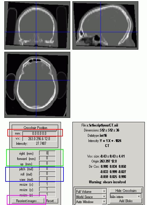
3 VOI format (just a gzipped NII file). You can use the VOI file with MRIcron or NPM. Troubleshootings: Manually setting the starting estimate MRI Normalization The previously described unified segmentation normalization can provide excellent registration, but it does require a high quality T1 scan (though anecdotally it can also work with good T2 scans or DWI B0 scans). In some clinical situations there are no T1 scans available (as DWI, PWI, FLAIR, T2 images are typically more clinically relevant). In these cases, you can try to normalize your MRI scans using the MRI normalization function. 1. With Matlab open, press the Batch button to show the Batch editor window. Then select SPM/Tools/Clinical/MRnormalize (see the CT Normalization section for a picture). 2. Make sure your images have a fairly accurate origin (see the Setting the starting estimate section). 3. Click on Anatomicals and press Select files choose your MRI (DWI, T2, FLAIR) image(s). Note you can select multiple files. For the tutorial, select T2nii 4. Optional: if you have lesion maps, click on the Lesions item and press Select files. If you are processing multiple files, they must be entered in the same order as the images from the previous step. For the tutorial, select the image lesiont2.nii 5. Optional: if the lesion maps are drawn on a different scan than selected for step 3, select the pathological scans used for lesion mapping. 6. Optional: The modality option lets you select the modality of your anatomical scans. The choices are T1, T2, FLAIR, and Other. Select the appropriate modality. For T1-weighted images, select T1. For B0 DWI images, and T2-weighted images, choose T2. For fluid-attenuated T2 scans, select FLAIR. Alternatively, you can try Other which attempts to use both the T1 and T2 templates. Note that this procedure always uses the default SPM templates, and therefore the brains will be aligned to the size and shape estimated from healthy young adults. The exception is the FLAIR option, which uses the images from the Glahn team based on 181 individuals (Mean age: 39.9y, std dev: 9.3y, range: 26-76y, 102 females, 7. Optional: By default this toolbox will not apply a template mask. If you select to use a brain mask the initial coarse normalization will get the image to be roughly aligned to the template, and then a brain mask will be applied so that regions outside the brain do not influence the normalization. This can help ensure that features like nose and bone shape do not influence the final normalization. However, the MR normalization option is often used for images with poor contrast and resolution, where the coarse fitting is not very accurate. 8. Optional: By default, images are normalized to 1x1x1mm resolution, but you can edit Bounding box and Voxel sizes options if you wish. 9. SPM will create a normalized bias corrected anatomical image (e.g. wt1.nii ) and a binarized, warped, smoothed lesion map ( bwslesiont2.nii ). It will also convert the lesion map to MRIcron s VOI format (just a gzipped NII file). You can use the VOI file with MRIcron or NPM. If this fails, see the "Troubleshootings: Manually setting the starting estimate" section. Troubleshootings: Manually setting the starting estimate Each of the normalization methods described above will default to automatically attempting to set the origin for a scan. If this fails, you should try manually setting the origin for your input data and re-running the normalization with the "Automatically Set Origin" option to false. This section describes how to manually set the origin. It is critical that normalization has a good starting estimate, otherwise the routines will get trapped in a local minimum resulting in a poor solution. For SPM, the starting estimate should point to the anterior commissure. We need to set this, because for most MRI scanners the starting estimate is the magnet isocenter, whereas for most CT scans it is the table center. This procedure is also described in 1. Start SPM8 (e.g. spm fmri from the Matlab command line). 2. Press the Display button in SPM s command window and select your CT scan (e.g. yourct.nii ).
. 4.")
4 3. Put into the mm edit and press enter (alternatively, click the horizontal bar between Crosshair Position and the mm edit). Blue crosshairs should appear (though you may not see them if your starting estimate is off). 4. Adjust the right {mm}, forward {mm} and up {mm} so that the blue crosshairs are centered on the anterior commissure. After entering a new number, press return to see the adjustment. For CT scans, you may have to shift the image by 100s of mms, as the image center is usually way off. 5. Press reorient images and select your CT scan (e.g. yourct.nii ). Note: if you have multiple scans from the same individual during the same session, reorient all of them with this single set of parameters (so they stay aligned with each other). 6. Put into the mm edit and press enter. Blue crosshairs should appear. 7. Adjust the pitch {rad}, roll {rad} and yaw {rad} so that the image is roughly aligned to stereotaxic space (e.g. the brain is not rotated relative to the blue crosshairs). Note these values are in Radians, so small values (e.g. 0.2) induce a large rotation.. 8. Press reorient images and select your CT scan (e.g. yourct.nii ). Note: if you have multiple scans from the same individual during the same session, reorient all of them with this single set of parameters (so they stay aligned with each other).
5

6 New features Version 8/8/2015 includes the following features By default each normalization routine automatically attempts to set the origin for each image. This reduces the tedious process of manually setting the origin. The automated process works by initially assuming the origin is located at the image's center of brightness. A linear coregisteration is then performed to approximately align the image to its template. This tends to be more robust than directly normalizing the images (as the normalization routines can get trapped in local minima). Some General Electric CT scanners fill portions outside the imaging circle with artificially dark pixels (-3024 Hounsfield units), which can disrupt normalization. The new version automatically detects these images and resets these regions to match air (-1024 Hounsfield). The MRI segmentation-normalization routine adds the option for enantiomorphic normalization, instead of the traditional lesion masked normalization. If you use enantiomorphic normalization you should cite the seminal work by Nachev et al., for example "Images were normalized to an ageappropriate template brain using the Clinical Toolbox for SPM (Rorden et al., 2012) using enantiomorphic normalization (Nachev et al., 2008)." If you use the traditional lesion masked normalization you should cite the seminal work by Brett et al. "Images were normalized to an ageappropriate template brain using the Clinical Toolbox for SPM (Rorden et al., 2012) using lesionmasked normalization (Brett et al., 2001).". The appropriate papers are Note that Nachev et al. do not provide routines for their process, and used several tools. Therefore, this toolbox attempts to mimic their effects. For our toolbox the anatomical scan's mirror is coregistered to the native anatomical scan. We then slightly dilate the lesion and apply a Gaussian blur. This blurred, dilated image is used to create a feathered mask for inserting tissue from the injured brain into the healthy brain. The images below show this effect with the sample T1 image provided in the tutorial. The left image shows the lesioned brain, the right image shows the same brain after the enantiomorphic fill.
GLM for fmri data analysis Lab Exercise 1
 GLM for fmri data analysis Lab Exercise 1 March 15, 2013 Medical Image Processing Lab Medical Image Processing Lab GLM for fmri data analysis Outline 1 Getting Started 2 AUDIO 1 st level Preprocessing
SPM Introduction. SPM : Overview. SPM: Preprocessing SPM! SPM: Preprocessing. Scott Peltier. FMRI Laboratory University of Michigan
 SPM Introduction Scott Peltier FMRI Laboratory University of Michigan! Slides adapted from T. Nichols SPM! SPM : Overview Library of MATLAB and C functions Graphical user interface Four main components:
SPM Introduction SPM! Scott Peltier. FMRI Laboratory University of Michigan. Software to perform computation, manipulation and display of imaging data
 SPM Introduction Scott Peltier FMRI Laboratory University of Michigan Slides adapted from T. Nichols SPM! Software to perform computation, manipulation and display of imaging data 1 1 SPM : Overview Library
An Introduction To Automatic Tissue Classification Of Brain MRI. Colm Elliott Mar 2014
 An Introduction To Automatic Tissue Classification Of Brain MRI Colm Elliott Mar 2014 Tissue Classification Tissue classification is part of many processing pipelines. We often want to classify each voxel
Methods for data preprocessing
 Methods for data preprocessing John Ashburner Wellcome Trust Centre for Neuroimaging, 12 Queen Square, London, UK. Overview Voxel-Based Morphometry Morphometry in general Volumetrics VBM preprocessing
Preprocessing II: Between Subjects John Ashburner
 Preprocessing II: Between Subjects John Ashburner Pre-processing Overview Statistics or whatever fmri time-series Anatomical MRI Template Smoothed Estimate Spatial Norm Motion Correct Smooth Coregister
Brain Extraction, Registration & EPI Distortion Correction
 Brain Extraction, Registration & EPI Distortion Correction What use is Registration? Some common uses of registration: Combining across individuals in group studies: including fmri & diffusion Quantifying
CHAPTER 2. Morphometry on rodent brains. A.E.H. Scheenstra J. Dijkstra L. van der Weerd
 CHAPTER 2 Morphometry on rodent brains A.E.H. Scheenstra J. Dijkstra L. van der Weerd This chapter was adapted from: Volumetry and other quantitative measurements to assess the rodent brain, In vivo NMR
Group (Level 2) fmri Data Analysis - Lab 4
 fmri Data Analysis - Lab 4") Group (Level 2) fmri Data Analysis - Lab 4 Index Goals of this Lab Before Getting Started The Chosen Ten Checking Data Quality Create a Mean Anatomical of the Group Group Analysis: One-Sample T-Test Examine
Group (Level 2) fmri Data Analysis - Lab 4 Index Goals of this Lab Before Getting Started The Chosen Ten Checking Data Quality Create a Mean Anatomical of the Group Group Analysis: One-Sample T-Test Examine
Supplementary methods
 Supplementary methods This section provides additional technical details on the sample, the applied imaging and analysis steps and methods. Structural imaging Trained radiographers placed all participants
ASAP_2.0 (Automatic Software for ASL Processing) USER S MANUAL
 USER S MANUAL") ASAP_2.0 (Automatic Software for ASL Processing) USER S MANUAL ASAP was developed as part of the COST Action "Arterial Spin Labelling Initiative in Dementia (AID)" by: Department of Neuroimaging, Institute
ASAP_2.0 (Automatic Software for ASL Processing) USER S MANUAL ASAP was developed as part of the COST Action "Arterial Spin Labelling Initiative in Dementia (AID)" by: Department of Neuroimaging, Institute
FROM IMAGE RECONSTRUCTION TO CONNECTIVITY ANALYSIS: A JOURNEY THROUGH THE BRAIN'S WIRING. Francesca Pizzorni Ferrarese
 FROM IMAGE RECONSTRUCTION TO CONNECTIVITY ANALYSIS: A JOURNEY THROUGH THE BRAIN'S WIRING Francesca Pizzorni Ferrarese Pipeline overview WM and GM Segmentation Registration Data reconstruction Tractography
Neuroimaging and mathematical modelling Lesson 2: Voxel Based Morphometry
 Neuroimaging and mathematical modelling Lesson 2: Voxel Based Morphometry Nivedita Agarwal, MD Nivedita.agarwal@apss.tn.it Nivedita.agarwal@unitn.it Volume and surface morphometry Brain volume White matter
This exercise uses one anatomical data set (ANAT1) and two functional data sets (FUNC1 and FUNC2).
 and two functional data sets (FUNC1 and FUNC2).") Exploring Brain Anatomy This week s exercises will let you explore the anatomical organization of the brain to learn some of its basic properties, as well as the location of different structures. The human
Exploring Brain Anatomy This week s exercises will let you explore the anatomical organization of the brain to learn some of its basic properties, as well as the location of different structures. The human
Fmri Spatial Processing
 Educational Course: Fmri Spatial Processing Ray Razlighi Jun. 8, 2014 Spatial Processing Spatial Re-alignment Geometric distortion correction Spatial Normalization Smoothing Why, When, How, Which Why is
Functional MRI data preprocessing. Cyril Pernet, PhD
 Functional MRI data preprocessing Cyril Pernet, PhD Data have been acquired, what s s next? time No matter the design, multiple volumes (made from multiple slices) have been acquired in time. Before getting
Image Registration + Other Stuff
 Image Registration + Other Stuff John Ashburner Pre-processing Overview fmri time-series Motion Correct Anatomical MRI Coregister m11 m 21 m 31 m12 m13 m14 m 22 m 23 m 24 m 32 m 33 m 34 1 Template Estimate
Machine Learning for Medical Image Analysis. A. Criminisi
 Machine Learning for Medical Image Analysis A. Criminisi Overview Introduction to machine learning Decision forests Applications in medical image analysis Anatomy localization in CT Scans Spine Detection
Single Subject Demo Data Instructions 1) click "New" and answer "No" to the "spatially preprocess" question.
 click New and answer No to the spatially preprocess question.") (1) conn - Functional connectivity toolbox v1.0 Single Subject Demo Data Instructions 1) click "New" and answer "No" to the "spatially preprocess" question. 2) in "Basic" enter "1" subject, "6" seconds
(1) conn - Functional connectivity toolbox v1.0 Single Subject Demo Data Instructions 1) click "New" and answer "No" to the "spatially preprocess" question. 2) in "Basic" enter "1" subject, "6" seconds
Whole Body MRI Intensity Standardization
 Whole Body MRI Intensity Standardization Florian Jäger 1, László Nyúl 1, Bernd Frericks 2, Frank Wacker 2 and Joachim Hornegger 1 1 Institute of Pattern Recognition, University of Erlangen, {jaeger,nyul,hornegger}@informatik.uni-erlangen.de
Spatial normalization of injured brains for neuroimaging research: An illustrative introduction of available options
 Spatial normalization of injured brains for neuroimaging research: An illustrative introduction of available options Junghoon Kim, PhD, Brian Avants, PhD, Sunil Patel, MS, and John Whyte, MD, PhD 1 Recent
fmri/dti analysis using Dynasuite
 fmri/dti analysis using Dynasuite Contents 1 Logging in 2 Finding patient session 3 Viewing and adjusting images 4 Checking brain segmentation 5 Checking image registration 6 Seeing fmri results 7 Saving
Computational Neuroanatomy
 Computational Neuroanatomy John Ashburner john@fil.ion.ucl.ac.uk Smoothing Motion Correction Between Modality Co-registration Spatial Normalisation Segmentation Morphometry Overview fmri time-series kernel
Introduction to fmri. Pre-processing
 Introduction to fmri Pre-processing Tibor Auer Department of Psychology Research Fellow in MRI Data Types Anatomical data: T 1 -weighted, 3D, 1/subject or session - (ME)MPRAGE/FLASH sequence, undistorted
CS/NEUR125 Brains, Minds, and Machines. Due: Wednesday, April 5
 CS/NEUR125 Brains, Minds, and Machines Lab 8: Using fmri to Discover Language Areas in the Brain Due: Wednesday, April 5 In this lab, you will analyze fmri data from an experiment that was designed to
Spatial Filtering Methods in MEG. Part 3: Template Normalization and Group Analysis"
 Spatial Filtering Methods in MEG Part 3: Template Normalization and Group Analysis" Douglas Cheyne, PhD" Program in Neurosciences and Mental Health" Hospital for Sick Children Research Institute " &" Department
Introduction to Neuroimaging Janaina Mourao-Miranda
 Introduction to Neuroimaging Janaina Mourao-Miranda Neuroimaging techniques have changed the way neuroscientists address questions about functional anatomy, especially in relation to behavior and clinical
Diffusion-MRI processing for group analysis
 Diffusion-MRI processing for group analysis Felix Renard IRMaGe: Inserm US 17 / CNRS UMS 3552 University Hospital of Grenoble - France 25/09/2015 felixrenard@gmail.com 1 Diffusion-MRI processing for group
Stroke Quantification Tool (Sonia) Ver User Manual
 Ver User Manual") Stroke Quantification Tool (Sonia) Ver. 1.0 User Manual English. 12/2016 Rev. 1.0 www.wakeup-stroke.eu 1 Table of Contents 1. Introduction...3 2. Installation...4 3. Data Import...5 4. Registration...7
Stroke Quantification Tool (Sonia) Ver. 1.0 User Manual English. 12/2016 Rev. 1.0 www.wakeup-stroke.eu 1 Table of Contents 1. Introduction...3 2. Installation...4 3. Data Import...5 4. Registration...7
Automated MR Image Analysis Pipelines
 Automated MR Image Analysis Pipelines Andy Simmons Centre for Neuroimaging Sciences, Kings College London Institute of Psychiatry. NIHR Biomedical Research Centre for Mental Health at IoP & SLAM. Neuroimaging
Functional MRI in Clinical Research and Practice Preprocessing
 Functional MRI in Clinical Research and Practice Preprocessing fmri Preprocessing Slice timing correction Geometric distortion correction Head motion correction Temporal filtering Intensity normalization
Concurrent Visualization of and Mapping between 2D and 3D Medical Images for Disease Pattern Analysis
 Concurrent Visualization of and Mapping between 2D and 3D Medical Images for Disease Pattern Analysis Mei Xiao 1, Jung Soh 1, Thao Do 1, Oscar Meruvia-Pastor 1 and Christoph W. Sensen 1 1 Department of
Automatic Registration-Based Segmentation for Neonatal Brains Using ANTs and Atropos
 Automatic Registration-Based Segmentation for Neonatal Brains Using ANTs and Atropos Jue Wu and Brian Avants Penn Image Computing and Science Lab, University of Pennsylvania, Philadelphia, USA Abstract.
SPM8 for Basic and Clinical Investigators. Preprocessing. fmri Preprocessing
 SPM8 for Basic and Clinical Investigators Preprocessing fmri Preprocessing Slice timing correction Geometric distortion correction Head motion correction Temporal filtering Intensity normalization Spatial
VBM Tutorial. 1 Getting Started. John Ashburner. March 12, 2015
 VBM Tutorial John Ashburner March 12, 2015 1 Getting Started The data provided are a selection of T1-weighted scans from the freely available IXI dataset 1. The overall plan will be to Start up SPM. Check
GLIRT: Groupwise and Longitudinal Image Registration Toolbox
 Software Release (1.0.1) Last updated: March. 30, 2011. GLIRT: Groupwise and Longitudinal Image Registration Toolbox Guorong Wu 1, Qian Wang 1,2, Hongjun Jia 1, and Dinggang Shen 1 1 Image Display, Enhancement,
fmri Preprocessing & Noise Modeling
 Translational Neuromodeling Unit fmri Preprocessing & Noise Modeling Lars Kasper September 25 th / October 17 th, 2015 MR-Technology Group & Translational Neuromodeling Unit An SPM Tutorial Institute for
Learn Image Segmentation Basics with Hands-on Introduction to ITK-SNAP. RSNA 2016 Courses RCB22 and RCB54
 Learn Image Segmentation Basics with Hands-on Introduction to ITK-SNAP RSNA 2016 Courses RCB22 and RCB54 RCB22 Mon, Nov 28 10:30-12:00 PM, Room S401CD RCB54 Thu, Dec 1 2:30-4:30 PM, Room S401CD Presenters:
LST: A lesion segmentation tool for SPM
 LST: A lesion segmentation tool for SPM Manual/Documentation for version 2.0.15 June 2017 Paul Schmidt Lucie Wink Contents 1 Getting started 3 1.1 License................................. 3 1.2 Installation...............................
Neuroimaging Group Pipeline Quick Start Manual
 Centre for Healthy Brain Ageing (CHeBA) Neuroimaging Group Pipeline Quick Start Manual UBO Detector UBO Detector is a cluster-based white matter hyperintensity (WMH) extraction pipeline based on k-nearest
SISCOM (Subtraction Ictal SPECT CO-registered to MRI)
") SISCOM (Subtraction Ictal SPECT CO-registered to MRI) Introduction A method for advanced imaging of epilepsy patients has been developed with Analyze at the Mayo Foundation which uses a combination of
SISCOM (Subtraction Ictal SPECT CO-registered to MRI) Introduction A method for advanced imaging of epilepsy patients has been developed with Analyze at the Mayo Foundation which uses a combination of
Advanced Visual Medicine: Techniques for Visual Exploration & Analysis
 Advanced Visual Medicine: Techniques for Visual Exploration & Analysis Interactive Visualization of Multimodal Volume Data for Neurosurgical Planning Felix Ritter, MeVis Research Bremen Multimodal Neurosurgical
Multiple Sclerosis Brain MRI Segmentation Workflow deployment on the EGEE grid
 Multiple Sclerosis Brain MRI Segmentation Workflow deployment on the EGEE grid Erik Pernod 1, Jean-Christophe Souplet 1, Javier Rojas Balderrama 2, Diane Lingrand 2, Xavier Pennec 1 Speaker: Grégoire Malandain
Automatic Generation of Training Data for Brain Tissue Classification from MRI
 MICCAI-2002 1 Automatic Generation of Training Data for Brain Tissue Classification from MRI Chris A. Cocosco, Alex P. Zijdenbos, and Alan C. Evans McConnell Brain Imaging Centre, Montreal Neurological
n o r d i c B r a i n E x Tutorial DTI Module
 m a k i n g f u n c t i o n a l M R I e a s y n o r d i c B r a i n E x Tutorial DTI Module Please note that this tutorial is for the latest released nordicbrainex. If you are using an older version please
MARS: Multiple Atlases Robust Segmentation
 Software Release (1.0.1) Last updated: April 30, 2014. MARS: Multiple Atlases Robust Segmentation Guorong Wu, Minjeong Kim, Gerard Sanroma, and Dinggang Shen {grwu, mjkim, gerard_sanroma, dgshen}@med.unc.edu
Zurich SPM Course Voxel-Based Morphometry. Ged Ridgway (Oxford & UCL) With thanks to John Ashburner and the FIL Methods Group
 With thanks to John Ashburner and the FIL Methods Group") Zurich SPM Course 2015 Voxel-Based Morphometry Ged Ridgway (Oxford & UCL) With thanks to John Ashburner and the FIL Methods Group Examples applications of VBM Many scientifically or clinically interesting
Zurich SPM Course 2015 Voxel-Based Morphometry Ged Ridgway (Oxford & UCL) With thanks to John Ashburner and the FIL Methods Group Examples applications of VBM Many scientifically or clinically interesting
Basic fmri Design and Analysis. Preprocessing
 Basic fmri Design and Analysis Preprocessing fmri Preprocessing Slice timing correction Geometric distortion correction Head motion correction Temporal filtering Intensity normalization Spatial filtering
Norbert Schuff VA Medical Center and UCSF
 Norbert Schuff Medical Center and UCSF Norbert.schuff@ucsf.edu Medical Imaging Informatics N.Schuff Course # 170.03 Slide 1/67 Objective Learn the principle segmentation techniques Understand the role
MR IMAGE SEGMENTATION
 MR IMAGE SEGMENTATION Prepared by : Monil Shah What is Segmentation? Partitioning a region or regions of interest in images such that each region corresponds to one or more anatomic structures Classification
EMSegmenter Tutorial (Advanced Mode)
") EMSegmenter Tutorial (Advanced Mode) Dominique Belhachemi Section of Biomedical Image Analysis Department of Radiology University of Pennsylvania 1/65 Overview The goal of this tutorial is to apply the
EMSegmenter Tutorial (Advanced Mode) Dominique Belhachemi Section of Biomedical Image Analysis Department of Radiology University of Pennsylvania 1/65 Overview The goal of this tutorial is to apply the
Image Processing for fmri John Ashburner. Wellcome Trust Centre for Neuroimaging, 12 Queen Square, London, UK.
 Iage Processing for fmri John Ashburner Wellcoe Trust Centre for Neuroiaging, 12 Queen Square, London, UK. Contents * Preliinaries * Rigid-Body and Affine Transforations * Optiisation and Objective Functions
EPI Data Are Acquired Serially. EPI Data Are Acquired Serially 10/23/2011. Functional Connectivity Preprocessing. fmri Preprocessing
 Functional Connectivity Preprocessing Geometric distortion Head motion Geometric distortion Head motion EPI Data Are Acquired Serially EPI Data Are Acquired Serially descending 1 EPI Data Are Acquired
Spatial Normalization of Brain Images with Focal Lesions Using Cost Function Masking
 NeuroImage 14, 486 500 (2001) doi:10.1006/nimg.2001.0845, available online at http://www.idealibrary.com on Spatial Normalization of Brain Images with Focal Lesions Using Cost Function Masking Matthew
Measuring longitudinal brain changes in humans and small animal models. Christos Davatzikos
 Measuring longitudinal brain changes in humans and small animal models Christos Davatzikos Section of Biomedical Image Analysis University of Pennsylvania (Radiology) http://www.rad.upenn.edu/sbia Computational
SPM8 for Basic and Clinical Investigators. Preprocessing
 SPM8 for Basic and Clinical Investigators Preprocessing fmri Preprocessing Slice timing correction Geometric distortion correction Head motion correction Temporal filtering Intensity normalization Spatial
Tutorial BOLD Module
 m a k i n g f u n c t i o n a l M R I e a s y n o r d i c B r a i n E x Tutorial BOLD Module Please note that this tutorial is for the latest released nordicbrainex. If you are using an older version please
Transforming Datasets to Talairach-Tournoux Coordinates
 -1- Transforming Datasets to Talairach-Tournoux Coordinates The original purpose of AFNI was to perform the transformation of datasets to Talairach-Tournoux (stereotaxic) coordinates The transformation
Quantitative MRI of the Brain: Investigation of Cerebral Gray and White Matter Diseases
 Quantities Measured by MR - Quantitative MRI of the Brain: Investigation of Cerebral Gray and White Matter Diseases Static parameters (influenced by molecular environment): T, T* (transverse relaxation)
fmri pre-processing Juergen Dukart
 fmri pre-processing Juergen Dukart Outline Why do we need pre-processing? fmri pre-processing Slice time correction Realignment Unwarping Coregistration Spatial normalisation Smoothing Overview fmri time-series
Evaluation of multiple voxel-based morphometry approaches and applications in the analysis of white matter changes in temporal lobe epilepsy
 Evaluation of multiple voxel-based morphometry approaches and applications in the analysis of white matter changes in temporal lobe epilepsy Wenjing Li a, Huiguang He a, Jingjing Lu b, Bin Lv a, Meng Li
MRI Segmentation MIDAS, 2007, 2010
 MRI Segmentation MIDAS, 2007, 2010 Lawrence O. Hall, Dmitry Goldgof, Yuhua Gu, Prodip Hore Dept. of Computer Science & Engineering University of South Florida CONTENTS: 1. Introduction... 1 2. Installing
syngo.mr Neuro 3D: Your All-In-One Post Processing, Visualization and Reporting Engine for BOLD Functional and Diffusion Tensor MR Imaging Datasets
 syngo.mr Neuro 3D: Your All-In-One Post Processing, Visualization and Reporting Engine for BOLD Functional and Diffusion Tensor MR Imaging Datasets Julien Gervais; Lisa Chuah Siemens Healthcare, Magnetic
Preprocessing of fmri data (basic)
") Preprocessing of fmri data (basic) Practical session SPM Course 2016, Zurich Andreea Diaconescu, Maya Schneebeli, Jakob Heinzle, Lars Kasper, and Jakob Sieerkus Translational Neuroodeling Unit (TNU) Institute
Preprocessing of fmri data (basic) Practical session SPM Course 2016, Zurich Andreea Diaconescu, Maya Schneebeli, Jakob Heinzle, Lars Kasper, and Jakob Sieerkus Translational Neuroodeling Unit (TNU) Institute
Analysis of fmri data within Brainvisa Example with the Saccades database
 Analysis of fmri data within Brainvisa Example with the Saccades database 18/11/2009 Note : All the sentences in italic correspond to informations relative to the specific dataset under study TP participants
This Time. fmri Data analysis
 This Time Reslice example Spatial Normalization Noise in fmri Methods for estimating and correcting for physiologic noise SPM Example Spatial Normalization: Remind ourselves what a typical functional image
How does the ROI affect the thresholding?
 How does the ROI affect the thresholding? Micro-computed tomography can be applied for the visualization of the inner structure of a material or biological tissue in a non-destructive manner. Besides visualization,
Preprocessing of fmri Data in SPM 12 - Lab 1
 Preprocessing of fmri Data in SPM 12 - Lab 1 Index Goals of this Lab Preprocessing Overview MATLAB, SPM, Data Setup Preprocessing I: Checking Motion Correction Preprocessing II: Coregistration Preprocessing
Statistical Analysis of Neuroimaging Data. Phebe Kemmer BIOS 516 Sept 24, 2015
 Statistical Analysis of Neuroimaging Data Phebe Kemmer BIOS 516 Sept 24, 2015 Review from last time Structural Imaging modalities MRI, CAT, DTI (diffusion tensor imaging) Functional Imaging modalities
Ischemic Stroke Lesion Segmentation Proceedings 5th October 2015 Munich, Germany
 0111010001110001101000100101010111100111011100100011011101110101101012 Ischemic Stroke Lesion Segmentation www.isles-challenge.org Proceedings 5th October 2015 Munich, Germany Preface Stroke is the second
Joint CI-JAI advanced accelerator lecture series Imaging and detectors for medical physics Lecture 1: Medical imaging
 Joint CI-JAI advanced accelerator lecture series Imaging and detectors for medical physics Lecture 1: Medical imaging Dr Barbara Camanzi barbara.camanzi@stfc.ac.uk Course layout Day AM 09.30 11.00 PM 15.30
NIH Public Access Author Manuscript Proc IEEE Int Symp Biomed Imaging. Author manuscript; available in PMC 2014 November 15.
 NIH Public Access Author Manuscript Published in final edited form as: Proc IEEE Int Symp Biomed Imaging. 2013 April ; 2013: 748 751. doi:10.1109/isbi.2013.6556583. BRAIN TUMOR SEGMENTATION WITH SYMMETRIC
Spatial Preprocessing
 Spatial Preprocessing Overview of SPM Analysis fmri time-series Design matrix Statistical Parametric Map John Ashburner john@fil.ion.ucl.ac.uk Motion Correction Smoothing General Linear Model Smoothing
Artifact Detection and Repair: Overview and Sample Outputs
 Artifact Detection and Repair: Overview and Sample Outputs Paul Mazaika February 2007 Programs originated in Gabrieli Neuroscience Laboratory, updated and enhanced at Center for Interdisciplinary Brain
fmri Analysis Sackler Ins2tute 2011
 fmri Analysis Sackler Ins2tute 2011 How do we get from this to this? How do we get from this to this? And what are those colored blobs we re all trying to see, anyway? Raw fmri data straight from the scanner
MIDAS Processing of Segmentation Data for Brain Lesions
 MIDAS Processing of Segmentation Data for Brain Lesions A. A. Maudsley 4/6/2012 For studies that contain lesions, the MIDAS processing has two steps that benefit from a modified processing pipeline to
Skull Segmentation of MR images based on texture features for attenuation correction in PET/MR
 Skull Segmentation of MR images based on texture features for attenuation correction in PET/MR CHAIBI HASSEN, NOURINE RACHID ITIO Laboratory, Oran University Algeriachaibih@yahoo.fr, nourine@yahoo.com
ANALYSIS OF FUNCTIONAL MAGNETIC RESONANCE IMAGING DATA USING SPM99: VOXEL-BASED MORPHOMETRY DONNA ROSE ADDIS
 Donna Rose Addis, TWRI, May 2004 1 ANALYSIS OF FUNCTIONAL MAGNETIC RESONANCE IMAGING DATA USING SPM99: VOXEL-BASED MORPHOMETRY DONNA ROSE ADDIS DEPT. OF PSYCHOLOGY, UNIVERSITY OF TORONTO TORONTO WESTERN
Appendix E1. Supplementary Methods. MR Image Acquisition. MR Image Analysis
 RSNA, 2015 10.1148/radiol.2015150532 Appendix E1 Supplementary Methods MR Image Acquisition By using a 1.5-T system (Avanto, Siemens Medical, Erlangen, Germany) under a program of regular maintenance (no
Object Identification in Ultrasound Scans
 Object Identification in Ultrasound Scans Wits University Dec 05, 2012 Roadmap Introduction to the problem Motivation Related Work Our approach Expected Results Introduction Nowadays, imaging devices like
The alignment process uses two alignment methods: (1) the mrvista function, rxalign, for a rough alignment, and then (2) KNK's alignment for
 the mrvista function, rxalign, for a rough alignment, and then (2) KNK's alignment for") Alignment (Allegra) Download example alignment script here. Anatomical images are collected at much higher resolution (isotropic 1 mm voxels) than functional images (isotropic 2 mm voxels; sometimes 2.5).
Alignment (Allegra) Download example alignment script here. Anatomical images are collected at much higher resolution (isotropic 1 mm voxels) than functional images (isotropic 2 mm voxels; sometimes 2.5).
Crop Counting and Metrics Tutorial
 Crop Counting and Metrics Tutorial The ENVI Crop Science platform contains remote sensing analytic tools for precision agriculture and agronomy. In this tutorial you will go through a typical workflow
Section 9. Human Anatomy and Physiology
 Section 9. Human Anatomy and Physiology 9.1 MR Neuroimaging 9.2 Electroencephalography Overview As stated throughout, electrophysiology is the key tool in current systems neuroscience. However, single-
Data pre-processing framework in SPM. Bogdan Draganski
 Data pre-processing fraework in SPM Bogdan Draganski Outline Why do we need pre-processing? Overview Structural MRI pre-processing fmri pre-processing Why do we need pre-processing? What do we want? Reason
Critique: Efficient Iris Recognition by Characterizing Key Local Variations
 Critique: Efficient Iris Recognition by Characterizing Key Local Variations Authors: L. Ma, T. Tan, Y. Wang, D. Zhang Published: IEEE Transactions on Image Processing, Vol. 13, No. 6 Critique By: Christopher
Pattern Recognition for Neuroimaging Toolbox: PRoNTo
 Click to edit Master title style Pattern Recognition for Neuroimaging Toolbox: PRoNTo Jessica Schrouff PRNI 2018 June 14 th NUS, Singapore Click Outline to edit Master title style PRoNTo s goals and history
Data Visualisation in SPM: An introduction
 Data Visualisation in SPM: An introduction Alexa Morcom Edinburgh SPM course, April 2015 SPMmip [-30, 3, -9] 3 Visualising results remembered vs. fixation contrast(s) < < After the results table - what
Version. Getting Started: An fmri-cpca Tutorial
 Version 11 Getting Started: An fmri-cpca Tutorial 2 Table of Contents Table of Contents... 2 Introduction... 3 Definition of fmri-cpca Data... 3 Purpose of this tutorial... 3 Prerequisites... 4 Used Terms
Image Registration I
 Image Registration I Comp 254 Spring 2002 Guido Gerig Image Registration: Motivation Motivation for Image Registration Combine images from different modalities (multi-modality registration), e.g. CT&MRI,
Data Visualisation in SPM: An introduction
 Data Visualisation in SPM: An introduction Alexa Morcom Edinburgh SPM course, April 2010 Centre for Cognitive & Neural Systems/ Department of Psychology University of Edinburgh Visualising results remembered
n o r d i c B r a i n E x Tutorial DSC Module
 m a k i n g f u n c t i o n a l M R I e a s y n o r d i c B r a i n E x Tutorial DSC Module Please note that this tutorial is for the latest released nordicbrainex. If you are using an older version please
fmri Basics: Spatial Pre-processing Workshop
 fmri Basics: Spatial Pre-processing Workshop Starting a VNC session: Most of your fmri analysis will be done on the central Linux machines accessed via a VNC (Virtual Network Computing) server. This is
Biomedical Image Processing
 Biomedical Image Processing Jason Thong Gabriel Grant 1 2 Motivation from the Medical Perspective MRI, CT and other biomedical imaging devices were designed to assist doctors in their diagnosis and treatment
BrainSpace V2.1: Geometric mapping and diffusion-based software for cross-subject multimodality brain imaging informatics
 Brief Introduction to BrainSpace V2.1 BrainSpace V2.1: Geometric mapping and diffusion-based software for cross-subject multimodality brain imaging informatics Graphics and Imaging Laboratory Wayne State
Data mining for neuroimaging data. John Ashburner
 Data mining for neuroimaging data John Ashburner MODELLING The Scientific Process MacKay, David JC. Bayesian interpolation. Neural computation 4, no. 3 (1992): 415-447. Model Selection Search for the best
SPM99 fmri Data Analysis Workbook
 SPM99 fmri Data Analysis Workbook This file is a description of the steps needed to use SPM99 analyze a fmri data set from a single subject using a simple on/off activation paradigm. There are two parts
arxiv: v1 [stat.ap] 1 Jun 2016
![arxiv: v1 [stat.ap] 1 Jun 2016](/thumbs/81/83094240.jpg "arxiv: v1 [stat.ap] 1 Jun 2016") Permutation-based cluster size correction for voxel-based lesion-symptom mapping arxiv:1606.00475v1 [stat.ap] 1 Jun 2016 June 3, 2016 Daniel Mirman a,b,1 Jon-Frederick Landrigan a Spiro Kokolis a Sean
Permutation-based cluster size correction for voxel-based lesion-symptom mapping arxiv:1606.00475v1 [stat.ap] 1 Jun 2016 June 3, 2016 Daniel Mirman a,b,1 Jon-Frederick Landrigan a Spiro Kokolis a Sean
Surface-based Analysis: Inter-subject Registration and Smoothing
 Surface-based Analysis: Inter-subject Registration and Smoothing Outline Exploratory Spatial Analysis Coordinate Systems 3D (Volumetric) 2D (Surface-based) Inter-subject registration Volume-based Surface-based
Where are we now? Structural MRI processing and analysis
 Where are we now? Structural MRI processing and analysis Pierre-Louis Bazin bazin@cbs.mpg.de Leipzig, Germany Structural MRI processing: why bother? Just use the standards? SPM FreeSurfer FSL However:
Methodological progress in image registration for ventilation estimation, segmentation propagation and multi-modal fusion
 Methodological progress in image registration for ventilation estimation, segmentation propagation and multi-modal fusion Mattias P. Heinrich Julia A. Schnabel, Mark Jenkinson, Sir Michael Brady 2 Clinical