Last class. This class. Single molecule imaging Deconvolution. FLIM Confocal
|
|
|
- Sara Miles
- 6 years ago
- Views:
Transcription
1 FLIM, Confocal
2 Last class Single molecule imaging Deconvolution This class FLIM Confocal
3 FLIM Fluorescence Lifetime IMaging
4 FLIM Fluorescence lifetime imaging Easier and more accurate quantitation Much harder to measure For a population of fluorophores BB = QQQQ εε FF tt = II 0 tt NN(tt) QQQQ(tt) εε γ(tt) γ(t) = photobleaching as a function of time N(t) = number of fluorophores ττ dd = QQQQ = QQQQ 1 kk ff + kk nnnn kk ff kk ff + kk nnnn = kk ff ττ dd k f is mostly constant for a given molecule k f -1 is the radiation lifetime how long it lives in the excited state before decay
![Fluorescence Lifetime Imaging (FLIM) Examine physical properties of fluorophores Can avoid complications like photobleaching and concentration effects Fluorophore Lifetime [ns] Excitation Max [nm]](/docs-images/76/74410136/images/5-0.jpg "Emission Max [nm] Solvent ATTO 655 3.6 655 690 Water Acridine Orange 2.0 500 530 PB ph 7.8 Alexa Fluor 488 4.1 494 519 PB ph 7.4 Alexa Fluor 647 1.0 651 672 Water BODIPY FL 5.")
5 Fluorescence Lifetime Imaging (FLIM) Examine physical properties of fluorophores Can avoid complications like photobleaching and concentration effects Fluorophore Lifetime [ns] Excitation Max [nm] Emission Max [nm] Solvent ATTO Water Acridine Orange PB ph 7.8 Alexa Fluor PB ph 7.4 Alexa Fluor Water BODIPY FL Methanol Coumarin Ethanol CY3B PBS CY PBS CY PBS Fluorescein PB ph 7.5 Oregon Green PB ph 9 FF tt = FF 0 ee tt/ττ Ru(bpy) 2 (dcpby)[pf 6 ] Water Pyrene > Water Indocyanine Green Water Rhodamine B PB 7.8


6 Measuring lifetime Need a very short excitation pulse Need to measure on very short time scales Collect image many times, at each delay to build up total image Takes a long time to image
7 Fitting multiple time constants Can only realistically fit up to 3 time constants. More than that will introduce artifacts
 1 MM = 1 + (ωω nn ττ) 2 ωω nn = Frequency of excitation")
8 Frequency domain measurements Instead of watching decay, apply sinusoidal light Lifetime can be calculated from the phase delay of the fluorescence relative to the excitation Typically only works on one lifetime species If τ phase is not equal to τ m, then there is more than one lifetime Δφφ = arctan(ωω nn ττ) 1 MM = 1 + (ωω nn ττ) 2 ωω nn = Frequency of excitation light

9 Frequency domain Can take images fast Somewhat reasonable for live cell

10 FLIM advantages Insensitive to concentration Insensitive to photobleaching Easily able to convert to absolute units In FRET, you only measure the donor lifetime, reduces quantification errors Much harder to analyze if there are multiple species of fluorophores


11 FLIM applications Measurements of ph use dye that has a lifetime dependent on the local environment FRET interactions in cells
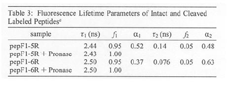
12 Peptide conformations
13 Confocal microscopy 3D sectioning Increased resolution

14 Widefield fluorescence Bright light source illuminates the entire field of view at the same time Fluorescence cube is placed between the objective and the camera In epi-illumination, the objective acts as both the condenser and the objective typically use high NA with low fluorescence glass

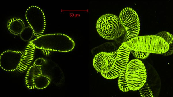
15 Widefield images
16 Confocal principle Image plane Tube lens Dichroic Obj Camera PMT


17 Confocal principle Light is focused to a point in the sample Emitted photons imaged on to a plane Aperture is placed at the image plane Single element detector placed behind aperture
 and emission (diffraction limited, but with different wavelength). dd xx,yy = 0.")
18 Improved resolution in confocal Resolution is now set both by excitation and emission light. New PSF is product of excitation (diffraction limited spot) and emission (diffraction limited, but with different wavelength). dd xx,yy = 0.4λλ NNNN Optimal diffraction Widefield Confocal


19 Scan to form an image Confocal only illuminates a single point Have to scan the point around the sample to build up spatial information Use fast moving galvo mirrors to rapidly move the laser across the sample Typically move in a raster pattern Tradeoff between time on pixel and frame rate Fast galvo can scan around 1 khz On a 512 x 512 pixel image -> 2 µs per pixel, 500 ms total exposure
20 Confocal principle Image plane Tube lens Dichroic Obj Camera PMT dd xx,yy = 0.4λλ NNNN

21 Confocal images Confocal advantages: -Blocks out of focus light rays, increasing resolution and contrast -Higher flexibility in image size and acquisition -Allows 3D sectioning dd xx,yy = 0.4λλ NNNN


22 3D PSF PSF has a width in x and y, we ve seen this before In confocal, now we can think about a z- detection PSF as well, set by the pinhole Narrower the pinhole, the better z resolution dd zz = 1.4λλn NNNN 2

23 3D imaging Use galvo mirrors to take X-Y image Motorized stage to move sample in Z Take another X-Y image Repeat
 z Voxel x Pixel")
24 Voxels Regular grid in 3D space Has dimensions in x, y, and z Z dimensions set by confocal system Each voxel is assigned an intensity value in each color Z resolution is lower than x,y resolution Sampling at least at the Nyquist frequency (1/2 size of smallest feature) z Voxel x Pixel
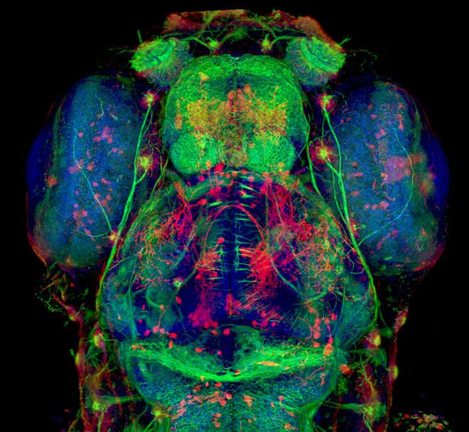
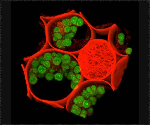
25 3D reconstruction
26 And on to Matlab
NDD FLIM Systems for Leica SP2 MP and SP5 MP Multiphoton Microscopes
 NDD FLIM Systems for Leica SP2 MP and SP5 MP Multiphoton Microscopes bh FLIM systems for the confocal and the multiphoton versions of the Leica SP2 and SP5 microscopes are available since 2002 [4]. These
Optical Sectioning. Bo Huang. Pharmaceutical Chemistry
 Optical Sectioning Bo Huang Pharmaceutical Chemistry Approaches to 3D imaging Physical cutting Technical difficulty Highest resolution Highest sensitivity Optical sectioning Simple sample prep. No physical
Introductory Guide to Light Microscopy - Biomedical Confocal Microscopy
 Introductory Guide to Light Microscopy - Biomedical Confocal Microscopy 7 May 2007 Michael Hooker Microscopy Facility Michael Chua microscopy@unc.edu 843-3268 6007 Thurston Bowles Wendy Salmon wendy_salmon@med.unc.edu
Samples Carolina sample slides (pollen, algae, ). Clean off oil with lens paper then OpticPad around lens (metal not glass) when done.
. Clean off oil with lens paper then OpticPad around lens (metal not glass) when done.") Bi/BE 227 Winter 2018 Assignment #1 Widefield and confocal laser scanning microscopy Schedule: Jan 3: Lecture Jan 3-12: Students get trained on how to use scopes, start on assignment Jan 3-17: Carrying
Bi/BE 227 Winter 2018 Assignment #1 Widefield and confocal laser scanning microscopy Schedule: Jan 3: Lecture Jan 3-12: Students get trained on how to use scopes, start on assignment Jan 3-17: Carrying
IMAGING PLATFORMS IN THE FACULTY OF MEDICINE. Guo Jing Lab Manager Faculty Core Facilty June
 IMAGING PLATFORMS IN THE FACULTY OF MEDICINE Guo Jing Lab Manager Faculty Core Facilty June 27 2011 http://www.med.hku.hk/corefac/ Mission Training and education Basic operation Advanced application Imaging
- Free fluorophores - Donors without partner acceptors - Acceptors without partner donors
 Determining Distances / Distance Changes Caveats Angular dependence (κ 2 ) Environment dependence (J, ϕd) Distance is a (complicated) average Probe is large, linkages can be long Construct complications
Nonlinear optics and two photon microscopy. Table of contents. Sam Whiteley and Seth Parker PHYS 173/BGGN 266 July 13, 2014
 Nonlinear optics and two photon microscopy Sam Whiteley and Seth Parker PHYS 173/BGGN 266 July 13, 2014 Table of contents 1. Introduction 2. Optical setup 3. Initial images and troubleshooting 4. Determining
Confocal Microscope Imaging of Single-Emitter Fluorescence and Hanbury Brown & Twiss Setup for Photon Antibunching. Edward Pei
 Confocal Microscope Imaging of Single-Emitter Fluorescence and Hanbury Brown & Twiss Setup for Photon Antibunching Edward Pei Abstract The purpose of these labs was to study single photon sources and measure
Leica TCS STED CW. The Fast Track to Superresolution. Leica TCS STED CW
 The Fast Track to Superresolution content Motivation Concept Realisation Applications - why do researchers need the TCS STED CW? - what is the TCS STED CW based on? - how does the TCS STED CW work? - what
Confocal vs. Deconvolution
 Confocal vs. Deconvolution Cesare Covino ALEMBIC Advanced Light and Electron Bio-Imaging Center Istituto Scientifico San Raffaele (Milano) www.hsr.it/research/alembic Fluorescence high contrast high sensibility
COLOCALISATION. Alexia Loynton-Ferrand. Imaging Core Facility Biozentrum Basel
 COLOCALISATION Alexia Loynton-Ferrand Imaging Core Facility Biozentrum Basel OUTLINE Introduction How to best prepare your samples for colocalisation How to acquire the images for colocalisation How to
DSU Start-Up instructions
 DSU Start-Up instructions Always: - start with the 10x objective - properly center the stage around the current objective before changing to another objective - when done, leave the 10x objective in standby
LIFA KEY FEATURES APPLICATIONS. Fluorescence Lifetime Attachment LIFA 15001A02 16/03/2015
 LIFA Fluorescence Lifetime Attachment LIFA 151A2 16/3/215 The LIFA is a dedicated system for Fluorescence Lifetime Imaging Microscopy (FLIM). It allows the generation of lifetime images on any widefield
CFIM MICROSCOPY COURSE TIMETABLE PRINCIPLES OF MICROSCOPY MONDAY 6 TH OF JANUARY 2014 FRIDAY 10 TH OF JANUARY 2014
 MICROSCOPY COURSE TIMETABLE PRINCIPLES OF MICROSCOPY MONDAY 6 TH OF JANUARY 2014 FRIDAY 10 TH OF JANUARY 2014 CONFOCAL AND FLUORESCENCE MICROSCOPY MONDAY 20 TH OF JANUARY 2014 FRIDAY 24 TH OF JANUARY 2014
Indiana Center for Biological Microscopy. BioRad MRC 1024 MP Confocal & Multi-Photon Microscope
 Indiana Center for Biological Microscopy BioRad MRC 1024 MP Confocal & Multi-Photon Microscope Microscope and the Attached Accessories A: B: C: D: E: F: G: H: Mercury Lamp Transmission Light Kr/Ar Laser
Single particle tracking: principles and applications
 Single particle tracking: principles and applications Valeria Levi Laboratorio de Dinámica Intracelular Facultad de Ciencias Exactas y Naturales Universidad de Buenos Aires vlevi1@gmail.com Why single
FastFLIMTm. For Olympus Laser Scanning Microscope Systems ( FV1000, FV1200, FV3000, FVMPE-RS )
") FastFLIMTm For Olympus Laser Scanning Microscope Systems ( FV1000, FV1200, FV3000, FVMPE-RS ) Add functionality to the Confocal Microscopes by Olympus with the Fluorescence Lifetime Imaging (FLIM) and
FastFLIMTm For Olympus Laser Scanning Microscope Systems ( FV1000, FV1200, FV3000, FVMPE-RS ) Add functionality to the Confocal Microscopes by Olympus with the Fluorescence Lifetime Imaging (FLIM) and
2, the coefficient of variation R 2, and properties of the photon counts traces
 Supplementary Figure 1 Quality control of FCS traces. (a) Typical trace that passes the quality control (QC) according to the parameters shown in f. The QC is based on thresholds applied to fitting parameters
Supplementary Figure 1 Guide stars of progressively longer wavelengths can be used for direct wavefront sensing at increasingly large depth in the
 Supplementary Figure 1 Guide stars of progressively longer wavelengths can be used for direct wavefront sensing at increasingly large depth in the cortex of the living mouse. Typical SH images of guide
COLOCALISATION. Alexia Ferrand. Imaging Core Facility Biozentrum Basel
 COLOCALISATION Alexia Ferrand Imaging Core Facility Biozentrum Basel OUTLINE Introduction How to best prepare your samples for colocalisation How to acquire the images for colocalisation How to analyse
Principles of Light Microscopy
 Monday 8 August 2011 Principles of Light Microscopy 09:00 09:30 Introduction 09:30 10:15 The story of the microscope / 10:15 Coffee 10:30 12:45 Limitations of the eye. Resolution, contrast, magnification.
LSM 5 MP, LSM 510 and LSM 510 META Laser Scanning Microscopes
 LSM 5 MP, LSM 510 and LSM 510 META Laser Scanning Microscopes Brief Operating Manual Release 4.2 January 2007 Contents Page Starting the System...3 Setting the microscope...6 Configuring the beam path
Non-Descanned FLIM Systems for Olympus FV-1000 and FV-300 Multiphoton Microscopes
 Non-Descanned FLIM Systems for Olympus FV-1000 and FV-300 Multiphoton Microscopes Abstract. Recently multiphoton versions of the Olympus FV 1000 and FV 300 laser scanning microscopes have become available.
LIFA. SPECIFICATIONs. Fluorescence Lifetime Attachment LIFA14001A02 25/02/2014
 LIFA Fluorescence Lifetime Attachment The LIFA is a dedicated system for Fluorescence Lifetime Imaging Microscopy (FLIM). It allows the generation of lifetime images on any widefield fluorescence microscope
Cell-based FLIM Energy-Transfer Measurements Using Alba
 Cell-based FLIM Energy-Transfer Measurements Using Alba ISS, Inc. Introduction This application note describes the use of Alba - Confocal Spectroscopy and Imaging Workstation for the acquisition and analysis
Confocal Microscopy Imaging of Single Emitter Fluorescence and Hanbury Brown, and Twiss Setup for Photon Antibunching. Abstract
 James Maslek 10/26/12 Confocal Microscopy Imaging of Single Emitter Fluorescence and Hanbury Brown, and Twiss Setup for Photon Antibunching Abstract The purpose of this experiment was to observe fluorescence
Diskovery. Multi-modal Imaging System
 Diskovery Multi-modal Imaging System Diskovery DISKOVERY SYSTEM CONFIGURATOR Diskover More Different tools are required to answer different questions. Now you can answer more questions about a sample during
FLUOVIEW FV1000/FV1200
 FLUOVIEW FV1000/FV1200 UPGRADE TO 3D NANOIMAGING AND SINGLE MOLECULE TRACKING FOR OLYMPUS FLUOVIEW FV1000/FV1200 Within the past few years, several methods have been devised in order to obtain images with
Building Your Own 2-Photon Microscope: Challenges, Advantages and Limitations
 Building Your Own 2-Photon : Challenges, Advantages and Limitations Roberto Weigert, Ph.D. Intracellular Membrane Trafficking Unit Oral and Pharyngeal Cancer Branch NIDCR-NIH Building Your Own 2-Photon
Confocal Raman Imaging with WITec Sensitivity - Resolution - Speed. Always - Provable - Routinely
 Confocal Raman Imaging with WITec Sensitivity - Resolution - Speed Always - Provable - Routinely WITec GmbH, Ulm, Germany, info@witec.de, www.witec.de A modular microscope series An Example: FLIM optical
Raster Image Correlation Spectroscopy and Number and Brightness Analysis
 Raster Image Correlation Spectroscopy and Number and Brightness Analysis 15 Principles of Fluorescence Course Paolo Annibale, PhD On behalf of Dr. Enrico Gratton lfd Raster Image Correlation Spectroscopy
Microscopy Specs. Media refractive index
 Zeiss AxioImager A2 EC Plan-Neofluar (420320-9901-000000) EC Plan-Neofluar (420330-9901-000000) Plan-Apochromat (420640-9900-000000) Plan-Apochromat (420650-9901-000000) Plan-Apochromat Korr (420660-9970-000)
Leica TCS SP8 X Confocal Microscope and Leica Application Suite X software DRAFT VERSION Room B123b
 Leica TCS SP8 X Confocal Microscope and Leica Application Suite X software DRAFT VERSION Room B123b User Guide Biomedicum Imaging Unit (BIU) University of Helsinki www.biu.helsinki.fi 11.11.2016 1 GENERAL...1
Chapter 4 Microscopy
 Chapter 4 Microscopy Gabriel Popescu University of Illinois at Urbana Champaign Beckman Institute Quantitative Light Imaging Laboratory http://light.ece.uiuc.edu Principles of Optical Imaging Electrical
DeltaVision OMX SR super-resolution microscope. Super-resolution doesn t need to be complicated
 DeltaVision OMX SR super-resolution microscope Super-resolution doesn t need to be complicated Live-cell super-resolution microscopy DeltaVision OMX SR is a compact super-resolution microscope system optimized
Leica TCS STED. The Fast Track to Superresolution Technical Documentation
 Leica TCS STED The Fast Track to Superresolution Technical Documentation 8 9 6 7 Inverted research microscope Leica DMI6000 CS Scan head Laser and power supply Computer table Air damped optical table 6
PH880 Topics in Physics
 PH880 Topics in Physics Modern Optical Imaging (Fall 2010) Overview of week 8 Monday Nonlinear Microscopy Wednesday No class (Mid term week) Quantum Optics Electro- magnetic Optics Wave Optics Ray Optics
Zeiss Universal SIM. A structured illumination system based on a Zeiss Universal microscope stand. by Nidhogg
 Zeiss Universal SIM A structured illumination system based on a Zeiss Universal microscope stand. by Nidhogg Structured illumination Structured illumination applied to fluorescence microscopy is a super
ONBI Practical 7: Comparison of techniques
 ONBI Practical 7: Comparison of techniques RM Parton 2014 Aims of practical 7: One of the most common issues confronting people new to microscopy is the confusing array of different techniques available.
Confocal Raman Microscope RAMOS
 Confocal Raman Microscope RAMOS 1 future`s Confocal Raman Microscope RAMOS 2 Confocal Raman Microscope RAMOS Ostec Corporate Group produces and offers hi-tech innovative scientific and analytical equipment.
Calibrate the Confocal Volume for FCS Using the FCS Calibration Script
 Tutorial Calibrate the Confocal Volume for FCS Using the FCS Calibration Script Summary This tutorial shows step-by-step, how to calibrate the confocal volume for FCS measurements with a calibration dye.
CM2002 Cerna Microscope
 CERNA MICROSCOPE FOR DODT CONTRAST IMAGING Equipped with Six-Cube Epi-Illuminator and Dodt Contrast Imaging Module Ready to Accept Objectives, Cameras, Filters, and Illumination Sources CM2002 Cerna Microscope
TissueFAXS SL Confocal high throughput configuration (actual appearance of the product may differ)
") TISSUEFAXS CONFOCAL TissueFAXS SL Confocal high throughput configuration (actual appearance of the product may differ) TissueFAXS Confocal provides a unique combination of digital slide scanning and laser
TISSUEFAXS CONFOCAL TissueFAXS SL Confocal high throughput configuration (actual appearance of the product may differ) TissueFAXS Confocal provides a unique combination of digital slide scanning and laser
with an incubator for study of live
 Light microscopy Support medium is glass. Choose this route for confocal imaging. Do you want to quickly check your cells for green, red or blue fluorescence or using phase contrast? (If so, refer to microscope
Dorigny resources MICROSCOPES - BIOPHORE. Confocal Microscope Zeiss LSM 700
 Dorigny resources Contributed by Yannick KREMPP Friday, 13 July 2007 Last Updated Monday, 30 October 2017 MICROSCOPES - BIOPHORE Confocal Microscope Zeiss LSM 700 This Zeiss LSM700 confocal is mounted
CM1002 Cerna Microscope
 CERNA MICROSCOPE WITH SIX-CUBE EPI-ILLUMINATOR Equipped with Six-Cube Epi-Illuminator Ready to Accept Objectives, Cameras, Filters, and Illumination Sources CM1002 Cerna Microscope (Optical Table Not Included)
SPCM Software Runs Online-FLIM at 10 Images per Second
 SPCM Software Runs Online-FLIM at 10 Images per Second Abstract: Version 9.72 SPCM software of the bh TCSPC/FLIM systems displays fluorescence lifetime images at a rate of 10 images per second. The calculation
Fluorescence Microscope with Spinning Disk Confocal and Total Internal Reflection Fluorescence Modules
 Open Tender Notification for the procurement of Fluorescence Microscope with Spinning Disk Confocal and Total Internal Reflection Fluorescence Modules at the Indian Institute of Science, Bangalore (Last
SENSITIVITY DIFFERENTLY TO SEE THINGS THE SPEED AND. Opera Phenix High Content Screening System
 THE SPEED AND SENSITIVITY TO SEE THINGS DIFFERENTLY Opera Phenix High Content Screening System For research use only. Not for use in diagnostic procedures. HIGH CONTENT SCREENING WITHOUT THE COMPROMISE
TN425: A study of fluorescence standards confirms that OptiGrid confocal images are suitable for quantitative microscopy
 TN425: A study of fluorescence standards confirms that OptiGrid confocal images are suitable for quantitative microscopy Introduction The OptiGrid converts the illumination system of a conventional wide
Prizmatix. Optical Specifications. Mic-LED-635 spectrum. BLCC-02 Benchtop LED Current Controller Specifications
 Mic-LED-635 High Power Collimated Red LED Light Source for Fluorescence Microscopy Featuring Prizmatix Modular Design for Multi-Wavelength and Fiberoptic Setup Ver. 01 Introduction The compact Mic-LED-635,
BD CARV II Confocal Imager. Real-time, full spectrum, personal confocal
 BD CARV II Confocal Imager Real-time, full spectrum, personal confocal Introduction to spinning disk confocal imaging Explore the benefits of BD CARV II Confocal Imager The BD CARV II confocal imager utilizes
Back Illuminated Scientific CMOS
 Prime 95B Scientific CMOS Camera Datasheet CMOS, EMCCD AND CCD CAMERAS FOR LIFE SCIENCES Back Illuminated Scientific CMOS Discovery depends on every photon Primary applications: Super-Resolution Microscopy
BSI scmos. High Quantum Efficiency. Cooled Scientific CMOS Camera
 95 BSI scmos High Quantum Efficiency Cooled Scientific CMOS Camera Backside-illuminated scmos technology Opening a new era of high sensitivity imaging applications! The Dhyana 95 is a highly sensitive
BD CARV II Confocal Imager. Real-time, full spectrum, personal confocal
 BD CARV II Confocal Imager Real-time, full spectrum, personal confocal Introduction to spinning disk confocal imaging The BD CARV II confocal imager utilizes a Nipkow spinning disk containing multiple
Time-resolved wavelet- based acquisitions using a single pixel camera
 Florian Rousset 1,2, Nicolas Ducros 1, Andrea Farina 2, Gianluca Valentini 2, Cosimo D Andrea 2, Françoise Peyrin 1 1 Univ Lyon, INSA Lyon, CNRS 5220, INSERM U1206, CREATIS Lyon, France 2 Politecnico di
Transmitted light Illuminator VIS-LED, color temperature. Epifluorescence lamp module X-Cite 120PC Q (Excelitas
 ELYRA PS.1 Multi-functional fluorescence inverted widefield microscope enabling live-cell imaging, TIRF or HILO illuminion and two super-resolution techniques: structured illuminion microscopy (SIM) and
Image restoration by deconvolution
 Image restoration by deconvolution chong.zhang@bioquant.uni-heidelberg.de 17/12/2014 (part) Slides courtesy: Sébastien Tosi (IRB Barcelona) A few concepts related to the topic Convolution Deconvolution
INFINITY-CORRECTED TUBE LENSES
 INFINITY-CORRECTED TUBE LENSES For use with Infinity-Corrected Objectives Available in Focal Lengths Used by Thorlabs, Nikon, Leica, Olympus, and Zeiss Designs for Widefield and Laser Scanning Applications
Manual ANGSTROM grid confocal microscope
 Manual ANGSTROM grid confocal microscope Switching on the system 1) Start by switching on the different components of the microscope system. When using live cell imaging also start the temperature and
Vutara 350. Innovation with Integrity. The Fastest, Super-Resolution Microscope Deep 3D Imaging on Live Cells, Quickly and Easily
 Vutara 350 The Fastest, Super-Resolution Microscope Deep 3D Imaging on Live Cells, Quickly and Easily Innovation with Integrity Fluorescence Microscopy Vutara 350 Don t Get Left Behind Bruker s Vutara
Lightsheet Z.1. Light Sheet Fluorescence Microscopy by Carl Zeiss. Fabrice Schmitt, Sales Manager Carl ZEISS France
 Lightsheet Z.1 Light Sheet Fluorescence Microscopy by Carl Zeiss Fabrice Schmitt, Sales Manager Carl ZEISS France 12.12.2012 Light Sheet Fluorescence Microscopy (LSFM) Principle The Principle of Light
Renishaw invia Raman Microscope (April 2006)
") Renishaw invia Raman Microscope (April 2006) I. Starting the System 1. The main system unit is ON all the time. 2. Switch on the Leica microscope and light source for reflective bright field (BF) imaging.
Renishaw invia Raman Microscope (April 2006) I. Starting the System 1. The main system unit is ON all the time. 2. Switch on the Leica microscope and light source for reflective bright field (BF) imaging.
Score 1: No backward movement, animal shows shrinker behavior.
 Supplementary Information Axon regeneration in C. elegans after femtosecond laser axotomy M. F. Yanik, H. Cinar, H. N. Cinar, A. D. Chisholm, Y. Jin, and A. Ben-Yakar Methods: The worms were maintained
EPFL SV PTBIOP BIOP COURSE 2015 OPTICAL SLICING METHODS
 BIOP COURSE 2015 OPTICAL SLICING METHODS OPTICAL SLICING METHODS Scanning Methods Wide field Methods Point Scanning Deconvolution Line Scanning Multiple Beam Scanning Single Photon Multiple Photon Total
Single Photon Counting System
 PHOTONSCORE LINCam Single Photon Counting System User Manual Contact: www.photonscore.de email@photonscore.de Page 1! of!21 PHOTONSCORE Contents. 1 Getting started 3 1.1 In the box... 4 1.2 Before you
1. Getting Started: Brief Step-By-Step Guide (PDF File)
") Page 1 of 5 1. Getting Started: Brief Step-By-Step Guide (PDF File) Leica SP2 confocal microscope is controlled via software LCS (Leica Confocal Software). The latest version is V2.5. Bellowing is a brief
Page 1 of 5 1. Getting Started: Brief Step-By-Step Guide (PDF File) Leica SP2 confocal microscope is controlled via software LCS (Leica Confocal Software). The latest version is V2.5. Bellowing is a brief
TRiCAM APPLICATIONS KEY FEATURES. Time Resolved intensified CAMera. TRiCAM 13001A01 31/10/2013
 TRiCAM Time Resolved intensified CAMera The TRiCAM is a compact Intensified CCD camera for scientific and industrial applications that require 1) lowlight level imaging, 2) ultra-short exposures through
Technology and equipment at the Bioimaging Center
 Technology and equipment at the Bioimaging Center Light microscopy / Widefields Name Type Applications and equipment LEICA AF6000LX Nikon BioStation Timelaps imaging, CO2 and temperature controlled (14
Development of automated ultraviolet laser beam profiling system using fluorometric technique
 Development of automated ultraviolet laser beam profiling system using fluorometric technique BB Shrivastava*, NS Benerji, P Bhatnagar, HS Vora a and U Nundy Chemical and Excimer Laser Section a Laser
Widefield High Content Screening System User Guide
 ImageXpress Micro Widefield High Content Screening System User Guide 5015321 C April 2015 This document is provided to customers who have purchased Molecular Devices equipment, software, reagents, and
Biological Image Information I: A Description of the Modalities
 2.771J BEH.453J HST.958J Spring 2005 Lecture 21 April 2005 Biological Image I: A Description of the Modalities The Modalities Direct photography SEM and TEM Cryo-EM Two-photon microscopy Confocal microscopy
Live-cell 3D super-resolution imaging in thick biological samples
 Nature Methods Live-cell 3D super-resolution imaging in thick biological samples Francesca Cella Zanacchi, Zeno Lavagnino, Michela Perrone Donnorso, Alessio Del Bue, Lauria Furia, Mario Faretta & Alberto
Bulletin 80C01C01-01E
 Bulletin 80C01C01-01E *1 *2 The CSU-X1 is the advanced model of our CSU-series, which are widely recognized as the most powerful tools for live cell imaging. Faster The world's fastest scanning speed (up
Light and tissue: 2. Two-photon, Optical Coherence Tomography, Photoacoustics
 Light and tissue: 2 Two-photon, Optical Coherence Tomography, Photoacoustics Last lecture: Absorption and Scattering Last lecture: Mean free path Mean free path Transport mean free path Ntziachristos (2010)
Ray Optics. Lecture 23. Chapter 23. Physics II. Course website:
 Lecture 23 Chapter 23 Physics II Ray Optics Course website: http://faculty.uml.edu/andriy_danylov/teaching/physicsii Let s finish talking about a diffraction grating Diffraction Grating Let s improve (more
Virtual Frap User Guide
 Virtual Frap User Guide http://wiki.vcell.uchc.edu/twiki/bin/view/vcell/vfrap Center for Cell Analysis and Modeling University of Connecticut Health Center 2010-1 - 1 Introduction Flourescence Photobleaching
Multi-Photon Training
 Multi-Photon Training Overview This training will take approximately 4 hours. The first 2 hours will be spent learning one-on-one with your trainer, while the following 2 hours will be your opportunity
Time Tagged Time-Resolved Fluorescence Data Collection in Life Sciences
 Technical Note Time Tagged Time-Resolved Fluorescence Data Collection in Life Sciences Michael Wahl, Sandra Orthaus-Müller PicoQuant GmbH, Rudower Chaussee 29, 12489 Berlin, Germany, info@picoquant.com
1. ABOUT INSTALLATION COMPATIBILITY SURESIM WORKFLOWS a. Workflow b. Workflow SURESIM TUTORIAL...
 SuReSim manual 1. ABOUT... 2 2. INSTALLATION... 2 3. COMPATIBILITY... 2 4. SURESIM WORKFLOWS... 2 a. Workflow 1... 3 b. Workflow 2... 4 5. SURESIM TUTORIAL... 5 a. Import Data... 5 b. Parameter Selection...
TuCam. Features and Benefits. Andor TuCam - High Performance, Two Camera Imaging Adapter. Specifications Summary. Applications Guide
 Ultra Sensitive Imaging Microscopy Systems Features and Benefits Largest aperture Unique 22 mm aperture for large format sensors e.g. Neo and Zyla scmos High quality achromatic lenses Image from 425 -
Leica TCS SPE. Spectacular Imaging! Technical Documentation
 Leica TCS SPE Spectacular Imaging! Technical Documentation Leica TCS SPE Spectacular Imaging Easy to Achieve A Reliable System Affordable Excellence The high resolution spectral confocal Leica TCS SPE
All-In-One. digital inverted microscope. High-quality Imaging Has Never Been Easier. evos-ca.com
 All-In-One digital inverted microscope High-quality Imaging Has Never Been Easier evos-ca.com Fluorescence Imaging Is Now Easier Than Ever ALL-IN-ONE, digital inverted fluorescence microscope The easiest
PHYSICS. Chapter 33 Lecture FOR SCIENTISTS AND ENGINEERS A STRATEGIC APPROACH 4/E RANDALL D. KNIGHT
 PHYSICS FOR SCIENTISTS AND ENGINEERS A STRATEGIC APPROACH 4/E Chapter 33 Lecture RANDALL D. KNIGHT Chapter 33 Wave Optics IN THIS CHAPTER, you will learn about and apply the wave model of light. Slide
TuCam. Andor TuCam - High Performance, Two Camera Imaging Adapter. Features and Benefits. Specifications Summary.
 Multi-Wavelength Imaging Features and Benefits Largest aperture Unique 22 mm aperture for large format sensors e.g. Neo and Zyla scmos High quality achromatic lenses Image from 425-700 nm with minimal
Phys 1020, Day 18: Questions? Cameras, Blmfld Reminders: Next Up: digital cameras finish Optics Note Final Project proposals next week!
 Lights. Action. Phys 1020, Day 18: Questions? Cameras, Blmfld 15.1 Reminders: Next Up: digital cameras finish Optics Note Final Project proposals next week! 1 What have we learned in this section: 1) Lasers
Supporting information for: A highly directional room-temperature single. photon device
 Supporting information for: A highly directional room-temperature single photon device Nitzan Livneh,, Moshe G. Harats,, Daniel Istrati, Hagai S. Eisenberg, and Ronen Rapaport,, Applied Physics Department,
Optics Final Exam Name
 Instructions: Place your name on all of the pages. Do all of your work in this booklet. Do not tear off any sheets. Show all of your steps in the problems for full credit. Be clear and neat in your work.
Note: FLIM-FRET is a robust method to determine the FRET efficiency of a suited donor acceptor pair.
 Tutorial FLIM-FRET-Calculation for Multi-Exponential Donors Summary This tutorial shows step-by-step, how the "Lifetime FRET Image" script of SymPhoTime 64 can be used to calculate pixel-by-pixel the average
FLUOSTAR Rhodamine B- encapsulating microspheres are seeding particles optimized for Particle Image Velocimetry. Technical handbook ver.
 www.ebm.vc FLUOSTAR Rhodamine B- encapsulating microspheres are seeding particles optimized for Particle Image Velocimetry Technical handbook ver.1, Feb, 2010 CONTENTS 1) Introduction of fluorescent PIV
Constructing System Matrices for SPECT Simulations and Reconstructions
 Constructing System Matrices for SPECT Simulations and Reconstructions Nirantha Balagopal April 28th, 2017 M.S. Report The University of Arizona College of Optical Sciences 1 Acknowledgement I would like
2011 Optical Science & Engineering PhD Qualifying Examination Optical Sciences Track: Advanced Optics Time allowed: 90 minutes
 2011 Optical Science & Engineering PhD Qualifying Examination Optical Sciences Track: Advanced Optics Time allowed: 90 minutes Answer all four questions. All questions count equally. 3(a) A linearly polarized
WAVELENGTH MANAGEMENT
 Camera Accessories WAVELENGTH MANAGEMENT UV CONVERTERS UV Converters take advantage of a phenomenon called fluorescence to extend the performance range of the Beamage beam profiling camera to ultraviolet
Colocalization in Confocal Microscopy. Quantitative Colocalization Analysis
 Quantitative Colocalization Analysis 1 Quantitative Colocalization Analysis Colocalization of fluorescently labeled molecules (immunostainings, fluorescent proteins, etc.) is often used as the first indicator
MAT 17C - DISCUSSION #4, Counting Proteins
 MAT 17C - DISCUSSION #4, Counting Proteins Visualization of molecules inside a living cell is one of the most important tools of molecular biology in the 21 st century. In fact, it was the subject of the
Models of Light The wave model: The ray model: The photon model:
 Models of Light The wave model: under many circumstances, light exhibits the same behavior as sound or water waves. The study of light as a wave is called wave optics. The ray model: The properties of
STEP-BY-STEP INSTRUCTIONS FOR BUILDING A FLUORESCENCE MICROSCOPE. TECHSPEC Optical Cage System
 STEP-BY-STEP INSTRUCTIONS FOR BUILDING A FLUORESCENCE MICROSCOPE TECHSPEC Optical Cage System INTRODUCTION 2 What is a Digital Fluorescence Microscope? Unlike traditional microscopes, which utilize an
New Olympus FV3000RS Laser Scanning Confocal System. Quotation Number V2
 New Olympus FV3000RS Laser Scanning Confocal System Quotation Number 00056013 V2 Quotation Date: 08/08/2018 Revised Date: 07/09/2018 Quotation number: 00056013 Quotation Expiry: 07/11/2018 Macquarie University
User Operation Manual
 User Operation Manual Manual Version: 1.3 For Single Cell Bioanalyzer (SCB-402) Doc# 102-SCB-402-1.3 Table of Contents 1. General Information... 1 1.1 How to Use This Manual... 1 1.2 Operation Safety...
Leica HyD for Confocal Imaging. All-Purpose Super-Sensitivity
 Leica HyD for Confocal Imaging All-Purpose Super-Sensitivity Reduce light dosage, increase cell viability Excitation 488 Emission 490-556 nm Scan frequency 0 Hz No accumulation No averaging PMT HyD 3 Leica