Deep learning for undersampled MRI reconstruction
|
|
|
- George Bailey
- 6 years ago
- Views:
Transcription
1 1 Deep learning for undersampled MRI reconstruction Chang Min Hyun, Hwa Pyung Kim, ung Min Lee, ungchul Lee and Jin Keun eo Department of Computational cience and Engineering, Yonsei University, eoul, 03722, outh Korea Department of Mathematics, Yonsei University, eoul, 03722, outh Korea arxiv: v2 [stat.ml] 11 ep 2017 Abstract This paper presents a deep learning method for faster magnetic resonance imaging (MRI) by reducing k-space data with sub-nyquist sampling strategies and provides a rationale for why the proposed approach works well. Uniform subsampling is used in the time-consuming phase-encoding direction to capture high-resolution image information, while permitting the image-folding problem dictated by the Poisson summation formula. To deal with the localization uncertainty due to image folding, very few low-frequency k-space data are added. Training the deep learning net involves input and output images that are pairs of Fourier transforms of the subsampled and fully sampled k-space data. Numerous experiments show the remarkable performance of the proposed method; only 29% of k-space data can generate images of high quality as effectively as standard MRI reconstruction with fully sampled data. Index Terms Magnetic resonance imaging, Fast MRI, Deep learning, Undersampling. I. INTRODUCTION Magnetic resonance imaging (MRI) produces crosssectional images with high spatial resolution by using strong nuclear magnetic resonances, gradient fields, and hydrogen atoms inside the human body [8], [15]. MRI does not use damaging ionizing radiation like X-rays, but the scan takes a long time and involves confining the subject in an uncomfortable narrow tube. While shortening the MRI scan time does not reduce radioactive iodine dosage. it might help increase patients treatment satisfaction and reduce the medical cost. The MRI scan time is roughly proportional to the number of time-consuming phase-encoding steps in k-space. Many efforts have sought to expedite MRI scans by skipping phase-encoding lines in k-space while eliminating aliasing, a serious consequence of the Nyquist criteria violation [11] caused by skipping. To eliminate or ease aliasing, we use prior information on MR images of the missing k-space data. Parallel MRI and compressed MRI are some of the techniques used to deal with these aliasing artifacts. Parallel MRI installs multiple receiver coils and uses space-dependent properties of receiver coils to ease aliasing [7], [12], [16]. This paper focuses solely on undersampled MRI; for simplicity, parallel MRI will not be discussed. Undersampled MRI aims to produce medically acceptable high quality images as good as fully sampled MRI using undersampled MRI data. Undersampled MRI consists of two parts, the subsampling and reconstruction, as shown in Fig.1. The subsampling systematically reduces the original MRI k- space data x full to the undersampled data x. Reconstruction involves learning the optimal reconstruction function f : x y, Manuscript received XXX; revised XXX. Corresponding author:. Lee (sungchul@yonsei.ac.kr)). x full x ubsampling y y Fig. 1. General strategy for undersampled MRI reconstruction problem. The inverse Fourier transform () of a fully sampled k-space data x full produces a reconstructed MRI image y. The goal is to find a subsampling function and learn an undersampled MRI reconstruction f from training data set. Here, y = (x) is an aliased image caused by the violation of Nyquist criteria. We use the U-net to find the function g which provides the mapping from the aliased image y to an anti-aliased image y. where y is the MR image of the original MRI k-space data x full. One should note that this reconstruction function f cannot fully recover the original MR image y from the undersampled data x. f(x) only approximates y. The reconstructed image f(x) should approximate the full MR image y with medically acceptable tolerance. To minimize the reconstruction error, we must design the subsampling such that it preserves the information in x full as much as possible. On the other hand, to shorten MRI scan time, we must design the subsampling with high skipping rate. In this paper we balance these two and propose a subsampling that uses only 29% of the original data. We use the deep learning technique, more specifically the U-net [13] with some domain knowledge to construct the optimal reconstruction function f. Compressive sensing (C) MRI can be viewed as a method beyond the Nyquist requirement, in which the image sparsity is enforced to compensate for highly undersampled data [1], [10]. C-MRI can be described roughly as a model-fitting method to reconstruct the MR image y by adding a regularization term that enforces the sparsity-inducing prior on y. It aims to reconstruct an image given by y = argmin y g Reconstruction x F(y) 2 l 2 + λ T (y) l1, (1) where F denotes the Fourier transform, is a subsampling, T (y) represents a transformation capturing the sparsity pattern f


 l2 forces the residual x F(y) to be small, whereas T (y) l1 enforces the sparsity of T (y).")

2 2 2-skip? g Reconstruction f 2-skip + low frequency g Reconstruction f Fig. 2. Feasibility of deep learning methods. Learning f requires separability: y 1 y 2 implies F(y 1 ) F(y 2 ). The figure on the left shows how uniform subsampling does not satisfy the separability condition. We consider two different MR images with small anomalies at position (n, m) and (n, m + N/2), respectively. Corresponding k-space data are different, but corresponding 2-skip k-space data are completely identical. There is no way to identify whether the anomaly is at the top or bottom from these two identical 2-skip k-space data. In contrast, the figure on the right shows how separability can be achieved by adding low frequency data. of y, is the symbol of composition, and λ is the regularization parameter controlling the trade-off between the residual norm and regularity. Here, the term x F(y) l2 forces the residual x F(y) to be small, whereas T (y) l1 enforces the sparsity of T (y). In C-MRI, a priori knowledge of MR images is converted to a sparsity of T (y) with a suitable choice of T. The most widely used C method is total variation denoising (i.e., y l1 ), which enforces piecewise constant images by uniformly penalizing image gradients. Although C-MRI with random sampling has gained great attention over the past decade, it has some limitations in preserving fine-scale details and noise-like textures that hold diagnostically important information in MR images. In contrast to the regularized least square approaches (1), our deep learning approach is a completely reversed paradigm. It aims to learn a function f : x y using many training data {(x (i), y (i) ) : i = 1,, N}. Roughly speaking, f is achieved by 1 N f = argmin f(x (i) ) y (i) 2, (2) f U net N i=1 where U net is a deep convolutional neural network with some domain knowledge. This reconstruction function f can be viewed as the inverse mapping of the forward model F subject to the constraint of MR images, which are assumed to exist in a low dimensional manifold. In the conventional regularized least square framework (1), it is very difficult to incorporate the very complicated MR image manifold in the regularization term. However, in the deep learning framework, the manifold constraint learned from the training set acts as a highly nonlinear compressed sensing to get an useful reconstruction f(x) by leveraging complex prior knowledge on y. This paper shows that a suitably chosen subsampling with deep learning produces high-quality MR images as effectively as regular MRI reconstruction from fully sampled k-space data. To be precise, we skip four phase-encoding lines so that the Fourier transform contains all detailed features in a 4- folded image, according to the Poisson summation formula. We include 4% low-frequency sampling to learn the overall structure of MR images. Numerous experiments show the remarkable performance of the proposed method. II. METHOD Let y C N N be the MR image to be reconstructed, where N 2 is the number of pixels and C is the set of complex numbers. In 2D Fourier imaging with conventional Cartesian k-space sampling, the MR image y can be reconstructed from the corresponding k-space data x full C N N : For n, m = 1 N/2,, 0,, N/2, y(n, m) = N/2 N/2 a=1 N/2 b=1 N/2 x full (a, b) e 2iπ(an+bm)/N, (3) where x full (a, b) is the MR-signal received at k-space position (2πa/N, 2πb/N). The frequency-encoding is along a-axis in k-space and the phase-encoding is along b-axis as per our convention. In undersampled MRI, we violate the Nyquist criteria and skip phase-encoding lines during MRI acquisition to speed up the time-consuming phase encoding. Further, we use prior information on MR images and reconstruct the image from undersampled data. However, sub-nyquist k-space data results in aliasing artifacts in the image space. For example, suppose we skip two phase-encoding lines and get an acceleration factor of 2. Then, the k-space data with zero padding is given by x full ( N 2 1, N 2 ) x full( N 2, N 2 ) 0 0 x full ( N 2 1, N 2 2) x full( N 2, N 2 2) (4)
 If the deep learning approach finds an unfolding map y 2-fold y, we can accelerate the data acquisition speed.")
 and (n, m + N/2), respectively. As a result, the corresponding k-space data are different.")
3 3 According to the Poisson summation formula, the discrete Fourier transform of the above skipped data produces the following two-folded image: y 2-fold (n, m) = y(n, m) + y(n, m + N/2). (5) If the deep learning approach finds an unfolding map y 2-fold y, we can accelerate the data acquisition speed. However, it is impossible to get this unfolding map even with sophisticated manifold learning for MR images. In the left panel of Fig. 2, we consider two different MR images with small anomalies at position (n, m) and (n, m + N/2), respectively. As a result, the corresponding k-space data are different. However, the corresponding 2-skip k-space data are completely identical. There is no way to identify whether the anomaly is at the top or bottom from these two identical 2-skip k-space data. Deep learning cannot make impossible possible. Now, we are ready to explain our undersampling strategy for deep learning. A. ubsampling trategy Let {(x (j), y (j) )} M j=1 be a training set of undersampled and ground-truth MR images. The vectors x (j) and y (j) exist in the space C N N. Fig. 1 shows a schematic diagram of our undersampled reconstruction method, where the corresponding inverse problem is to solve the underdetermined linear system, F(y) = x. (6) Given undersampled data x, there are infinitely many solutions in the space C N N satisfying (6). In other word, it is impossible to invert the ill-conditioned system F : C N N R F, where R F is the range space of operator F and its dimension is much lower than N 2. We must use the fact that the MR images of humans exist in a much lower dimensional manifold M embedded in the space C N N. With this constraint M which is unknown, there is a possibility that there exists a practically meaningful inverse f in the sense that f ( F(y)) = y for y M. (7) The major issue is what types of subsampling allow to learn f from the corresponding training data. To learn f that meets the condition (7), the subsampling must satisfy the following separability condition: for y 1, y 2 M, y 1 y 2 implies F(y 1 ) F(y 2 ). (8) The image on the left in Fig. 2 shows the case what is the 2-skipped subsampling. With this choice of, two different images y 1 y 2 produce the same F(y 1 ) = F(y 2 ). This means that the 2-skipped subsampling is inappropriate to learn f satisfying (7). Here, y 1 is the standard Logan phantom image and y 2 is a modified image of y 1 obtained by moving three small anomalies to their symmetric positions with respect to the middle horizontal line. On the other hand, if we add a few low frequencies to 2-skipped subsampling as shown in the image on the right in Fig. 2, the situation is dramatically changed and separability (8) may be achieved. In Fig. 3, we demonstrate the separability again using the real data. (b) is the ground truth and the tumor is at the bottom. (a) is the reconstructed image using the 2-skipped subsampling; the tumors are found at both top and bottom, and the 2-skipped subsampling is not separable. However, in the reconstructed image (c) using the 2-skipped subsampling with added low frequencies the tumor is clearly located at the bottom and separability (8) may be achieved. This crucial observation is validated by various numerical simulations as shown in Fig. 6. (a) (b) (c) Fig. 3. MR images of human brain with tumor at the bottom. Images are reconstructed from (a) 50% uniform sampling, (b) full sampling, and (c) 50 % uniform sampling with some low frequencies added. In (a) the tumors are found at both top and bottom, and there exists a location uncertainty in uniform sampling. However, in the reconstructed image (c) using the 2- skipped subsampling with added low frequencies, the tumor is clearly located at the bottom and the location uncertainty can be removed by adding a few low frequencies in k-space. In our subsampling strategy, we use the 4-skip subsampling (25% k-space data), add a few low frequencies (4% k-space data), and obtain an acceleration factor of approximately 3.4. Owing to the Poisson summation formula, the 4-skipped data provides the detailed structure of the folded image of y as y 4-fold (n, m) = 3 y(n, m + jn ). (9) 4 j=0 However, the folded image may not contain location information of small anomalies. We fix the anomaly location uncertainty by adding a minimal amount of low frequency k-space data. Adding low frequency data provides a highly blurred image along the phase-encoding direction. However, this lowresolution image seems to be sufficient to compensate for the location uncertainty of the high-resolution folded image. B. Image Reconstruction Function Given a training set {y (j) } M j=1 of ground-truth MR images, we apply our subsampling strategy to each ground-truth MR image y (j) and get the training set {(x (j), y (j) )} M j=1 of subsampled data and ground-truth MR images. As shown in Fig. 4, the input x (j) of the image reconstruction function f is an undersampled k-space data and the output y (j) is our reconstruction image. In this subsection, we describe the design of the image reconstruction function f using deep learning. We first apply the inverse Fourier transform to the undersampled k-space data x (j), extract absolute values,and obtain folded images y (j). Our goal is then to recover the ground truth images y (j) from folded images y (j). There is an actively developing deep learning area called the super resolution,
4 4 : Deep Learning Process : k-space Correction Process : 3 3 Convolution, ReLU : 2 2 Max Pooling : 2 2 Avg Unpooling : 1 1 Convolution : Copy and Concat 256 U-net y = F 1 (x) ỹ = f d (y ) folded image U-net image Input x F(ỹ) undersampled data k-space data from the U-net ŷ = f cor (F(ỹ)) Output y = F 1 (ŷ) New k-space data our reconstruction image Fig. 4. Overall process of the proposed method including architecture of U-net. The image is first reconstructed by deep learning technique. The U-net image is further tuned by k-space correction. The final reconstruction is obtained by the inverse Fourier transform of the restored k-space data.

 as our activation function, and 2 2 max pooing with stride 2. We use zero-padding to equalize the input and label image size.")
5 5 which enhances the resolution of the poor original images. Our data set is different from the usual data sets handled by the super resolution, but the basic underlying ideas are identical. We search available tools and choose to use U-net, which has quite promising records in the medical imaging field. The architecture of our U-net is illustrated in Fig. 4. The first half of the net is contraction and the last half is expansion. Each step in contraction comprises two convolutions with 3 3 windows, a stride 1 in both directions,and zero-padding. After each convolution, we use a rectified linear unit(relu) as our activation function, and 2 2 max pooing with stride 2. We use zero-padding to equalize the input and label image size. During expansion, we use average unpooling instead of max pooling. After average unpooling, we concatenate a contraction feature map of the same size to the unpooling feature map. The input of the net is y (j), the weights is W, the net as a function is f net (, W ), and the output is denoted as f net (y (j), W ). To train the net we use the L2 loss and find the optimal weight set W 0 such that 1 M W 0 = argmin f net (y (j) W M, W ) y(j) 2 L2. (10) j=1 After training, the net as a function is f d = f net (, W 0 ), and the output is denoted by ỹ (j) = f d (y (j)) = f net(y (j), W 0). We input the folded image y (j) into the trained net and obtain the U-net image ỹ (j). We apply the Fourier transform to the U-net image ỹ (j) and get the k-space data F(ỹ (j) ) from the U-net. Recall that the input x (j) is undersampled k-space data and 71% of x (j) are zero-padded. The k-space data F(ỹ (j) ) from the U-net recovers the zero-padded part of the information and fills up the numbers. During this recovery, F(ỹ (j) ) also modifies the non zero-padded parts and distorts the numbers. It is reasonable to keep the original data x (j) in the non zero-padded part, change the zeros in the non zeropadded part and fill up the numbers from the k-space data F(ỹ (j) ) from the U-net. We call this k-space correction as f cor and the new k-space data ŷ (j) = f cor (F(ỹ (j) )). ince the original input data is preserved, we expect to obtain a more satisfactory reconstruction image. Our experiments show that the k-space correction is very effective. Finally, we apply Fourier transform to ŷ (j) and get our reconstruction image F(ŷ (j) ). In summary, our image reconstruction function f : x y is given by f = f cor F f d, (11) where f d is the trained U-net and f cor indicates the k-space correction. III. EXPERIMENT In our experiment, the ground-truth MR image y was normalized to be in the range [0, 1] and the undersampled data x was subsampled to 29% k-space data as described in ection II. We trained our model using a training set of 1,400 images from 30 patients. Fig. 5 shows the overall structure of our image reconstruction. To train our deep neural network, all weights were initialized by a zero-centered normal distribution with standard ubsampling g Reconstruction Fig. 5. chematic diagram of the proposed undersampled reconstruction. deviation 0.01 without bias term. The loss function was minimized using the RMPropOptimizer [6] with a learning rate of 0.001, weight decay of 0.9, and mini-batch size of 32 at each epoch. The number of epoch was 2,000. Training was implemented using Tensorflow [4] on an Intel(R) Core(TM) i7-6850k, 3.60GHz CPU and four NVIDIA GTX-1080, 8GB GPU system. The network required approximately 6 hours for training. Fig. 6 shows the performance of the proposed method for five different brain images in the test set. The first, second and third columns show the ground-truth, aliased and corrected images, respectively. The aliased images are folded four times. ome folds are quite strong and visible,while others are not. The proposed method suppresses these artifacts, but provides surprisingly sharp and natural-looking images. Fig. 7 displays the impact of k-space correction. The first, second, third, and last columns represent the ground-truth, aliased, U-net, and k-space correction images, respectively. The images (c) before and (d) after k-space correction are visually indistinguishable. In the second row, we display the difference between the images in the column and the ground truth. (e) is the difference between the ground truth and ground truth, and is the ground zero. (f) is the difference between the aliased image and ground truth. One can clearly see the folding artifacts. (g) is the difference between the U-net image and ground truth. The U-net eases the folding artifacts dramatically. However, one can still see the folding artifacts. (h) is the difference between the corrected image using k-space correction and ground truth. The k-space correction removes the remaining folding artifacts further. All our observations are supported by the quantitative evaluation. After training, we use the test set of 400 images, and measure and report their mean-squared error (ME) and tructural imilarity Index (IM) [17] in Table I. All these indices strongly support the effectiveness of both the U-net and k-space correction. In particular, the validity of k-space f

6 6 Ground Truth Aliased Image Corrected Image Fig. 6. Numerical simulation results of five different brain MR images. The first, second and third columns show the ground-truth, aliased and corrected images, respectively. The proposed method significantly reduces the undersampling artifacts while preserving morphological information.
-(h) depict the difference image to the image (a). correction is justified further. TABLE I QUANTITATIVE EVALUATION REULT IN TERM OF ME AND IM UING THE TET ET OF 400 IMAGE.")


7 7 (a) (b) (c) (d) (e) (f) (g) (h) Fig. 7. imulation result using the proposed method : (a) ground-truth image; (b) aliased image; (c) output from the trained network; (d) k-space corrected image; Fig. (e)-(h) depict the difference image to the image (a). correction is justified further. TABLE I QUANTITATIVE EVALUATION REULT IN TERM OF ME AND IM UING THE TET ET OF 400 IMAGE. Aliased U-net Proposed ME ± ± ± IM ± ± ± IV. DICUION AND CONCLUION Deep learning techniques exhibit surprisingly good performances in various challenging fields and our case is not an exception. In our case, it generates the reconstruction function f using the U-net, providing a very competitive performance over the existing methods. It should be mentioned that, at the time of writing this paper, we found that there is another work that proposes almost the same approach as [5], [9], [18]. However, in contrast to our paper, no theoretical explanation as to why the undersampled MRI reconstruction works well has been provided. Our inverse problem of undersampled MRI reconstruction is ill-posed in the sense that there are fewer equations than unknowns. The underdetermined system in ection III has unknowns and equations. The dimension of the set {y R : F(y) = 0} is (256 76) 256, and therefore it is impossible to have an explicit reconstruction formula for solving (6), without imposing a strong constraint of a solution manifold. For the uniqueness, the Hausdorff dimension of the solution manifold must be less than the number of equations (i.e., ). Unfortunately, it is extremely hard to find a mathematical expression for the complex structure of MR images in terms of parameters, because of its highly nonlinearity characteristic. The deep learning approach is a feasible way to capture MRI image structure as dimensionality reduction. We learned the kind of subsampling strategy necessary to have a medically acceptable image reconstruction function after extensive effort. Initially we used the 4-skip subsampling, but realized that it could not satisfy the separability condition. We added some low frequencies hoping to satisfy separability and this turned out to guarantee separability in a practical sense. Once the data set satisfies the separability condition, we have many deep learning tools to recover the images from the folded images. We chose to use the U-net. The trained U- net successfully unfolded and recovered the images from the folded images. The U-net eases the folding artifacts; however, one can still see them. The k-space correction eases the folding artifacts further. Numerous experiments show that our learned function f appears to have highly expressive representation capturing anatomical geometry as well as small anomalies. We also observed that the learned function f using MRI images solved the undetermined problem (6) for CT images that are never trained. Describing the process of recognizing the features and roles of functions in the learning network with mathematical rigorous analysis will be taken up in future research.
8 8 ACKNOWLEDGMENT This research was supported by the National Research Foundation of Korea No. NRF-2017R1A2B REFERENCE [1] E.J. Candès, J. Romberg and T. Tao, Robust Uncertainty Principles: Exact ignal Reconstruction from Highly Incomplete Frequency Information, IEEE Trans. Inf. Theory, vol. 52, no. 2, pp , [2] D.L. Donoho, Compressed sensing, IEEE Trans. Inf. Theory, vol. 52, pp , [3] D.L. Donoho, For most large underdetermined systems of linear equations the minimal 1-norm solution is also the sparsest solution, Communications on pure and applied mathematics, vol. 59, pp , [4] Google, TensorFlow: Large-scale machine learning on heterogeneous systems, URL [5] K. Hammernik, T. Klatzer, E. Kobler, M.P. Recht, D.K. odickson, T. Pock and F. Knoll, Learning ad Variational Network for Reconstruction of Accelerated MRI Data, arxiv preprint, arxiv: , [6] T. Tieleman and G. Hinton, Lecture 6.5-rmsprop: Divide the gradient by a running average of its recent magnitude, COURERA: Neural Networks for Machine Learning, [7] D.J. Larkman and R.G. Nunes, Parallel magnetic resonance imaging, Phys. Med. Biol., vol. 52, pp. R15 R55, [8] P.C. Lauterbur, Image Formation by Induced Local Interactions: Examples of Employing Nuclear Magnetic Resonance, Nature, vol. 242, pp , [9] D. Lee, J. Yoo, J.C. Ye, Deep artifact learning for compressed sensing and parallel MRI, arxiv preprint, arxiv: , [10] M. Lustig, D.L. Donoho and J.M. Pauly, parse MRI: The Application of Compressed ensing for Rapid MR Imaging, Magnetic Resonance in Medicine, Vol. 58, pp , [11] H. Nyquist, Certain topics in telegraph transmission theory, Trans. AIEE, vol. 47, pp , [12] K.P. Pruessmann, M. Weiger, M.B. cheidegger and P. Boesiger, ENE: sensitivity encoding for fast MRI, Magn. Reson. Med., vol. 42, pp , [13] O. Ronneberger, P. Fischer, and T. Brox, U-net: Convolutional networks for biomedical image segmentation, in Int. Conf. on Medical Image Computing and Computer-Assisted Intervention, pringer, pp , [14] J. K. eo and E. J. Woo, Nonlinear inverse problems in imaging, Chichester, U.K.: John Wiley & ons, [15] J. K. eo, E. J. Woo, U. Katscher, and Y. Wang, Electro-Magnetic Tissue Properties MRI, Imperial College Press, [16] D.K. odickson and W.J. Manning, imultaneous acquisition of spatial harmonics (MAH): fast imaging with radiofrequency coil arrays, Magn. Reson. Med., vol. 38, pp , [17] Z. Wang, A. C. Bovik, H.R. heikh, E.P. imoncelli, Image Quality Assessment: From Error Visibility to tructural imilarity, IEEE Trans. on Image Processing Vol. 13, no. 4, pp , [18]. Wang, Z. u, L. Ying, X. Peng,. Zhu, F. Liang, D. Feng and D. Liang, Accelerating magnetic resonance imaging via deep learning, in Proc. IEEE 13th Int. Conf. Biomedical Imaging, pp , 2016.

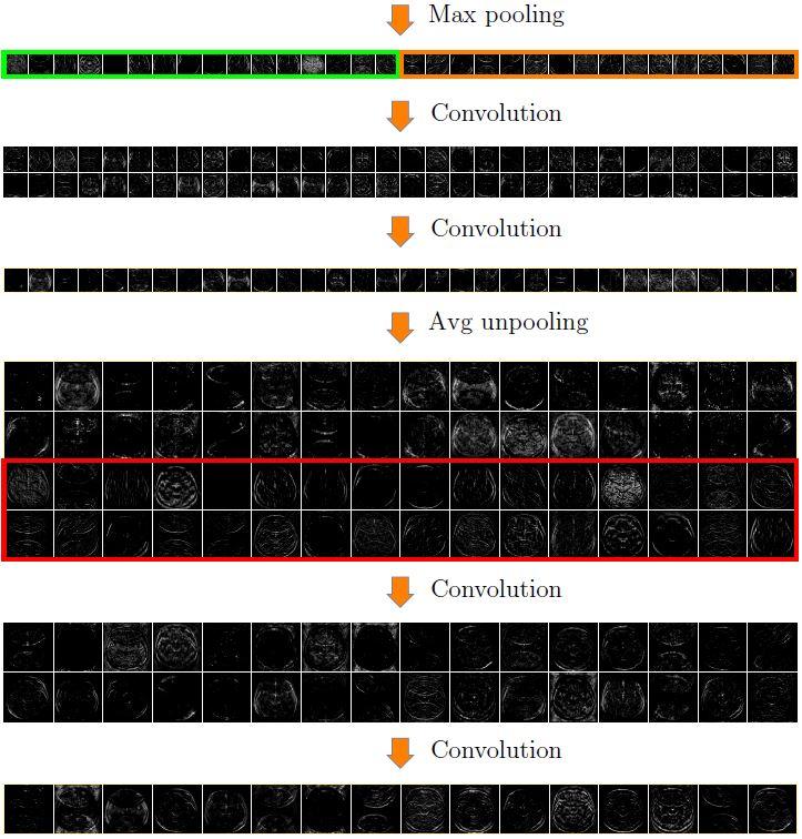
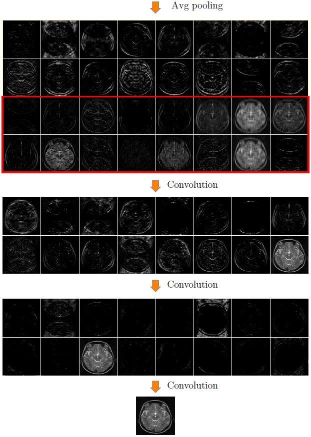
9 APPENDIX. This appendix presents the reconstruction process intuitively using a simplified version of the U-net. 9
10 10
11 11
Weighted-CS for reconstruction of highly under-sampled dynamic MRI sequences
 Weighted- for reconstruction of highly under-sampled dynamic MRI sequences Dornoosh Zonoobi and Ashraf A. Kassim Dept. Electrical and Computer Engineering National University of Singapore, Singapore E-mail:
Compressed Sensing Reconstructions for Dynamic Contrast Enhanced MRI
 1 Compressed Sensing Reconstructions for Dynamic Contrast Enhanced MRI Kevin T. Looby klooby@stanford.edu ABSTRACT The temporal resolution necessary for dynamic contrast enhanced (DCE) magnetic resonance
Introduction. Wavelets, Curvelets [4], Surfacelets [5].
![Introduction. Wavelets, Curvelets [4], Surfacelets [5].](/thumbs/87/95249322.jpg "Introduction. Wavelets, Curvelets [4], Surfacelets [5].") Introduction Signal reconstruction from the smallest possible Fourier measurements has been a key motivation in the compressed sensing (CS) research [1]. Accurate reconstruction from partial Fourier data
Introduction Signal reconstruction from the smallest possible Fourier measurements has been a key motivation in the compressed sensing (CS) research [1]. Accurate reconstruction from partial Fourier data
G Practical Magnetic Resonance Imaging II Sackler Institute of Biomedical Sciences New York University School of Medicine. Compressed Sensing
 G16.4428 Practical Magnetic Resonance Imaging II Sackler Institute of Biomedical Sciences New York University School of Medicine Compressed Sensing Ricardo Otazo, PhD ricardo.otazo@nyumc.org Compressed
Optimal Sampling Geometries for TV-Norm Reconstruction of fmri Data
 Optimal Sampling Geometries for TV-Norm Reconstruction of fmri Data Oliver M. Jeromin, Student Member, IEEE, Vince D. Calhoun, Senior Member, IEEE, and Marios S. Pattichis, Senior Member, IEEE Abstract
Enhao Gong, PhD Candidate, Electrical Engineering, Stanford University Dr. John Pauly, Professor in Electrical Engineering, Stanford University Dr.
 Enhao Gong, PhD Candidate, Electrical Engineering, Stanford University Dr. John Pauly, Professor in Electrical Engineering, Stanford University Dr. Greg Zaharchuk, Associate Professor in Radiology, Stanford
Compressed Sensing for Rapid MR Imaging
 Compressed Sensing for Rapid Imaging Michael Lustig1, Juan Santos1, David Donoho2 and John Pauly1 1 Electrical Engineering Department, Stanford University 2 Statistics Department, Stanford University rapid
MODEL-BASED FREE-BREATHING CARDIAC MRI RECONSTRUCTION USING DEEP LEARNED & STORM PRIORS: MODL-STORM
 MODEL-BASED FREE-BREATHING CARDIAC MRI RECONSTRUCTION USING DEEP LEARNED & STORM PRIORS: MODL-STORM Sampurna Biswas, Hemant K. Aggarwal, Sunrita Poddar, and Mathews Jacob Department of Electrical and Computer
NIH Public Access Author Manuscript Med Phys. Author manuscript; available in PMC 2009 March 13.
 NIH Public Access Author Manuscript Published in final edited form as: Med Phys. 2008 February ; 35(2): 660 663. Prior image constrained compressed sensing (PICCS): A method to accurately reconstruct dynamic
ECE 8201: Low-dimensional Signal Models for High-dimensional Data Analysis
 ECE 8201: Low-dimensional Signal Models for High-dimensional Data Analysis Yuejie Chi Departments of ECE and BMI The Ohio State University September 24, 2015 Time, location, and office hours Time: Tue/Thu
Deconvolution with curvelet-domain sparsity Vishal Kumar, EOS-UBC and Felix J. Herrmann, EOS-UBC
 Deconvolution with curvelet-domain sparsity Vishal Kumar, EOS-UBC and Felix J. Herrmann, EOS-UBC SUMMARY We use the recently introduced multiscale and multidirectional curvelet transform to exploit the
Incoherent noise suppression with curvelet-domain sparsity Vishal Kumar, EOS-UBC and Felix J. Herrmann, EOS-UBC
 Incoherent noise suppression with curvelet-domain sparsity Vishal Kumar, EOS-UBC and Felix J. Herrmann, EOS-UBC SUMMARY The separation of signal and noise is a key issue in seismic data processing. By
REGULARIZATION PARAMETER TRIMMING FOR ITERATIVE IMAGE RECONSTRUCTION
 REGULARIZATION PARAMETER TRIMMING FOR ITERATIVE IMAGE RECONSTRUCTION Haoyi Liang and Daniel S. Weller University of Virginia, Department of ECE, Charlottesville, VA, 2294, USA ABSTRACT Conventional automatic
Iterative CT Reconstruction Using Curvelet-Based Regularization
 Iterative CT Reconstruction Using Curvelet-Based Regularization Haibo Wu 1,2, Andreas Maier 1, Joachim Hornegger 1,2 1 Pattern Recognition Lab (LME), Department of Computer Science, 2 Graduate School in
P257 Transform-domain Sparsity Regularization in Reconstruction of Channelized Facies
 P257 Transform-domain Sparsity Regularization in Reconstruction of Channelized Facies. azemi* (University of Alberta) & H.R. Siahkoohi (University of Tehran) SUMMARY Petrophysical reservoir properties,
Sparse sampling in MRI: From basic theory to clinical application. R. Marc Lebel, PhD Department of Electrical Engineering Department of Radiology
 Sparse sampling in MRI: From basic theory to clinical application R. Marc Lebel, PhD Department of Electrical Engineering Department of Radiology Objective Provide an intuitive overview of compressed sensing
Accelerated MRI Techniques: Basics of Parallel Imaging and Compressed Sensing
 Accelerated MRI Techniques: Basics of Parallel Imaging and Compressed Sensing Peng Hu, Ph.D. Associate Professor Department of Radiological Sciences PengHu@mednet.ucla.edu 310-267-6838 MRI... MRI has low
Deep Learning of Compressed Sensing Operators with Structural Similarity Loss
 Deep Learning of Compressed Sensing Operators with Structural Similarity Loss Y. Zur and A. Adler Abstract Compressed sensing CS is a signal processing framework for efficiently reconstructing a signal
MODEL-based recovery of images from noisy and sparse. MoDL: Model Based Deep Learning Architecture for Inverse Problems
 1 MoDL: Model Based Deep Learning Architecture for Inverse Problems Hemant K. Aggarwal, Member, IEEE, Merry P. Mani, and Mathews Jacob, Senior Member, IEEE arxiv:1712.02862v3 [cs.cv] 10 Aug 2018 Abstract
Compressed Sensing MRI with Multichannel Data Using Multicore Processors
 Compressed Sensing MRI with Multichannel Data Using Multicore Processors Ching-Hua Chang and Jim Ji* Magnetic Resonance in Medicine 64:1135 1139 (2010) Compressed sensing (CS) is a promising method to
29 th NATIONAL RADIO SCIENCE CONFERENCE (NRSC 2012) April 10 12, 2012, Faculty of Engineering/Cairo University, Egypt
 April 10 12, 2012, Faculty of Engineering/Cairo University, Egypt") K1. High Performance Compressed Sensing MRI Image Reconstruction Ahmed Abdel Salam, Fadwa Fawzy, Norhan Shaker, Yasser M.Kadah Biomedical Engineering, Cairo University, Cairo, Egypt, ymk@k-space.org Computer
K1. High Performance Compressed Sensing MRI Image Reconstruction Ahmed Abdel Salam, Fadwa Fawzy, Norhan Shaker, Yasser M.Kadah Biomedical Engineering, Cairo University, Cairo, Egypt, ymk@k-space.org Computer
Deep convolutional neural networks for accelerated dynamic magnetic resonance imaging
 Deep convolutional neural networks for accelerated dynamic magnetic resonance imaging Christopher M. Sandino 1 Neerav Dixit 1 Joseph Y. Cheng 2 Shreyas S. Vasanawala 2 1 Department of Electrical Engineering,
Recovery of Piecewise Smooth Images from Few Fourier Samples
 Recovery of Piecewise Smooth Images from Few Fourier Samples Greg Ongie*, Mathews Jacob Computational Biomedical Imaging Group (CBIG) University of Iowa SampTA 2015 Washington, D.C. 1. Introduction 2.
Compressive Sensing for Multimedia. Communications in Wireless Sensor Networks
 Compressive Sensing for Multimedia 1 Communications in Wireless Sensor Networks Wael Barakat & Rabih Saliba MDDSP Project Final Report Prof. Brian L. Evans May 9, 2008 Abstract Compressive Sensing is an
Compressed Sensing Algorithm for Real-Time Doppler Ultrasound Image Reconstruction
 Mathematical Modelling and Applications 2017; 2(6): 75-80 http://www.sciencepublishinggroup.com/j/mma doi: 10.11648/j.mma.20170206.14 ISSN: 2575-1786 (Print); ISSN: 2575-1794 (Online) Compressed Sensing
Deep Learning for Fast and Spatially- Constrained Tissue Quantification from Highly-Undersampled Data in Magnetic Resonance Fingerprinting (MRF)
") Deep Learning for Fast and Spatially- Constrained Tissue Quantification from Highly-Undersampled Data in Magnetic Resonance Fingerprinting (MRF) Zhenghan Fang 1, Yong Chen 1, Mingxia Liu 1, Yiqiang Zhan
Deep Learning for Fast and Spatially- Constrained Tissue Quantification from Highly-Undersampled Data in Magnetic Resonance Fingerprinting (MRF) Zhenghan Fang 1, Yong Chen 1, Mingxia Liu 1, Yiqiang Zhan
Detecting Burnscar from Hyperspectral Imagery via Sparse Representation with Low-Rank Interference
 Detecting Burnscar from Hyperspectral Imagery via Sparse Representation with Low-Rank Interference Minh Dao 1, Xiang Xiang 1, Bulent Ayhan 2, Chiman Kwan 2, Trac D. Tran 1 Johns Hopkins Univeristy, 3400
arxiv: v1 [cs.cv] 3 Mar 2017
![arxiv: v1 [cs.cv] 3 Mar 2017](/thumbs/86/93233283.jpg "arxiv: v1 [cs.cv] 3 Mar 2017") Deep artifact learning for compressed sensing and parallel MRI Dongwook Lee 1, Jaejun Yoo 1 and Jong Chul Ye 1, arxiv:1703.01120v1 [cs.cv] 3 Mar 2017 1 Dept. of Bio and Brain Engineering Korea Advanced
Deep artifact learning for compressed sensing and parallel MRI Dongwook Lee 1, Jaejun Yoo 1 and Jong Chul Ye 1, arxiv:1703.01120v1 [cs.cv] 3 Mar 2017 1 Dept. of Bio and Brain Engineering Korea Advanced
Collaborative Sparsity and Compressive MRI
 Modeling and Computation Seminar February 14, 2013 Table of Contents 1 T2 Estimation 2 Undersampling in MRI 3 Compressed Sensing 4 Model-Based Approach 5 From L1 to L0 6 Spatially Adaptive Sparsity MRI
Development of fast imaging techniques in MRI From the principle to the recent development
 980-8575 2-1 2012 10 13 Development of fast imaging techniques in MRI From the principle to the recent development Yoshio MACHIDA and Issei MORI Health Sciences, Tohoku University Graduate School of Medicine
Dynamic Autocalibrated Parallel Imaging Using Temporal GRAPPA (TGRAPPA)
") Magnetic Resonance in Medicine 53:981 985 (2005) Dynamic Autocalibrated Parallel Imaging Using Temporal GRAPPA (TGRAPPA) Felix A. Breuer, 1 * Peter Kellman, 2 Mark A. Griswold, 1 and Peter M. Jakob 1 Current
Magnetic Resonance in Medicine 53:981 985 (2005) Dynamic Autocalibrated Parallel Imaging Using Temporal GRAPPA (TGRAPPA) Felix A. Breuer, 1 * Peter Kellman, 2 Mark A. Griswold, 1 and Peter M. Jakob 1 Current
Off-the-Grid Compressive Imaging: Recovery of Piecewise Constant Images from Few Fourier Samples
 Off-the-Grid Compressive Imaging: Recovery of Piecewise Constant Images from Few Fourier Samples Greg Ongie PhD Candidate Department of Applied Math and Computational Sciences University of Iowa April
TITLE: Regularized Reconstruction of Dynamic Contrast-Enhanced MR Images for Evaluation of Breast Lesions
 AD Award Number: W81XWH-08-1-0273 TITLE: Regularized Reconstruction of Dynamic Contrast-Enhanced MR Images for Evaluation of Breast Lesions PRINCIPAL INVESTIGATOR: Kimberly A. Khalsa CONTRACTING ORGANIZATION:
Improving the quality of compressed sensing MRI that exploits adjacent slice similarity
 Improving the quality of compressed sensing MRI that exploits adjacent slice similarity Norihito Inamuro and Akira Hirabayashi College of Information Science and Engineering, Ritsumeikan University, Kusatsu,
Reconstruction of CT Images from Sparse-View Polyenergetic Data Using Total Variation Minimization
 1 Reconstruction of CT Images from Sparse-View Polyenergetic Data Using Total Variation Minimization T. Humphries and A. Faridani Abstract Recent work in CT image reconstruction has seen increasing interest
Speed up a Machine-Learning-based Image Super-Resolution Algorithm on GPGPU
 Speed up a Machine-Learning-based Image Super-Resolution Algorithm on GPGPU Ke Ma 1, and Yao Song 2 1 Department of Computer Sciences 2 Department of Electrical and Computer Engineering University of Wisconsin-Madison
2D and 3D Far-Field Radiation Patterns Reconstruction Based on Compressive Sensing
 Progress In Electromagnetics Research M, Vol. 46, 47 56, 206 2D and 3D Far-Field Radiation Patterns Reconstruction Based on Compressive Sensing Berenice Verdin * and Patrick Debroux Abstract The measurement
Efficient MR Image Reconstruction for Compressed MR Imaging
 Efficient MR Image Reconstruction for Compressed MR Imaging Junzhou Huang, Shaoting Zhang, and Dimitris Metaxas Division of Computer and Information Sciences, Rutgers University, NJ, USA 08854 Abstract.
Role of Parallel Imaging in High Field Functional MRI
 Role of Parallel Imaging in High Field Functional MRI Douglas C. Noll & Bradley P. Sutton Department of Biomedical Engineering, University of Michigan Supported by NIH Grant DA15410 & The Whitaker Foundation
An Automated Image-based Method for Multi-Leaf Collimator Positioning Verification in Intensity Modulated Radiation Therapy
 An Automated Image-based Method for Multi-Leaf Collimator Positioning Verification in Intensity Modulated Radiation Therapy Chenyang Xu 1, Siemens Corporate Research, Inc., Princeton, NJ, USA Xiaolei Huang,
A MORPHOLOGY-BASED FILTER STRUCTURE FOR EDGE-ENHANCING SMOOTHING
 Proceedings of the 1994 IEEE International Conference on Image Processing (ICIP-94), pp. 530-534. (Austin, Texas, 13-16 November 1994.) A MORPHOLOGY-BASED FILTER STRUCTURE FOR EDGE-ENHANCING SMOOTHING
arxiv: v1 [cs.cv] 23 Sep 2017
![arxiv: v1 [cs.cv] 23 Sep 2017](/thumbs/93/113283137.jpg "arxiv: v1 [cs.cv] 23 Sep 2017") Adaptive Measurement Network for CS Image Reconstruction Xuemei Xie, Yuxiang Wang, Guangming Shi, Chenye Wang, Jiang Du, and Zhifu Zhao Xidian University, Xi an, China xmxie@mail.xidian.edu.cn arxiv:1710.01244v1
Adaptive Measurement Network for CS Image Reconstruction Xuemei Xie, Yuxiang Wang, Guangming Shi, Chenye Wang, Jiang Du, and Zhifu Zhao Xidian University, Xi an, China xmxie@mail.xidian.edu.cn arxiv:1710.01244v1
An Intuitive Explanation of Fourier Theory
 An Intuitive Explanation of Fourier Theory Steven Lehar slehar@cns.bu.edu Fourier theory is pretty complicated mathematically. But there are some beautifully simple holistic concepts behind Fourier theory
Clustered Compressive Sensing: Application on Medical Imaging
 Clustered Compressive Sensing: Application on Medical Imaging Solomon A. Tesfamicael, IACSIT Member and Faraz Barzideh Abstract This paper provides clustered compressive sensing (CCS) based image processing
Total Variation Regularization Method for 3D Rotational Coronary Angiography
 Total Variation Regularization Method for 3D Rotational Coronary Angiography Haibo Wu 1,2, Christopher Rohkohl 1,3, Joachim Hornegger 1,2 1 Pattern Recognition Lab (LME), Department of Computer Science,
Randomized sampling strategies
 Randomized sampling strategies Felix J. Herrmann SLIM Seismic Laboratory for Imaging and Modeling the University of British Columbia SLIM Drivers & Acquisition costs impediments Full-waveform inversion
Sparse wavelet expansions for seismic tomography: Methods and algorithms
 Sparse wavelet expansions for seismic tomography: Methods and algorithms Ignace Loris Université Libre de Bruxelles International symposium on geophysical imaging with localized waves 24 28 July 2011 (Joint
An Iterative Approach for Reconstruction of Arbitrary Sparsely Sampled Magnetic Resonance Images
 An Iterative Approach for Reconstruction of Arbitrary Sparsely Sampled Magnetic Resonance Images Hamed Pirsiavash¹, Mohammad Soleymani², Gholam-Ali Hossein-Zadeh³ ¹Department of electrical engineering,
ComputerLab: compressive sensing and application to MRI
 Compressive Sensing, 207-8 ComputerLab: compressive sensing and application to MRI Aline Roumy This computer lab addresses the implementation and analysis of reconstruction algorithms for compressive sensing.
ADVANCED IMAGE PROCESSING METHODS FOR ULTRASONIC NDE RESEARCH C. H. Chen, University of Massachusetts Dartmouth, N.
 ADVANCED IMAGE PROCESSING METHODS FOR ULTRASONIC NDE RESEARCH C. H. Chen, University of Massachusetts Dartmouth, N. Dartmouth, MA USA Abstract: The significant progress in ultrasonic NDE systems has now
Sparse Reconstruction / Compressive Sensing
 Sparse Reconstruction / Compressive Sensing Namrata Vaswani Department of Electrical and Computer Engineering Iowa State University Namrata Vaswani Sparse Reconstruction / Compressive Sensing 1/ 20 The
Image reconstruction based on back propagation learning in Compressed Sensing theory
 Image reconstruction based on back propagation learning in Compressed Sensing theory Gaoang Wang Project for ECE 539 Fall 2013 Abstract Over the past few years, a new framework known as compressive sampling
K-Space Trajectories and Spiral Scan
 K-Space and Spiral Scan Presented by: Novena Rangwala nrangw2@uic.edu 1 Outline K-space Gridding Reconstruction Features of Spiral Sampling Pulse Sequences Mathematical Basis of Spiral Scanning Variations
Blind Sparse Motion MRI with Linear Subpixel Interpolation
 Blind Sparse Motion MRI with Linear Subpixel Interpolation Anita Möller, Marco Maaß, Alfred Mertins Institute for Signal Processing, University of Lübeck moeller@isip.uni-luebeck.de Abstract. Vital and
DYNAMIC MR imaging is a non-invasive imaging technique
 1 CRDN: Cascaded Residual Dense Networks for Dynamic MR Imaging with Edge-enhanced Loss Constraint Ziwen Ke, Shanshan Wang, Member, IEEE, Huitao Cheng, Qiegen Liu, Leslie Ying, Hairong Zheng and Dong Liang,
Steen Moeller Center for Magnetic Resonance research University of Minnesota
 Steen Moeller Center for Magnetic Resonance research University of Minnesota moeller@cmrr.umn.edu Lot of material is from a talk by Douglas C. Noll Department of Biomedical Engineering Functional MRI Laboratory
Parallel Imaging. Marcin.
 Parallel Imaging Marcin m.jankiewicz@gmail.com Parallel Imaging initial thoughts Over the last 15 years, great progress in the development of pmri methods has taken place, thereby producing a multitude
Spread Spectrum Using Chirp Modulated RF Pulses for Incoherent Sampling Compressive Sensing MRI
 Spread Spectrum Using Chirp Modulated RF Pulses for Incoherent Sampling Compressive Sensing MRI Sulaiman A. AL Hasani Department of ECSE, Monash University, Melbourne, Australia Email: sulaiman.alhasani@monash.edu
MRI reconstruction from partial k-space data by iterative stationary wavelet transform thresholding
 MRI reconstruction from partial k-space data by iterative stationary wavelet transform thresholding Mohammad H. Kayvanrad 1,2, Charles A. McKenzie 1,2,3, Terry M. Peters 1,2,3 1 Robarts Research Institute,
High dynamic range magnetic resonance flow imaging in the abdomen
 High dynamic range magnetic resonance flow imaging in the abdomen Christopher M. Sandino EE 367 Project Proposal 1 Motivation Time-resolved, volumetric phase-contrast magnetic resonance imaging (also known
Computer Graphics. Sampling Theory & Anti-Aliasing. Philipp Slusallek
 Computer Graphics Sampling Theory & Anti-Aliasing Philipp Slusallek Dirac Comb (1) Constant & δ-function flash Comb/Shah function 2 Dirac Comb (2) Constant & δ-function Duality f(x) = K F(ω) = K (ω) And
Institute of Cardiovascular Science, UCL Centre for Cardiovascular Imaging, London, United Kingdom, 2
 Grzegorz Tomasz Kowalik 1, Jennifer Anne Steeden 1, Bejal Pandya 1, David Atkinson 2, Andrew Taylor 1, and Vivek Muthurangu 1 1 Institute of Cardiovascular Science, UCL Centre for Cardiovascular Imaging,
WEINER FILTER AND SUB-BLOCK DECOMPOSITION BASED IMAGE RESTORATION FOR MEDICAL APPLICATIONS
 WEINER FILTER AND SUB-BLOCK DECOMPOSITION BASED IMAGE RESTORATION FOR MEDICAL APPLICATIONS ARIFA SULTANA 1 & KANDARPA KUMAR SARMA 2 1,2 Department of Electronics and Communication Engineering, Gauhati
Spiral keyhole imaging for MR fingerprinting
 Spiral keyhole imaging for MR fingerprinting Guido Buonincontri 1, Laura Biagi 1,2, Pedro A Gómez 3,4, Rolf F Schulte 4, Michela Tosetti 1,2 1 IMAGO7 Research Center, Pisa, Italy 2 IRCCS Stella Maris,
DOWNWARD SPATIALLY-SCALABLE IMAGE RECONSTRUCTION BASED ON COMPRESSED SENSING
 DOWNWARD SPATIALLY-SCALABLE IMAGE RECONSTRUCTION BASED ON COMPRESSED SENSING Shuyuan Zhu 1 Bing Zeng 1 Lu Fang 2 and Moncef Gabbouj 3 1 Institute of Image Processing University of Electronic Science and
GRID WARPING IN TOTAL VARIATION IMAGE ENHANCEMENT METHODS. Andrey Nasonov, and Andrey Krylov
 GRID WARPING IN TOTAL VARIATION IMAGE ENHANCEMENT METHODS Andrey Nasonov, and Andrey Krylov Lomonosov Moscow State University, Moscow, Department of Computational Mathematics and Cybernetics, e-mail: nasonov@cs.msu.ru,
Evaluations of k-space Trajectories for Fast MR Imaging for project of the course EE591, Fall 2004
 Evaluations of k-space Trajectories for Fast MR Imaging for project of the course EE591, Fall 24 1 Alec Chi-Wah Wong Department of Electrical Engineering University of Southern California 374 McClintock
XI Signal-to-Noise (SNR)
") XI Signal-to-Noise (SNR) Lecture notes by Assaf Tal n(t) t. Noise. Characterizing Noise Noise is a random signal that gets added to all of our measurements. In D it looks like this: while in D
XI Signal-to-Noise (SNR) Lecture notes by Assaf Tal n(t) t. Noise. Characterizing Noise Noise is a random signal that gets added to all of our measurements. In D it looks like this: while in D
Consistency in Tomographic Reconstruction by Iterative Methods
 Consistency in Tomographic Reconstruction by Iterative Methods by M. Reha Civanlar and H.J. Trussell Center for Communications and Signal Processing Department of Electrical and Computer Engineering North
4D Computed Tomography Reconstruction from Few-Projection Data via Temporal Non-local Regularization
 4D Computed Tomography Reconstruction from Few-Projection Data via Temporal Non-local Regularization Xun Jia 1, Yifei Lou 2, Bin Dong 3, Zhen Tian 1,4, and Steve Jiang 1 1 Department of Radiation Oncology
Uniqueness in Bilinear Inverse Problems with Applications to Subspace and Sparsity Models
 Uniqueness in Bilinear Inverse Problems with Applications to Subspace and Sparsity Models Yanjun Li Joint work with Kiryung Lee and Yoram Bresler Coordinated Science Laboratory Department of Electrical
Total Variation Regularization Method for 3-D Rotational Coronary Angiography
 Total Variation Regularization Method for 3-D Rotational Coronary Angiography Haibo Wu 1,2, Christopher Rohkohl 1,3, Joachim Hornegger 1,2 1 Pattern Recognition Lab (LME), Department of Computer Science,
Image Denoising Based on Hybrid Fourier and Neighborhood Wavelet Coefficients Jun Cheng, Songli Lei
 Image Denoising Based on Hybrid Fourier and Neighborhood Wavelet Coefficients Jun Cheng, Songli Lei College of Physical and Information Science, Hunan Normal University, Changsha, China Hunan Art Professional
(a Scrhon5 R2iwd b. P)jc%z 5. ivcr3. 1. I. ZOms Xn,s. 1E IDrAS boms. EE225E/BIOE265 Spring 2013 Principles of MRI. Assignment 8 Solutions
jc%z 5. ivcr3. 1. I. ZOms Xn,s. 1E IDrAS boms. EE225E/BIOE265 Spring 2013 Principles of MRI. Assignment 8 Solutions") EE225E/BIOE265 Spring 2013 Principles of MRI Miki Lustig Assignment 8 Solutions 1. Nishimura 7.1 P)jc%z 5 ivcr3. 1. I Due Wednesday April 10th, 2013 (a Scrhon5 R2iwd b 0 ZOms Xn,s r cx > qs 4-4 8ni6 4
EE225E/BIOE265 Spring 2013 Principles of MRI Miki Lustig Assignment 8 Solutions 1. Nishimura 7.1 P)jc%z 5 ivcr3. 1. I Due Wednesday April 10th, 2013 (a Scrhon5 R2iwd b 0 ZOms Xn,s r cx > qs 4-4 8ni6 4
Tomographic reconstruction: the challenge of dark information. S. Roux
 Tomographic reconstruction: the challenge of dark information S. Roux Meeting on Tomography and Applications, Politecnico di Milano, 20-22 April, 2015 Tomography A mature technique, providing an outstanding
Robust Face Recognition via Sparse Representation
 Robust Face Recognition via Sparse Representation Panqu Wang Department of Electrical and Computer Engineering University of California, San Diego La Jolla, CA 92092 pawang@ucsd.edu Can Xu Department of
COMP 551 Applied Machine Learning Lecture 16: Deep Learning
 COMP 551 Applied Machine Learning Lecture 16: Deep Learning Instructor: Ryan Lowe (ryan.lowe@cs.mcgill.ca) Slides mostly by: Class web page: www.cs.mcgill.ca/~hvanho2/comp551 Unless otherwise noted, all
Content Based Image Retrieval Using Curvelet Transform
 Content Based Image Retrieval Using Curvelet Transform Ishrat Jahan Sumana, Md. Monirul Islam, Dengsheng Zhang and Guojun Lu Gippsland School of Information Technology, Monash University Churchill, Victoria
DENOISING OF COMPUTER TOMOGRAPHY IMAGES USING CURVELET TRANSFORM
 VOL. 2, NO. 1, FEBRUARY 7 ISSN 1819-6608 6-7 Asian Research Publishing Network (ARPN). All rights reserved. DENOISING OF COMPUTER TOMOGRAPHY IMAGES USING CURVELET TRANSFORM R. Sivakumar Department of Electronics
A Curvelet based Sinogram Correction Method for Metal Artifact Reduction
 A based Sinogram Correction Method for Metal Artifact Reduction Kiwan Jeon 1 and Hyoung Suk Park 1 More info about this article: http://www.ndt.net/?id=3715 1 National Institute for Mathematical Sciences,
Novel Lossy Compression Algorithms with Stacked Autoencoders
 Novel Lossy Compression Algorithms with Stacked Autoencoders Anand Atreya and Daniel O Shea {aatreya, djoshea}@stanford.edu 11 December 2009 1. Introduction 1.1. Lossy compression Lossy compression is
Central Slice Theorem
 Central Slice Theorem Incident X-rays y f(x,y) R x r x Detected p(, x ) The thick line is described by xcos +ysin =R Properties of Fourier Transform F [ f ( x a)] F [ f ( x)] e j 2 a Spatial Domain Spatial
PERFORMANCE ANALYSIS BETWEEN TWO SPARSITY-CONSTRAINED MRI METHODS: HIGHLY CONSTRAINED BACKPROJECTION (HYPR) AND
 AND") PERFORMANCE ANALYSIS BETWEEN TWO SPARSITY-CONSTRAINED MRI METHODS: HIGHLY CONSTRAINED BACKPROJECTION (HYPR) AND COMPRESSED SENSING (CS) FOR DYNAMIC IMAGING A Thesis by NIBAL ARZOUNI Submitted to the Office
PERFORMANCE ANALYSIS BETWEEN TWO SPARSITY-CONSTRAINED MRI METHODS: HIGHLY CONSTRAINED BACKPROJECTION (HYPR) AND COMPRESSED SENSING (CS) FOR DYNAMIC IMAGING A Thesis by NIBAL ARZOUNI Submitted to the Office
Zigzag Sampling for Improved Parallel Imaging
 Magnetic Resonance in Medicine 60:474 478 (2008) Zigzag Sampling for Improved Parallel Imaging Felix A. Breuer, 1 * Hisamoto Moriguchi, 2 Nicole Seiberlich, 3 Martin Blaimer, 1 Peter M. Jakob, 1,3 Jeffrey
REMOVAL OF THE EFFECT OF COMPTON SCATTERING IN 3-D WHOLE BODY POSITRON EMISSION TOMOGRAPHY BY MONTE CARLO
 REMOVAL OF THE EFFECT OF COMPTON SCATTERING IN 3-D WHOLE BODY POSITRON EMISSION TOMOGRAPHY BY MONTE CARLO Abstract C.S. Levin, Y-C Tai, E.J. Hoffman, M. Dahlbom, T.H. Farquhar UCLA School of Medicine Division
Introduction to Topics in Machine Learning
 Introduction to Topics in Machine Learning Namrata Vaswani Department of Electrical and Computer Engineering Iowa State University Namrata Vaswani 1/ 27 Compressed Sensing / Sparse Recovery: Given y :=
Constrained Reconstruction of Sparse Cardiac MR DTI Data
 Constrained Reconstruction of Sparse Cardiac MR DTI Data Ganesh Adluru 1,3, Edward Hsu, and Edward V.R. DiBella,3 1 Electrical and Computer Engineering department, 50 S. Central Campus Dr., MEB, University
Lecture 19. Lecturer: Aleksander Mądry Scribes: Chidambaram Annamalai and Carsten Moldenhauer
 CS-621 Theory Gems November 21, 2012 Lecture 19 Lecturer: Aleksander Mądry Scribes: Chidambaram Annamalai and Carsten Moldenhauer 1 Introduction We continue our exploration of streaming algorithms. First,
Supplemental Material for Efficient MR Image Reconstruction for Compressed MR Imaging
 Supplemental Material for Efficient MR Image Reconstruction for Compressed MR Imaging Paper ID: 1195 No Institute Given 1 More Experiment Results 1.1 Visual Comparisons We apply all methods on four 2D
M R I Physics Course
 M R I Physics Course Multichannel Technology & Parallel Imaging Nathan Yanasak, Ph.D. Jerry Allison Ph.D. Tom Lavin, B.S. Department of Radiology Medical College of Georgia References: 1) The Physics of
EE290T: Advanced Reconstruction Methods for Magnetic Resonance Imaging. Martin Uecker
 EE290T: Advanced Reconstruction Methods for Magnetic Resonance Imaging Martin Uecker Tentative Syllabus 01: Jan 27 Introduction 02: Feb 03 Parallel Imaging as Inverse Problem 03: Feb 10 Iterative Reconstruction
Image denoising in the wavelet domain using Improved Neigh-shrink
 Image denoising in the wavelet domain using Improved Neigh-shrink Rahim Kamran 1, Mehdi Nasri, Hossein Nezamabadi-pour 3, Saeid Saryazdi 4 1 Rahimkamran008@gmail.com nasri_me@yahoo.com 3 nezam@uk.ac.ir
Neural Networks for Machine Learning. Lecture 15a From Principal Components Analysis to Autoencoders
 Neural Networks for Machine Learning Lecture 15a From Principal Components Analysis to Autoencoders Geoffrey Hinton Nitish Srivastava, Kevin Swersky Tijmen Tieleman Abdel-rahman Mohamed Principal Components
Stacked Denoising Autoencoders for Face Pose Normalization
 Stacked Denoising Autoencoders for Face Pose Normalization Yoonseop Kang 1, Kang-Tae Lee 2,JihyunEun 2, Sung Eun Park 2 and Seungjin Choi 1 1 Department of Computer Science and Engineering Pohang University
Theoretically Perfect Sensor
 Sampling 1/60 Sampling The ray tracer samples the geometry, only gathering information from the parts of the world that interact with a finite number of rays In contrast, a scanline renderer can push all
Computer Vision 2. SS 18 Dr. Benjamin Guthier Professur für Bildverarbeitung. Computer Vision 2 Dr. Benjamin Guthier
 Computer Vision 2 SS 18 Dr. Benjamin Guthier Professur für Bildverarbeitung Computer Vision 2 Dr. Benjamin Guthier 1. IMAGE PROCESSING Computer Vision 2 Dr. Benjamin Guthier Content of this Chapter Non-linear
The Benefit of Tree Sparsity in Accelerated MRI
 The Benefit of Tree Sparsity in Accelerated MRI Chen Chen and Junzhou Huang Department of Computer Science and Engineering, The University of Texas at Arlington, TX, USA 76019 Abstract. The wavelet coefficients
CoE4TN4 Image Processing. Chapter 5 Image Restoration and Reconstruction
 CoE4TN4 Image Processing Chapter 5 Image Restoration and Reconstruction Image Restoration Similar to image enhancement, the ultimate goal of restoration techniques is to improve an image Restoration: a
Higher Degree Total Variation for 3-D Image Recovery
 Higher Degree Total Variation for 3-D Image Recovery Greg Ongie*, Yue Hu, Mathews Jacob Computational Biomedical Imaging Group (CBIG) University of Iowa ISBI 2014 Beijing, China Motivation: Compressed
Adaptive Waveform Inversion: Theory Mike Warner*, Imperial College London, and Lluís Guasch, Sub Salt Solutions Limited
 Adaptive Waveform Inversion: Theory Mike Warner*, Imperial College London, and Lluís Guasch, Sub Salt Solutions Limited Summary We present a new method for performing full-waveform inversion that appears