Automated Image Analysis Software for Quality Assurance of a Radiotherapy CT Simulator
|
|
|
- Ross Hunter
- 6 years ago
- Views:
Transcription
1 Automated Image Analysis Software for Quality Assurance of a Radiotherapy CT Simulator Andrew J Reilly Imaging Physicist Oncology Physics Edinburgh Cancer Centre Western General Hospital EDINBURGH EH4 2XU Phone: Fax: andrew.reilly@luht.scot.nhs.uk Web:
2 Overview Radiotherapy imaging RT Imaging QA: problems and solution Describe features of auto analysis software Demonstrate application to CT-Sim and Sim-CT Outline experience to date
3 Imaging Modalities for RT Common Simulator (fluoroscopy) CT-simulator Digitally Reconstructed Radiographs (DRRs) Simulator-CT (single slice and cone-beam) Electronic Portal Imaging Devices (EPIDs) Emerging Ultrasound MRI PET On treatment cone-beam CT and kv radiography Integrated System
4 RT Imaging QA: Essential Tests Geometric Accuracy in 3D In and out of image plane (pixel size, couch travel) Mechanical alignments Laser alignment Image quality Sufficient for purpose? Consistent over time Accurate physical information CT number / HU calibration -> electron density Testing of overall system Geometrical co-registration Transfer of image data
5 The Problems Different tests are specified for different modalities Range of equivalent test objects Most tests are only semi-quantitative Operator dependency Frequent (daily/fortnightly) comprehensive testing is required BUT most tests are time-consuming Some imaging equipment performs too well! Difficult to test integrated system.
6 The Solution Develop single, uniform approach for all RT imaging modalities + display devices, film processors, etc. Robust, fully objective and quantitative Analysis performed by computer Results automatically stored in database for trend analysis, etc.
7 The Approach 1. Develop Appropriate Phantom 2. Acquire Image of Phantom Signal s 1 s 2 Signal s 1 s 2 SNR in = s 1 / s 2 SNR out = s 1 / s 2 2 SNR out = SNRin DQE( f )
8 Determining the DQE Modulation Transfer Function (Phantom) DQE ( f ) = K D MTF 2 NPS Dose and acquisition setting dependent. Noise Power Spectrum (Phantom)

9

10 Additional Collimators Varian Ximatron EX Sim-CT
11 Varian Performance Phantom A A A A WATER 1 2 R L INNER INNER BONE BONE MTF WATER LUNG R L R R 3 θ L L CORT BONE MTF AIR θ P P P P
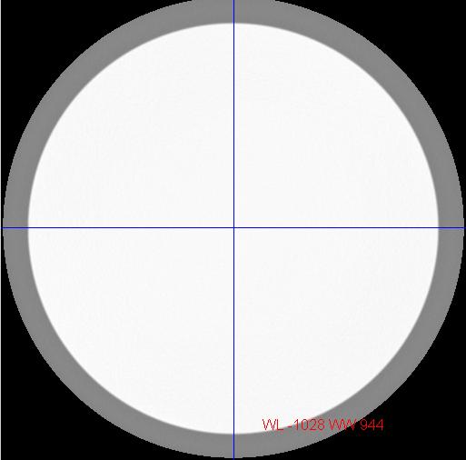
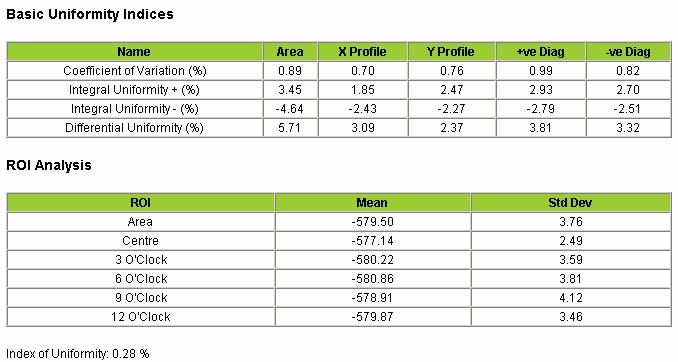
12 Varian Uniformity Phantoms 44 cm 34 cm Polyurethane Casting HU -580

13 Geometry: Phantom Alignment Detect phantom edge Threshold at 580 Trace edges and choose largest contour Calculate COM Compare against CT zero position

14 Geometry: Pixel Size Measure distance between holes Use centre of phantom A and expected pixel size to identify seek area Local minimum is centre of hole R θ L P
15 Hounsfield Unit Calibration Baseline Values Measured During Commissioning ICRU 42 Ax, 80kV, 150mA Ax, 80kV, 300mA Ax, 120kV, 150mA Ax, 120kV, 300mA Ax, 140kV, 150mA Ax, 140kV, 250mA 1000 CT Number Electron Density Rel to Water

16 Hounsfield Unit Calibration A WATER R SOFT BONE MTF LUNG L HARD BONE AIR P
17 Modulation Transfer Function Calculate from impulse object ( f ) FT{ PSF( x) } MTF = OSF ( x) = PSF( x) DSF( x) Finite size (DSF) { OSF( x) } = FT{ PSF( x) } FT{ DSF( x) } FT ( f ) MTF = FT FT { OSF( x) } { DSF( x) }

18 Calculation from Impulse Object Object Spread Function (From ALL pixels in ROI)
19
20 Uniformity Phantom Analysis Define Useful FOV (UFOV) as 90% FOV Calculate: std dev Coefficien t of Variation, CoV = mean pmax mean Integral Uniformity, U + = mean pmin mean Integral Uniformity, U = mean pmax Differential Uniformity,U d = mean centre periphery Uniformity Index, U CT = 1000
21 Uniformity Phantom Analysis

22 Uniformity Profiles CT Sim: 50 cm FOV Sim-CT Urethane Norm Air Norm
23 Noise Power Spectrum Region of Interest from Uniformity Phantom Remove DC component (subtract mean value) Perform 2D FFT NPS ( u, v) = Re ( ) 2 u, v + Im( u, v) area 2 Separation of stochastic noise NPS s = n NPS NPS n n ROI n

24 NPS Example 100 images of Uniformity Phantom, 50 cm FOV
25
26 Production of DRRs Ray trace from virtual source of x-rays through stack of CT slices and model attenuation of beam. X-ray source SAD 100 cm isocentre Imaging Plane Reference: Milickovic et al, Physics in Medicine and Biology (2000) 45:10; Projected back to isocentre


27 DRR Production Example CT Slices 3D array of voxels DRR

28 Edinburgh DRR Phantom
29 Software Demo
30 Experience & Conclusions New approach appears complicated, but Significantly faster than previous methods More robust, fully objective and quantitative Greater confidence in results New ability to follow trends Need to finalise DRR phantom Expand to include other RT imaging modalities
Evaluation of AutoQA Lite TM Image Quality Measurement Software
 Evaluation of AutoQA Lite TM Image Quality Measurement Software Andrew J Reilly Imaging Physicist Oncology Physics Edinburgh Cancer Centre Western General Hospital EDINBURGH EH4 2XU Phone: 0131 537 1161
Quality control phantoms and protocol for a tomography system
 Quality control phantoms and protocol for a tomography system Lucía Franco 1 1 CT AIMEN, C/Relva 27A O Porriño Pontevedra, Spain, lfranco@aimen.es Abstract Tomography systems for non-destructive testing
Ch. 4 Physical Principles of CT
 Ch. 4 Physical Principles of CT CLRS 408: Intro to CT Department of Radiation Sciences Review: Why CT? Solution for radiography/tomography limitations Superimposition of structures Distinguishing between
7/31/2011. Learning Objective. Video Positioning. 3D Surface Imaging by VisionRT
 CLINICAL COMMISSIONING AND ACCEPTANCE TESTING OF A 3D SURFACE MATCHING SYSTEM Hania Al-Hallaq, Ph.D. Assistant Professor Radiation Oncology The University of Chicago Learning Objective Describe acceptance
IAEA-TECDOC-1583 Commissioning of Radiotherapy Treatment Planning Systems: Testing for Typical External Beam Treatment Techniques
 IAEA-TECDOC-1583 Commissioning of Radiotherapy Treatment Planning Systems: Testing for Typical External Beam Treatment Techniques Report of the Coordinated Research Project (CRP) on Development of Procedures
S. Guru Prasad, Ph.D., DABR
 PURPOSE S. Guru Prasad, Ph.D., DABR Director of Medical Physics IAEA Consultant NorthShore University Health System and University of Chicago, Pritzker School of Medicine Current TPS utilize more information
Basics of treatment planning II
 Basics of treatment planning II Sastry Vedam PhD DABR Introduction to Medical Physics III: Therapy Spring 2015 Dose calculation algorithms! Correction based! Model based 1 Dose calculation algorithms!
Virtual Phantoms for IGRT QA
 TM Virtual Phantoms for IGRT QA Why ImSimQA? ImSimQA was developed to overcome the limitations of physical phantoms for testing modern medical imaging and radiation therapy software systems, when there
Image Acquisition Systems
 Image Acquisition Systems Goals and Terminology Conventional Radiography Axial Tomography Computer Axial Tomography (CAT) Magnetic Resonance Imaging (MRI) PET, SPECT Ultrasound Microscopy Imaging ITCS
Introduction to Biomedical Imaging
 Alejandro Frangi, PhD Computational Imaging Lab Department of Information & Communication Technology Pompeu Fabra University www.cilab.upf.edu X-ray Projection Imaging Computed Tomography Digital X-ray
Introduction. Quality Assurance for Image- Guided Radiation Therapy. Justification for IGRT. Image-Guided Radiation Therapy
 Introduction Quality Assurance for Image- Guided Radiation Therapy Jean-Pierre Bissonnette, Ph.D., MCCPM Princess Margaret Hospital, Toronto, Canada IGRT What is it? Rationale Equipment Quality Assurance
ImPACT. Information Leaflet No. 1: CT Scanner Acceptance Testing
 ImPACT Information Leaflet No. 1: CT Scanner Acceptance Testing Version 1.02, 18/05/01 CONTENTS: 1. SCOPE OF LEAFLET 2. GENERAL PRINCIPLES OF ACCEPTANCE AND COMMISSIONING 2.1 PHANTOMS 2.2 EXPOSURE AND
Protocol. Technical evaluation of X-ray tomographic image-guided radiotherapy devices CEP10070
 Protocol Technical evaluation of X-ray tomographic image-guided radiotherapy devices CEP10070 March 2010 Contents 2 Introduction... 3 Protocol design and validation... 4 General information... 7 Technical
Lucy Phantom MR Grid Evaluation
 Lucy Phantom MR Grid Evaluation Anil Sethi, PhD Loyola University Medical Center, Maywood, IL 60153 November 2015 I. Introduction: The MR distortion grid, used as an insert with Lucy 3D QA phantom, is
CBCT: Past, Present and Future. Disclosures. Computed Tomography. ATec Education Course Feb 28-Mar3, Douglas Moseley PhD, DABR
 CBCT: Past, Present and Future Douglas Moseley PhD, DABR Disclosures License Agreement Modus Medical Educational Consultant Elekta Oncology Systems Computed Tomography First CT Scanner Third Generation
ISO ISO ISO OHSAS ISO
 ISO 9001 ISO 13485 ISO 14001 OHSAS 18001 ISO 27001 Pro-NM Performance 08-101 - standard version 08-103 - version with the PET Lid Phantom for NM and PET systems performance evaluation (collimator, artifacts,
Evaluation report. X-ray tomographic image guided radiotherapy systems CEP10071
 Evaluation report X-ray tomographic image guided radiotherapy systems CEP10071 March 2010 Contents 2 Summary... 3 Introduction... 6 Product description... 9 Methods... 14 Technical performance... 23 Purchasing...
Automated Quality Assurance for Image-Guided Radiation Therapy
 JOURNAL OF APPLIED CLINICAL MEDICAL PHYSICS, VOLUME 10, NUMBER 1, WINTER 2009 Automated Quality Assurance for Image-Guided Radiation Therapy Eduard Schreibmann, a Eric Elder, Tim Fox Department of Radiation
Implementation and evaluation of a fully 3D OS-MLEM reconstruction algorithm accounting for the PSF of the PET imaging system
 Implementation and evaluation of a fully 3D OS-MLEM reconstruction algorithm accounting for the PSF of the PET imaging system 3 rd October 2008 11 th Topical Seminar on Innovative Particle and Radiation
BME I5000: Biomedical Imaging
 1 Lucas Parra, CCNY BME I5000: Biomedical Imaging Lecture 4 Computed Tomography Lucas C. Parra, parra@ccny.cuny.edu some slides inspired by lecture notes of Andreas H. Hilscher at Columbia University.
Facility Questionnaire PART I (General Information for 3DCRT and IMRT)
") Facility Questionnaire PART I (General Information for 3DCRT and IMRT) The following items are required before you can enter cases on any RTOG protocol that requires data submission to the Image-Guided
Facility Questionnaire PART I (General Information for 3DCRT and IMRT) The following items are required before you can enter cases on any RTOG protocol that requires data submission to the Image-Guided
Future Topics. Projection Imaging Dose Reporting, XA 3D Volume Objects. for DICOM WG-02. Presented by Heinz Blendinger, Siemens Medical Solutions
 Future Topics for Projection Imaging Dose Reporting, XA 3D Volume Objects DICOM WG-02 Presented by Heinz Blendinger, Siemens Medical Solutions 1 Presentation outline Dose Reporting Why Dose Reporting?
C a t p h a n / T h e P h a n t o m L a b o r a t o r y
 C a t p h a n 5 0 0 / 6 0 0 T h e P h a n t o m L a b o r a t o r y C a t p h a n 5 0 0 / 6 0 0 Internationally recognized for measuring the maximum obtainable performance of axial, spiral and multi-slice
Thank-You Members of TG147 TG 147: QA for nonradiographic
 Thank-You Members of TG147 TG 147: QA for nonradiographic localization and positioning systems Twyla Willoughby, M.S. Medical Physicist Clinical AAPM Meeting March 2013 Department of Radiation Oncology
GPU applications in Cancer Radiation Therapy at UCSD. Steve Jiang, UCSD Radiation Oncology Amit Majumdar, SDSC Dongju (DJ) Choi, SDSC
 Choi, SDSC") GPU applications in Cancer Radiation Therapy at UCSD Steve Jiang, UCSD Radiation Oncology Amit Majumdar, SDSC Dongju (DJ) Choi, SDSC Conventional Radiotherapy SIMULATION: Construciton, Dij Days PLANNING:
GPU applications in Cancer Radiation Therapy at UCSD Steve Jiang, UCSD Radiation Oncology Amit Majumdar, SDSC Dongju (DJ) Choi, SDSC Conventional Radiotherapy SIMULATION: Construciton, Dij Days PLANNING:
Shadow casting. What is the problem? Cone Beam Computed Tomography THE OBJECTIVES OF DIAGNOSTIC IMAGING IDEAL DIAGNOSTIC IMAGING STUDY LIMITATIONS
 Cone Beam Computed Tomography THE OBJECTIVES OF DIAGNOSTIC IMAGING Reveal pathology Reveal the anatomic truth Steven R. Singer, DDS srs2@columbia.edu IDEAL DIAGNOSTIC IMAGING STUDY Provides desired diagnostic
Spiral CT. Protocol Optimization & Quality Assurance. Ge Wang, Ph.D. Department of Radiology University of Iowa Iowa City, Iowa 52242, USA
 Spiral CT Protocol Optimization & Quality Assurance Ge Wang, Ph.D. Department of Radiology University of Iowa Iowa City, Iowa 52242, USA Spiral CT Protocol Optimization & Quality Assurance Protocol optimization
A study on image quality provided by a kilovoltage cone-beam computed tomography
 JOURNAL OF APPLIED CLINICAL MEDICAL PHYSICS, VOLUME 14, NUMBER 1, 2013 A study on image quality provided by a kilovoltage cone-beam computed tomography Julia Garayoa a and Pablo Castro Servicio de Radiofísica,
Quick Reference Datasheet For All RIT113 Packages
 Quick Reference Datasheet For All RIT113 Packages For Rotational Therapies, IMRT & TG142 Highlights and selected product information only. A complete TG142 brochure is available. For more information on
CT NOISE POWER SPECTRUM FOR FILTERED BACKPROJECTION AND ITERATIVE RECONSTRUCTION
 CT NOISE POWER SPECTRUM FOR FILTERED BACKPROJECTION AND ITERATIVE RECONSTRUCTION Frank Dong, PhD, DABR Diagnostic Physicist, Imaging Institute Cleveland Clinic Foundation and Associate Professor of Radiology
ADVANCING CANCER TREATMENT
 3 ADVANCING CANCER TREATMENT SUPPORTING CLINICS WORLDWIDE RaySearch is advancing cancer treatment through pioneering software. We believe software has un limited potential, and that it is now the driving
TEP Hounsfield units. Related topics Attenuation coefficient, Hounsfield units
 Hounsfield units TEP Related topics Attenuation coefficient, Hounsfield units Principle Depending on the type of CT scanner and the settings, the result of a CT scan of the same material can be different
Basic Radiation Oncology Physics
 Basic Radiation Oncology Physics T. Ganesh, Ph.D., DABR Chief Medical Physicist Fortis Memorial Research Institute Gurgaon Acknowledgment: I gratefully acknowledge the IAEA resources of teaching slides
8/2/2016. Measures the degradation/distortion of the acquired image (relative to an ideal image) using a quantitative figure-of-merit
 using a quantitative figure-of-merit") Ke Li Assistant Professor Department of Medical Physics and Department of Radiology School of Medicine and Public Health, University of Wisconsin-Madison This work is partially supported by an NIH Grant
Ke Li Assistant Professor Department of Medical Physics and Department of Radiology School of Medicine and Public Health, University of Wisconsin-Madison This work is partially supported by an NIH Grant
Patient Set-ups and Tumor Localizations
 Patient Set-ups and Tumor Localizations Amy S. Harrison Patient Positioning Prior to starting any localization or simulation procedure patients need to be positioned and immobilized Patients disease location
Conflicts of Interest Nuclear Medicine and PET physics reviewer for the ACR Accreditation program
 James R Halama, PhD Loyola University Medical Center Conflicts of Interest Nuclear Medicine and PET physics reviewer for the ACR Accreditation program Learning Objectives 1. Be familiar with recommendations
Hidenobu Tachibana The Cancer Institute Hospital of JFCR, Radiology Dept. The Cancer Institute of JFCR, Physics Dept.
 2-D D Dose-CT Mapping in Geant4 Hidenobu Tachibana The Cancer Institute Hospital of JFCR, Radiology Dept. The Cancer Institute of JFCR, Physics Dept. Table of Contents Background & Purpose Materials Methods
Financial disclosure. Onboard imaging modality for IGRT
 Tetrahedron Beam Computed Tomography Based On Multi-Pixel X- Ray Source and Its Application in Image Guided Radiotherapy Tiezhi Zhang, Ph.D. Advanced X-ray imaging Lab Financial disclosure Patent royalty
DUE to beam polychromacity in CT and the energy dependence
 1 Empirical Water Precorrection for Cone-Beam Computed Tomography Katia Sourbelle, Marc Kachelrieß, Member, IEEE, and Willi A. Kalender Abstract We propose an algorithm to correct for the cupping artifact
TomoTherapy Related Projects. An image guidance alternative on Tomo Low dose MVCT reconstruction Patient Quality Assurance using Sinogram
 TomoTherapy Related Projects An image guidance alternative on Tomo Low dose MVCT reconstruction Patient Quality Assurance using Sinogram Development of A Novel Image Guidance Alternative for Patient Localization
Assessment of 3D performance metrics. X-ray based Volumetric imaging systems: Fourier-based imaging metrics. The MTF in CT
 Assessment of 3D performance metrics D and 3D Metrics of Performance Towards Quality Index: Volumetric imaging systems X-ray based Volumetric imaging systems: CBCT/CT Tomosynthesis Samuel Richard and Ehsan
ICARO Vienna April Implementing 3D conformal radiotherapy and IMRT in clinical practice: Recommendations of IAEA- TECDOC-1588
 ICARO Vienna April 27-29 2009 Implementing 3D conformal radiotherapy and IMRT in clinical practice: Recommendations of IAEA- TECDOC-1588 M. Saiful Huq, Ph.D., Professor and Director, Dept. of Radiation
ADVANCING CANCER TREATMENT
 The RayPlan treatment planning system makes proven, innovative RayStation technology accessible to clinics that need a cost-effective and streamlined solution. Fast, efficient and straightforward to use,
A simple method to test geometrical reliability of digital reconstructed radiograph (DRR)
") JOURNAL OF APPLIED CLINICAL MEDICAL PHYSICS, VOLUME 11, NUMBER 1, WINTER 2010 A simple method to test geometrical reliability of digital reconstructed radiograph (DRR) Stefania Pallotta, a Marta Bucciolini
JOURNAL OF APPLIED CLINICAL MEDICAL PHYSICS, VOLUME 11, NUMBER 1, WINTER 2010 A simple method to test geometrical reliability of digital reconstructed radiograph (DRR) Stefania Pallotta, a Marta Bucciolini
James R Halama, PhD Loyola University Medical Center
 James R Halama, PhD Loyola University Medical Center Conflicts of Interest Nuclear Medicine and PET physics reviewer for the ACR Accreditation program Learning Objectives Be familiar with the tests recommended
Slide 1. Technical Aspects of Quality Control in Magnetic Resonance Imaging. Slide 2. Annual Compliance Testing. of MRI Systems.
 Slide 1 Technical Aspects of Quality Control in Magnetic Resonance Imaging Slide 2 Compliance Testing of MRI Systems, Ph.D. Department of Radiology Henry Ford Hospital, Detroit, MI Slide 3 Compliance Testing
INTRODUCTION TO MEDICAL IMAGING- 3D LOCALIZATION LAB MANUAL 1. Modifications for P551 Fall 2013 Medical Physics Laboratory
 INTRODUCTION TO MEDICAL IMAGING- 3D LOCALIZATION LAB MANUAL 1 Modifications for P551 Fall 2013 Medical Physics Laboratory Introduction Following the introductory lab 0, this lab exercise the student through
Radiology. Marta Anguiano Millán. Departamento de Física Atómica, Molecular y Nuclear Facultad de Ciencias. Universidad de Granada
 Departamento de Física Atómica, Molecular y Nuclear Facultad de Ciencias. Universidad de Granada Overview Introduction Overview Introduction Tecniques of imaging in Overview Introduction Tecniques of imaging
Acknowledgments. High Performance Cone-Beam CT of Acute Traumatic Brain Injury
 A. Sisniega et al. (presented at RSNA 214) High Performance Cone-Beam CT of Acute Traumatic Brain Injury A. Sisniega 1 W. Zbijewski 1, H. Dang 1, J. Xu 1 J. W. Stayman 1, J. Yorkston 2, N. Aygun 3 V. Koliatsos
Radon Transform and Filtered Backprojection
 Radon Transform and Filtered Backprojection Jørgen Arendt Jensen October 13, 2016 Center for Fast Ultrasound Imaging, Build 349 Department of Electrical Engineering Center for Fast Ultrasound Imaging Department
8/3/2016. Image Guidance Technologies. Introduction. Outline
 8/3/26 Session: Image Guidance Technologies and Management Strategies Image Guidance Technologies Jenghwa Chang, Ph.D.,2 Department of Radiation Medicine, Northwell Health 2 Hofstra Northwell School of
Michael Speiser, Ph.D.
 IMPROVED CT-BASED VOXEL PHANTOM GENERATION FOR MCNP MONTE CARLO Michael Speiser, Ph.D. Department of Radiation Oncology UT Southwestern Medical Center Dallas, TX September 1 st, 2012 CMPWG Workshop Medical
1. Learn to incorporate QA for surface imaging
 Hania Al-Hallaq, Ph.D. Assistant Professor Radiation Oncology The University of Chicago ***No disclosures*** 1. Learn to incorporate QA for surface imaging into current QA procedures for IGRT. 2. Understand
CLASS HOURS: 4 CREDIT HOURS: 4 LABORATORY HOURS: 0
 Revised 10/10 COURSE SYLLABUS TM 220 COMPUTED TOMOGRAPHY PHYSICS CLASS HOURS: 4 CREDIT HOURS: 4 LABORATORY HOURS: 0 CATALOG COURSE DESCRIPTION: This course is one of a three course set in whole body Computed
URGENT IMPORTANT FIELD SAFETY NOTIFICATION
 Subject: Incorrect Movement of the Treatment Table Product: MOSAIQ Scope: Sites affected will be those: 1. Running MOSAIQ and, 2. Treating on linear accelerators with the RATM license Notification Released:
REAL-TIME ADAPTIVITY IN HEAD-AND-NECK AND LUNG CANCER RADIOTHERAPY IN A GPU ENVIRONMENT
 REAL-TIME ADAPTIVITY IN HEAD-AND-NECK AND LUNG CANCER RADIOTHERAPY IN A GPU ENVIRONMENT Anand P Santhanam Assistant Professor, Department of Radiation Oncology OUTLINE Adaptive radiotherapy for head and
Developments in Dimensional Metrology in X-ray Computed Tomography at NPL
 Developments in Dimensional Metrology in X-ray Computed Tomography at NPL Wenjuan Sun and Stephen Brown 10 th May 2016 1 Possible factors influencing XCT measurements Components Influencing variables Possible
Comparison of Scatter Correction Methods for CBCT. Author(s): Suri, Roland E.; Virshup, Gary; Kaissl, Wolfgang; Zurkirchen, Luis
: Suri, Roland E.; Virshup, Gary; Kaissl, Wolfgang; Zurkirchen, Luis") Research Collection Working Paper Comparison of Scatter Correction Methods for CBCT Author(s): Suri, Roland E.; Virshup, Gary; Kaissl, Wolfgang; Zurkirchen, Luis Publication Date: 2010 Permanent Link:
Research Collection Working Paper Comparison of Scatter Correction Methods for CBCT Author(s): Suri, Roland E.; Virshup, Gary; Kaissl, Wolfgang; Zurkirchen, Luis Publication Date: 2010 Permanent Link:
Optimisation of Toshiba Aquilion ONE Volume Imaging
 Optimisation of Toshiba Aquilion ONE Volume Imaging Jane Edwards, RPRSG Royal Free London NHS Foundation Trust Dr Mufudzi Maviki, Plymouth Hospitals NHS Trust Background In 2011/12 Radiology at RFH was
Micro-CT Methodology Hasan Alsaid, PhD
 Micro-CT Methodology Hasan Alsaid, PhD Preclinical & Translational Imaging LAS, PTS, GlaxoSmithKline 20 April 2015 Provide basic understanding of technical aspects of the micro-ct Statement: All procedures
Philips SPECT/CT Systems
 Philips SPECT/CT Systems Ling Shao, PhD Director, Imaging Physics & System Analysis Nuclear Medicine, Philips Healthcare June 14, 2008 *Presented SNM08 Categorical Seminar - Quantitative SPECT and PET
INDUSTRIAL SYSTEM DEVELOPMENT FOR VOLUMETRIC INTEGRITY
 INDUSTRIAL SYSTEM DEVELOPMENT FOR VOLUMETRIC INTEGRITY VERIFICATION AND ANALYSIS M. L. Hsiao and J. W. Eberhard CR&D General Electric Company Schenectady, NY 12301 J. B. Ross Aircraft Engine - QTC General
Digital phantoms for the evaluation of a software used for an automatic analysis of the Winston-Lutz test in image guided radiation therapy
 Author manuscript, published in "Medical Imaging 008: Physics of Medical Imaging, San Diego, CA, USA : United States (008)" DOI : 10.1117/1.768668 Digital phantoms for the evaluation of a software used
8/3/2016. Outline. The EPID Strikes Back: Future EPID Technology and Applications. Active Matrix Flat-Panel Imagers (AMFPIs)
") 8//6 The EPID Strikes Back: Future EPID Technology and Applications Larry E. Antonuk Department of Radiation Oncology University of Michigan, Ann Arbor Acknowledgements: Youcef El-Mohri, Qihua Zhao (U.
8//6 The EPID Strikes Back: Future EPID Technology and Applications Larry E. Antonuk Department of Radiation Oncology University of Michigan, Ann Arbor Acknowledgements: Youcef El-Mohri, Qihua Zhao (U.
Effects of the difference in tube voltage of the CT scanner on. dose calculation
 Effects of the difference in tube voltage of the CT scanner on dose calculation Dong Joo Rhee, Sung-woo Kim, Dong Hyeok Jeong Medical and Radiological Physics Laboratory, Dongnam Institute of Radiological
RITtrend allows you to effectively manage all your physics QA data in one powerful and customizable package.
 So much is asked of medical physicists these days We Can Help Automated Radiation Therapy Phantom Analysis in Seconds QC for Therapy OBI s and CT simulators. Radia s Catphan/OBI module performs analysis
Background. Outline. Radiographic Tomosynthesis: Image Quality and Artifacts Reduction 1 / GE /
 Radiographic Tomosynthesis: Image Quality and Artifacts Reduction Baojun Li, Ph.D Department of Radiology Boston University Medical Center 2012 AAPM Annual Meeting Background Linear Trajectory Tomosynthesis
SYSTEM LINEARITY LAB MANUAL: 2 Modifications for P551 Fall 2013 Medical Physics Laboratory
 SYSTEM LINEARITY LAB MANUAL: 2 Modifications for P551 Fall 2013 Medical Physics Laboratory Introduction In this lab exercise, you will investigate the linearity of the DeskCAT scanner by making measurements
MEDICAL IMAGE ANALYSIS
 SECOND EDITION MEDICAL IMAGE ANALYSIS ATAM P. DHAWAN g, A B IEEE Engineering in Medicine and Biology Society, Sponsor IEEE Press Series in Biomedical Engineering Metin Akay, Series Editor +IEEE IEEE PRESS
CT vs. VolumeScope: image quality and dose comparison
 CT vs. VolumeScope: image quality and dose comparison V.N. Vasiliev *a, A.F. Gamaliy **b, M.Yu. Zaytsev b, K.V. Zaytseva ***b a Russian Sci. Center of Roentgenology & Radiology, 86, Profsoyuznaya, Moscow,
MEDICAL EQUIPMENT: COMPUTED TOMOGRAPHY. Prof. Yasser Mostafa Kadah
 MEDICAL EQUIPMENT: COMPUTED TOMOGRAPHY Prof. Yasser Mostafa Kadah www.k-space.org Recommended Textbook X-Ray Computed Tomography in Biomedical Engineering, by Robert Cierniak, Springer, 211 Computed Tomography
CT Basics Principles of Spiral CT Dose. Always Thinking Ahead.
 1 CT Basics Principles of Spiral CT Dose 2 Who invented CT? 1963 - Alan Cormack developed a mathematical method of reconstructing images from x-ray projections Sir Godfrey Hounsfield worked for the Central
Effect of CT number stopping power conversion uncertainties on dose distributions
 Effect of CT number stopping power conversion uncertainties on dose distributions Stella Flampouri Roelf Slopsema, Daniel Yeung, Zuofeng Li 23 May 2007 PTCOG Intro Treatment planning system dose calculations
3/27/2012 WHY SPECT / CT? SPECT / CT Basic Principles. Advantages of SPECT. Advantages of CT. Dr John C. Dickson, Principal Physicist UCLH
 3/27/212 Advantages of SPECT SPECT / CT Basic Principles Dr John C. Dickson, Principal Physicist UCLH Institute of Nuclear Medicine, University College London Hospitals and University College London john.dickson@uclh.nhs.uk
Integrated proton-photon treatment planning
 Pinnacle 3 Proton Planning Integrated proton-photon treatment planning Philips Pinnacle 3 Proton Planning specifications Pinnacle 3 Proton Planning is designed to simplify treatment planning for proton
Cone-beam computed tomography with a flat-panel imager: Initial performance characterization
 Cone-beam computed tomography with a flat-panel imager: Initial performance characterization D. A. Jaffray a) and J. H. Siewerdsen Department of Radiation Oncology, William Beaumont Hospital, Royal Oak,
Computer-Tomography II: Image reconstruction and applications
 Computer-Tomography II: Image reconstruction and applications Prof. Dr. U. Oelfke DKFZ Heidelberg Department of Medical Physics (E040) Im Neuenheimer Feld 280 69120 Heidelberg, Germany u.oelfke@dkfz.de
Version 5.6. Quick Start Guide
 WWW..COM Version 5.6 Quick Start Guide STANDARD IMAGING, INC. 3120 Deming Way Middleton, WI 53562-1461 Jul / 2018 2018 Standard Imaging, Inc. TEL 800.261.4446 TEL 608.831.0025 FAX 608.831.2202 DOC # 80714-05
Arion: a realistic projection simulator for optimizing laboratory and industrial micro-ct
 Arion: a realistic projection simulator for optimizing laboratory and industrial micro-ct J. DHAENE* 1, E. PAUWELS 1, T. DE SCHRYVER 1, A. DE MUYNCK 1, M. DIERICK 1, L. VAN HOOREBEKE 1 1 UGCT Dept. Physics
Technical aspects of SPECT and SPECT-CT. John Buscombe
 Technical aspects of SPECT and SPECT-CT John Buscombe What does the clinician need to know? For SPECT What factors affect SPECT How those factors should be sought Looking for artefacts For SPECT-CT Issues
The IORT Treatment Planning System. radiance. GMV, 2012 Property of GMV All rights reserved
 The IORT Treatment Planning System radiance Property of GMV All rights reserved WHY RADIANCE? JUSTIFICATION Property of GMV All rights reserved ADVANTAGES OF IORT PRECISION: RT guided by direct vision.
Digital Image Processing
 Digital Image Processing SPECIAL TOPICS CT IMAGES Hamid R. Rabiee Fall 2015 What is an image? 2 Are images only about visual concepts? We ve already seen that there are other kinds of image. In this lecture
Modifications for P551 Fall 2014
 LAB DEMONSTRATION COMPUTED TOMOGRAPHY USING DESKCAT 1 Modifications for P551 Fall 2014 Introduction This lab demonstration explores the physics and technology of Computed Tomography (CT) and guides the
Applications of Cone Beam Computed Tomography in Radiotherapy Treatment Planning
 Applications of Cone Beam Computed Tomography in Radiotherapy Treatment Planning Kavitha Srinivasan Thesis submitted for the degree of Master of Philosophy (Science) in the School of Chemistry and Physics
Transitioning from pencil beam to Monte Carlo for electron dose calculations
 Transitioning from pencil beam to Monte Carlo for electron dose calculations Jessie Huang-Vredevoogd (jyhuang4@wisc.edu) University of Wisconsin NCC AAPM October 12, 2019 1 Topics to cover Background RayStation
TG-148 overview. Introduction. System Overview. System Overview. QA for Helical Tomotherapy: Report of the AAPM Task Group 148. Conflict of Interest:
 QA or Helical Tomotherapy: Report o the AAPM Tas Group 148 Members: Conlict o Interest: r. John Balog owns TomoTherapy stoc. Katja Langen (Co-chair) Nio Papaniolaou (Co-chair) Walter Grant Richard Crilly
Good Morning! Thank you for joining us
 Good Morning! Thank you for joining us Deformable Registration, Contour Propagation and Dose Mapping: 101 and 201 Marc Kessler, PhD, FAAPM The University of Michigan Conflict of Interest I receive direct
Tomotherapy Physics. Machine Twinning and Quality Assurance. Emilie Soisson, MS
 Tomotherapy Physics Machine Twinning and Quality Assurance Emilie Soisson, MS Tomotherapy at UW- Madison Treating for nearly 5 years Up to ~45 patients a day on 2 tomo units Units twinned to facilitate
A Radiometry Tolerant Method for Direct 3D/2D Registration of Computed Tomography Data to X-ray Images
 A Radiometry Tolerant Method for Direct 3D/2D Registration of Computed Tomography Data to X-ray Images Transfer Function Independent Registration Boris Peter Selby 1, Georgios Sakas 2, Stefan Walter 1,
Design and performance characteristics of a Cone Beam CT system for Leksell Gamma Knife Icon
 Design and performance characteristics of a Cone Beam CT system for Leksell Gamma Knife Icon WHITE PAPER Introduction Introducing an image guidance system based on Cone Beam CT (CBCT) and a mask immobilization
Abbie M. Diak, PhD Loyola University Medical Center Dept. of Radiation Oncology
 Abbie M. Diak, PhD Loyola University Medical Center Dept. of Radiation Oncology Outline High Spectral and Spatial Resolution MR Imaging (HiSS) What it is How to do it Ways to use it HiSS for Radiation
Proton dose calculation algorithms and configuration data
 Proton dose calculation algorithms and configuration data Barbara Schaffner PTCOG 46 Educational workshop in Wanjie, 20. May 2007 VARIAN Medical Systems Agenda Broad beam algorithms Concept of pencil beam
Enhanced material contrast by dual-energy microct imaging
 Enhanced material contrast by dual-energy microct imaging Method note Page 1 of 12 2 Method note: Dual-energy microct analysis 1. Introduction 1.1. The basis for dual energy imaging Micro-computed tomography
Corso di laurea in Fisica A.A Fisica Medica 4 TC
 Corso di laurea in Fisica A.A. 2007-2008 Fisica Medica 4 TC Computed Tomography Principles 1. Projection measurement 2. Scanner systems 3. Scanning modes Basic Tomographic Principle The internal structure
PURE. ViSION Edition PET/CT. Patient Comfort Put First.
 PURE ViSION Edition PET/CT Patient Comfort Put First. 2 System features that put patient comfort and safety first. Oncology patients deserve the highest levels of safety and comfort during scans. Our Celesteion
Medical Imaging BMEN Spring 2016
 Name Medical Imaging BMEN 420-501 Spring 2016 Homework #4 and Nuclear Medicine Notes All questions are from the introductory Powerpoint (based on Chapter 7) and text Medical Imaging Signals and Systems,
Agenda : Lung Density Breakout Session
 Agenda : Lung Density Breakout Session 1. : Mathew Fuld and Bernice Hoppel 2. Automatic Exposure Control (AEC) Evaluation : Sean Fain Round 4 Project 3. Dose reduction effects on emphysema metrics : Philip
Validation of GEANT4 for Accurate Modeling of 111 In SPECT Acquisition
 Validation of GEANT4 for Accurate Modeling of 111 In SPECT Acquisition Bernd Schweizer, Andreas Goedicke Philips Technology Research Laboratories, Aachen, Germany bernd.schweizer@philips.com Abstract.
Some reference material
 Some reference material Physics reference book on medical imaging: A good one is The Essential Physics of Medical Imaging, 3 rd Ed. by Bushberg et al. ($170! new). However, there are several similar books
Position accuracy analysis of the stereotactic reference defined by the CBCT on Leksell Gamma Knife Icon
 Position accuracy analysis of the stereotactic reference defined by the CBCT on Leksell Gamma Knife Icon WHITE PAPER Introduction An image guidance system based on Cone Beam CT (CBCT) is included in Leksell