MR IMAGE SEGMENTATION
|
|
|
- Isaac Bridges
- 5 years ago
- Views:
Transcription
1 MR IMAGE SEGMENTATION Prepared by : Monil Shah What is Segmentation? Partitioning a region or regions of interest in images such that each region corresponds to one or more anatomic structures Classification Classification means to assign to each point in the image a tissue class, where the classes are agreed in advance Note that the problems are inter-linked: a classifier implicitly segments an image, and a segmentation implies a classification 1


2 An image can be represented as: F is the set of all pixels and P() is a uniformity (homogeneity) predicate defined on groups of connected pixels, then segmentation is a partitioning of the set F into a set of connected subsets or regions (S1,S2,..Sn) such that The uniformity predicate P(Si) = true for all regions P (SiυSj) = false when Si is adjacent to Sj. Radiotherapy planning Surgical planning Clinical drug trials Multiple Sclerosis, MS Lesion quantification Determine Brain tumor volumetrices Artery Vein separation in MR Angiography Preprocessing for multimodality Image Registration 2

3 MR Image These two features facilitate segmentation Excellent contrast between soft tissues Brain images are approximately piecewise constant; with small number of classes Practical Conditions There are image distortions (e.g. motion artifact, bias field, Partial volume effect (Many pixels contain more than one tissue type) structures of interest (tumours, temporal lobes, ) have complex shapes The noisy MRI image of the brain slice shown left is ideally piecewise constant, comprising grey matter, white matter, air, ventricles. The right image is a segmentation of the image at left. 3
4 Edge based Segmentation Mainly used for segmentation of large well defined structures such as brain Parenchyma but not to distinguish individual tissue types. Segmentation of White matter - Gray matter Edge: boundary between two regions with relatively distinct gray levels 4
5 Basic idea: computation of a local derivative operator. The first order derivative can be used to detect the presence of edge at a point in an image The second order derivative is used to determine whether an edge pixel lies on the dark or light side of an edge An imaginary line joining the extreme positive and negative values would cross zero near the midpoint of the edge. 5
6 6


7 In this method, the image is first smoothed to reduce noise. Then, the second derivative (Laplacian) of the smoothed images is computed. The obtained image will have positive and negative regions. The boundary between the positive and negative regions is taken as the image edges. This process is the same as convolving the image with the Laplacian of a 2-D Gaussian and finding its zero-crossings. Zero crossings are approximated by thresholding the LoG image by setting all positive values to white and all negative values to black, as zero crossing occurs between positive and negative values of the Laplacian. Advantages of Zero Crossing Edges are thinner Noise is reduced Rugged Performance 7

8 Region Based Segmentation Let R represent the entire image. Segmentation is a process that partitions R into n sub regions R1,R2,,Rn such that: Region Growing Pixel aggregation: starts with a set of seed point and from these grows regions by appending to each seed point those neighboring pixels that have similar properties (e.g., gray-level, texture, color). 8

9 Region Growing A T-2 weighted MR brain image (left) and the segmented ventricles (right) using the region-growing method. Problems: 1. Selection of initial seeds that properly represent regions of interest 2. Selection of suitable properties 3. Formulation of a stopping rule. 4. Only well defined regions can be robustly identified 9

10 Intensity Thresholding Gray level histogram of an image f(x,y) composed of a light object on a dark background. To extract the object: select a threshold T that separates the gray levels of the background and the object. Threshold in general can be calculated as: T= T(x,y,p(x,y),f(x,y)) T depends only on f(x,y): global threshold T depends on f(x,y) and p(x,y): local threshold The method is limited: Clinical Applications are hindered Variability of anatomy in brain MRI 10


11 Intensity distributions of white matter, gray matter and CSF are modeled as Gaussian distributions mean = average intensity variance = variation around the average intensity The threshold value is computed by: Detecting the valley between the modes in the histogram of the image. Once the mean and variance of each tissue type is known, voxels can be classified based on their intensity 11
12 12
13 Drawbacks of previous Methods Edge detection algorithms are vulnerable to artifacts and noise Region growing algorithms depend on seed selection and whether the regions are well defined and therfore is not robust Thresholding is only good for well separated intensities Classification of Images The labels we wish to assign to objects are typically few and known in advance e.g. WM, GM, CSF and air for brain MRI Objects of interest usually form coherent continuous shapes If a pixel has label c, then its neighbors are also likely to have label c Boundaries between regions labeled c, d are continuous Image noise means that the label to be assigned initially at any pixel is probabilistic, not certain One way to accommodate these considerations is Hidden Markov Random Fields 13
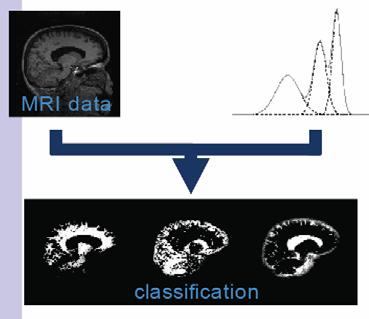
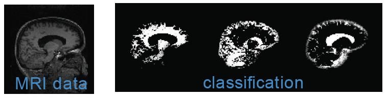
14 Segmentation by classification of voxels Every pixel is classified according to its probability of being a member of a particular tissue class. The Maximum Likelihood (ML) estimator assigns to each voxel x that class which maximizes The Maximum a Posteriori (MAP) estimator Normalizes intensities In practice do not work well on brain Each class is defined as a probability density function Each class, say the one with label (= GM say,) has an associated PDF, with parameters,often it is a Gaussian 14

15 Tissue Probability Density Function Note the huge overlap between the Gaussian pdf For GM and that for WM This means that there are many misclassifications of GM pixels as WM and vice versa. Even small amounts of noise can change the ML classification. HMRF Model Hidden Markov random field model (HMRF) is a stochastic process generated by a Markov random field whose state sequence is observed through a field of observations. Markov random field theory (MRF) encodes the spatial information of an image through contextual constraints of neighboring pixels. Neighboring pixels have same class labels or similar intensities. A statistical model is incomplete without its parameters being determined This is solved by using the Expectation-maximization (EM) algorithm. 15

16 MRF Model Pair wise Independence 16
 and, = = where, We call this the hidden Markov random field (HMRF) model.")
17 MRF equivalent to Gibbs distribution According to the local characteristics of MRFs, the joint probability of any pair of (Xi, Yi), given Xi's neighborhood configuration is: Thus, we can compute the marginal probability distribution of Yi dependent on the parameter set (random variable) and, = = where, We call this the hidden Markov random field (HMRF) model. Note, the concept of an HMRF is different from that of an MRF in the sense that the former is defined with respect to a pair of random variable families (X,Y) while the latter is only defined with respect to X. More precisely, an HMRF model can be described by the following: 17
 FGM model; (f)-(h) GHMRF model with standard deviation 0.3, 0.47, 0.55, respectively.")
18 Model Simulation Image simulation by the FGM model and the GHMRF model. The first row shows the 3-class case. (a) FGM model; (b)-(d) GHMRF mode with standard deviation 0.23, 0.4, 0.5, respectively. The second row shows the 5-class case. (e) FGM model; (f)-(h) GHMRF model with standard deviation 0.3, 0.47, 0.55, respectively. MRF-MAP estimation 18

19 Pixel wise independence & ICM Expectation-maximization statistical model is complete only if both its functional form and its parameters are determined. The procedure for estimating the unknown parameters is known as model fitting. We have assumed that we know the model parameters ahead of time. Unfortunately, this is rarely the case. The next idea is to estimate the parameters and do the segmentation cooperatively using the EM algorithm 19


20 Expectation Step Maximization step 20
-(e) show the same images with added Gaussian noise with standard deviation of 28, 47, 66, and 95.")
21 Summarize EM Experiment Figure 2(a) shows a simulated 3-class image sampled from an MRF model using the Gibbs sampler. Figure 2(b)-(e) show the same images with added Gaussian noise with standard deviation of 28, 47, 66, and 95. Because image contrast is what we are most interested in for examining qualities of an image, a measurement of the noise is more meaningful with image contrast being taken into account. 21


22 The standard HMRF-EM algorithm are then applied to the four test images until there is no significant change in the value of the Q-function. To measure the segmentation accuracy, we also define the misclassification ratio (MCR), which is the HMRF-EM algorithm gives more accurate estimates for parameters; the HMRF-EM algorithm provides automatic segmentation with much lower MCR. Automated Segmentation The prior probability for tissue type k is provided by the statistical brain atlas No unknown spatial model parameters to be estimated 22

23 Bias field Correction Since it is a complete approach to segmenting piecewise-constant images, the HMRF-EM framework can be applied to brain MR images. However, MR images are often corrupted by a low-frequency spatially varying artifact known as the bias field. This is mainly caused by the variation of the interaction between the human body and the magnetic field. The bias field can also be due to the differential attenuation of signals and nonlinearities of the sensitivities of the receiver coils, etc. Although such an artifact has little impact on visual diagnosis, the performance of most automatic image analysis techniques, especially intensity-based segmentation, can degrade dramatically. Therefore, a robust, automatic, and inexpensive way of correcting for this artifact is required. Bias field affects Histogram 23


24 Include an explicit model for the bias field in the intensity model Bias field is usually assumed to be multiplicative After logarithmic transformation => bias field becomes additive Gain field I i measured original = I γ + i i N i measured original y i = yi + β i Applying EM to bias field correction developed by W. M. Wells Given the class labels x, it is further assumed that the ideal intensity value at pixel i follows a Gaussian distribution with parameter With the bias field bi taken into account, the above distribution can be written in terms of the observed intensity yi as : Thus, the intensity distribution is modelled as a Gaussian mixture, given the bias field. It follows that 24

25 Applying EM to bias field correction developed by W. M. Wells The MAP principle is then employed to obtain the optimal estimate of the bias field, given the observed intensity values: A zero-gradient condition is then used to assess this maximum, Wij = bi = Wij is the posterior probability that pixel i belongs to class j given the bias field estimate. R is the residual and F is a low pass filter The E step assumes that the bias field is known and calculates the posterior tissue class probability Wij. In the M step, the bias field B is estimated given the estimated Wij in the E step. Once the bias field is obtained, the original intensity I* is restored by dividing I by the inverse log of B. Initially, the bias field is assumed to be zero everywhere. 25


26 Partial Volume Segmentation Assumed so far that each voxel belongs to one single tissue type In reality, many voxels in brain MR images are a mixture of several tissue types at the same time -Complex shape of the tissue interfaces in the brain -Limited spatial resolution of MRI Consistently misplacing the tissue borders in a 1 mm isotropic brain MRI with a single pixel in each slice results in large volume errors [Niessen et al., 1999]: ~ 30% for white matter ~ 40% for gray matter ~ 60% for CSF Partial volume voxels make lesion segmentation more difficult 26


27 Expectation step: find the function Involves a partial volume image classification: Not only probability for pure tissuesbut also probability for e.g. 22% of tissue1 and 78% of tissue 2 Mixing combination in a voxel is independent of the mixing combinations in other voxels All non-pure mixing combinations are equally probable Markov random field model Clustered regions of the same tissue type before downsampling Homogeneous regions of pure tissues bordered by partial volume voxels after downsampling 27
28 Drawbacks of HMRF-EM MRF model tends to minimize the boundary length between tissues. This discourages classifications from accurately following the complex shape of the tissue interfaces MRF over-smooths the classifications in cases where the intensity information doesn't prevent it 28
An Introduction To Automatic Tissue Classification Of Brain MRI. Colm Elliott Mar 2014
 An Introduction To Automatic Tissue Classification Of Brain MRI Colm Elliott Mar 2014 Tissue Classification Tissue classification is part of many processing pipelines. We often want to classify each voxel
Norbert Schuff VA Medical Center and UCSF
 Norbert Schuff Medical Center and UCSF Norbert.schuff@ucsf.edu Medical Imaging Informatics N.Schuff Course # 170.03 Slide 1/67 Objective Learn the principle segmentation techniques Understand the role
Histograms. h(r k ) = n k. p(r k )= n k /NM. Histogram: number of times intensity level rk appears in the image
 = n k. p(r k )= n k /NM. Histogram: number of times intensity level rk appears in the image") Histograms h(r k ) = n k Histogram: number of times intensity level rk appears in the image p(r k )= n k /NM normalized histogram also a probability of occurence 1 Histogram of Image Intensities Create
Histograms h(r k ) = n k Histogram: number of times intensity level rk appears in the image p(r k )= n k /NM normalized histogram also a probability of occurence 1 Histogram of Image Intensities Create
Norbert Schuff Professor of Radiology VA Medical Center and UCSF
 Norbert Schuff Professor of Radiology Medical Center and UCSF Norbert.schuff@ucsf.edu 2010, N.Schuff Slide 1/67 Overview Definitions Role of Segmentation Segmentation methods Intensity based Shape based
Region-based Segmentation
 Region-based Segmentation Image Segmentation Group similar components (such as, pixels in an image, image frames in a video) to obtain a compact representation. Applications: Finding tumors, veins, etc.
Methods for data preprocessing
 Methods for data preprocessing John Ashburner Wellcome Trust Centre for Neuroimaging, 12 Queen Square, London, UK. Overview Voxel-Based Morphometry Morphometry in general Volumetrics VBM preprocessing
mritc: A Package for MRI Tissue Classification
 mritc: A Package for MRI Tissue Classification Dai Feng 1 Luke Tierney 2 1 Merck Research Labratories 2 University of Iowa July 2010 Feng & Tierney (Merck & U of Iowa) MRI Tissue Classification July 2010
Dept of CSE, CIT Gubbi, Tumkur, Mysore, India
 Volume 5, Issue 6, June 2015 ISSN: 2277 128X International Journal of Advanced Research in Computer Science and Software Engineering Research Paper Available online at: www.ijarcsse.com MRI Tissue Segmentation
MEDICAL IMAGE COMPUTING (CAP 5937) LECTURE 4: Pre-Processing Medical Images (II)
 LECTURE 4: Pre-Processing Medical Images (II)") SPRING 2016 1 MEDICAL IMAGE COMPUTING (CAP 5937) LECTURE 4: Pre-Processing Medical Images (II) Dr. Ulas Bagci HEC 221, Center for Research in Computer Vision (CRCV), University of Central Florida (UCF),
SPRING 2016 1 MEDICAL IMAGE COMPUTING (CAP 5937) LECTURE 4: Pre-Processing Medical Images (II) Dr. Ulas Bagci HEC 221, Center for Research in Computer Vision (CRCV), University of Central Florida (UCF),
Norbert Schuff Professor of Radiology VA Medical Center and UCSF
 Norbert Schuff Professor of Radiology Medical Center and UCSF Norbert.schuff@ucsf.edu Slide 1/67 Overview Definitions Role of Segmentation Segmentation methods Intensity based Shape based Texture based
Introduction to Medical Image Processing
 Introduction to Medical Image Processing Δ Essential environments of a medical imaging system Subject Image Analysis Energy Imaging System Images Image Processing Feature Images Image processing may be
Neuroimaging and mathematical modelling Lesson 2: Voxel Based Morphometry
 Neuroimaging and mathematical modelling Lesson 2: Voxel Based Morphometry Nivedita Agarwal, MD Nivedita.agarwal@apss.tn.it Nivedita.agarwal@unitn.it Volume and surface morphometry Brain volume White matter
Preprocessing II: Between Subjects John Ashburner
 Preprocessing II: Between Subjects John Ashburner Pre-processing Overview Statistics or whatever fmri time-series Anatomical MRI Template Smoothed Estimate Spatial Norm Motion Correct Smooth Coregister
Comparison Study of Clinical 3D MRI Brain Segmentation Evaluation
 Comparison Study of Clinical 3D MRI Brain Segmentation Evaluation Ting Song 1, Elsa D. Angelini 2, Brett D. Mensh 3, Andrew Laine 1 1 Heffner Biomedical Imaging Laboratory Department of Biomedical Engineering,
Computer Vision & Digital Image Processing. Image segmentation: thresholding
 Computer Vision & Digital Image Processing Image Segmentation: Thresholding Dr. D. J. Jackson Lecture 18-1 Image segmentation: thresholding Suppose an image f(y) is composed of several light objects on
CHAPTER 3 IMAGE ENHANCEMENT IN THE SPATIAL DOMAIN
 CHAPTER 3 IMAGE ENHANCEMENT IN THE SPATIAL DOMAIN CHAPTER 3: IMAGE ENHANCEMENT IN THE SPATIAL DOMAIN Principal objective: to process an image so that the result is more suitable than the original image
Supplementary methods
 Supplementary methods This section provides additional technical details on the sample, the applied imaging and analysis steps and methods. Structural imaging Trained radiographers placed all participants
Chapter 3: Intensity Transformations and Spatial Filtering
 Chapter 3: Intensity Transformations and Spatial Filtering 3.1 Background 3.2 Some basic intensity transformation functions 3.3 Histogram processing 3.4 Fundamentals of spatial filtering 3.5 Smoothing
ADAPTIVE GRAPH CUTS WITH TISSUE PRIORS FOR BRAIN MRI SEGMENTATION
 ADAPTIVE GRAPH CUTS WITH TISSUE PRIORS FOR BRAIN MRI SEGMENTATION Abstract: MIP Project Report Spring 2013 Gaurav Mittal 201232644 This is a detailed report about the course project, which was to implement
Object Identification in Ultrasound Scans
 Object Identification in Ultrasound Scans Wits University Dec 05, 2012 Roadmap Introduction to the problem Motivation Related Work Our approach Expected Results Introduction Nowadays, imaging devices like
PET Image Reconstruction using Anatomical Information through Mutual Information Based Priors
 2005 IEEE Nuclear Science Symposium Conference Record M11-354 PET Image Reconstruction using Anatomical Information through Mutual Information Based Priors Sangeetha Somayajula, Evren Asma, and Richard
Basic relations between pixels (Chapter 2)
") Basic relations between pixels (Chapter 2) Lecture 3 Basic Relationships Between Pixels Definitions: f(x,y): digital image Pixels: q, p (p,q f) A subset of pixels of f(x,y): S A typology of relations:
Basic relations between pixels (Chapter 2) Lecture 3 Basic Relationships Between Pixels Definitions: f(x,y): digital image Pixels: q, p (p,q f) A subset of pixels of f(x,y): S A typology of relations:
CHAPTER 6 DETECTION OF MASS USING NOVEL SEGMENTATION, GLCM AND NEURAL NETWORKS
 130 CHAPTER 6 DETECTION OF MASS USING NOVEL SEGMENTATION, GLCM AND NEURAL NETWORKS A mass is defined as a space-occupying lesion seen in more than one projection and it is described by its shapes and margin
Chapter 10: Image Segmentation. Office room : 841
 Chapter 10: Image Segmentation Lecturer: Jianbing Shen Email : shenjianbing@bit.edu.cn Office room : 841 http://cs.bit.edu.cn/shenjianbing cn/shenjianbing Contents Definition and methods classification
AN EFFICIENT SKULL STRIPPING ALGORITHM USING CONNECTED REGIONS AND MORPHOLOGICAL OPERATION
 AN EFFICIENT SKULL STRIPPING ALGORITHM USING CONNECTED REGIONS AND MORPHOLOGICAL OPERATION Shijin Kumar P. S. 1 and Dharun V. S. 2 1 Department of Electronics and Communication Engineering, Noorul Islam
Segmenting Lesions in Multiple Sclerosis Patients James Chen, Jason Su
 Segmenting Lesions in Multiple Sclerosis Patients James Chen, Jason Su Radiologists and researchers spend countless hours tediously segmenting white matter lesions to diagnose and study brain diseases.
CHAPTER 2. Morphometry on rodent brains. A.E.H. Scheenstra J. Dijkstra L. van der Weerd
 CHAPTER 2 Morphometry on rodent brains A.E.H. Scheenstra J. Dijkstra L. van der Weerd This chapter was adapted from: Volumetry and other quantitative measurements to assess the rodent brain, In vivo NMR
Automatic Registration-Based Segmentation for Neonatal Brains Using ANTs and Atropos
 Automatic Registration-Based Segmentation for Neonatal Brains Using ANTs and Atropos Jue Wu and Brian Avants Penn Image Computing and Science Lab, University of Pennsylvania, Philadelphia, USA Abstract.
Automated segmentation methods for liver analysis in oncology applications
 University of Szeged Department of Image Processing and Computer Graphics Automated segmentation methods for liver analysis in oncology applications Ph. D. Thesis László Ruskó Thesis Advisor Dr. Antal
Modern Medical Image Analysis 8DC00 Exam
 Parts of answers are inside square brackets [... ]. These parts are optional. Answers can be written in Dutch or in English, as you prefer. You can use drawings and diagrams to support your textual answers.
Image Enhancement: To improve the quality of images
 Image Enhancement: To improve the quality of images Examples: Noise reduction (to improve SNR or subjective quality) Change contrast, brightness, color etc. Image smoothing Image sharpening Modify image
Automatic segmentation of the cortical grey and white matter in MRI using a Region Growing approach based on anatomical knowledge
 Automatic segmentation of the cortical grey and white matter in MRI using a Region Growing approach based on anatomical knowledge Christian Wasserthal 1, Karin Engel 1, Karsten Rink 1 und André Brechmann
Automatic Optimization of Segmentation Algorithms Through Simultaneous Truth and Performance Level Estimation (STAPLE)
") Automatic Optimization of Segmentation Algorithms Through Simultaneous Truth and Performance Level Estimation (STAPLE) Mahnaz Maddah, Kelly H. Zou, William M. Wells, Ron Kikinis, and Simon K. Warfield
Automatic Optimization of Segmentation Algorithms Through Simultaneous Truth and Performance Level Estimation (STAPLE) Mahnaz Maddah, Kelly H. Zou, William M. Wells, Ron Kikinis, and Simon K. Warfield
Performance Evaluation of the TINA Medical Image Segmentation Algorithm on Brainweb Simulated Images
 Tina Memo No. 2008-003 Internal Memo Performance Evaluation of the TINA Medical Image Segmentation Algorithm on Brainweb Simulated Images P. A. Bromiley Last updated 20 / 12 / 2007 Imaging Science and
Where are we now? Structural MRI processing and analysis
 Where are we now? Structural MRI processing and analysis Pierre-Louis Bazin bazin@cbs.mpg.de Leipzig, Germany Structural MRI processing: why bother? Just use the standards? SPM FreeSurfer FSL However:
ISSN: X Impact factor: 4.295
 ISSN: 2454-132X Impact factor: 4.295 (Volume3, Issue1) Available online at: www.ijariit.com Performance Analysis of Image Clustering Algorithm Applied to Brain MRI Kalyani R.Mandlik 1, Dr. Suresh S. Salankar
Operators-Based on Second Derivative double derivative Laplacian operator Laplacian Operator Laplacian Of Gaussian (LOG) Operator LOG
 Operator LOG") Operators-Based on Second Derivative The principle of edge detection based on double derivative is to detect only those points as edge points which possess local maxima in the gradient values. Laplacian
Operators-Based on Second Derivative The principle of edge detection based on double derivative is to detect only those points as edge points which possess local maxima in the gradient values. Laplacian
IMAGE SEGMENTATION. Václav Hlaváč
 IMAGE SEGMENTATION Václav Hlaváč Czech Technical University in Prague Faculty of Electrical Engineering, Department of Cybernetics Center for Machine Perception http://cmp.felk.cvut.cz/ hlavac, hlavac@fel.cvut.cz
Digital Image Analysis and Processing
 Digital Image Analysis and Processing CPE 0907544 Image Segmentation Part II Chapter 10 Sections : 10.3 10.4 Dr. Iyad Jafar Outline Introduction Thresholdingh Fundamentals Basic Global Thresholding Optimal
Image Processing. BITS Pilani. Dr Jagadish Nayak. Dubai Campus
 Image Processing BITS Pilani Dubai Campus Dr Jagadish Nayak Image Segmentation BITS Pilani Dubai Campus Fundamentals Let R be the entire spatial region occupied by an image Process that partitions R into
Available Online through
 Available Online through www.ijptonline.com ISSN: 0975-766X CODEN: IJPTFI Research Article ANALYSIS OF CT LIVER IMAGES FOR TUMOUR DIAGNOSIS BASED ON CLUSTERING TECHNIQUE AND TEXTURE FEATURES M.Krithika
Lesion Segmentation and Bias Correction in Breast Ultrasound B-mode Images Including Elastography Information
 Lesion Segmentation and Bias Correction in Breast Ultrasound B-mode Images Including Elastography Information Gerard Pons a, Joan Martí a, Robert Martí a, Mariano Cabezas a, Andrew di Battista b, and J.
Computational Neuroanatomy
 Computational Neuroanatomy John Ashburner john@fil.ion.ucl.ac.uk Smoothing Motion Correction Between Modality Co-registration Spatial Normalisation Segmentation Morphometry Overview fmri time-series kernel
Level-set MCMC Curve Sampling and Geometric Conditional Simulation
 Level-set MCMC Curve Sampling and Geometric Conditional Simulation Ayres Fan John W. Fisher III Alan S. Willsky February 16, 2007 Outline 1. Overview 2. Curve evolution 3. Markov chain Monte Carlo 4. Curve
Segmentation of Images
 Segmentation of Images SEGMENTATION If an image has been preprocessed appropriately to remove noise and artifacts, segmentation is often the key step in interpreting the image. Image segmentation is a
Surface-based Analysis: Inter-subject Registration and Smoothing
 Surface-based Analysis: Inter-subject Registration and Smoothing Outline Exploratory Spatial Analysis Coordinate Systems 3D (Volumetric) 2D (Surface-based) Inter-subject registration Volume-based Surface-based
Application of fuzzy set theory in image analysis. Nataša Sladoje Centre for Image Analysis
 Application of fuzzy set theory in image analysis Nataša Sladoje Centre for Image Analysis Our topics for today Crisp vs fuzzy Fuzzy sets and fuzzy membership functions Fuzzy set operators Approximate
Lecture 6: Edge Detection
 #1 Lecture 6: Edge Detection Saad J Bedros sbedros@umn.edu Review From Last Lecture Options for Image Representation Introduced the concept of different representation or transformation Fourier Transform
Machine Learning for Medical Image Analysis. A. Criminisi
 Machine Learning for Medical Image Analysis A. Criminisi Overview Introduction to machine learning Decision forests Applications in medical image analysis Anatomy localization in CT Scans Spine Detection
Image Processing. Traitement d images. Yuliya Tarabalka Tel.
 Traitement d images Yuliya Tarabalka yuliya.tarabalka@hyperinet.eu yuliya.tarabalka@gipsa-lab.grenoble-inp.fr Tel. 04 76 82 62 68 Noise reduction Image restoration Restoration attempts to reconstruct an
C E N T E R A T H O U S T O N S C H O O L of H E A L T H I N F O R M A T I O N S C I E N C E S. Image Operations I
 T H E U N I V E R S I T Y of T E X A S H E A L T H S C I E N C E C E N T E R A T H O U S T O N S C H O O L of H E A L T H I N F O R M A T I O N S C I E N C E S Image Operations I For students of HI 5323
Color Image Segmentation
 Color Image Segmentation Yining Deng, B. S. Manjunath and Hyundoo Shin* Department of Electrical and Computer Engineering University of California, Santa Barbara, CA 93106-9560 *Samsung Electronics Inc.
Digital Image Processing. Image Enhancement (Point Processing)
") Digital Image Processing Image Enhancement (Point Processing) 2 Contents In this lecture we will look at image enhancement point processing techniques: What is point processing? Negative images Thresholding
Digital Image Processing Image Enhancement (Point Processing) 2 Contents In this lecture we will look at image enhancement point processing techniques: What is point processing? Negative images Thresholding
Segmenting Glioma in Multi-Modal Images using a Generative Model for Brain Lesion Segmentation
 Segmenting Glioma in Multi-Modal Images using a Generative Model for Brain Lesion Segmentation Bjoern H. Menze 1,2, Koen Van Leemput 3, Danial Lashkari 4 Marc-André Weber 5, Nicholas Ayache 2, and Polina
Mixture Models and EM
 Mixture Models and EM Goal: Introduction to probabilistic mixture models and the expectationmaximization (EM) algorithm. Motivation: simultaneous fitting of multiple model instances unsupervised clustering
A Model-Independent, Multi-Image Approach to MR Inhomogeneity Correction
 Tina Memo No. 2007-003 Published in Proc. MIUA 2007 A Model-Independent, Multi-Image Approach to MR Inhomogeneity Correction P. A. Bromiley and N.A. Thacker Last updated 13 / 4 / 2007 Imaging Science and
Image Segmentation Based on Watershed and Edge Detection Techniques
 0 The International Arab Journal of Information Technology, Vol., No., April 00 Image Segmentation Based on Watershed and Edge Detection Techniques Nassir Salman Computer Science Department, Zarqa Private
Biometrics Technology: Image Processing & Pattern Recognition (by Dr. Dickson Tong)
") Biometrics Technology: Image Processing & Pattern Recognition (by Dr. Dickson Tong) References: [1] http://homepages.inf.ed.ac.uk/rbf/hipr2/index.htm [2] http://www.cs.wisc.edu/~dyer/cs540/notes/vision.html
Biometrics Technology: Image Processing & Pattern Recognition (by Dr. Dickson Tong) References: [1] http://homepages.inf.ed.ac.uk/rbf/hipr2/index.htm [2] http://www.cs.wisc.edu/~dyer/cs540/notes/vision.html
BAYESIAN SEGMENTATION OF THREE DIMENSIONAL IMAGES USING THE EM/MPM ALGORITHM. A Thesis. Submitted to the Faculty.
 BAYESIAN SEGMENTATION OF THREE DIMENSIONAL IMAGES USING THE EM/MPM ALGORITHM A Thesis Submitted to the Faculty of Purdue University by Lauren Christopher In Partial Fulfillment of the Requirements for
Idea. Found boundaries between regions (edges) Didn t return the actual region
 Didn t return the actual region") Region Segmentation Idea Edge detection Found boundaries between regions (edges) Didn t return the actual region Segmentation Partition image into regions find regions based on similar pixel intensities,
Region Segmentation Idea Edge detection Found boundaries between regions (edges) Didn t return the actual region Segmentation Partition image into regions find regions based on similar pixel intensities,
Digital Image Processing
 Digital Image Processing Intensity Transformations (Point Processing) Christophoros Nikou cnikou@cs.uoi.gr University of Ioannina - Department of Computer Science and Engineering 2 Intensity Transformations
Segmentation and Grouping
 Segmentation and Grouping How and what do we see? Fundamental Problems ' Focus of attention, or grouping ' What subsets of pixels do we consider as possible objects? ' All connected subsets? ' Representation
EECS490: Digital Image Processing. Lecture #22
 Lecture #22 Gold Standard project images Otsu thresholding Local thresholding Region segmentation Watershed segmentation Frequency-domain techniques Project Images 1 Project Images 2 Project Images 3 Project
Region & edge based Segmentation
 INF 4300 Digital Image Analysis Region & edge based Segmentation Fritz Albregtsen 06.11.2018 F11 06.11.18 IN5520 1 Today We go through sections 10.1, 10.4, 10.5, 10.6.1 We cover the following segmentation
Image Segmentation. Shengnan Wang
 Image Segmentation Shengnan Wang shengnan@cs.wisc.edu Contents I. Introduction to Segmentation II. Mean Shift Theory 1. What is Mean Shift? 2. Density Estimation Methods 3. Deriving the Mean Shift 4. Mean
Image Analysis Image Segmentation (Basic Methods)
") Image Analysis Image Segmentation (Basic Methods) Christophoros Nikou cnikou@cs.uoi.gr Images taken from: R. Gonzalez and R. Woods. Digital Image Processing, Prentice Hall, 2008. Computer Vision course
Image Analysis Image Segmentation (Basic Methods) Christophoros Nikou cnikou@cs.uoi.gr Images taken from: R. Gonzalez and R. Woods. Digital Image Processing, Prentice Hall, 2008. Computer Vision course
Automatic Generation of Training Data for Brain Tissue Classification from MRI
 Automatic Generation of Training Data for Brain Tissue Classification from MRI Chris A. COCOSCO, Alex P. ZIJDENBOS, and Alan C. EVANS http://www.bic.mni.mcgill.ca/users/crisco/ McConnell Brain Imaging
Global Thresholding Techniques to Classify Dead Cells in Diffusion Weighted Magnetic Resonant Images
 Global Thresholding Techniques to Classify Dead Cells in Diffusion Weighted Magnetic Resonant Images Ravi S 1, A. M. Khan 2 1 Research Student, Department of Electronics, Mangalore University, Karnataka
Topic 4 Image Segmentation
 Topic 4 Image Segmentation What is Segmentation? Why? Segmentation important contributing factor to the success of an automated image analysis process What is Image Analysis: Processing images to derive
Image Restoration using Markov Random Fields
 Image Restoration using Markov Random Fields Based on the paper Stochastic Relaxation, Gibbs Distributions and Bayesian Restoration of Images, PAMI, 1984, Geman and Geman. and the book Markov Random Field
Segmentation algorithm for monochrome images generally are based on one of two basic properties of gray level values: discontinuity and similarity.
 Chapter - 3 : IMAGE SEGMENTATION Segmentation subdivides an image into its constituent s parts or objects. The level to which this subdivision is carried depends on the problem being solved. That means
Modified Expectation Maximization Method for Automatic Segmentation of MR Brain Images
 Modified Expectation Maximization Method for Automatic Segmentation of MR Brain Images R.Meena Prakash, R.Shantha Selva Kumari 1 P.S.R.Engineering College, Sivakasi, Tamil Nadu, India 2 Mepco Schlenk Engineering
EEM 463 Introduction to Image Processing. Week 3: Intensity Transformations
 EEM 463 Introduction to Image Processing Week 3: Intensity Transformations Fall 2013 Instructor: Hatice Çınar Akakın, Ph.D. haticecinarakakin@anadolu.edu.tr Anadolu University Enhancement Domains Spatial
Chapter 3 Set Redundancy in Magnetic Resonance Brain Images
 16 Chapter 3 Set Redundancy in Magnetic Resonance Brain Images 3.1 MRI (magnetic resonance imaging) MRI is a technique of measuring physical structure within the human anatomy. Our proposed research focuses
Discriminative, Semantic Segmentation of Brain Tissue in MR Images
 Discriminative, Semantic Segmentation of Brain Tissue in MR Images Zhao Yi 1, Antonio Criminisi 2, Jamie Shotton 2, and Andrew Blake 2 1 University of California, Los Angeles, USA. zyi@ucla.edu. 2 Microsoft
Lecture 7: Most Common Edge Detectors
 #1 Lecture 7: Most Common Edge Detectors Saad Bedros sbedros@umn.edu Edge Detection Goal: Identify sudden changes (discontinuities) in an image Intuitively, most semantic and shape information from the
Multiple Sclerosis Brain MRI Segmentation Workflow deployment on the EGEE grid
 Multiple Sclerosis Brain MRI Segmentation Workflow deployment on the EGEE grid Erik Pernod 1, Jean-Christophe Souplet 1, Javier Rojas Balderrama 2, Diane Lingrand 2, Xavier Pennec 1 Speaker: Grégoire Malandain
UNIT - 5 IMAGE ENHANCEMENT IN SPATIAL DOMAIN
 UNIT - 5 IMAGE ENHANCEMENT IN SPATIAL DOMAIN Spatial domain methods Spatial domain refers to the image plane itself, and approaches in this category are based on direct manipulation of pixels in an image.
Babu Madhav Institute of Information Technology Years Integrated M.Sc.(IT)(Semester - 7)
(Semester - 7)") 5 Years Integrated M.Sc.(IT)(Semester - 7) 060010707 Digital Image Processing UNIT 1 Introduction to Image Processing Q: 1 Answer in short. 1. What is digital image? 1. Define pixel or picture element?
5 Years Integrated M.Sc.(IT)(Semester - 7) 060010707 Digital Image Processing UNIT 1 Introduction to Image Processing Q: 1 Answer in short. 1. What is digital image? 1. Define pixel or picture element?
Ulrik Söderström 16 Feb Image Processing. Segmentation
 Ulrik Söderström ulrik.soderstrom@tfe.umu.se 16 Feb 2011 Image Processing Segmentation What is Image Segmentation? To be able to extract information from an image it is common to subdivide it into background
Image Processing. Bilkent University. CS554 Computer Vision Pinar Duygulu
 Image Processing CS 554 Computer Vision Pinar Duygulu Bilkent University Today Image Formation Point and Blob Processing Binary Image Processing Readings: Gonzalez & Woods, Ch. 3 Slides are adapted from
White Pixel Artifact. Caused by a noise spike during acquisition Spike in K-space <--> sinusoid in image space
 White Pixel Artifact Caused by a noise spike during acquisition Spike in K-space sinusoid in image space Susceptibility Artifacts Off-resonance artifacts caused by adjacent regions with different
GENERAL AUTOMATED FLAW DETECTION SCHEME FOR NDE X-RAY IMAGES
 GENERAL AUTOMATED FLAW DETECTION SCHEME FOR NDE X-RAY IMAGES Karl W. Ulmer and John P. Basart Center for Nondestructive Evaluation Department of Electrical and Computer Engineering Iowa State University
The organization of the human cerebral cortex estimated by intrinsic functional connectivity
 1 The organization of the human cerebral cortex estimated by intrinsic functional connectivity Journal: Journal of Neurophysiology Author: B. T. Thomas Yeo, et al Link: https://www.ncbi.nlm.nih.gov/pubmed/21653723
Image Segmentation Techniques
 A Study On Image Segmentation Techniques Palwinder Singh 1, Amarbir Singh 2 1,2 Department of Computer Science, GNDU Amritsar Abstract Image segmentation is very important step of image analysis which
In this lecture. Background. Background. Background. PAM3012 Digital Image Processing for Radiographers
 PAM3012 Digital Image Processing for Radiographers Image Enhancement in the Spatial Domain (Part I) In this lecture Image Enhancement Introduction to spatial domain Information Greyscale transformations
Vivekananda. Collegee of Engineering & Technology. Question and Answers on 10CS762 /10IS762 UNIT- 5 : IMAGE ENHANCEMENT.
 Vivekananda Collegee of Engineering & Technology Question and Answers on 10CS762 /10IS762 UNIT- 5 : IMAGE ENHANCEMENT Dept. Prepared by Harivinod N Assistant Professor, of Computer Science and Engineering,
An ICA based Approach for Complex Color Scene Text Binarization
 An ICA based Approach for Complex Color Scene Text Binarization Siddharth Kherada IIIT-Hyderabad, India siddharth.kherada@research.iiit.ac.in Anoop M. Namboodiri IIIT-Hyderabad, India anoop@iiit.ac.in
Fmri Spatial Processing
 Educational Course: Fmri Spatial Processing Ray Razlighi Jun. 8, 2014 Spatial Processing Spatial Re-alignment Geometric distortion correction Spatial Normalization Smoothing Why, When, How, Which Why is
Markov Random Fields and Gibbs Sampling for Image Denoising
 Markov Random Fields and Gibbs Sampling for Image Denoising Chang Yue Electrical Engineering Stanford University changyue@stanfoed.edu Abstract This project applies Gibbs Sampling based on different Markov
Correction of Partial Volume Effects in Arterial Spin Labeling MRI
 Correction of Partial Volume Effects in Arterial Spin Labeling MRI By: Tracy Ssali Supervisors: Dr. Keith St. Lawrence and Udunna Anazodo Medical Biophysics 3970Z Six Week Project April 13 th 2012 Introduction
Adaptive Fuzzy Connectedness-Based Medical Image Segmentation
 Adaptive Fuzzy Connectedness-Based Medical Image Segmentation Amol Pednekar Ioannis A. Kakadiaris Uday Kurkure Visual Computing Lab, Dept. of Computer Science, Univ. of Houston, Houston, TX, USA apedneka@bayou.uh.edu
Image Registration + Other Stuff
 Image Registration + Other Stuff John Ashburner Pre-processing Overview fmri time-series Motion Correct Anatomical MRI Coregister m11 m 21 m 31 m12 m13 m14 m 22 m 23 m 24 m 32 m 33 m 34 1 Template Estimate
Introduction to Medical Imaging (5XSA0) Module 5
 Module 5") Introduction to Medical Imaging (5XSA0) Module 5 Segmentation Jungong Han, Dirk Farin, Sveta Zinger ( s.zinger@tue.nl ) 1 Outline Introduction Color Segmentation region-growing region-merging watershed
Introduction to Medical Imaging (5XSA0) Module 5 Segmentation Jungong Han, Dirk Farin, Sveta Zinger ( s.zinger@tue.nl ) 1 Outline Introduction Color Segmentation region-growing region-merging watershed
Automatic MS Lesion Segmentation by Outlier Detection and Information Theoretic Region Partitioning Release 0.00
 Automatic MS Lesion Segmentation by Outlier Detection and Information Theoretic Region Partitioning Release 0.00 Marcel Prastawa 1 and Guido Gerig 1 Abstract July 17, 2008 1 Scientific Computing and Imaging
Contents.
 Contents Brief introduction to Image segmentation Types of Image segmentation Region growing and Shrinking (split /merge ) method Applications of Image segmentation Results 1 http://astro.temple.edu/~siddu
K-Means Clustering Using Localized Histogram Analysis
 K-Means Clustering Using Localized Histogram Analysis Michael Bryson University of South Carolina, Department of Computer Science Columbia, SC brysonm@cse.sc.edu Abstract. The first step required for many
MEDICAL IMAGE COMPUTING (CAP 5937) LECTURE 9: Medical Image Segmentation (III) (Fuzzy Connected Image Segmentation)
 LECTURE 9: Medical Image Segmentation (III) (Fuzzy Connected Image Segmentation)") SPRING 2017 1 MEDICAL IMAGE COMPUTING (CAP 5937) LECTURE 9: Medical Image Segmentation (III) (Fuzzy Connected Image Segmentation) Dr. Ulas Bagci HEC 221, Center for Research in Computer Vision (CRCV),
SPRING 2017 1 MEDICAL IMAGE COMPUTING (CAP 5937) LECTURE 9: Medical Image Segmentation (III) (Fuzzy Connected Image Segmentation) Dr. Ulas Bagci HEC 221, Center for Research in Computer Vision (CRCV),
Image Segmentation. Segmentation is the process of partitioning an image into regions
 Image Segmentation Segmentation is the process of partitioning an image into regions region: group of connected pixels with similar properties properties: gray levels, colors, textures, motion characteristics
MEDICAL IMAGE COMPUTING (CAP 5937) LECTURE 10: Medical Image Segmentation as an Energy Minimization Problem
 LECTURE 10: Medical Image Segmentation as an Energy Minimization Problem") SPRING 06 MEDICAL IMAGE COMPUTING (CAP 97) LECTURE 0: Medical Image Segmentation as an Energy Minimization Problem Dr. Ulas Bagci HEC, Center for Research in Computer Vision (CRCV), University of Central
SPRING 06 MEDICAL IMAGE COMPUTING (CAP 97) LECTURE 0: Medical Image Segmentation as an Energy Minimization Problem Dr. Ulas Bagci HEC, Center for Research in Computer Vision (CRCV), University of Central