Diagnostic imaging techniques. Krasznai Zoltán. University of Debrecen Medical and Health Science Centre Department of Biophysics and Cell Biology
|
|
|
- James Mosley
- 5 years ago
- Views:
Transcription
1 Diagnostic imaging techniques Krasznai Zoltán University of Debrecen Medical and Health Science Centre Department of Biophysics and Cell Biology
2 1. Computer tomography (CT) 2. Gamma camera 3. Single Photon Emission Computer Tomography (SPECT) 4. Pozitron Emission Tomography (PET)
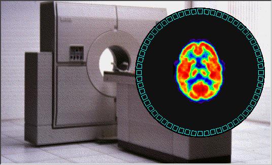
3 Computer tomography (CT) CT is a diagnostic imaging technique that provides information about a slice perpendicular to the longitudinal axis of the examined body.
4 The a, b and c squares can not be distinguished on the base of their projection to the y axis.
5 If we take into consideration their projection to the x axis, the c square can be properly drown, but the a and b squares still look uniform.
6 Modeling the density matrix radiation source detector
7 The attenuation of the I 0 intensity of X-ray can be discribed as: I x = I 0 e -μx The I A-D intensities are the following: I A = I 0 e -(D1+D2) I B = I 0 e -(D3+D4) I C = I 0 e -(D2+D4) I D = I 0 e -(D1+D3) where D k = μx
8 Good resolution requires small voxel size! For all measured I k intensities it is determined which voxels are between the rotating radiation source and the detector. That is called back projection.
9 Following it using Furier transformation the density matrix can be reconstucted. I k = I 0 e - μl where I 0 is the X-ray intensity entering to the body, l a distance the X-ray travels in the body and μ the average attenuation coefficient for the given distance μ=( μ i Δl) /n = D i /n where n is the number of voxels on the given distance.
10 In the energy range used in CT ( kv X-ray tube voltage) The X-ray attenuates mainly by Compton effect (85%) and by fotoeffect (15%). Pair production can not occur. The attenuation in a voxel has two components: μ x = τ x + σ x where τ = absorption coefficient σ = scattering coefficient
11 Both have additional components: μ x = p ρ x Z n eff,x + s ρ x (Z/A) eff,x ρ = density Z = atomic number Z eff,x = Effectiv atomic number n = exponential power (appr. 3) s = scattering constant at a given voltage A = mass number
12 The attenuation coefficient depends upon the atomic and mass number of the material Element Z A Z 3 Z/A H C N O Ca Fe I Ba
13 The Ba and I atoms (because the 3. power of their atomic number is high) shift the attenuation to higher absorption! The contrast material selectively modify the absorption coefficients of the voxels in different tissues/organs. The most frequently applied contrast material is the iodin bound to different organic carries/metabolites. CT angiography (CTA) Renotrop and hepatotrop contrast materials. Dinamyc CT examinations.
14 The density values in CT are expressed in HOUNSFIELD (HU) units. The attenuation of the air and the water are constant (-1000 HU and 0 HU) The density values of few tissues/organs in HU units: Tissue/organ Compact bones Spongy bones Liver Kidney Plasma Lung HU value between between ± 5 30 ± ± 2 between
15 Block scheme of the different generations of CT a detector Radiation source
16 b detector radiation source
17 c detector radiation source
18 d d detector Wolfram ring elektron beam deflecting coil
19 deflecting coil elektron beam Wolfram ring
20 Possible development of CT: Faster speed of the X-ray tube Inceased number of detectors Smaller size of detectors 1 mm slices 3 dimensional secondary image reconstruction. Unravel secondary image reconstruction (so called Janus projection) (data generated from the digital image)
21 Spiral CT Dynamic Volume Scanning, DVS Using continuously moving X-ray tube and patient table in helical (spirális) arrangement within sec all voxel densities of a relatively thick slice of body cylinder can be determined. This method results in an excellent 3 dimension secondary image reconstruction which coupled with Janus projection, using contrast material makes CT angiography possible.


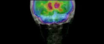
22 CT images of a healthy human brain and after stroke
23 Gamma-camera Using gamma camera the two dimensional projections of γ radiation of radioactive isotope labeled pharmacons from the human body can be detected.
24 Principle of gamma scintillation examinations
25 Upper view of the scintillation crystal and the connected photoelectron amplifiers
26 Renogram showing the kidneys function Counts/sec furosemid injection Right kidney Left kidney Time (min)
27 Block scheme of the scintillation gamma camera Matrix circuit Differential discriminator ADC ADC
28 Scanning trajectory following the body contour
29
30 Positron Emission Tomography PET A PET is a functional imaging method, that provides information about the distribution of the radioactive isotope labeled metabolite administered into the body for diagnostic purpose.

31 Positron-electron annihilation electron/positron annihilation β β + photon γ Conservation of momentum Before: annihilation the momentum of the system is ~ 0 After:two photons of the same energy travelling to opposite directions are created photon γ decay by positron emission Conservation of energy Before: two electrons, with mass equivalent 2 x 511 kev After: two photons with 511 kev energy each
32
33 Block diagram of a PETexamination Production of positron emitting isotope (ciklotron) Injection of the radiopharmacon Data collection Processing of data Synthesis of the radiopharnacon Image reconstruction Interpretation
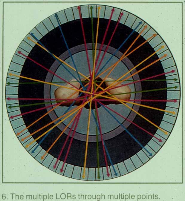
34 Data collection
35 Data collection
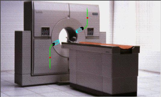
36 Data collection
37 Data collection

38 Image construction
39 Image construction
40 Characteristics of the PET-method Advantages: high sensitivity high spatial resolution high selectivity characteristic for the applied radiopharmacon low radiation dose Disadvantages: low accessibility time consuming high cost


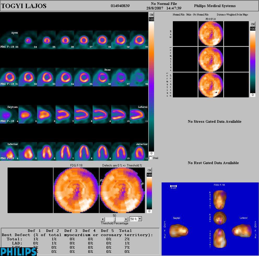
41 Commonly used radiopharmacons 18 FDG Image construction is on the base of glucose metabolism [ 11 C]-metionin Image construction is on the base of protein synthesis (helps in differentiating malignant tumours from inflammations)

42 The PET-camera in Debrecen GE
43
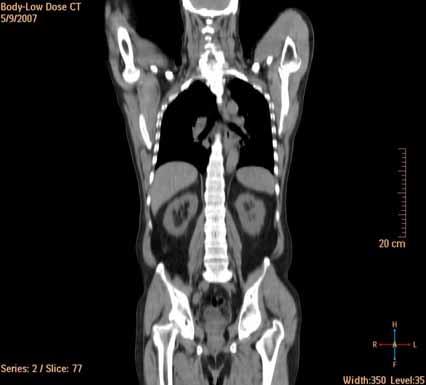
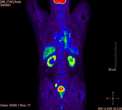
44 Image registration: anatomically equivalent sections CT FDG-PET

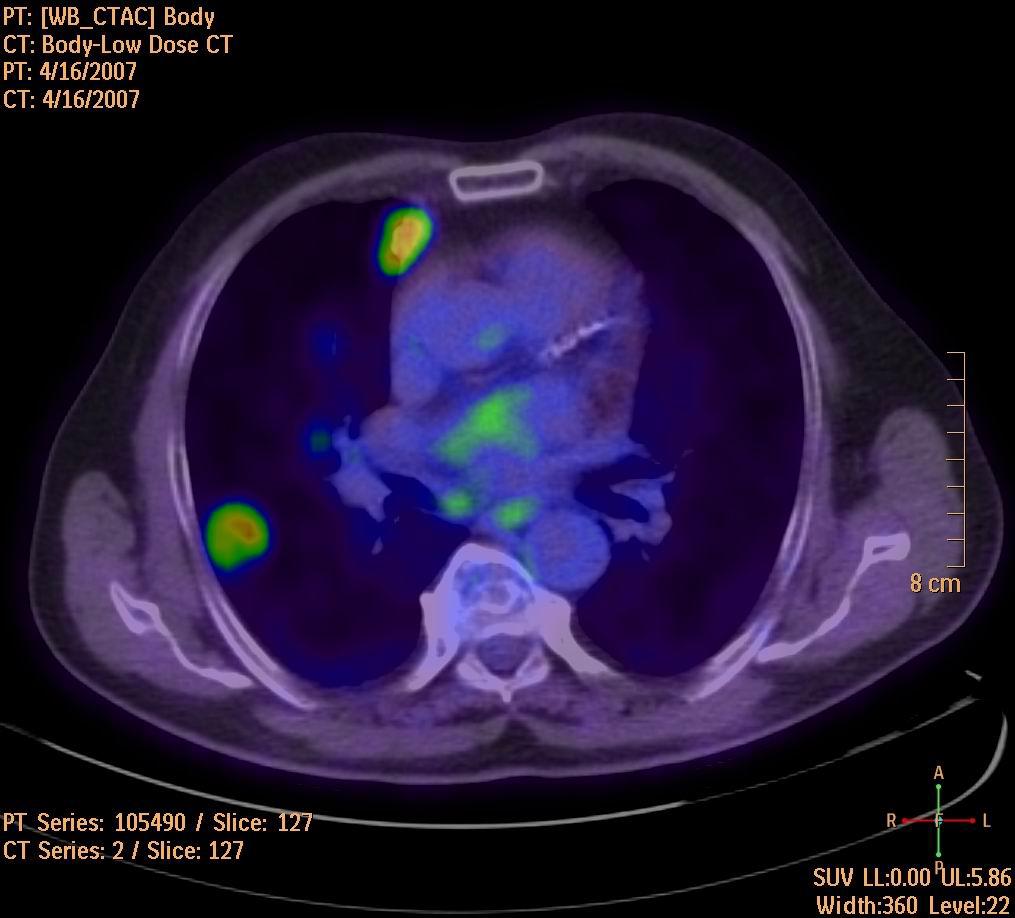
45 Image fusion: overlayed visualization
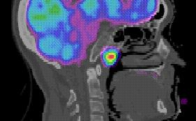
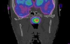
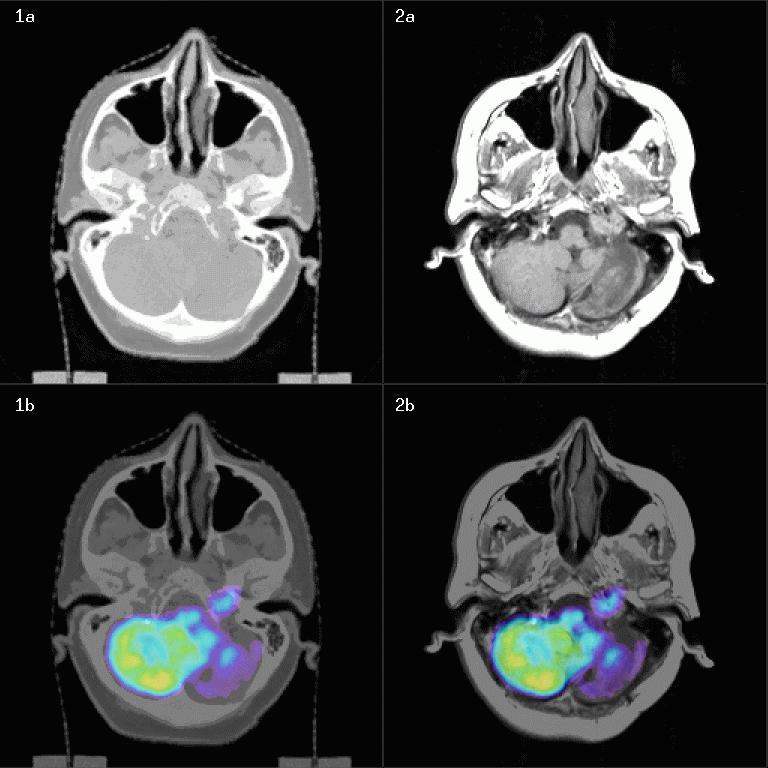
46 Epipharynx-tumour & 3D fusion axial sagittal coronal

47 Image fusion based 3D radiotherapy planning
48
49
50
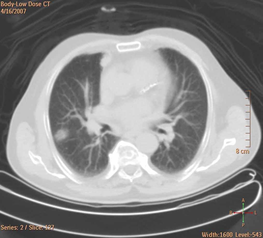
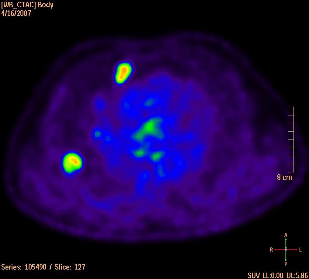
51
52

53 Whole body 3D fusion
54 The main metabolic differences between normal tissue and cancer increased glycolysis (FDG uptake) increased protein synthesis (C11methionine uptake) increased amino acid transport (C11methionine uptake) increased or decreased receptor densities (radionuclid labeled ligands show receptor densities) increased DNA synthesis (C11 thymidine uptake) increased blood flow (O15 butanol/or water uptake) more anoxic and hypoxic cells (F18 labeled ligand uptake)

55 Low-grade astrocytoma FDG METHIONINE

56 Low-grade recurrent glioma (FDG)

57 Low-grade recurrent glioma (MET)

58 Recurrent colorectal cc. & metastases

59 Malignant melanoma Before chemotherapy After chemotherapy

60 Search for unknown tumour No. 1 Metastatic lymph node on the right side of the neck CT [ 11 C]Methionine-PET CT-PET image fusion
BME I5000: Biomedical Imaging
 1 Lucas Parra, CCNY BME I5000: Biomedical Imaging Lecture 4 Computed Tomography Lucas C. Parra, parra@ccny.cuny.edu some slides inspired by lecture notes of Andreas H. Hilscher at Columbia University.
Corso di laurea in Fisica A.A Fisica Medica 5 SPECT, PET
 Corso di laurea in Fisica A.A. 2007-2008 Fisica Medica 5 SPECT, PET Step 1: Inject Patient with Radioactive Drug Drug is labeled with positron (β + ) emitting radionuclide. Drug localizes
Physical bases of X-ray diagnostics
 Physical bases of X-ray diagnostics Dr. István Voszka Possibilities of X-ray production (X-ray is produced, when charged particles of high velocity are stopped) X-ray tube: Relatively low accelerating
Emission Computed Tomography Notes
 Noll (24) ECT Notes: Page 1 Emission Computed Tomography Notes Introduction Emission computed tomography (ECT) is the CT applied to nuclear medicine. There are two varieties of ECT: 1. SPECT single-photon
Ch. 4 Physical Principles of CT
 Ch. 4 Physical Principles of CT CLRS 408: Intro to CT Department of Radiation Sciences Review: Why CT? Solution for radiography/tomography limitations Superimposition of structures Distinguishing between
Medical Imaging BMEN Spring 2016
 Name Medical Imaging BMEN 420-501 Spring 2016 Homework #4 and Nuclear Medicine Notes All questions are from the introductory Powerpoint (based on Chapter 7) and text Medical Imaging Signals and Systems,
Positron Emission Tomography
 Physics 656 Seminar on Physical Fundamentals of Medical Imaging Positron Emission Tomography Ahmed Qamesh Outline What is PET? PET mechanism Radionuclide and its synthesis Detection concept and Development
Spiral CT. Protocol Optimization & Quality Assurance. Ge Wang, Ph.D. Department of Radiology University of Iowa Iowa City, Iowa 52242, USA
 Spiral CT Protocol Optimization & Quality Assurance Ge Wang, Ph.D. Department of Radiology University of Iowa Iowa City, Iowa 52242, USA Spiral CT Protocol Optimization & Quality Assurance Protocol optimization
Tomographic Reconstruction
 Tomographic Reconstruction 3D Image Processing Torsten Möller Reading Gonzales + Woods, Chapter 5.11 2 Overview Physics History Reconstruction basic idea Radon transform Fourier-Slice theorem (Parallel-beam)
Computational Medical Imaging Analysis
 Computational Medical Imaging Analysis Chapter 2: Image Acquisition Systems Jun Zhang Laboratory for Computational Medical Imaging & Data Analysis Department of Computer Science University of Kentucky
Corso di laurea in Fisica A.A Fisica Medica 4 TC
 Corso di laurea in Fisica A.A. 2007-2008 Fisica Medica 4 TC Computed Tomography Principles 1. Projection measurement 2. Scanner systems 3. Scanning modes Basic Tomographic Principle The internal structure
3/27/2012 WHY SPECT / CT? SPECT / CT Basic Principles. Advantages of SPECT. Advantages of CT. Dr John C. Dickson, Principal Physicist UCLH
 3/27/212 Advantages of SPECT SPECT / CT Basic Principles Dr John C. Dickson, Principal Physicist UCLH Institute of Nuclear Medicine, University College London Hospitals and University College London john.dickson@uclh.nhs.uk
ANALYSIS OF CT AND PET/SPECT IMAGES FOR DOSIMETRY CALCULATION
 2009 International Nuclear Atlantic Conference - INAC 2009 Rio de Janeiro,RJ, Brazil, September27 to October 2, 2009 ASSOCIAÇÃO BRASILEIRA DE ENERGIA NUCLEAR - ABEN ISBN: 978-85-99141-03-8 ANALYSIS OF
Shadow casting. What is the problem? Cone Beam Computed Tomography THE OBJECTIVES OF DIAGNOSTIC IMAGING IDEAL DIAGNOSTIC IMAGING STUDY LIMITATIONS
 Cone Beam Computed Tomography THE OBJECTIVES OF DIAGNOSTIC IMAGING Reveal pathology Reveal the anatomic truth Steven R. Singer, DDS srs2@columbia.edu IDEAL DIAGNOSTIC IMAGING STUDY Provides desired diagnostic
Medical Images Analysis and Processing
 Medical Images Analysis and Processing - 25642 Emad Course Introduction Course Information: Type: Graduated Credits: 3 Prerequisites: Digital Image Processing Course Introduction Reference(s): Insight
Radiology. Marta Anguiano Millán. Departamento de Física Atómica, Molecular y Nuclear Facultad de Ciencias. Universidad de Granada
 Departamento de Física Atómica, Molecular y Nuclear Facultad de Ciencias. Universidad de Granada Overview Introduction Overview Introduction Tecniques of imaging in Overview Introduction Tecniques of imaging
Image Acquisition Systems
 Image Acquisition Systems Goals and Terminology Conventional Radiography Axial Tomography Computer Axial Tomography (CAT) Magnetic Resonance Imaging (MRI) PET, SPECT Ultrasound Microscopy Imaging ITCS
Introduction to Biomedical Imaging
 Alejandro Frangi, PhD Computational Imaging Lab Department of Information & Communication Technology Pompeu Fabra University www.cilab.upf.edu X-ray Projection Imaging Computed Tomography Digital X-ray
Introduction to Positron Emission Tomography
 Planar and SPECT Cameras Summary Introduction to Positron Emission Tomography, Ph.D. Nuclear Medicine Basic Science Lectures srbowen@uw.edu System components: Collimator Detector Electronics Collimator
Implementation and evaluation of a fully 3D OS-MLEM reconstruction algorithm accounting for the PSF of the PET imaging system
 Implementation and evaluation of a fully 3D OS-MLEM reconstruction algorithm accounting for the PSF of the PET imaging system 3 rd October 2008 11 th Topical Seminar on Innovative Particle and Radiation
Cherenkov Radiation. Doctoral Thesis. Rok Dolenec. Supervisor: Prof. Dr. Samo Korpar
 Doctoral Thesis Time-of-Flight Time-of-Flight Positron Positron Emission Emission Tomography Tomography Using Using Cherenkov Cherenkov Radiation Radiation Rok Dolenec Supervisor: Prof. Dr. Samo Korpar
Cardiac Dual Energy CT: Technique
 RSNA 2013, VSCA51-01, Chicago, Dec. 5, 2013 Cardiac Radiology Series Cardiac Dual Energy CT: Technique Willi A. Kalender, Ph.D. Institute of Medical Physics University of Erlangen www.imp.uni-erlangen.de
MEDICAL IMAGING 2nd Part Computed Tomography
 MEDICAL IMAGING 2nd Part Computed Tomography Introduction 2 In the last 30 years X-ray Computed Tomography development produced a great change in the role of diagnostic imaging in medicine. In convetional
Computer-Tomography II: Image reconstruction and applications
 Computer-Tomography II: Image reconstruction and applications Prof. Dr. U. Oelfke DKFZ Heidelberg Department of Medical Physics (E040) Im Neuenheimer Feld 280 69120 Heidelberg, Germany u.oelfke@dkfz.de
CT Basics Principles of Spiral CT Dose. Always Thinking Ahead.
 1 CT Basics Principles of Spiral CT Dose 2 Who invented CT? 1963 - Alan Cormack developed a mathematical method of reconstructing images from x-ray projections Sir Godfrey Hounsfield worked for the Central
Medical Imaging Modalities
 Image Science Introduction Medical Imaging Modalities Ho Kyung Kim hokyung@pusan.ac.kr Pusan National University Projection Radiography Routine diagnostic radiography Chest x rays, fluoroscopy, mammography,
Positron. MillenniumVG. Emission Tomography Imaging with the. GE Medical Systems
 Positron Emission Tomography Imaging with the MillenniumVG GE Medical Systems Table of Contents Introduction... 3 PET Imaging With Gamma Cameras PET Overview... 4 Coincidence Detection on Gamma Cameras...
Introduction to Emission Tomography
 Introduction to Emission Tomography Gamma Camera Planar Imaging Robert Miyaoka, PhD University of Washington Department of Radiology rmiyaoka@u.washington.edu Gamma Camera: - collimator - detector (crystal
Digital Image Processing
 Digital Image Processing SPECIAL TOPICS CT IMAGES Hamid R. Rabiee Fall 2015 What is an image? 2 Are images only about visual concepts? We ve already seen that there are other kinds of image. In this lecture
Some reference material
 Some reference material Physics reference book on medical imaging: A good one is The Essential Physics of Medical Imaging, 3 rd Ed. by Bushberg et al. ($170! new). However, there are several similar books
Moscow-Bavarian Joint Advanced Student School 2006 / Medical Imaging Principles of Computerized Tomographic Imaging and Cone-Beam Reconstruction
 Line Integrals Line integrals represent the integral of some parameter of the object along the line (e.g. attenuation of x-rays) Object: f(x,y) Line: x cosθ + y sinθ = t Line integral / Radon transform:
DICOM. Supplement 188 Multi-Energy CT Imaging. DICOM Working Group 21 Computed Tomography
 DICOM Supplement 188 Multi-Energy CT Imaging DICOM Working Group 21 Computed Tomography Rationale Short introduction of Multi Energy (ME) s Overview: Imaging techniques, including scanning, reconstruction,
Validation of PET/CT dataset for radiation treatment planning
 Louisiana State University LSU Digital Commons LSU Master's Theses Graduate School 2004 Validation of PET/CT dataset for radiation treatment planning Rajesh Manoharan Louisiana State University and Agricultural
8/7/2017. Disclosures. MECT Systems Overview and Quantitative Opportunities. Overview. Computed Tomography (CT) CT Numbers. Polyenergetic Acquisition
 CT Numbers. Polyenergetic Acquisition") Quantitative Multi-Energy Computed Tomography: Imaging and Therapy Advancements Disclosures MECT Systems Overview and Quantitative Opportunities The speaker receives research funding from GE Healthcare
Quantitative Multi-Energy Computed Tomography: Imaging and Therapy Advancements Disclosures MECT Systems Overview and Quantitative Opportunities The speaker receives research funding from GE Healthcare
Constructing System Matrices for SPECT Simulations and Reconstructions
 Constructing System Matrices for SPECT Simulations and Reconstructions Nirantha Balagopal April 28th, 2017 M.S. Report The University of Arizona College of Optical Sciences 1 Acknowledgement I would like
Fits you like no other
 Fits you like no other BrightView X and XCT specifications The new BrightView X system is a fully featured variableangle camera that is field-upgradeable to BrightView XCT without any increase in room
Fundamentals of CT imaging
 SECTION 1 Fundamentals of CT imaging I History In the early 1970s Sir Godfrey Hounsfield s research produced the first clinically useful CT scans. Original scanners took approximately 6 minutes to perform
Medical Image Analysis
 Computer assisted Image Analysis VT04 29 april 2004 Medical Image Analysis Lecture 10 (part 1) Xavier Tizon Medical Image Processing Medical imaging modalities XRay,, CT Ultrasound MRI PET, SPECT Generic
Fits you like no other
 Fits you like no other Philips BrightView X and XCT specifications The new BrightView X system is a fully featured variableangle camera that is field-upgradeable to BrightView XCT without any increase
Computer-Tomography I: Principles, History, Technology
 Computer-Tomography I: Principles, History, Technology Prof. Dr. U. Oelfke DKFZ Heidelberg Department of Medical Physics (E040) Im Neuenheimer Feld 280 69120 Heidelberg, Germany u.oelfke@dkfz.de History
Nuclear Medicine Imaging
 Introduction to Medical Engineering (Medical Imaging) Suetens 5 Nuclear Medicine Imaging Ho Kyung Kim Pusan National University Introduction Use of radioactive isotopes for medical purposes since 1920
Review of PET Physics. Timothy Turkington, Ph.D. Radiology and Medical Physics Duke University Durham, North Carolina, USA
 Review of PET Physics Timothy Turkington, Ph.D. Radiology and Medical Physics Duke University Durham, North Carolina, USA Chart of Nuclides Z (protons) N (number of neutrons) Nuclear Data Evaluation Lab.
A dedicated tool for PET scanner simulations using FLUKA
 A dedicated tool for PET scanner simulations using FLUKA P. G. Ortega FLUKA meeting June 2013 1 Need for in-vivo treatment monitoring Particles: The good thing is that they stop... Tumour Normal tissue/organ
PET/CT Image Segmentation using the Channeler Ant Model for Lymphoma Therapy Assessment
 Univeristy of Turin School of Natural Sciences Master of Science in Physics Nuclear and Biomedical Physics Academic Year 2013-2014 PET/CT Image Segmentation using the Channeler Ant Model for Lymphoma Therapy
Enhanced material contrast by dual-energy microct imaging
 Enhanced material contrast by dual-energy microct imaging Method note Page 1 of 12 2 Method note: Dual-energy microct analysis 1. Introduction 1.1. The basis for dual energy imaging Micro-computed tomography
Extremely Fast Detector for 511 kev Gamma
 Extremely Fast Detector for 511 kev Gamma V. Sharyy, D. Yvon, G. Tauzin, E.Delagnes, Ph. Abbon, J P. Bard, M. Kebbiri, M. Alokhina, C. Canot IRFU, CEA D. Breton, J. Maalmi LAL,IN2P3 Journée 2015 du Labex
Principles of Computerized Tomographic Imaging
 Principles of Computerized Tomographic Imaging Parallel CT, Fanbeam CT, Helical CT and Multislice CT Marjolein van der Glas August 29, 2000 Abstract The total attenuation suffered by one beam of x-rays
SNIC Symposium, Stanford, California April The Hybrid Parallel Plates Gas Counter for Medical Imaging
 The Hybrid Parallel Plates Gas Counter for Medical Imaging F. Anulli, G. Bencivenni, C. D Ambrosio, D. Domenici, G. Felici, F. Murtas Laboratori Nazionali di Frascati - INFN, Via E. Fermi 40, I-00044 Frascati,
Continuation Format Page
 C.1 PET with submillimeter spatial resolution Figure 2 shows two views of the high resolution PET experimental setup used to acquire preliminary data [92]. The mechanics of the proposed system are similar
MEDICAL IMAGING 2nd Part Computed Tomography
 MEDICAL IMAGING 2nd Part Computed Tomography Introduction 2 In the last 30 years X-ray Computed Tomography development produced a great change in the role of diagnostic imaging in medicine. In convetional
Reconstruction in CT and relation to other imaging modalities
 Reconstruction in CT and relation to other imaging modalities Jørgen Arendt Jensen November 1, 2017 Center for Fast Ultrasound Imaging, Build 349 Department of Electrical Engineering Center for Fast Ultrasound
Detection of Lesions in Positron Emission Tomography
 Detection of Lesions in Positron Emission Tomography Bachelor Thesis Nina L.F. Bezem Study: Physics and Astronomy Faculty of Science Supervised by: Dr. Andre Mischke Utrecht University, Institute for Subatomic
REMOVAL OF THE EFFECT OF COMPTON SCATTERING IN 3-D WHOLE BODY POSITRON EMISSION TOMOGRAPHY BY MONTE CARLO
 REMOVAL OF THE EFFECT OF COMPTON SCATTERING IN 3-D WHOLE BODY POSITRON EMISSION TOMOGRAPHY BY MONTE CARLO Abstract C.S. Levin, Y-C Tai, E.J. Hoffman, M. Dahlbom, T.H. Farquhar UCLA School of Medicine Division
Biomedical Imaging. Computed Tomography. Patrícia Figueiredo IST
 Biomedical Imaging Computed Tomography Patrícia Figueiredo IST 2013-2014 Overview Basic principles X ray attenuation projection Slice selection and line projections Projection reconstruction Instrumentation
James R Halama, PhD Loyola University Medical Center
 James R Halama, PhD Loyola University Medical Center Conflicts of Interest Nuclear Medicine and PET physics reviewer for the ACR Accreditation program Learning Objectives Be familiar with the tests recommended
INVESTIGATION OF ACCURACY IN QUANTITATION OF 18 F-FDG CONCENTRATION OF PET/CT. A Thesis
 INVESTIGATION OF ACCURACY IN QUANTITATION OF 18 F-FDG CONCENTRATION OF PET/CT A Thesis Submitted to the Graduate Faculty of the Louisiana State University and Agricultural and Mechanical College in partial
FRONT-END DATA PROCESSING OF NEW POSITRON EMIS- SION TOMOGRAPHY DEMONSTRATOR
 SOUDABEH MORADI FRONT-END DATA PROCESSING OF NEW POSITRON EMIS- SION TOMOGRAPHY DEMONSTRATOR Master of Science Thesis Examiners: Prof. Ulla Ruotsalainen MSc Defne Us Examiners and topic approved by the
Computational Medical Imaging Analysis
 Computational Medical Imaging Analysis Chapter 1: Introduction to Imaging Science Jun Zhang Laboratory for Computational Medical Imaging & Data Analysis Department of Computer Science University of Kentucky
Semi-Quantitative Metrics in Positron Emission Tomography. Michael Adams. Department of Biomedical Engineering Duke University.
 Semi-Quantitative Metrics in Positron Emission Tomography by Michael Adams Department of Biomedical Engineering Duke University Date: Approved: Timothy G. Turkington, Supervisor Adam P. Wax Terence Z.
Medical Image Processing: Image Reconstruction and 3D Renderings
 Medical Image Processing: Image Reconstruction and 3D Renderings 김보형 서울대학교컴퓨터공학부 Computer Graphics and Image Processing Lab. 2011. 3. 23 1 Computer Graphics & Image Processing Computer Graphics : Create,
CT vs. VolumeScope: image quality and dose comparison
 CT vs. VolumeScope: image quality and dose comparison V.N. Vasiliev *a, A.F. Gamaliy **b, M.Yu. Zaytsev b, K.V. Zaytseva ***b a Russian Sci. Center of Roentgenology & Radiology, 86, Profsoyuznaya, Moscow,
TEP Hounsfield units. Related topics Attenuation coefficient, Hounsfield units
 Hounsfield units TEP Related topics Attenuation coefficient, Hounsfield units Principle Depending on the type of CT scanner and the settings, the result of a CT scan of the same material can be different
Basics of treatment planning II
 Basics of treatment planning II Sastry Vedam PhD DABR Introduction to Medical Physics III: Therapy Spring 2015 Dose calculation algorithms! Correction based! Model based 1 Dose calculation algorithms!
Comparison of internal and external dose conversion factors using ICRP adult male and MEET Man voxel model phantoms.
 Comparison of internal and external dose conversion factors using ICRP adult male and MEET Man voxel model phantoms. D.Leone, A.Häußler Intitute for Nuclear Waste Disposal, Karlsruhe Institute for Technology,
Conflicts of Interest Nuclear Medicine and PET physics reviewer for the ACR Accreditation program
 James R Halama, PhD Loyola University Medical Center Conflicts of Interest Nuclear Medicine and PET physics reviewer for the ACR Accreditation program Learning Objectives 1. Be familiar with recommendations
COMPARATIVE STUDIES OF DIFFERENT SYSTEM MODELS FOR ITERATIVE CT IMAGE RECONSTRUCTION
 COMPARATIVE STUDIES OF DIFFERENT SYSTEM MODELS FOR ITERATIVE CT IMAGE RECONSTRUCTION BY CHUANG MIAO A Thesis Submitted to the Graduate Faculty of WAKE FOREST UNIVERSITY GRADUATE SCHOOL OF ARTS AND SCIENCES
UNIVERSITY OF SOUTHAMPTON
 UNIVERSITY OF SOUTHAMPTON PHYS2007W1 SEMESTER 2 EXAMINATION 2014-2015 MEDICAL PHYSICS Duration: 120 MINS (2 hours) This paper contains 10 questions. Answer all questions in Section A and only two questions
DUE to beam polychromacity in CT and the energy dependence
 1 Empirical Water Precorrection for Cone-Beam Computed Tomography Katia Sourbelle, Marc Kachelrieß, Member, IEEE, and Willi A. Kalender Abstract We propose an algorithm to correct for the cupping artifact
Effect of Scattering on the Image. Reducing Compton Scatter with a Grid
 Effect of Scattering on the Image Increasing Compton scattering degrades image. Webb 21 Reducing Compton Scatter with a Grid Grids Parallel (focused at infinity) Linear Focused (see figure) Moving grids
Introduction to Medical Image Analysis
 Introduction to Medical Image Analysis Rasmus R. Paulsen DTU Compute rapa@dtu.dk http://courses.compute.dtu.dk/02511 http://courses.compute.dtu.dk/02511 Plenty of slides adapted from Thomas Moeslunds lectures
Joint CI-JAI advanced accelerator lecture series Imaging and detectors for medical physics Lecture 1: Medical imaging
 Joint CI-JAI advanced accelerator lecture series Imaging and detectors for medical physics Lecture 1: Medical imaging Dr Barbara Camanzi barbara.camanzi@stfc.ac.uk Course layout Day AM 09.30 11.00 PM 15.30
X-ray simulation and applications
 Computerized Tomography for Industrial Applications and Image Processing in Radiology March 15-17, 1999, Berlin, Germany DGZfP Proceedings BB 67-CD Paper 17 X-ray simulation and applications P. Hugonnard,
Midterm Review. Yao Wang Polytechnic University, Brooklyn, NY 11201
 Midterm Review Yao Wang Polytechnic University, Brooklyn, NY 11201 Based on J. L. Prince and J. M. Links, Medical maging Signals and Systems, and lecture notes by Prince. Figures are from the textbook.
RADIOLOGY AND DIAGNOSTIC IMAGING
 Day 2 part 2 RADIOLOGY AND DIAGNOSTIC IMAGING Dr hab. Zbigniew Serafin, MD, PhD serafin@cm.umk.pl 2 3 4 5 CT technique CT technique 6 CT system Kanal K: RSNA/AAPM web module: CT Systems & CT Image Quality
Hidenobu Tachibana The Cancer Institute Hospital of JFCR, Radiology Dept. The Cancer Institute of JFCR, Physics Dept.
 2-D D Dose-CT Mapping in Geant4 Hidenobu Tachibana The Cancer Institute Hospital of JFCR, Radiology Dept. The Cancer Institute of JFCR, Physics Dept. Table of Contents Background & Purpose Materials Methods
Hybrid Imaging for Patient-Specific Dosimetry in Radionuclide Therapy
 Hybrid Imaging for Patient-Specific Dosimetry in Radionuclide Therapy Ljungberg, Michael; Sjögreen Gleisner, Katarina Published in: Diagnostics DOI: 10.3390/diagnostics5030296 Published: 2015-01-01 Link
I. The digital image MEDICAL IMAGING METHODS. Optical illusions - intensity (color density) Optical illusions space. Optical illusions size, direction
 Optical illusions space. Optical illusions size, direction") I. The digital image Seeing is believing!? Vision is not only the detection of image information, but complex processing occurs as well MEDICAL IMAGING METHODS Optical illusions size, direction Miklós
I. The digital image Seeing is believing!? Vision is not only the detection of image information, but complex processing occurs as well MEDICAL IMAGING METHODS Optical illusions size, direction Miklós
Philips SPECT/CT Systems
 Philips SPECT/CT Systems Ling Shao, PhD Director, Imaging Physics & System Analysis Nuclear Medicine, Philips Healthcare June 14, 2008 *Presented SNM08 Categorical Seminar - Quantitative SPECT and PET
Positron Emission Tomography for Pre-Clinical Sub-Volume Dose Escalation
 Virginia Commonwealth University VCU Scholars Compass Theses and Dissertations Graduate School 2013 Positron Emission Tomography for Pre-Clinical Sub-Volume Dose Escalation Christopher Bass Virginia Commonwealth
664 IEEE TRANSACTIONS ON NUCLEAR SCIENCE, VOL. 52, NO. 3, JUNE 2005
 664 IEEE TRANSACTIONS ON NUCLEAR SCIENCE, VOL. 52, NO. 3, JUNE 2005 Attenuation Correction for the NIH ATLAS Small Animal PET Scanner Rutao Yao, Member, IEEE, Jürgen Seidel, Jeih-San Liow, Member, IEEE,
Biomedical Imaging and Image Analysis
 Biomedical Imaging and Image Analysis Lecture in Medical Informatics Course Ewert Bengtsson Professor of computerized image analysis Centrum för bildanalys The theme Images are of central importance in
Contrast Enhancement with Dual Energy CT for the Assessment of Atherosclerosis
 Contrast Enhancement with Dual Energy CT for the Assessment of Atherosclerosis Stefan C. Saur 1, Hatem Alkadhi 2, Luca Regazzoni 1, Simon Eugster 1, Gábor Székely 1, Philippe Cattin 1,3 1 Computer Vision
Optimization of CT Simulation Imaging. Ingrid Reiser Dept. of Radiology The University of Chicago
 Optimization of CT Simulation Imaging Ingrid Reiser Dept. of Radiology The University of Chicago Optimization of CT imaging Goal: Achieve image quality that allows to perform the task at hand (diagnostic
Validation of GEANT4 for Accurate Modeling of 111 In SPECT Acquisition
 Validation of GEANT4 for Accurate Modeling of 111 In SPECT Acquisition Bernd Schweizer, Andreas Goedicke Philips Technology Research Laboratories, Aachen, Germany bernd.schweizer@philips.com Abstract.
Phase-Contrast Imaging and Tomography at 60 kev using a Conventional X-ray Tube
 Phase-Contrast Imaging and Tomography at 60 kev using a Conventional X-ray Tube T. Donath* a, F. Pfeiffer a,b, O. Bunk a, W. Groot a, M. Bednarzik a, C. Grünzweig a, E. Hempel c, S. Popescu c, M. Hoheisel
Medical Imaging Projects
 NSF REU MedIX Summer 2006 Medical Imaging Projects Daniela Stan Raicu, PhD http://facweb.cs.depaul.edu/research draicu@cs.depaul.edu Outline Medical Informatics Imaging Modalities Computed Tomography Medical
CT Systems and their standards
 CT Systems and their standards Stephen Brown Engineering Measurement 11 th April 2012 Industrial X-ray computed tomography: The future of co-ordinate metrology? Burleigh Court, Loughborough University
A closer look at CT scanning
 Vet Times The website for the veterinary profession https://www.vettimes.co.uk A closer look at CT scanning Author : Charissa Lee, Natalie Webster Categories : General, Vets Date : April 3, 2017 A basic
Lecture 6: Medical imaging and image-guided interventions
 ME 328: Medical Robotics Winter 2019 Lecture 6: Medical imaging and image-guided interventions Allison Okamura Stanford University Updates Assignment 3 Due this Thursday, Jan. 31 Note that this assignment
Effects of the difference in tube voltage of the CT scanner on. dose calculation
 Effects of the difference in tube voltage of the CT scanner on dose calculation Dong Joo Rhee, Sung-woo Kim, Dong Hyeok Jeong Medical and Radiological Physics Laboratory, Dongnam Institute of Radiological
C a t p h a n / T h e P h a n t o m L a b o r a t o r y
 C a t p h a n 5 0 0 / 6 0 0 T h e P h a n t o m L a b o r a t o r y C a t p h a n 5 0 0 / 6 0 0 Internationally recognized for measuring the maximum obtainable performance of axial, spiral and multi-slice
Electron Dose Kernels (EDK) for Secondary Particle Transport in Deterministic Simulations
 for Secondary Particle Transport in Deterministic Simulations") Electron Dose Kernels (EDK) for Secondary Particle Transport in Deterministic Simulations A. Al-Basheer, G. Sjoden, M. Ghita Computational Medical Physics Team Nuclear & Radiological Engineering University
Electron Dose Kernels (EDK) for Secondary Particle Transport in Deterministic Simulations A. Al-Basheer, G. Sjoden, M. Ghita Computational Medical Physics Team Nuclear & Radiological Engineering University
The Monte Carlo simulation of a Package formed by the combination of three scintillators: Brillance380, Brillance350, and Prelude420.
 EURONS I3 506065 JRA9 RHIB Report made during stay IEM-CSIC Madrid december 2006 MINISTERIO DE ASUNTOS EXTERIORES Y DE COOPERACIÓN AECI VICESECRETARÍA GENERAL The Monte Carlo simulation of a Package formed
Master of Science Thesis. Activity quantification of planar gamma camera images
 Master of Science Thesis Activity quantification of planar gamma camera images Marie Sydoff Supervisor: Sigrid Leide-Svegborn The work has been performed at Department of Radiation Physics Malmö University
Low-Dose Dual-Energy CT for PET Attenuation Correction with Statistical Sinogram Restoration
 Low-Dose Dual-Energy CT for PET Attenuation Correction with Statistical Sinogram Restoration Joonki Noh, Jeffrey A. Fessler EECS Department, The University of Michigan Paul E. Kinahan Radiology Department,
Michael Speiser, Ph.D.
 IMPROVED CT-BASED VOXEL PHANTOM GENERATION FOR MCNP MONTE CARLO Michael Speiser, Ph.D. Department of Radiation Oncology UT Southwestern Medical Center Dallas, TX September 1 st, 2012 CMPWG Workshop Medical
Mathematical methods and simulations tools useful in medical radiation physics
 Mathematical methods and simulations tools useful in medical radiation physics Michael Ljungberg, professor Department of Medical Radiation Physics Lund University SE-221 85 Lund, Sweden Major topic 1:
CLASS HOURS: 4 CREDIT HOURS: 4 LABORATORY HOURS: 0
 Revised 10/10 COURSE SYLLABUS TM 220 COMPUTED TOMOGRAPHY PHYSICS CLASS HOURS: 4 CREDIT HOURS: 4 LABORATORY HOURS: 0 CATALOG COURSE DESCRIPTION: This course is one of a three course set in whole body Computed
RADIOMICS: potential role in the clinics and challenges
 27 giugno 2018 Dipartimento di Fisica Università degli Studi di Milano RADIOMICS: potential role in the clinics and challenges Dr. Francesca Botta Medical Physicist Istituto Europeo di Oncologia (Milano)
First CT Scanner. How it Works. Contemporary CT. Before and After CT. Computer Tomography: How It Works. Medical Imaging and Pattern Recognition
 Computer Tomography: How t Works Medical maging and Pattern Recognition Lecture 7 Computed Tomography Oleh Tretiak Only one plane is illuminated. Source-subject motion provides added information. 2 How