Medical Image Processing: Image Reconstruction and 3D Renderings
|
|
|
- Theodore Lang
- 5 years ago
- Views:
Transcription


1 Medical Image Processing: Image Reconstruction and 3D Renderings 김보형 서울대학교컴퓨터공학부 Computer Graphics and Image Processing Lab


2 Computer Graphics & Image Processing Computer Graphics : Create, store and manipulate models and images using computers Image Processing : any form of signal processing for which the input is an image, the output of image processing can be either an image or a set of characteristics or parameters related to the image. 2
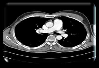
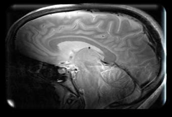
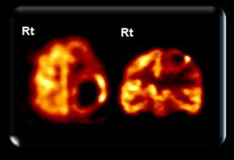
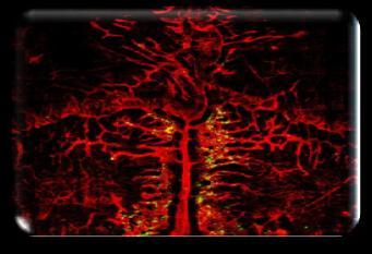
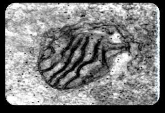
3 Medical Applications Multi-modality images CT MR PET CONFOCAL TEM 3
4 4 Capability of Radiologists
5 Evolution of Spiral CT Scanners TOSHIBA Philips Siemens Siemens GE TOSHIBA Brilliance ict Dual Source Dual Source LightSpeed HD Row thickness Detector rows Spatial resolution 0.5 mm mm 0.6 mm 0.6 mm mm 0.5 mm mm 0.34 mm 0.4 mm 0.4 mm 0.24 mm 0.35 mm Detector Coverage 32 mm 80 mm 19.2 mm 38.4 mm 44 mm 160 mm Rotation Speed 350 ms 270 ms 330 ms 280 ms 350 ms 350 ms 5
6 6


7 Need of 3D Imaging Limitations of diagnosis by radiologist according to a large of amount of images Additional cost due to filming of a large of amount of images Data overload due to multi-slice CT/MR Need 3D imaging 7
8 8

9 General CT System X-ray Tube Scan (Data acquisition) Console Monitor Detector Reconstructor Data Acquisition System 9


10 3D Reconstruction Sampling point During the rotation 3D Volume Data reconstruction Re Filtering & Back projection Projected images 10

 P ( Y( r), Z( r)) rz d r z D z P Φ Y r(x,y,z) Source to object : d O x Z Beam Source y Object")
11 Back-projection Every voxel in the object is calculated using geometric information. r is a position vector in object space P Φ is a position vector in projection space r is back-projected from the value at P Φ of projected image Projection space ry Y( r) D, Z( r) d ry P ( r) P ( Y( r), Z( r)) rz d r z D z P Φ Y r(x,y,z) Source to object : d O x Z Beam Source y Object space Source to detector : D 11
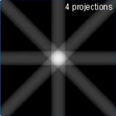
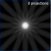
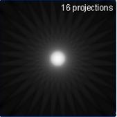
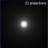
12 Back-projection Feldkamp, Davis, and Kress Practical cone-beam algorithm 12
13 GPU based 3D Reconstruction Projection image : , 360 slices Volume Size : Test machine CPU : Intel Core2Quad Q6600 GPU : NVIDIA GTX 280 1GB CPU Backprojection (BP) 352 unit : second GPU Upload projections BP on GPU Download result


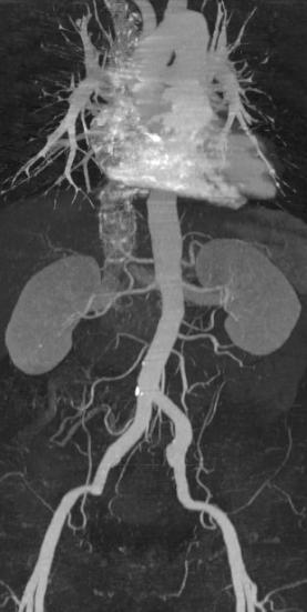
14 How we see 3D Volume Data? DICOM PACS Server CT Non-DICOM DICOM Gateway (Converter) MR 3D Visualization & CAD 2D MPR VR MIP/MinIP SSD Endoscopy Colonoscopy BSA Fusion CAD 14


15 How we get 3D images from a stack of many slices? MPR VR MIP MinIP 15
16 Thin- vs. Thick-section Scans Scans with a large section thickness (e.g., 5mm) Excellent low-contrast detectability For primary interpretation Scans with a small section thickness (e.g., 0.67, 1mm) Excellent through-plane spatial resolution But, grainy noise For Multiplanar refomations (MPR) or 3D renderings (VR, MIP/MinIP, SSD) routinely reconstruct images with two or more different STs from a single multi-helical projection data 16


17 Thin- vs. Thick-section Scans Thick-section scan image Thin-section scan image 17
 Reconstruction")
, Coronal,")

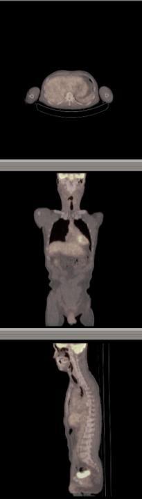
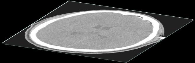
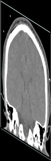
18 Multiplanar Reformation (MPR) Reconstruction planes : Axial (Transverse), Coronal, Sagittal Interactive viewing : Freehand drawing Axial Coronal Sagittal 18

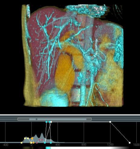
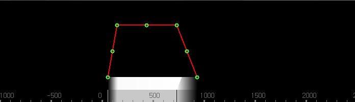
19 Multiplanar Reformation (MPR) Window level: image brightness Window width: image contrast Lung window Window level: -600 HU Window width: 1600 HU Window level: 40 HU Window width: 400 HU Bone window Window level: 300 HU Window width: 2000 HU

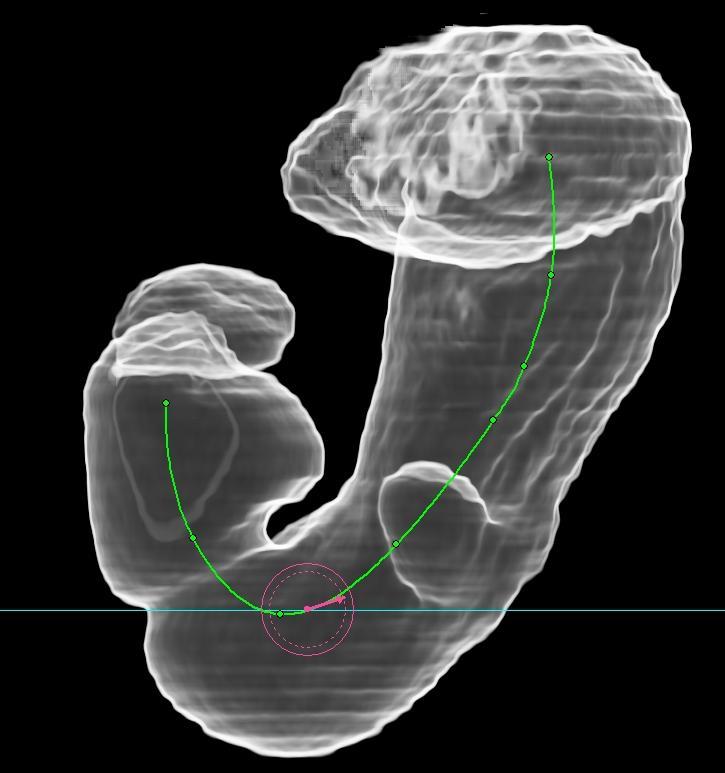
20 Multiplanar Reformation (MPR) Interactive viewing : Curve, Freehand drawings 20
 2D image & lack of overall feature A: Anterior P: Posterior R: Right L: Left H: Head F: Foot")
21 Multiplanar Reformation (MPR) MPR images imposed on volume rendering (+) Real-time & entire information without loss of data (-) 2D image & lack of overall feature A: Anterior P: Posterior R: Right L: Left H: Head F: Foot 21


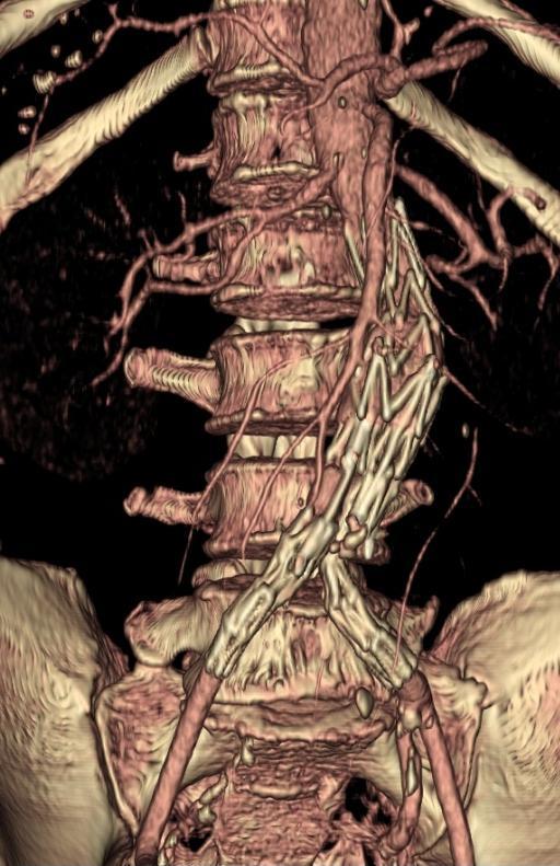
22 Maximum Intensity Projection (MIP) How do we see only organs with high density values such as bones, contrasted vessels, and calcification? Vessels, Calcifications Bones 22
 Maximum attenuation")

23 Maximum Intensity Projection (MIP) Maximum attenuation value is mapped to a 2D gray scale image Entry point Exit point 23
")


24 Minimum Intensity Projection (MinIP) How do we see only organs with low density values such as lung and airway? Line of Sight Minimum attenuation value is mapped to a 2D gray scale image 24


25 Ray Sum Technique Entry point Adds the pixel values along the line of sight Resultant display is similar to an X-Ray image Excellent for displaying overlapping vessels Exit point MIP Ray Sum 25


26 MIP / MinIP / Ray Sum Pros Good for representing organs with high/low/average intensity values Cons 2D image Lack of depth information Overlap of objects with similar intensities Contrasted vessels, parenchyma, bones Low performance Need to process the whole data sets 26


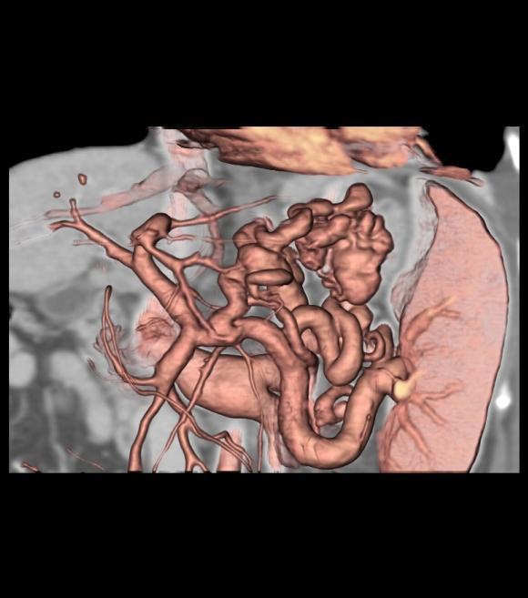
27 Volume Rendering Data Set Volume Data Image A method of displaying volumetric data as a two-dimensional image Render volume without extracting any surfaces (Direct Volume Rendering, DVR) Surface Shaded Display, SSD 27

28 Theoretical Background on Volume Rendering Refer to Chapter 1 of the book Real-Time Volume Graphics by K. Engel et al. 28

29 29 Theoretical Background on Volume Rendering Opacity Correction Compositing schemes assumes an equidistant sampling When the sampling rate needs to be changed The discretized opacity and color need to be modified accordingly. t k dt t k e e T t s i i s ) ( t k e T ~ ~ t t T T ~ ~ t t ~ ) (1 1 ~ A segment length of t A segment length of t ~ t q t t q C ) ( t q C ~ ~ A segment length of t A segment length of t ~ t t C C ~ ~

30 30 Volume Rendering Parameters


31 Volume Rendering Pros High quality Natural representation of CT/MRI images. Good detectability of subtle lesions Minimal editing Cons Huge data sets Computationally expensive Operator dependent Cannot be embedded easily into a polygonal scene 31

32 GPU based Volume Rendering Texture-mapping Ray Casting 32


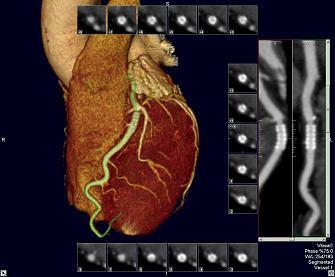
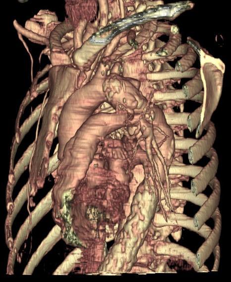
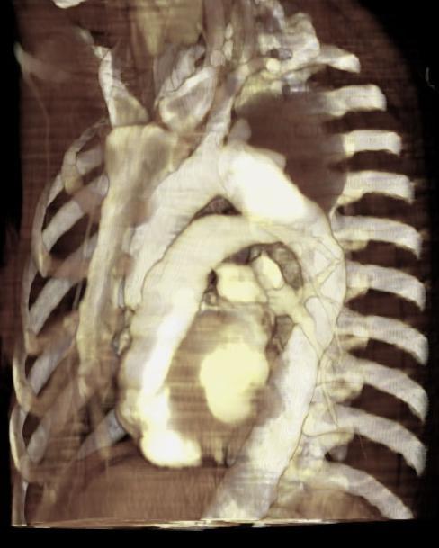
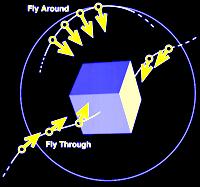
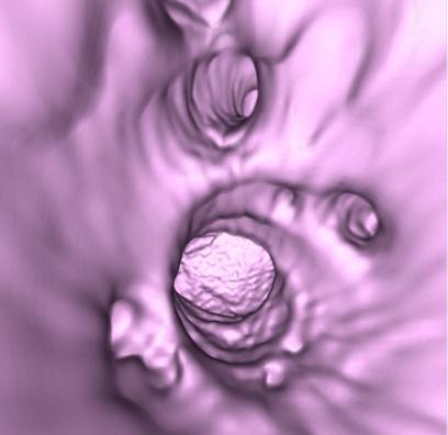
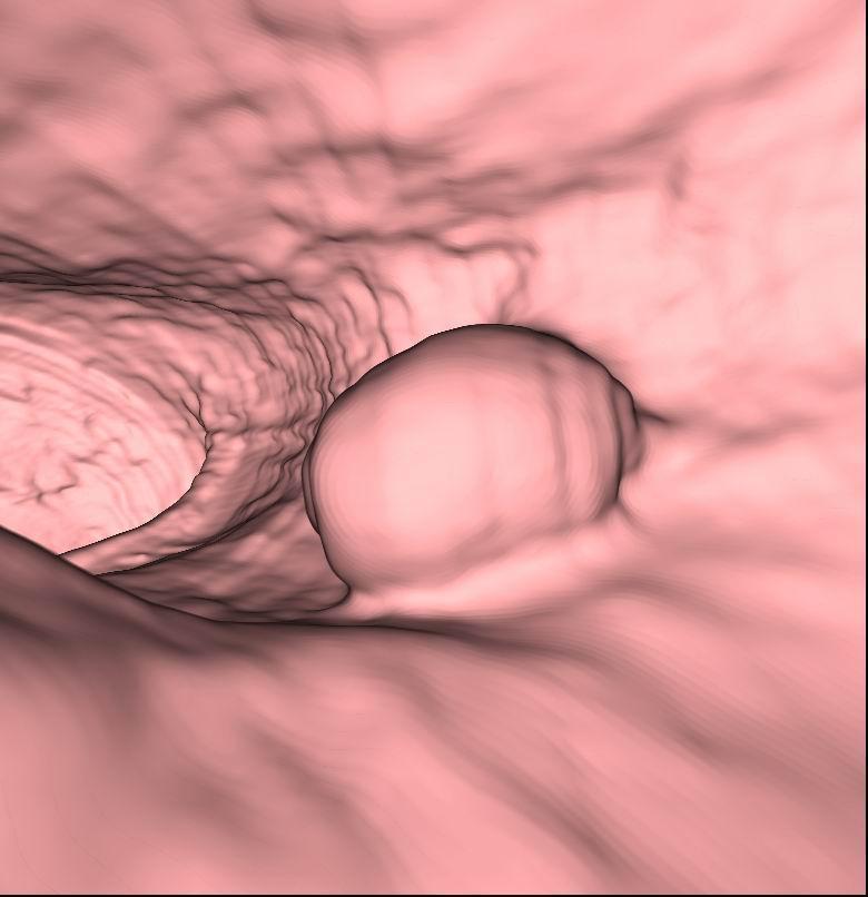
33 Perspective Volume Rendering Perspective vs. Non-perspective Non-perspective : parallel ray Perspective : divergent ray Perspective viewing Inside (Virtual Endoscopy) Applied fields Angioscopy Bronchoscopy Gastroscopy Colonoscopy Cisternoscopy Non-perspective Perspective 33


34 GPU based Perspective VR Texture-based VR with perspective view Set the viewing matrix for perspective view Proxy geometry is changed to hemisphere mesh for uniform sampling interval eye proxy geometry mesh proxy geometry perspective texture-based VR 34

35 35 Virtual Angioscopy


36 36 Virtual Gastroscopy


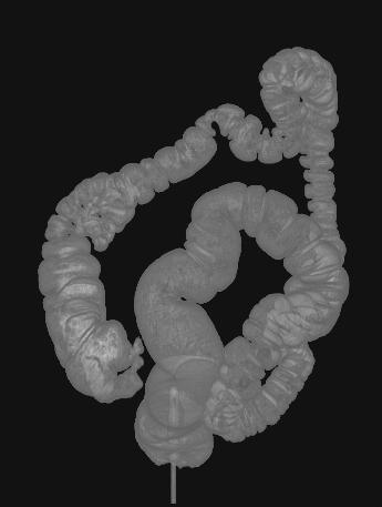
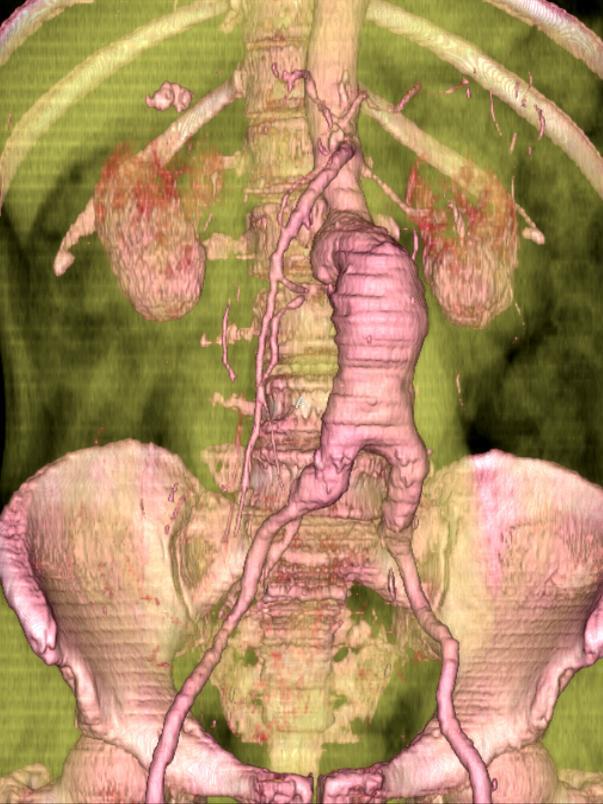
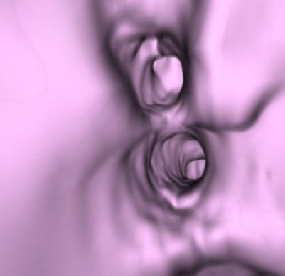
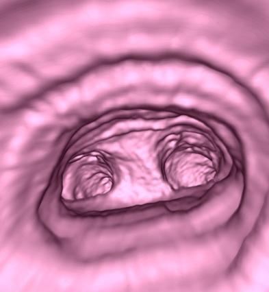
37 Virtual Colonoscopy Virtual colonoscopy A non-invasive computerized medical procedure for examining the entire colon to detect polyps. More comfortable than conventional colonoscopy because it does not use a colonoscope. Low risks, low cost, unlimited number of viewing angles Convenient to localize the three dimensional position of polyps 37
 Mammography CAD Colon CAD Lung CAD Image processing Volume measure Automatic segmentation Fusion Real time 3D")
38 The Future of Medical Imaging More advanced visualization techniques Large volume data processing Real time rendering 4D rendering Computer Assisted Diagnosis (CAD) Mammography CAD Colon CAD Lung CAD Image processing Volume measure Automatic segmentation Fusion Real time 3D reconstruction 38


39 39 Thanks!
Ch. 4 Physical Principles of CT
 Ch. 4 Physical Principles of CT CLRS 408: Intro to CT Department of Radiation Sciences Review: Why CT? Solution for radiography/tomography limitations Superimposition of structures Distinguishing between
CT Basics Principles of Spiral CT Dose. Always Thinking Ahead.
 1 CT Basics Principles of Spiral CT Dose 2 Who invented CT? 1963 - Alan Cormack developed a mathematical method of reconstructing images from x-ray projections Sir Godfrey Hounsfield worked for the Central
First CT Scanner. How it Works. Contemporary CT. Before and After CT. Computer Tomography: How It Works. Medical Imaging and Pattern Recognition
 Computer Tomography: How t Works Medical maging and Pattern Recognition Lecture 7 Computed Tomography Oleh Tretiak Only one plane is illuminated. Source-subject motion provides added information. 2 How
Shadow casting. What is the problem? Cone Beam Computed Tomography THE OBJECTIVES OF DIAGNOSTIC IMAGING IDEAL DIAGNOSTIC IMAGING STUDY LIMITATIONS
 Cone Beam Computed Tomography THE OBJECTIVES OF DIAGNOSTIC IMAGING Reveal pathology Reveal the anatomic truth Steven R. Singer, DDS srs2@columbia.edu IDEAL DIAGNOSTIC IMAGING STUDY Provides desired diagnostic
Digital Image Processing
 Digital Image Processing SPECIAL TOPICS CT IMAGES Hamid R. Rabiee Fall 2015 What is an image? 2 Are images only about visual concepts? We ve already seen that there are other kinds of image. In this lecture
CUDA and OpenCL Implementations of 3D CT Reconstruction for Biomedical Imaging
 CUDA and OpenCL Implementations of 3D CT Reconstruction for Biomedical Imaging Saoni Mukherjee, Nicholas Moore, James Brock and Miriam Leeser September 12, 2012 1 Outline Introduction to CT Scan, 3D reconstruction
Arbitrary cut planes Slab control with slab thickness Projection plane adjustment Box cropping Mandible detection MPR cross-section linked views
 Provides unparalleled quality and performance no need to sacrifice one for the other Enables high fidelity viewing with interactive deep supersampling Requires no GPUs or hardware accelerators a true,
DEVELOPMENT OF CONE BEAM TOMOGRAPHIC RECONSTRUCTION SOFTWARE MODULE
 Rajesh et al. : Proceedings of the National Seminar & Exhibition on Non-Destructive Evaluation DEVELOPMENT OF CONE BEAM TOMOGRAPHIC RECONSTRUCTION SOFTWARE MODULE Rajesh V Acharya, Umesh Kumar, Gursharan
Multi-slice CT Image Reconstruction Jiang Hsieh, Ph.D.
 Multi-slice CT Image Reconstruction Jiang Hsieh, Ph.D. Applied Science Laboratory, GE Healthcare Technologies 1 Image Generation Reconstruction of images from projections. textbook reconstruction advanced
VieW 3D. 3D Post-Processing WorKstation THE THIRD DIMENSION. Version 3.1
 VieW 3D 3D Post-Processing WorKstation THE THIRD DIMENSION Version 3.1 iq-view 3D THE FULLY-FEATURED 3D MEDICAL IMAGING SOLUTION FOR RADIOLOGISTS iq-view 3D contains all components of iq-view with the
VIEW PRO 4D. four dimensions in one workstation
 VIEW PRO 4D THE complete 3D/4D workstation for automatic image processing four dimensions in one workstation VERSiON 1.0 iq-view PRO 4D FROM HEAD TO TOE IN 4D iq-view PRO 4D is the perfect solution for
Scalar Data. Visualization Torsten Möller. Weiskopf/Machiraju/Möller
 Scalar Data Visualization Torsten Möller Weiskopf/Machiraju/Möller Overview Basic strategies Function plots and height fields Isolines Color coding Volume visualization (overview) Classification Segmentation
INDUSTRIAL SYSTEM DEVELOPMENT FOR VOLUMETRIC INTEGRITY
 INDUSTRIAL SYSTEM DEVELOPMENT FOR VOLUMETRIC INTEGRITY VERIFICATION AND ANALYSIS M. L. Hsiao and J. W. Eberhard CR&D General Electric Company Schenectady, NY 12301 J. B. Ross Aircraft Engine - QTC General
Corso di laurea in Fisica A.A Fisica Medica 4 TC
 Corso di laurea in Fisica A.A. 2007-2008 Fisica Medica 4 TC Computed Tomography Principles 1. Projection measurement 2. Scanner systems 3. Scanning modes Basic Tomographic Principle The internal structure
icatvision Quick Reference
 icatvision Quick Reference Navigating the i-cat Interface This guide shows how to: View reconstructed images Use main features and tools to optimize an image. REMINDER Images are displayed as if you are
Scalar Data. CMPT 467/767 Visualization Torsten Möller. Weiskopf/Machiraju/Möller
 Scalar Data CMPT 467/767 Visualization Torsten Möller Weiskopf/Machiraju/Möller Overview Basic strategies Function plots and height fields Isolines Color coding Volume visualization (overview) Classification
multimodality image processing workstation Visualizing your SPECT-CT-PET-MRI images
 multimodality image processing workstation Visualizing your SPECT-CT-PET-MRI images FUSION FUSION is a new visualization and evaluation software developed by Mediso built on state of the art technology,
Annexure XII SPECIFICATIONS FOR A NEW STATE OF ART 16 SLICE ALL PURPOSE C. T. SCANNER
 Annexure XII SPECIFICATIONS FOR A NEW STATE OF ART 16 SLICE ALL PURPOSE C. T. SCANNER A) Scanner Design X-Ray generator and tube: 1. Scanner: Whole body spiral CT scanner (16 slices) of latest technology.
Computer-Tomography II: Image reconstruction and applications
 Computer-Tomography II: Image reconstruction and applications Prof. Dr. U. Oelfke DKFZ Heidelberg Department of Medical Physics (E040) Im Neuenheimer Feld 280 69120 Heidelberg, Germany u.oelfke@dkfz.de
Medical Image Analysis
 Computer assisted Image Analysis VT04 29 april 2004 Medical Image Analysis Lecture 10 (part 1) Xavier Tizon Medical Image Processing Medical imaging modalities XRay,, CT Ultrasound MRI PET, SPECT Generic
CT Basics: Image Processing and Reconstruction Module 4
 Module 4 For educational and institutional use. This transcript is licensed for noncommercial, educational inhouse or online educational course use only in educational and corporate institutions. Any broadcast,
Display. Introduction page 67 2D Images page 68. All Orientations page 69 Single Image page 70 3D Images page 71
 Display Introduction page 67 2D Images page 68 All Orientations page 69 Single Image page 70 3D Images page 71 Intersecting Sections page 71 Cube Sections page 72 Render page 73 1. Tissue Maps page 77
Computational Medical Imaging Analysis Chapter 4: Image Visualization
 Computational Medical Imaging Analysis Chapter 4: Image Visualization Jun Zhang Laboratory for Computational Medical Imaging & Data Analysis Department of Computer Science University of Kentucky Lexington,
Tomographic Reconstruction
 Tomographic Reconstruction 3D Image Processing Torsten Möller Reading Gonzales + Woods, Chapter 5.11 2 Overview Physics History Reconstruction basic idea Radon transform Fourier-Slice theorem (Parallel-beam)
DICOM Correction Item
 DICOM Correction Item Correction Number CP-668 Log Summary: Type of Modification Addition Name of Standard PS 3.3, 3.17 2006 Rationale for Correction The term axial is common in practice, but is incorrectly
Computed Tomography. Principles, Design, Artifacts, and Recent Advances. Jiang Hsieh THIRD EDITION. SPIE PRESS Bellingham, Washington USA
 Computed Tomography Principles, Design, Artifacts, and Recent Advances THIRD EDITION Jiang Hsieh SPIE PRESS Bellingham, Washington USA Table of Contents Preface Nomenclature and Abbreviations xi xv 1 Introduction
Equipment Specification
 MULTISLICE CT SCANNER ( 16 Slices ) Merk : Hitachi Japan Model : SUPRIA 5 MHU Price : Rp 6.512.360.215,27 No Equipment Specification 1 2 3 Scanner Gantry Object for scanning : Whole body including head
CT NOISE POWER SPECTRUM FOR FILTERED BACKPROJECTION AND ITERATIVE RECONSTRUCTION
 CT NOISE POWER SPECTRUM FOR FILTERED BACKPROJECTION AND ITERATIVE RECONSTRUCTION Frank Dong, PhD, DABR Diagnostic Physicist, Imaging Institute Cleveland Clinic Foundation and Associate Professor of Radiology
L1 - Introduction. Contents. Introduction of CAD/CAM system Components of CAD/CAM systems Basic concepts of graphics programming
 L1 - Introduction Contents Introduction of CAD/CAM system Components of CAD/CAM systems Basic concepts of graphics programming 1 Definitions Computer-Aided Design (CAD) The technology concerned with the
INTRODUCTION TO MEDICAL IMAGING- 3D LOCALIZATION LAB MANUAL 1. Modifications for P551 Fall 2013 Medical Physics Laboratory
 INTRODUCTION TO MEDICAL IMAGING- 3D LOCALIZATION LAB MANUAL 1 Modifications for P551 Fall 2013 Medical Physics Laboratory Introduction Following the introductory lab 0, this lab exercise the student through
Improvement of Efficiency and Flexibility in Multi-slice Helical CT
 J. Shanghai Jiaotong Univ. (Sci.), 2008, 13(4): 408 412 DOI: 10.1007/s12204-008-0408-x Improvement of Efficiency and Flexibility in Multi-slice Helical CT SUN Wen-wu 1 ( ), CHEN Si-ping 2 ( ), ZHUANG Tian-ge
BME I5000: Biomedical Imaging
 1 Lucas Parra, CCNY BME I5000: Biomedical Imaging Lecture 4 Computed Tomography Lucas C. Parra, parra@ccny.cuny.edu some slides inspired by lecture notes of Andreas H. Hilscher at Columbia University.
Image Acquisition Systems
 Image Acquisition Systems Goals and Terminology Conventional Radiography Axial Tomography Computer Axial Tomography (CAT) Magnetic Resonance Imaging (MRI) PET, SPECT Ultrasound Microscopy Imaging ITCS
RADIOLOGY AND DIAGNOSTIC IMAGING
 Day 2 part 2 RADIOLOGY AND DIAGNOSTIC IMAGING Dr hab. Zbigniew Serafin, MD, PhD serafin@cm.umk.pl 2 3 4 5 CT technique CT technique 6 CT system Kanal K: RSNA/AAPM web module: CT Systems & CT Image Quality
The VesselGlyph: Focus & Context Visualization in CT-Angiography
 The VesselGlyph: Focus & Context Visualization in CT-Angiography Matúš Straka M. Šrámek, A. La Cruz E. Gröller, D. Fleischmann Contents Motivation:» Why again a new visualization method for vessel data?
June 05, 2018, Version 3.0.6
 June 05, 2018, Version 3.0.6 VolViCon is an advanced application for reconstruction of computed tomography (CT), magnetic resonance (MR), ultrasound, and x-rays images. It gives features for exporting
Fundamentals of CT imaging
 SECTION 1 Fundamentals of CT imaging I History In the early 1970s Sir Godfrey Hounsfield s research produced the first clinically useful CT scans. Original scanners took approximately 6 minutes to perform
Computed tomography - outline
 Computed tomography - outline Computed Tomography Systems Jørgen Arendt Jensen and Mikael Jensen (DTU Nutech) October 6, 216 Center for Fast Ultrasound Imaging, Build 349 Department of Electrical Engineering
Volume Graphics Introduction
 High-Quality Volume Graphics on Consumer PC Hardware Volume Graphics Introduction Joe Kniss Gordon Kindlmann Markus Hadwiger Christof Rezk-Salama Rüdiger Westermann Motivation (1) Motivation (2) Scientific
Assessment of 3D performance metrics. X-ray based Volumetric imaging systems: Fourier-based imaging metrics. The MTF in CT
 Assessment of 3D performance metrics D and 3D Metrics of Performance Towards Quality Index: Volumetric imaging systems X-ray based Volumetric imaging systems: CBCT/CT Tomosynthesis Samuel Richard and Ehsan
Volume Visualization
 Volume Visualization Part 1 (out of 3) Overview: Volume Visualization Introduction to volume visualization On volume data Surface vs. volume rendering Overview: Techniques Simple methods Slicing, cuberille
Toshiba Aquilion 4 3d Manual READ ONLINE
 Toshiba Aquilion 4 3d Manual READ ONLINE TOSHIBA AQUILION VISION: Head. EP No: 4. Boost 3D: OFF: OSR *** Filter: OFF. SureStart - Manual trigger as soon as contrast is seen: Scan mode: TOSHIBA AQUILION
FINDING THE TRUE EDGE IN CTA
 FINDING THE TRUE EDGE IN CTA by: John A. Rumberger, PhD, MD, FACC Your patient has chest pain. The Cardiac CT Angiography shows plaque in the LAD. You adjust the viewing window trying to evaluate the stenosis
MEDICAL IMAGING 2nd Part Computed Tomography
 MEDICAL IMAGING 2nd Part Computed Tomography Introduction 2 In the last 30 years X-ray Computed Tomography development produced a great change in the role of diagnostic imaging in medicine. In convetional
Image Post-Processing, Workflow, & Interpretation
 Image Post-Processing, Workflow, & Interpretation Richard L. Hallett, MD Chief, Cardiovascular Imaging Northwest Radiology Network Indianapolis, IN Adjunct Assistant Professor Stanford University Stanford,
4DM Packages. 4DM Packages & License Types. Information to help you order the appropriate licenses for your site.
 4DM Packages 4DM Packages & License Types. Information to help you order the appropriate licenses for your site. Nuclear Cardiac Quantification, Review, and Reporting Select Your 4DM Package and corresponding
Previously... contour or image rendering in 2D
 Volume Rendering Visualisation Lecture 10 Taku Komura Institute for Perception, Action & Behaviour School of Informatics Volume Rendering 1 Previously... contour or image rendering in 2D 2D Contour line
Volume Rendering. Computer Animation and Visualisation Lecture 9. Taku Komura. Institute for Perception, Action & Behaviour School of Informatics
 Volume Rendering Computer Animation and Visualisation Lecture 9 Taku Komura Institute for Perception, Action & Behaviour School of Informatics Volume Rendering 1 Volume Data Usually, a data uniformly distributed
CLASS HOURS: 4 CREDIT HOURS: 4 LABORATORY HOURS: 0
 Revised 10/10 COURSE SYLLABUS TM 220 COMPUTED TOMOGRAPHY PHYSICS CLASS HOURS: 4 CREDIT HOURS: 4 LABORATORY HOURS: 0 CATALOG COURSE DESCRIPTION: This course is one of a three course set in whole body Computed
3DMMVR REFERENCE MANUAL V 0.81
 3DMMVR REFERENCE MANUAL V 0.81 Page 1 of 30 Index: 1.0 System Requirements...5 1.1 System Processor...5 1.2 System RAM...5 1.3 Graphics Card...5 1.4 Operating System...5 2.0 Conventions...6 2.1 Typographic
Introduction to Biomedical Imaging
 Alejandro Frangi, PhD Computational Imaging Lab Department of Information & Communication Technology Pompeu Fabra University www.cilab.upf.edu X-ray Projection Imaging Computed Tomography Digital X-ray
LAB DEMONSTRATION COMPUTED TOMOGRAPHY USING DESKCAT Lab Manual: 0
 LAB DEMONSTRATION COMPUTED TOMOGRAPHY USING DESKCAT Lab Manual: 0 Introduction This lab demonstration explores the physics and technology of Computed Tomography (CT) and guides the student and instructor
Computer-Tomography I: Principles, History, Technology
 Computer-Tomography I: Principles, History, Technology Prof. Dr. U. Oelfke DKFZ Heidelberg Department of Medical Physics (E040) Im Neuenheimer Feld 280 69120 Heidelberg, Germany u.oelfke@dkfz.de History
Enhancement Image Quality of CT Using Single Slice Spiral Technique
 Enhancement Image Quality of CT Using Single Slice Spiral Technique Doaa. N. Al Sheack 1 and Dr.Mohammed H. Ali Al Hayani 2 1 2 Electronic and Communications Engineering Department College of Engineering,
New Enhanced Multi-frame DICOM CT and MR Objects to Enhance Performance and Image Processing on PACS and Workstations
 New Enhanced Multi-frame DICOM CT and MR Objects to Enhance Performance and Image Processing on PACS and Workstations SCAR 2004 Hot Topics - 22 May 2004 David Clunie, RadPharm Charles Parisot,, GE Healthcare
Volume Visualization. Part 1 (out of 3) Volume Data. Where do the data come from? 3D Data Space How are volume data organized?
 Volume Data. Where do the data come from? 3D Data Space How are volume data organized?") Volume Data Volume Visualization Part 1 (out of 3) Where do the data come from? Medical Application Computed Tomographie (CT) Magnetic Resonance Imaging (MR) Materials testing Industrial-CT Simulation
Volume Data Volume Visualization Part 1 (out of 3) Where do the data come from? Medical Application Computed Tomographie (CT) Magnetic Resonance Imaging (MR) Materials testing Industrial-CT Simulation
A closer look at CT scanning
 Vet Times The website for the veterinary profession https://www.vettimes.co.uk A closer look at CT scanning Author : Charissa Lee, Natalie Webster Categories : General, Vets Date : April 3, 2017 A basic
CTA HEAD Perfusion AqONE without and with IV Contrast
 CTA HEAD Perfusion AqONE without and with IV Contrast Patient Position Adult Contrast Adult Injection Rate Supine IOML perpendicular to table top. IV: 100 ml with helical head CTA 50 ml without helical
Visualisation : Lecture 1. So what is visualisation? Visualisation
 So what is visualisation? UG4 / M.Sc. Course 2006 toby.breckon@ed.ac.uk Computer Vision Lab. Institute for Perception, Action & Behaviour Introducing 1 Application of interactive 3D computer graphics to
Spiral ASSR Std p = 1.0. Spiral EPBP Std. 256 slices (0/300) Kachelrieß et al., Med. Phys. 31(6): , 2004
 Kachelrieß et al., Med. Phys. 31(6): , 2004") Spiral ASSR Std p = 1.0 Spiral EPBP Std p = 1.0 Kachelrieß et al., Med. Phys. 31(6): 1623-1641, 2004 256 slices (0/300) Advantages of Cone-Beam Spiral CT Image quality nearly independent of pitch Increase
Spiral ASSR Std p = 1.0 Spiral EPBP Std p = 1.0 Kachelrieß et al., Med. Phys. 31(6): 1623-1641, 2004 256 slices (0/300) Advantages of Cone-Beam Spiral CT Image quality nearly independent of pitch Increase
Introduction to 3D Graphics
 Graphics Without Polygons Volume Rendering May 11, 2010 So Far Volumetric Rendering Techniques Misc. So Far Extended the Fixed Function Pipeline with a Programmable Pipeline Programming the pipeline is
IMPAX Volume Viewing 3D Visualization & Segmentation
 Getting started guide IMPAX Volume Viewing 3D Visualization & Segmentation This guide outlines the basic steps to perform and manipulate a 3D reconstruction of volumetric image data using IMPAX Volume
Spiral CT. Protocol Optimization & Quality Assurance. Ge Wang, Ph.D. Department of Radiology University of Iowa Iowa City, Iowa 52242, USA
 Spiral CT Protocol Optimization & Quality Assurance Ge Wang, Ph.D. Department of Radiology University of Iowa Iowa City, Iowa 52242, USA Spiral CT Protocol Optimization & Quality Assurance Protocol optimization
A Study of Medical Image Analysis System
 Indian Journal of Science and Technology, Vol 8(25), DOI: 10.17485/ijst/2015/v8i25/80492, October 2015 ISSN (Print) : 0974-6846 ISSN (Online) : 0974-5645 A Study of Medical Image Analysis System Kim Tae-Eun
CSE 167: Lecture #17: Volume Rendering. Jürgen P. Schulze, Ph.D. University of California, San Diego Fall Quarter 2012
 CSE 167: Introduction to Computer Graphics Lecture #17: Volume Rendering Jürgen P. Schulze, Ph.D. University of California, San Diego Fall Quarter 2012 Announcements Thursday, Dec 13: Final project presentations
Optimisation of Toshiba Aquilion ONE Volume Imaging
 Optimisation of Toshiba Aquilion ONE Volume Imaging Jane Edwards, RPRSG Royal Free London NHS Foundation Trust Dr Mufudzi Maviki, Plymouth Hospitals NHS Trust Background In 2011/12 Radiology at RFH was
Modifications for P551 Fall 2014
 LAB DEMONSTRATION COMPUTED TOMOGRAPHY USING DESKCAT 1 Modifications for P551 Fall 2014 Introduction This lab demonstration explores the physics and technology of Computed Tomography (CT) and guides the
CIS 467/602-01: Data Visualization
 CIS 467/60-01: Data Visualization Isosurfacing and Volume Rendering Dr. David Koop Fields and Grids Fields: values come from a continuous domain, infinitely many values - Sampled at certain positions to
Athena Radiology Medical Workstation
 High productivity and integration Athena DICOM Viewer is being designed according with the suggestions and the necessities of medical radiologists. From the design of its interface to the advanced image
Medical Imaging and Beyond
 Medical Imaging and Beyond Jesus J. Caban Schedule! Today:! Lecture: Medical Imaging and Beyond! Wednesday:! No Class (Thanksgiving Eve)! Final presentations:! Nov 29 th : W. Griffin, F. Zafar! Dec 1 st
The Near Future in Cardiac CT Image Reconstruction
 SCCT 2010 The Near Future in Cardiac CT Image Reconstruction Marc Kachelrieß Institute of Medical Physics (IMP) Friedrich-Alexander Alexander-University Erlangen-Nürnberg rnberg www.imp.uni-erlangen.de
Operator s manual. Operator s Manual WhiteFox Imaging V0C (15) 10/2017 NCBCEN020C
 10/2017 NCBCEN020C") Operator s manual Operator s Manual WhiteFox Imaging V0C (15) 10/2017 NCBCEN020C MANUFACTURER de Götzen S.r.l. - a company of ACTEON Group Via Roma, 45 21057 OLGIATE OLONA (VA) ITALY Tel. +39 0331 376760
Operator s manual Operator s Manual WhiteFox Imaging V0C (15) 10/2017 NCBCEN020C MANUFACTURER de Götzen S.r.l. - a company of ACTEON Group Via Roma, 45 21057 OLGIATE OLONA (VA) ITALY Tel. +39 0331 376760
User-friendly Environment for Virtual Endoscopy
 User-friendly Environment for Virtual Endoscopy Jeong-Jin Lee 1, Yun-Mo Koo 2 and Yeong Gil Shin 1 1 School of Computer Science and Engineering, Seoul National University San 56-1, Shilim-Dong, Kwanak-Gu,
MEDICAL IMAGING 2nd Part Computed Tomography
 MEDICAL IMAGING 2nd Part Computed Tomography Introduction 2 In the last 30 years X-ray Computed Tomography development produced a great change in the role of diagnostic imaging in medicine. In convetional
Copyright 2017 Medical IP - Tutorial Medip v /2018, Revision
 Copyright 2017 Medical IP - Tutorial Medip v.1.0.0.9 01/2018, Revision 1.0.0.2 List of Contents 1. Introduction......................................................... 2 2. Overview..............................................................
Technical Publications
 GE Medical Systems Technical Publications Direction 2188003-100 Revision 0 Tissue Volume Analysis DICOM for DICOM V3.0 Copyright 1997 By General Electric Co. Do not duplicate REVISION HISTORY REV DATE
Interoperability Issues in Image Registration and ROI Generation
 1 DICOM 2005 International Conference, Budapest, Hungary Interoperability Issues in Image Registration and ROI Generation Todd Kantchev PhD, Siemens Molecular Imaging, Oxford, UK 2 Scope The following
Lucy Phantom MR Grid Evaluation
 Lucy Phantom MR Grid Evaluation Anil Sethi, PhD Loyola University Medical Center, Maywood, IL 60153 November 2015 I. Introduction: The MR distortion grid, used as an insert with Lucy 3D QA phantom, is
Technical Publications
 g GE Medical Systems Technical Publications Direction 2275362-100 Revision 0 DICOM for DICOM V3.0 Copyright 2000 By General Electric Co. Do not duplicate REVISION HISTORY REV DATE REASON FOR CHANGE 0 May
Introduction to Medical Imaging. Cone-Beam CT. Klaus Mueller. Computer Science Department Stony Brook University
 Introduction to Medical Imaging Cone-Beam CT Klaus Mueller Computer Science Department Stony Brook University Introduction Available cone-beam reconstruction methods: exact approximate algebraic Our discussion:
RADIOMICS: potential role in the clinics and challenges
 27 giugno 2018 Dipartimento di Fisica Università degli Studi di Milano RADIOMICS: potential role in the clinics and challenges Dr. Francesca Botta Medical Physicist Istituto Europeo di Oncologia (Milano)
Data Visualization (DSC 530/CIS )
") Data Visualization (DSC 530/CIS 60-01) Scalar Visualization Dr. David Koop Online JavaScript Resources http://learnjsdata.com/ Good coverage of data wrangling using JavaScript Fields in Visualization Scalar
Data Visualization (DSC 530/CIS 60-01) Scalar Visualization Dr. David Koop Online JavaScript Resources http://learnjsdata.com/ Good coverage of data wrangling using JavaScript Fields in Visualization Scalar
Quick Reference Guide
 Key functions Quick Reference Guide Philips isite Enterprise Version 3.6 Enhance mouse cursor mode Function Wheel mouse 2-button mouse Adjust WW/WL Left click and drag Left click and drag Scroll/Cine Wheel
Digital Imaging and Communications in Medicine (DICOM) Supplement 167: X-Ray 3D Angiographic IOD Informative Annex
 Supplement 167: X-Ray 3D Angiographic IOD Informative Annex") 5 Digital Imaging and Communications in Medicine (DICOM) Supplement 167: X-Ray 3D Angiographic IOD Informative Annex 10 15 Prepared by: DICOM Standards Committee, Working Group 2, Projection Radiography
5 Digital Imaging and Communications in Medicine (DICOM) Supplement 167: X-Ray 3D Angiographic IOD Informative Annex 10 15 Prepared by: DICOM Standards Committee, Working Group 2, Projection Radiography
ACQUIRING AND PROCESSING SUSCEPTIBILITY WEIGHTED IMAGING (SWI) DATA ON GE 3.0T
 DATA ON GE 3.0T") ACQUIRING AND PROCESSING SUSCEPTIBILITY WEIGHTED IMAGING (SWI) DATA ON GE 3.0T Revision date: 12/13/2010 Overview Susceptibility Weighted Imaging (SWI) is a relatively new data acquisition and processing
ACQUIRING AND PROCESSING SUSCEPTIBILITY WEIGHTED IMAGING (SWI) DATA ON GE 3.0T Revision date: 12/13/2010 Overview Susceptibility Weighted Imaging (SWI) is a relatively new data acquisition and processing
Data Visualization (CIS/DSC 468)
") Data Visualization (CIS/DSC 46) Volume Rendering Dr. David Koop Visualizing Volume (3D) Data 2D visualization slice images (or multi-planar reformating MPR) Indirect 3D visualization isosurfaces (or surface-shaded
Data Visualization (CIS/DSC 46) Volume Rendering Dr. David Koop Visualizing Volume (3D) Data 2D visualization slice images (or multi-planar reformating MPR) Indirect 3D visualization isosurfaces (or surface-shaded
GPU implementation for rapid iterative image reconstruction algorithm
 GPU implementation for rapid iterative image reconstruction algorithm and its applications in nuclear medicine Jakub Pietrzak Krzysztof Kacperski Department of Medical Physics, Maria Skłodowska-Curie Memorial
Radiology. Marta Anguiano Millán. Departamento de Física Atómica, Molecular y Nuclear Facultad de Ciencias. Universidad de Granada
 Departamento de Física Atómica, Molecular y Nuclear Facultad de Ciencias. Universidad de Granada Overview Introduction Overview Introduction Tecniques of imaging in Overview Introduction Tecniques of imaging
Direct Volume Rendering
 Direct Volume Rendering CMPT 467/767 Visualization Torsten Möller Weiskopf/Machiraju/Möller Overview Volume rendering equation Compositing schemes Ray casting Acceleration techniques for ray casting Texture-based
An Iterative Approach to the Beam Hardening Correction in Cone Beam CT (Proceedings)
") Marquette University e-publications@marquette Biomedical Engineering Faculty Research and Publications Engineering, College of 1-1-1999 An Iterative Approach to the Beam Hardening Correction in Cone Beam
Marquette University e-publications@marquette Biomedical Engineering Faculty Research and Publications Engineering, College of 1-1-1999 An Iterative Approach to the Beam Hardening Correction in Cone Beam
Medical Images Analysis and Processing
 Medical Images Analysis and Processing - 25642 Emad Course Introduction Course Information: Type: Graduated Credits: 3 Prerequisites: Digital Image Processing Course Introduction Reference(s): Insight
High-performance tomographic reconstruction using graphics processing units
 18 th World IMACS / MODSIM Congress, Cairns, Australia 13-17 July 29 http://mssanz.org.au/modsim9 High-performance tomographic reconstruction using graphics processing units Ya.I. esterets and T.E. Gureyev
Volume Rendering. Lecture 21
 Volume Rendering Lecture 21 Acknowledgements These slides are collected from many sources. A particularly valuable source is the IEEE Visualization conference tutorials. Sources from: Roger Crawfis, Klaus
RT_Image v0.2β User s Guide
 RT_Image v0.2β User s Guide RT_Image is a three-dimensional image display and analysis suite developed in IDL (ITT, Boulder, CO). It offers a range of flexible tools for the visualization and quantitation
Japan Foundry Society, Inc. Application of Recent X-ray CT Technology to Investment Casting field. Kouichi Inagaki ICC / IHI Corporation
 Japan Foundry Society, Inc. Application of Recent X-ray CT Technology to Investment Casting field Kouichi Inagaki ICC / IHI Corporation 13 th WORLD CONFERENCE ON INVESTMENT CASTING Paper: T3 Copyright
Visage 7 Clinical Training Basic Features
 Visage 7 Clinical Training Basic Features Contents Overview... 4 Usage... 4 Client Server Architecture... 5 Client Login... 6 Study Browser... 7 Query Section... 8 Study Labels... 10 Query Labeled Studies...
Extraction and recognition of the thoracic organs based on 3D CT images and its application
 1 Extraction and recognition of the thoracic organs based on 3D CT images and its application Xiangrong Zhou, PhD a, Takeshi Hara, PhD b, Hiroshi Fujita, PhD b, Yoshihiro Ida, RT c, Kazuhiro Katada, MD
3/27/2012 WHY SPECT / CT? SPECT / CT Basic Principles. Advantages of SPECT. Advantages of CT. Dr John C. Dickson, Principal Physicist UCLH
 3/27/212 Advantages of SPECT SPECT / CT Basic Principles Dr John C. Dickson, Principal Physicist UCLH Institute of Nuclear Medicine, University College London Hospitals and University College London john.dickson@uclh.nhs.uk
TOLERANCE ANALYSIS OF THIN-WALL CFRP STRUCTURAL ELEMENTS USING TOMOGRAPHIC IMAGING
 Proceedings of the 6th International Conference on Mechanics and Materials in Design, Editors: J.F. Silva Gomes & S.A. Meguid, P.Delgada/Azores, 26-30 July 2015 PAPER REF: 5495 TOLERANCE ANALYSIS OF THIN-WALL
Hardware Accelerated Volume Visualization. Leonid I. Dimitrov & Milos Sramek GMI Austrian Academy of Sciences
 Hardware Accelerated Volume Visualization Leonid I. Dimitrov & Milos Sramek GMI Austrian Academy of Sciences A Real-Time VR System Real-Time: 25-30 frames per second 4D visualization: real time input of