MEDICAL IMAGE COMPUTING (CAP 5937) LECTURE 9: Medical Image Segmentation (III) (Fuzzy Connected Image Segmentation)
|
|
|
- Josephine Phyllis Gregory
- 5 years ago
- Views:
Transcription
1 SPRING MEDICAL IMAGE COMPUTING (CAP 5937) LECTURE 9: Medical Image Segmentation (III) (Fuzzy Connected Image Segmentation) Dr. Ulas Bagci HEC 221, Center for Research in Computer Vision (CRCV), University of Central Florida (UCF), Orlando, FL bagci@ucf.edu or bagci@crcv.ucf.edu
2 2 Outline Fuzzy Connectivity (FC) Affinity functions Absolute FC Relative FC (and Iterative Relative FC) Successful example applications of FC in medical imaging Segmentation of Airway and Airway Walls using RFC based method

 image data using iterative relative fuzzy connectivity Slide")
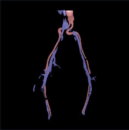
3 3 Motivation Connectivity: a popularly used tool for region growing Applications: image segmentation, object tracking, object separation A fuzzy model for connectivity analysis is essential to capture the global extent of an object using local hanging togetherness and path connectivity CE-MRA Image data Segmented vasculature Separated arteries/veins Separation of arteries and veins in a contrastenhanced magnetic resonance angiographic (CE-MRA) image data using iterative relative fuzzy connectivity Slide credit: P. Saha
4 4 Hard-coded & Fuzzy-coded Many image segmentation algorithms are based on hardcoded relationship between individual regions (or within regions)
5 5 Hard-coded & Fuzzy-coded Many image segmentation algorithms are based on hardcoded relationship between individual regions (or within regions) Fuzzy algorithms take into consideration various uncertainties such as noise, uneven illumination/brightness/contrast differences, etc.
6 6 Hard-coded & Fuzzy-coded Many image segmentation algorithms are based on hardcoded relationship between individual regions (or within regions) Fuzzy algorithms take into consideration various uncertainties such as noise, uneven illumination/brightness/contrast differences, etc. Example: If two regions have about same gray-scale and if they are relatively close to each other in space, then they likely to belong to the same object.
7 7 Hard-coded & Fuzzy-coded Many image segmentation algorithms are based on hardcoded relationship between individual regions (or within regions) Fuzzy algorithms take into consideration various uncertainties such as noise, uneven illumination/brightness/contrast differences, etc. Example: If two regions have about same gray-scale and if they are relatively close to each other in space, then they likely to belong to the same object.
8 8 Fuzzy Connected (FC) Image Segmentation FC has been used with considerable success in medical (and other) images. Udupa and Samarasekera were the first to use FC in medical images. (Graphical Models and Image Processing, 1996)
 FC segmentation is a methodology for finding M objects in a digital image based on user-specified seed points and user-specified functions, called")
9 9 Fuzzy Connected (FC) Image Segmentation FC has been used with considerable success in medical (and other) images. Udupa and Samarasekera were the first to use FC in medical images. (Graphical Models and Image Processing, 1996) FC segmentation is a methodology for finding M objects in a digital image based on user-specified seed points and user-specified functions, called (fuzzy) affinities, which map each pair of image points to a value in the real interval [0, 1].
10 10 FC Family Absolute FC Scale-based FC (b-, t-, g-scale based) Relative FC Iterative Relative FC Vectorial FC Hierarchical FC Model-based FC
11 FC Medical Image Segmentation Examples 11
 d x Spatial location intensity value")
12 12 Object Characteristics in the Images c local hanging togetherness (affinity) d x Spatial location intensity value (-derived)
:")
13 13 FC is a global relation! Effectiveness of the FC algorithm is dependent on the choice of the affinity function, and the general setup can be divided into three components (for any voxels p and q): Adjacency Homogeneity Object Feature FC is a global fuzzy relation between voxels! All voxels are assessed via defined affinity functions for labelling.
14 14 Affinity Definition: local relation between every two image elements u and v
15 15 Affinity Definition: local relation between every two image elements u and v If u and v are apart, affinity should be small (or zero) If u and v are close, affinity should be large
 If u and v are close, affinity should be large p and q1 hang-together (than p and q2) Green path is stronger than red")
16 16 Affinity Definition: local relation between every two image elements u and v If u and v are apart, affinity should be small (or zero) If u and v are close, affinity should be large p and q1 hang-together (than p and q2) Green path is stronger than red path.
17 17 Fuzzy Adjacency A local fuzzy relation α to indicate how near two voxels a and b are spatially.
18 18 Fuzzy Adjacency A local fuzzy relation α to indicate how near two voxels a and b are spatially. Its strength α (a, b): 1, if a= b α a, b = g a b, if a b D 0, if a b > D1 ( ) ( ) 1 D 1 is a distance (known) g is a function mapping between [0,1]
19 19 Homogeneity and Object Feature Affinities µ (p, q) =min e µ (p, q) =e f(p) m 2 2 2,e f(p) f(q) 2 2 2,! f(q) m
20 20 Fuzzy Affinity A local fuzzy relation κ to indicate how voxels a and b hang together locally in scene S = (C, f).
21 21 Fuzzy Affinity A local fuzzy relation κ to indicate how voxels a and b hang together locally in scene S = (C, f). Its strength κ(a, b) depends on: (1) α (a, b) - Fuzzy adjacency (2) homogeneity of intensity at a and b. (3) how close intensity features at a and b are to be expected object features -
22 22 Fuzzy Affinity A local fuzzy relation κ to indicate how voxels a and b hang together locally in scene S = (C, f). Its strength κ(a, b) depends on: (1) α (a, b) - Fuzzy adjacency (2) homogeneity of intensity at a and b. (3) how close intensity features at a and b are to be expected object features - κ( a, b) = h α( a, b), ψ( a, b), φ( a, b)
 and f(b): intensity values at voxel location a, b.")
23 23 Different Affinity Functions can be devised! f(a) and f(b): intensity values at voxel location a, b. : expected object intensity
24 24 Fuzzy Affinity and Path Strength Fuzzy Affinity (κ): local hanging-togetherness between two spels (i.e., space elements) κ p, q [0,1] κ p, q is zero if p, q are non-adjacent κ p, p = 1, i.e., reflexive κ p, q = κ q, p, i.e. symmetric Strength ( Π ) of a path ( π = p -,p., p 0 ) Π π = the affinity of the weakest link on the path, i.e., Π π = min κ p 5, p 57-
25 25 Fuzzy Connectivity Fuzzy connectedness is a global fuzzy relation Κ among voxels. Its strength Κ (c, d) for any c, d is defined as: (1) Every path π between c and d has a strength which is the smallest affinity along π. c d (2) Κ (c, d) is the strength of the strongest path. { } i i 1 Κ( c, d) = max min κ ( c, c ) + π i
26 26 Weakest affinity=0.1 Numerical Example Path 1 Path 2 Path Path N (assuming there are N paths between voxels c and d)
27 27 (Absolute) FC Algorithm 1. Define properties of fuzzy adjacency α and fuzzy affinity κ
28 28 (Absolute) FC Algorithm 1. Define properties of fuzzy adjacency α and fuzzy affinity κ 2. Determine the affinity values for all pairs of fuzzy adjacent voxels
29 29 (Absolute) FC Algorithm 1. Define properties of fuzzy adjacency α and fuzzy affinity κ 2. Determine the affinity values for all pairs of fuzzy adjacent voxels 3. Determine the segmentation seed element c
30 30 (Absolute) FC Algorithm 1. Define properties of fuzzy adjacency α and fuzzy affinity κ 2. Determine the affinity values for all pairs of fuzzy adjacent voxels 3. Determine the segmentation seed element c 4. Determine all possible paths between the seed c and all other voxels d i in the image domain considering the fuzzy adjacency relation
31 31 (Absolute) FC Algorithm 1. Define properties of fuzzy adjacency α and fuzzy affinity κ 2. Determine the affinity values for all pairs of fuzzy adjacent voxels 3. Determine the segmentation seed element c 4. Determine all possible paths between the seed c and all other voxels d i in the image domain considering the fuzzy adjacency relation 5. For each path, determine its strength using minimum affinity along the path
32 32 (Absolute) FC Algorithm 1. Define properties of fuzzy adjacency α and fuzzy affinity κ 2. Determine the affinity values for all pairs of fuzzy adjacent voxels 3. Determine the segmentation seed element c 4. Determine all possible paths between the seed c and all other voxels d i in the image domain considering the fuzzy adjacency relation 5. For each path, determine its strength using minimum affinity along the path 6. For each voxel d i, determine its fuzzy connectedness to the seed point c as the maximum strength of all possible paths < c,, d i > and form connectedness map.
33 33 (Absolute) FC Algorithm 1. Define properties of fuzzy adjacency α and fuzzy affinity κ 2. Determine the affinity values for all pairs of fuzzy adjacent voxels 3. Determine the segmentation seed element c 4. Determine all possible paths between the seed c and all other voxels d i in the image domain considering the fuzzy adjacency relation 5. For each path, determine its strength using minimum affinity along the path 6. For each voxel d i, determine its fuzzy connectedness to the seed point c as the maximum strength of all possible paths < c,, d i > and form connectedness map. 7. Threshold connected map to obtain object containing c
) specified in a soft tissue region of the scene in (a).")
34 Illustration of equivalent affinities. (a) A 2D scene a CT slice of a human knee. (b), (c) Connectivity scenes corresponding to affinities ψ σ with σ = 1 and σ = 10.8, respectively, and the same seed spel (indicated by + in (a)) specified in a soft tissue region of the scene in (a). (d), (e) Identical AFC objects obtained from the scenes in (b) and (c), respectively. 34

35 Quantifying Breast Density 35

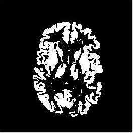

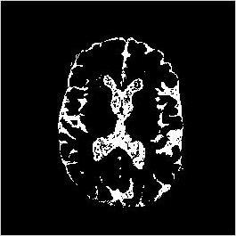

36 36 Brain MS Lesion Quantification T2 PD GM WM CSF MS


37 Upper Airway Study in Children with Obstructive Sleep Apnea 37

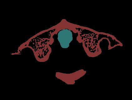
38 CT Skull Extraction 38

39 Brain Tumor Quantification - MRI 39
40 40 Relative Fuzzy Connected (RFC) Image Segmentation Main contribution of this approach is to eliminate connectedness map thresholding step (Saha and Udupa, 2000)
41 41 Relative Fuzzy Connected (RFC) Image Segmentation Main contribution of this approach is to eliminate connectedness map thresholding step (Saha and Udupa, 2000) Instead of extracting a single object at a time, two objects are extracted at the same time
42 42 Relative Fuzzy Connected (RFC) Image Segmentation Main contribution of this approach is to eliminate connectedness map thresholding step (Saha and Udupa, 2000) Instead of extracting a single object at a time, two objects are extracted at the same time During the segmentation, these two objects compete against each other with each individual voxel (seed) assigned to the object with a stronger affinity to this voxel
43 43 Relative Fuzzy Connected (RFC) Image Segmentation Main contribution of this approach is to eliminate connectedness map thresholding step (Saha and Udupa, 2000) Instead of extracting a single object at a time, two objects are extracted at the same time During the segmentation, these two objects compete against each other with each individual voxel (seed) assigned to the object with a stronger affinity to this voxel These 2-object RFC was extended into multiple-object RFC by the same authors
44 44 Motivation for RFC (and IRFC) FC may fail to identify objects in this situation. -Objects O1 and O2 are located very close to each other. Due to limited resolution, border Between O1 and O2 may be weak, Causing homogeneity between d and e, and Homogeneity between c and e be similar!
45 45 Motivation for RFC (and IRFC) FC may fail to identify objects in this situation. -Objects O1 and O2 are located very close to each other. Due to limited resolution, border Between O1 and O2 may be weak, Causing homogeneity between d and e, and Homogeneity between c and e be similar! Solution: If O1 is segmented first, paths between e and d are omitted! It will be iterative process, IRFC.

46 46 Artery-vein separation MRA Motivation for IRFC

47 47 RFC and IRFC RFC IRFC

.")
48 48 Airway and Airway Wall Segmentation with RFC Airways are the airconducting structures (bronchi and bronchioles) bringing air into and out of the lungs from sites of gas exchange (alveoli). Credit: healthhype.com
, often resulting from chronic infection (Bagci et al.")
49 49 Airway and Airway Wall Segmentation with RFC Airways are pathologically involved in various lung diseases. As examples, bronchiectasis is the dilation of airways (enlarged lumen), often resulting from chronic infection (Bagci et al., CMIG 2012), obstruction, and inflammation. Credit: Corehealthclub
50 50 Airway and Airway Wall Segmentation with RFC Airway wall thickening can be associated with airway narrowing, such as asthma and bronchitis. Tumors on airway walls can also form obstructions
51 51 Airway and Airway Wall Segmentation with RFC Airway wall thickening can be associated with airway narrowing, such as asthma and bronchitis. Tumors on airway walls can also form obstructions CT imaging provides in-vivo anatomical information of lung structures in a non-invasive manner, which enables a quantitative investigation of airway pathologies
52 52 Airway and Airway Wall Segmentation with RFC Airway wall thickening can be associated with airway narrowing, such as asthma and bronchitis. Tumors on airway walls can also form obstructions CT imaging provides in-vivo anatomical information of lung structures in a non-invasive manner, which enables a quantitative investigation of airway pathologies Due to the inherent complexity of airway structures and the resolution limitations of CT, manually tracing and analyzing airways is an extremely challenging task, taking more than 7 h of intensive work per image
53 53 Airway and Airway Wall Segmentation with RFC Airway wall thickening can be associated with airway narrowing, such as asthma and bronchitis. Tumors on airway walls can also form obstructions CT imaging provides in-vivo anatomical information of lung structures in a non-invasive manner, which enables a quantitative investigation of airway pathologies Due to the inherent complexity of airway structures and the resolution limitations of CT, manually tracing and analyzing airways is an extremely challenging task, taking more than 7 h of intensive work per image A precise method for segmentation of airways and its walls may facilitate better quantification of airway pathologies (and understanding of disease progression)

54 54 Airway and Airway Wall Segmentation with RFC (Credit: Xu, Bagci, et al. Medical Image Analysis The state of the art method)
55 55 Airway Segmentation Morphological operations Vesselness
56 56 Airway Segmentation Morphological operations Vesselness Good for large airways, Small airways can be detected to some extent, but limited. computationally expensive Good for small airways, But numerous false positives
57 FC can combine these two methods within a single framework! Large airways 57 small airways Where ls denotes local scale, k is a weight parameter, and D shows morphologically processed Image, V indicates vesselness image.

58 Airway and Airway Wall Segmentation with RFC 58

59 59 Airway and Airway Wall Segmentation with RFC Segmentation results Without fine tuning of parameters Segmentation results With fine tuning Reference segmentation results EXACT 09 Segmentation Challenge, CASE36

60 60 Airway and Airway Wall Segmentation with RFC Manual 1 Manual 2 Random Walk RFC Fused
61 61 Summary FC is a strong segmentation tool fit for many biomedical image segmentation problems Affinity functions are the key stones for FC FC family has different version of FC, suitable for challenging tasks RFC and IRFC are quite successful in segmenting complex shaped objects
62 62 Slide Credits and References Jayaram K. Udupa, MIPG of University of Pennsylvania, PA. Saha, Punam, University of Iowa, IA. Udupa and Samarasekera, GMIP, Udupa et al., IEEE TMI, Saha and Udupa, CVIU Udupa et al., IEEE PAMI Saha and Udupa, CVIU Herman and Carvalho, IEEE PAMI G. Moonis, et al., AJNR Ciesielski et al., CVIU Z.Xu et al., CMMI-MICCAI, Springer Z.Xu et al, Medical Image Analysis 2015.
Fuzzy Sets and Fuzzy Techniques. Outline. Motivation. Fuzzy sets (recap) Fuzzy connectedness theory. FC variants and details. Applications.
 Fuzzy connectedness theory. FC variants and details. Applications.") Sets and Sets and 1 sets Sets and Lecture 13 Department of Image Processing and Computer Graphics University of Szeged 2007-03-06 sets 2 sets 3 digital connected 4 5 Sets and Object characteristics in
Sets and Sets and 1 sets Sets and Lecture 13 Department of Image Processing and Computer Graphics University of Szeged 2007-03-06 sets 2 sets 3 digital connected 4 5 Sets and Object characteristics in
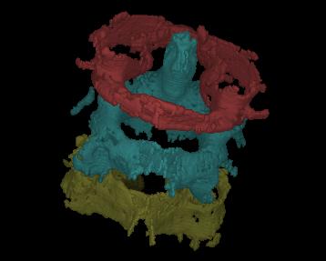
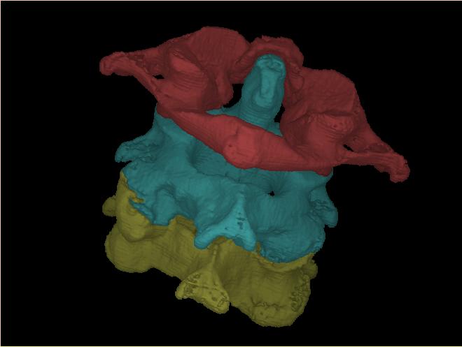
3D VISUALIZATION OF SEGMENTED CRUCIATE LIGAMENTS 1. INTRODUCTION
 JOURNAL OF MEDICAL INFORMATICS & TECHNOLOGIES Vol. 10/006, ISSN 164-6037 Paweł BADURA * cruciate ligament, segmentation, fuzzy connectedness,3d visualization 3D VISUALIZATION OF SEGMENTED CRUCIATE LIGAMENTS
Application of fuzzy set theory in image analysis. Nataša Sladoje Centre for Image Analysis
 Application of fuzzy set theory in image analysis Nataša Sladoje Centre for Image Analysis Our topics for today Crisp vs fuzzy Fuzzy sets and fuzzy membership functions Fuzzy set operators Approximate
MEDICAL IMAGE COMPUTING (CAP 5937) LECTURE 4: Pre-Processing Medical Images (II)
 LECTURE 4: Pre-Processing Medical Images (II)") SPRING 2016 1 MEDICAL IMAGE COMPUTING (CAP 5937) LECTURE 4: Pre-Processing Medical Images (II) Dr. Ulas Bagci HEC 221, Center for Research in Computer Vision (CRCV), University of Central Florida (UCF),
SPRING 2016 1 MEDICAL IMAGE COMPUTING (CAP 5937) LECTURE 4: Pre-Processing Medical Images (II) Dr. Ulas Bagci HEC 221, Center for Research in Computer Vision (CRCV), University of Central Florida (UCF),
MEDICAL IMAGE COMPUTING (CAP 5937) LECTURE 10: Medical Image Segmentation as an Energy Minimization Problem
 LECTURE 10: Medical Image Segmentation as an Energy Minimization Problem") SPRING 06 MEDICAL IMAGE COMPUTING (CAP 97) LECTURE 0: Medical Image Segmentation as an Energy Minimization Problem Dr. Ulas Bagci HEC, Center for Research in Computer Vision (CRCV), University of Central
SPRING 06 MEDICAL IMAGE COMPUTING (CAP 97) LECTURE 0: Medical Image Segmentation as an Energy Minimization Problem Dr. Ulas Bagci HEC, Center for Research in Computer Vision (CRCV), University of Central
GPU-Based Iterative Relative Fuzzy Connectedness Image Segmentation
 GPU-Based Iterative Relative Fuzzy Connectedness Image Segmentation Ying Zhuge a, Jayaram K. Udupa b, Krzysztof C. Ciesielski b,c, Alexandre X. Falcão d, Paulo A. V. Miranda e, and Robert W. Miller a a
Comparison Study of Clinical 3D MRI Brain Segmentation Evaluation
 Comparison Study of Clinical 3D MRI Brain Segmentation Evaluation Ting Song 1, Elsa D. Angelini 2, Brett D. Mensh 3, Andrew Laine 1 1 Heffner Biomedical Imaging Laboratory Department of Biomedical Engineering,
MEDICAL IMAGE COMPUTING (CAP 5937) LECTURE 10: Medical Image Segmentation as an Energy Minimization Problem
 LECTURE 10: Medical Image Segmentation as an Energy Minimization Problem") SPRING 07 MEDICAL IMAGE COMPUTING (CAP 97) LECTURE 0: Medical Image Segmentation as an Energy Minimization Problem Dr. Ulas Bagci HEC, Center for Research in Computer Vision (CRCV), University of Central
SPRING 07 MEDICAL IMAGE COMPUTING (CAP 97) LECTURE 0: Medical Image Segmentation as an Energy Minimization Problem Dr. Ulas Bagci HEC, Center for Research in Computer Vision (CRCV), University of Central
CANDIDATE TREE-IN-BUD PATTERN SELECTION AND CLASSIFICATION USING BALL SCALE ENCODING ALGORITHM
 ISSN: 9-6956 (ONLINE) DOI: 0.97/ijsc.03.009 ICTACT JOURNAL ON SOFT COMPUTING, OCTOBER 03, VOLUME: 04, ISSUE: 0 CANDIDATE TREE-IN-BUD PATTERN SELECTION AND CLASSIFICATION USING BALL SCALE ENCODING ALGORITHM
MEDICAL IMAGE COMPUTING (CAP 5937) LECTURE 19: Machine Learning in Medical Imaging (A Brief Introduction)
 LECTURE 19: Machine Learning in Medical Imaging (A Brief Introduction)") SPRING 2016 1 MEDICAL IMAGE COMPUTING (CAP 5937) LECTURE 19: Machine Learning in Medical Imaging (A Brief Introduction) Dr. Ulas Bagci HEC 221, Center for Research in Computer Vision (CRCV), University
SPRING 2016 1 MEDICAL IMAGE COMPUTING (CAP 5937) LECTURE 19: Machine Learning in Medical Imaging (A Brief Introduction) Dr. Ulas Bagci HEC 221, Center for Research in Computer Vision (CRCV), University
Department of Image Processing and Computer Graphics University of Szeged. Fuzzy Techniques for Image Segmentation. Outline.
 László G. Nyúl systems sets image László G. Nyúl Department of Processing and Computer Graphics University of Szeged 2009-07-07 systems sets image 1 systems 2 sets 3 image thresholding clustering 4 Dealing
Adaptive Fuzzy Connectedness-Based Medical Image Segmentation
 Adaptive Fuzzy Connectedness-Based Medical Image Segmentation Amol Pednekar Ioannis A. Kakadiaris Uday Kurkure Visual Computing Lab, Dept. of Computer Science, Univ. of Houston, Houston, TX, USA apedneka@bayou.uh.edu
Vascular segmentation in hepatic CT images using adaptive threshold fuzzy connectedness method
 DOI 10.1186/s12938-015-0055-z RESEARCH Open Access Vascular segmentation in hepatic CT images using adaptive threshold fuzzy connectedness method Xiaoxi Guo 1,2, Shaohui Huang 1*, Xiaozhu Fu 1, Boliang
Automated segmentation methods for liver analysis in oncology applications
 University of Szeged Department of Image Processing and Computer Graphics Automated segmentation methods for liver analysis in oncology applications Ph. D. Thesis László Ruskó Thesis Advisor Dr. Antal
Image segmentation via fuzzy object extraction and edge detection and its medical application
 Journal of X-Ray Science and Technology 10 (2002) 95 106 95 IOS Press Image segmentation via fuzzy object extraction and edge detection and its medical application Yao Lin, Jie Tian and Huiguang He AI
Dr. Ulas Bagci
 Lecture 9: Deformable Models and Segmentation CAP-Computer Vision Lecture 9-Deformable Models and Segmentation Dr. Ulas Bagci bagci@ucf.edu Lecture 9: Deformable Models and Segmentation Motivation A limitation
GPU-Based Relative Fuzzy Connectedness Image Segmentation
 GPU-Based Relative Fuzzy Connectedness Image Segmentation Ying Zhuge a) Radiation Oncology Branch, National Cancer Institute, National Institutes of Health, Bethesda, MD 20892 5 10 15 Krzysztof C. Ciesielski
Improved Fuzzy Connectedness Segmentation Method for Medical Images with Multiple Seeds in MRI
 Improved Fuzzy Connectedness Segmentation Method for Medical Images with Multiple Seeds in MRI Yunping Zheng 1*, Tong Chang 1, Mudar Sarem 2 1 School of Computer Science and Engineering, South China University
MR IMAGE SEGMENTATION
 MR IMAGE SEGMENTATION Prepared by : Monil Shah What is Segmentation? Partitioning a region or regions of interest in images such that each region corresponds to one or more anatomic structures Classification
mritc: A Package for MRI Tissue Classification
 mritc: A Package for MRI Tissue Classification Dai Feng 1 Luke Tierney 2 1 Merck Research Labratories 2 University of Iowa July 2010 Feng & Tierney (Merck & U of Iowa) MRI Tissue Classification July 2010
Segmentation of Medical Images. Application Context. Image Segmentation in SSIP 04 Projects. X-ray. Magnetic Resonance (MR) Computed Tomography (CT)
 Computed Tomography (CT)") Segmentation of Medical Images László Nyúl Department of Image Processing and Computer Graphics University of Szeged Hungary Image Segmentation in SSIP 04 Projects 24 projects suggested 1 pure mathematical
Segmentation of Medical Images László Nyúl Department of Image Processing and Computer Graphics University of Szeged Hungary Image Segmentation in SSIP 04 Projects 24 projects suggested 1 pure mathematical
Image Segmentation Based on Fuzzy Connectedness Using Dynamic Weights
 Image Segmentation Based on Fuzzy Connectedness Using Dynamic Weights Amol S. Pednekar and Ioannis A. Kakadiaris Department of Computer Science University of Houston Houston, TX, 77204, USA http://www.cs.uh.edu
arxiv: v1 [cs.cv] 26 Jun 2011
![arxiv: v1 [cs.cv] 26 Jun 2011](/thumbs/72/66652932.jpg "arxiv: v1 [cs.cv] 26 Jun 2011") LEARNING SHAPE AND TEXTURE CHARACTERISTICS OF CT TREE-IN-BUD OPACITIES FOR CAD SYSTEMS ULAŞ BAĞCI, JIANHUA YAO, JESUS CABAN, ANTHONY F. SUFFREDINI, TARA N. PALMORE, DANIEL J. MOLLURA arxiv:1106.5186v1
LEARNING SHAPE AND TEXTURE CHARACTERISTICS OF CT TREE-IN-BUD OPACITIES FOR CAD SYSTEMS ULAŞ BAĞCI, JIANHUA YAO, JESUS CABAN, ANTHONY F. SUFFREDINI, TARA N. PALMORE, DANIEL J. MOLLURA arxiv:1106.5186v1
Fuzzy Image Segmentation using Membership Connectedness
 Fuzzy Image Segmentation using Membership Connectedness Maryam Hasanzadeh and Shohreh Kasaei Computer Engineering Department, Sharif University of Technology, Tehran, Iran hasanzadeh@ce.sharif.edu, skasaei@sharif.edu
Interactive iterative relative fuzzy connectedness lung segmentation on thoracic 4D dynamic MR images
 Interactive iterative relative fuzzy connectedness lung segmentation on thoracic 4D dynamic MR images Yubing Tong 1, Jayaram K. Udupa 1, Dewey Odhner 1, Caiyun Wu 1, Yue Zhao 1, Joseph M. McDonough 2,
Learning Shape and Texture Characteristics of CT Tree-in-Bud Opacities for CAD Systems
 Learning Shape and Texture Characteristics of CT Tree-in-Bud Opacities for CAD Systems Ulaş Bağcı 1, Jianhua Yao 1, Jesus Caban 2, Anthony F. Suffredini 3, Tara N. Palmore 4, and Daniel J. Mollura 1 1
MEDICAL IMAGE COMPUTING (CAP 5937) LECTURE 20: Machine Learning in Medical Imaging II (deep learning and decision forests)
 LECTURE 20: Machine Learning in Medical Imaging II (deep learning and decision forests)") SPRING 2016 1 MEDICAL IMAGE COMPUTING (CAP 5937) LECTURE 20: Machine Learning in Medical Imaging II (deep learning and decision forests) Dr. Ulas Bagci HEC 221, Center for Research in Computer Vision (CRCV),
SPRING 2016 1 MEDICAL IMAGE COMPUTING (CAP 5937) LECTURE 20: Machine Learning in Medical Imaging II (deep learning and decision forests) Dr. Ulas Bagci HEC 221, Center for Research in Computer Vision (CRCV),
Norbert Schuff VA Medical Center and UCSF
 Norbert Schuff Medical Center and UCSF Norbert.schuff@ucsf.edu Medical Imaging Informatics N.Schuff Course # 170.03 Slide 1/67 Objective Learn the principle segmentation techniques Understand the role
Generalized Scale-Based Image Filtering
 Generalized Scale-Based Image Filtering Andre Souza a, Jayaram K. Udupa a, and Anant Madabhushi a a Medical Image Processing Group, Department of Radiology University of Pennsylvania, Philadelphia, PA
Learning-based Neuroimage Registration
 Learning-based Neuroimage Registration Leonid Teverovskiy and Yanxi Liu 1 October 2004 CMU-CALD-04-108, CMU-RI-TR-04-59 School of Computer Science Carnegie Mellon University Pittsburgh, PA 15213 Abstract
A Simple Centricity-based Region Growing Algorithm for the Extraction of Airways
 EXACT'09-309- A Simple Centricity-based Region Growing Algorithm for the Extraction of Airways Rafael Wiemker, Thomas Bülow, Cristian Lorenz Philips Research Lab Hamburg, Röntgenstrasse 24, 22335 Hamburg
Robust Region Growing Based Intrathoracic Airway Tree Segmentation
 EXACT'09-261- Robust Region Growing Based Intrathoracic Airway Tree Segmentation Rômulo Pinho, Sten Luyckx, and Jan Sijbers University of Antwerp, Physics Department, VisionLab, Belgium {romulo.pinho;
Norbert Schuff Professor of Radiology VA Medical Center and UCSF
 Norbert Schuff Professor of Radiology Medical Center and UCSF Norbert.schuff@ucsf.edu 2010, N.Schuff Slide 1/67 Overview Definitions Role of Segmentation Segmentation methods Intensity based Shape based
Medical Images Analysis and Processing
 Medical Images Analysis and Processing - 25642 Emad Course Introduction Course Information: Type: Graduated Credits: 3 Prerequisites: Digital Image Processing Course Introduction Reference(s): Insight
Histograms. h(r k ) = n k. p(r k )= n k /NM. Histogram: number of times intensity level rk appears in the image
 = n k. p(r k )= n k /NM. Histogram: number of times intensity level rk appears in the image") Histograms h(r k ) = n k Histogram: number of times intensity level rk appears in the image p(r k )= n k /NM normalized histogram also a probability of occurence 1 Histogram of Image Intensities Create
Histograms h(r k ) = n k Histogram: number of times intensity level rk appears in the image p(r k )= n k /NM normalized histogram also a probability of occurence 1 Histogram of Image Intensities Create
GPU-based relative fuzzy connectedness image segmentation
 GPU-based relative fuzzy connectedness image segmentation Ying Zhuge a) Radiation Oncology Branch, National Cancer Institute, National Institutes of Health, Bethesda, Maryland 20892 Krzysztof C. Ciesielski
Application of fuzzy set theory in image analysis
 Application of fuzzy set theory in image analysis Analysis and defuzzification of discrete fuzzy spatial sets Who am I? Nataša Sladoje Teaching assistant in mathematics Department of fundamental disciplines
Methods for data preprocessing
 Methods for data preprocessing John Ashburner Wellcome Trust Centre for Neuroimaging, 12 Queen Square, London, UK. Overview Voxel-Based Morphometry Morphometry in general Volumetrics VBM preprocessing
INTERPOLATION is a commonly used operation in image
 IEEE TRANSACTIONS ON MEDICAL IMAGING, VOL. 18, NO. 2, FEBRUARY 1999 137 A Task-Specific Evaluation of Three-Dimensional Image Interpolation Techniques George J. Grevera, Jayaram K. Udupa,* Senior Member,
CHAPTER 2. Morphometry on rodent brains. A.E.H. Scheenstra J. Dijkstra L. van der Weerd
 CHAPTER 2 Morphometry on rodent brains A.E.H. Scheenstra J. Dijkstra L. van der Weerd This chapter was adapted from: Volumetry and other quantitative measurements to assess the rodent brain, In vivo NMR
New Technology Allows Multiple Image Contrasts in a Single Scan
 These images were acquired with an investigational device. PD T2 T2 FLAIR T1 MAP T1 FLAIR PSIR T1 New Technology Allows Multiple Image Contrasts in a Single Scan MR exams can be time consuming. A typical
Subvoxel Segmentation and Representation of Brain Cortex Using Fuzzy Clustering and Gradient Vector Diffusion
 Subvoxel Segmentation and Representation of Brain Cortex Using Fuzzy Clustering and Gradient Vector Diffusion Ming-Ching Chang Xiaodong Tao GE Global Research Center {changm, taox} @ research.ge.com SPIE
Correction of Partial Volume Effects in Arterial Spin Labeling MRI
 Correction of Partial Volume Effects in Arterial Spin Labeling MRI By: Tracy Ssali Supervisors: Dr. Keith St. Lawrence and Udunna Anazodo Medical Biophysics 3970Z Six Week Project April 13 th 2012 Introduction
Available Online through
 Available Online through www.ijptonline.com ISSN: 0975-766X CODEN: IJPTFI Research Article ANALYSIS OF CT LIVER IMAGES FOR TUMOUR DIAGNOSIS BASED ON CLUSTERING TECHNIQUE AND TEXTURE FEATURES M.Krithika
Fuzzy-Based Extraction of Vascular Structures from Time-of-Flight MR Images
 816 Medical Informatics in a United and Healthy Europe K.-P. Adlassnig et al. (Eds.) IOS Press, 2009 2009 European Federation for Medical Informatics. All rights reserved. doi:10.3233/978-1-60750-044-5-816
Segmentation and Evaluation of Adipose Tissue from Whole Body MRI Scans
 Segmentation and Evaluation of Adipose Tissue from Whole Body MRI Scans Yinpeng Jin 1, Celina Z. Imielińska 2, Andrew F. Laine 1, Jayaram Udupa 3, Wei Shen 4, Steven B. Heymsfield 4 1 Department of Biomedical
Chapter 3. Image Processing Methods. (c) 2008 Prof. Dr. Michael M. Richter, Universität Kaiserslautern
 2008 Prof. Dr. Michael M. Richter, Universität Kaiserslautern") Chapter 3 Image Processing Methods The Role of Image Processing Methods (1) An image is an nxn matrix of gray or color values An image processing method is algorithm transforming such matrices or assigning
Chapter 3 Image Processing Methods The Role of Image Processing Methods (1) An image is an nxn matrix of gray or color values An image processing method is algorithm transforming such matrices or assigning
Dr. Ulas Bagci
 CAP5415-Computer Vision Lecture 11-Image Segmentation (BASICS): Thresholding, Region Growing, Clustering Dr. Ulas Bagci bagci@ucf.edu 1 Image Segmentation Aim: to partition an image into a collection of
Application of level set based method for segmentation of blood vessels in angiography images
 Lodz University of Technology Faculty of Electrical, Electronic, Computer and Control Engineering Institute of Electronics PhD Thesis Application of level set based method for segmentation of blood vessels
Automatic MS Lesion Segmentation by Outlier Detection and Information Theoretic Region Partitioning Release 0.00
 Automatic MS Lesion Segmentation by Outlier Detection and Information Theoretic Region Partitioning Release 0.00 Marcel Prastawa 1 and Guido Gerig 1 Abstract July 17, 2008 1 Scientific Computing and Imaging
Introduction to Medical Image Processing
 Introduction to Medical Image Processing Δ Essential environments of a medical imaging system Subject Image Analysis Energy Imaging System Images Image Processing Feature Images Image processing may be
Deep Learning in Pulmonary Image Analysis with Incomplete Training Samples
 Deep Learning in Pulmonary Image Analysis with Incomplete Training Samples Ziyue Xu, Staff Scientist, National Institutes of Health Nov. 2nd, 2017 (GTC DC Talk DC7137) Image Analysis Arguably the most
GLIRT: Groupwise and Longitudinal Image Registration Toolbox
 Software Release (1.0.1) Last updated: March. 30, 2011. GLIRT: Groupwise and Longitudinal Image Registration Toolbox Guorong Wu 1, Qian Wang 1,2, Hongjun Jia 1, and Dinggang Shen 1 1 Image Display, Enhancement,
Chapter 3 Set Redundancy in Magnetic Resonance Brain Images
 16 Chapter 3 Set Redundancy in Magnetic Resonance Brain Images 3.1 MRI (magnetic resonance imaging) MRI is a technique of measuring physical structure within the human anatomy. Our proposed research focuses
Preprocessing II: Between Subjects John Ashburner
 Preprocessing II: Between Subjects John Ashburner Pre-processing Overview Statistics or whatever fmri time-series Anatomical MRI Template Smoothed Estimate Spatial Norm Motion Correct Smooth Coregister
MICRO CT LUNG SEGMENTATION. Using Analyze
 MICRO CT LUNG SEGMENTATION Using Analyze 2 Table of Contents 1. Introduction page 3 2. Lung Segmentation page 4 3. Lung Volume Measurement page 13 4. References page 16 3 Introduction Mice are often used
Bioimage Informatics
 Bioimage Informatics Lecture 14, Spring 2012 Bioimage Data Analysis (IV) Image Segmentation (part 3) Lecture 14 March 07, 2012 1 Outline Review: intensity thresholding based image segmentation Morphological
Automatic System For Brain Tumor Detection And Classification Using Level Set And ANN
 Automatic System For Brain Tumor Detection And Classification Using Level Set And ANN Nisha Babu A Computer Science Department LBSITW Trivandrum, India nisthesweet@gmail.com Agoma Martin Computer Science
Brain Portion Peeling from T2 Axial MRI Head Scans using Clustering and Morphological Operation
 159 Brain Portion Peeling from T2 Axial MRI Head Scans using Clustering and Morphological Operation K. Somasundaram Image Processing Lab Dept. of Computer Science and Applications Gandhigram Rural Institute
On Standardizing the MR Image Intensity Scale
 On Standardizing the MR Image Intensity Scale László G. Nyúl and Jayaram K. Udupa* Magnetic Resonance in Medicine 42:1072 1081 (1999) The lack of a standard image intensity scale in MRI causes many difficulties
CAD SYSTEM FOR AUTOMATIC DETECTION OF BRAIN TUMOR THROUGH MRI BRAIN TUMOR DETECTION USING HPACO CHAPTER V BRAIN TUMOR DETECTION USING HPACO
 CHAPTER V BRAIN TUMOR DETECTION USING HPACO 145 CHAPTER 5 DETECTION OF BRAIN TUMOR REGION USING HYBRID PARALLEL ANT COLONY OPTIMIZATION (HPACO) WITH FCM (FUZZY C MEANS) 5.1 PREFACE The Segmentation of
Neuroimaging and mathematical modelling Lesson 2: Voxel Based Morphometry
 Neuroimaging and mathematical modelling Lesson 2: Voxel Based Morphometry Nivedita Agarwal, MD Nivedita.agarwal@apss.tn.it Nivedita.agarwal@unitn.it Volume and surface morphometry Brain volume White matter
FINDING THE TRUE EDGE IN CTA
 FINDING THE TRUE EDGE IN CTA by: John A. Rumberger, PhD, MD, FACC Your patient has chest pain. The Cardiac CT Angiography shows plaque in the LAD. You adjust the viewing window trying to evaluate the stenosis
Automatic Lung Segmentation of Volumetric Low-Dose CT Scans Using Graph Cuts
 Automatic Lung Segmentation of Volumetric Low-Dose CT Scans Using Graph Cuts Asem M. Ali and Aly A. Farag Computer Vision and Image Processing Laboratory (CVIP Lab) University of Louisville, Louisville,
Norbert Schuff Professor of Radiology VA Medical Center and UCSF
 Norbert Schuff Professor of Radiology Medical Center and UCSF Norbert.schuff@ucsf.edu Slide 1/67 Overview Definitions Role of Segmentation Segmentation methods Intensity based Shape based Texture based
Automated 3D Segmentation of the Lung Airway Tree Using Gain-Based Region Growing Approach
 Automated 3D Segmentation of the Lung Airway Tree Using Gain-Based Region Growing Approach Harbir Singh 1, Michael Crawford, 2, John Curtin 2, and Reyer Zwiggelaar 1 1 School of Computing Sciences, University
Advanced Visual Medicine: Techniques for Visual Exploration & Analysis
 Advanced Visual Medicine: Techniques for Visual Exploration & Analysis Interactive Visualization of Multimodal Volume Data for Neurosurgical Planning Felix Ritter, MeVis Research Bremen Multimodal Neurosurgical
Learning to Identify Fuzzy Regions in Magnetic Resonance Images
 Learning to Identify Fuzzy Regions in Magnetic Resonance Images Sarah E. Crane and Lawrence O. Hall Department of Computer Science and Engineering, ENB 118 University of South Florida 4202 E. Fowler Ave.
SEGMENTATION OF IMAGES USING GRADIENT METHODS AND POLYNOMIAL APPROXIMATION
 JOURNAL OF MEDICAL INFORMATICS & TECHNOLOGIES Vol. 23/2014, ISSN 1642-6037 segmentation, gradient methods, polynomial approximation Ewelina PIEKAR 1, Michal MOMOT 1, Alina MOMOT 2 SEGMENTATION OF IMAGES
Global Journal of Engineering Science and Research Management
 ADVANCED K-MEANS ALGORITHM FOR BRAIN TUMOR DETECTION USING NAIVE BAYES CLASSIFIER Veena Bai K*, Dr. Niharika Kumar * MTech CSE, Department of Computer Science and Engineering, B.N.M. Institute of Technology,
The MAGIC-5 CAD for nodule detection in low dose and thin slice lung CT. Piergiorgio Cerello - INFN
 The MAGIC-5 CAD for nodule detection in low dose and thin slice lung CT Piergiorgio Cerello - INFN Frascati, 27/11/2009 Computer Assisted Detection (CAD) MAGIC-5 & Distributed Computing Infrastructure
Processing math: 100% Intensity Normalization
 Intensity Normalization Overall Pipeline 2/21 Intensity normalization Conventional MRI intensites (T1-w, T2-w, PD, FLAIR) are acquired in arbitrary units Images are not comparable across scanners, subjects,
11/18/ CPT Preauthorization Groupings Effective January 1, Computerized Tomography (CT) Abdomen 6. CPT Description SEGR CT01
 Abdomen 6. CPT Description SEGR CT01") Computerized Tomography (CT) 6 & 101 5 Upper Extremity 11 Lower Extremity 12 Head 3 Orbit 1 Sinus 2 Neck 4 7 Cervical Spine 8 Thoracic Spine 9 Lumbar Spine 10 Colon 13 CPT Description SEGR 74150 74160
Computerized Tomography (CT) 6 & 101 5 Upper Extremity 11 Lower Extremity 12 Head 3 Orbit 1 Sinus 2 Neck 4 7 Cervical Spine 8 Thoracic Spine 9 Lumbar Spine 10 Colon 13 CPT Description SEGR 74150 74160
Three-dimensional Image Processing Techniques to Perform Landmarking and Segmentation of Computed Tomographic Images
 Three-dimensional Image Processing Techniques to Perform Landmarking and Segmentation of Computed Tomographic Images Rangaraj M. Rangayyan, Shantanu Banik, Graham S. Boag Department of Electrical and Computer
High Accuracy Region Growing Segmentation Technique for Magnetic Resonance and Computed Tomography Images with Weak Boundaries
 High Accuracy Region Growing Segmentation Technique for Magnetic Resonance and Computed Tomography Images with Weak Boundaries Ahmed Ayman * Takuya Funatomi Michihiko Minoh Academic Center for Computing
Ulas Bagci
 CAP5415-Computer Vision Lecture 14-Decision Forests for Computer Vision Ulas Bagci bagci@ucf.edu 1 Readings Slide Credits: Criminisi and Shotton Z. Tu R.Cipolla 2 Common Terminologies Randomized Decision
Segmentation Using Deformable Models With. Department of Computer and Information Science, University of Pennsylvania,
 Segmentation Using Deformable Models With Anity-Based Localization? Timothy N. Jones tnj@graphics.cis.upenn.edu Dimitris N. Metaxas dnm@central.cis.upenn.edu Department of Computer and Information Science,
Computational Medical Imaging Analysis Chapter 4: Image Visualization
 Computational Medical Imaging Analysis Chapter 4: Image Visualization Jun Zhang Laboratory for Computational Medical Imaging & Data Analysis Department of Computer Science University of Kentucky Lexington,
6 credits. BMSC-GA Practical Magnetic Resonance Imaging II
 BMSC-GA 4428 - Practical Magnetic Resonance Imaging II 6 credits Course director: Ricardo Otazo, PhD Course description: This course is a practical introduction to image reconstruction, image analysis
Using multimodal MR data for segmentation and topology recovery of the cerebral superficial venous tree
 Using multimodal MR data for segmentation and topology recovery of the cerebral superficial venous tree Nicolas Passat, Christian Ronse, Joseph Baruthio, Jean-Paul Armspach, Marcel Bosc, Jack Foucher To
Spatio-Temporal Registration of Biomedical Images by Computational Methods
 Spatio-Temporal Registration of Biomedical Images by Computational Methods Francisco P. M. Oliveira, João Manuel R. S. Tavares tavares@fe.up.pt, www.fe.up.pt/~tavares Outline 1. Introduction 2. Spatial
EE795: Computer Vision and Intelligent Systems
 EE795: Computer Vision and Intelligent Systems Spring 2012 TTh 17:30-18:45 WRI C225 Lecture 04 130131 http://www.ee.unlv.edu/~b1morris/ecg795/ 2 Outline Review Histogram Equalization Image Filtering Linear
Level Set Evolution with Region Competition: Automatic 3-D Segmentation of Brain Tumors
 1 Level Set Evolution with Region Competition: Automatic 3-D Segmentation of Brain Tumors 1 Sean Ho, 2 Elizabeth Bullitt, and 1,3 Guido Gerig 1 Department of Computer Science, 2 Department of Surgery,
Hybrid Approach for MRI Human Head Scans Classification using HTT based SFTA Texture Feature Extraction Technique
 Volume 118 No. 17 2018, 691-701 ISSN: 1311-8080 (printed version); ISSN: 1314-3395 (on-line version) url: http://www.ijpam.eu ijpam.eu Hybrid Approach for MRI Human Head Scans Classification using HTT
Where are we now? Structural MRI processing and analysis
 Where are we now? Structural MRI processing and analysis Pierre-Louis Bazin bazin@cbs.mpg.de Leipzig, Germany Structural MRI processing: why bother? Just use the standards? SPM FreeSurfer FSL However:
NIH Public Access Author Manuscript Proc IEEE Int Symp Biomed Imaging. Author manuscript; available in PMC 2014 November 15.
 NIH Public Access Author Manuscript Published in final edited form as: Proc IEEE Int Symp Biomed Imaging. 2013 April ; 2013: 748 751. doi:10.1109/isbi.2013.6556583. BRAIN TUMOR SEGMENTATION WITH SYMMETRIC
Interactive Differential Segmentation of the Prostate using Graph-Cuts with a Feature Detector-based Boundary Term
 MOSCHIDIS, GRAHAM: GRAPH-CUTS WITH FEATURE DETECTORS 1 Interactive Differential Segmentation of the Prostate using Graph-Cuts with a Feature Detector-based Boundary Term Emmanouil Moschidis emmanouil.moschidis@postgrad.manchester.ac.uk
Local or Global Minima: Flexible Dual-Front Active Contours
 Local or Global Minima: Flexible Dual-Front Active Contours Hua Li 1,2 and Anthony Yezzi 1 1 School of ECE, Georgia Institute of Technology, Atlanta, GA, USA 2 Dept. of Elect. & Info. Eng., Huazhong Univ.
Airway Segmentation Framework for Clinical Environments
 EXACT'09-227- Airway Segmentation Framework for Clinical Environments Juerg Tschirren 1, Tarunashree Yavarna 2, and Joseph M. Reinhardt 2 1 VIDA Diagnostics, Inc. 100 Oakdale Campus, 225 TIC Iowa City,
Interactive segmentation of vascular structures in CT images for liver surgery planning
 Interactive segmentation of vascular structures in CT images for liver surgery planning L. Wang¹, C. Hansen¹, S.Zidowitz¹, H. K. Hahn¹ ¹ Fraunhofer MEVIS, Institute for Medical Image Computing, Bremen,
HYBRID MULTISCALE LANDMARK AND DEFORMABLE IMAGE REGISTRATION. Dana Paquin. Doron Levy. Lei Xing. (Communicated by Yang Kuang)
") MATHEMATICAL BIOSCIENCES http://www.mbejournal.org/ AND ENGINEERING Volume 4, Number 4, October 2007 pp. 711 737 HYBRID MULTISCALE LANDMARK AND DEFORMABLE IMAGE REGISTRATION Dana Paquin Department of Mathematics,
MATHEMATICAL BIOSCIENCES http://www.mbejournal.org/ AND ENGINEERING Volume 4, Number 4, October 2007 pp. 711 737 HYBRID MULTISCALE LANDMARK AND DEFORMABLE IMAGE REGISTRATION Dana Paquin Department of Mathematics,
MARS: Multiple Atlases Robust Segmentation
 Software Release (1.0.1) Last updated: April 30, 2014. MARS: Multiple Atlases Robust Segmentation Guorong Wu, Minjeong Kim, Gerard Sanroma, and Dinggang Shen {grwu, mjkim, gerard_sanroma, dgshen}@med.unc.edu
MRI Brain image segmentation using graph cuts
 MRI Brain image segmentation using graph cuts Master of Science Thesis in Communication Engineering Mohammad Shajib Khadem Signal Processing Group Department of Signals and Systems CHALMERS UNIVERSITY
Fuzzy model based object delineation via energy minimization
 Fuzzy model based object delineation via energy minimization Krzysztof Chris Ciesielski, a,b Jayaram K. Udupa, b Dewey Odhner, b and Liming Zhao b a Department of Mathematics, West Virginia University,
The Anatomical Equivalence Class Formulation and its Application to Shape-based Computational Neuroanatomy
 The Anatomical Equivalence Class Formulation and its Application to Shape-based Computational Neuroanatomy Sokratis K. Makrogiannis, PhD From post-doctoral research at SBIA lab, Department of Radiology,
CHAPTER-1 INTRODUCTION
 CHAPTER-1 INTRODUCTION 1.1 Fuzzy concept, digital image processing and application in medicine With the advancement of digital computers, it has become easy to store large amount of data and carry out
GPU Based Region Growth and Vessel Tracking. Supratik Moulik M.D. Jason Walsh
 GPU Based Region Growth and Vessel Tracking Supratik Moulik M.D. (supratik@moulik.com) Jason Walsh Conflict of Interest Dr. Supratik Moulik does not have a significant financial stake in any company, nor
TWO and higher-dimensional images are currently available
 IEEE TRANSACTIONS ON PATTERN ANALYSIS AND MACHINE INTELLIGENCE, VOL. 24, NO. 11, NOVEMBER 2002 1485 Relative Fuzzy Connectedness and Object Definition: Theory, Algorithms, and Applications in Image Segmentation
Image Segmentation and Registration
 Image Segmentation and Registration Dr. Christine Tanner (tanner@vision.ee.ethz.ch) Computer Vision Laboratory, ETH Zürich Dr. Verena Kaynig, Machine Learning Laboratory, ETH Zürich Outline Segmentation
Human Heart Coronary Arteries Segmentation
 Human Heart Coronary Arteries Segmentation Qian Huang Wright State University, Computer Science Department Abstract The volume information extracted from computed tomography angiogram (CTA) datasets makes