Positron Emission Tomography
|
|
|
- Kimberly Norris
- 5 years ago
- Views:
Transcription


1 Physics 656 Seminar on Physical Fundamentals of Medical Imaging Positron Emission Tomography Ahmed Qamesh
2 Outline What is PET? PET mechanism Radionuclide and its synthesis Detection concept and Development Mathematical Model Photon Detection Time of flight Coincidence processing Detector Configuration 2D Vs 3D acquisition Data correction Application Summary 7/3/17 Universität Bonn 2


3 What is PET? Positron Emission Tomography A molecular imaging technique used to obtain images of biological systems How? by measuring metabolic activity of cells. Examples: Brain, kidney, Tumors, Heart disease, 7/3/17 Universität Bonn 3
4 What is PET? Method : Injecting the body with a radioactive trace A camera is then used to monitor the distribution of the substance in the body to study accurately the organ s function. Advantages: 2D and 3D images. Resolution (5-6 mm) in all directions. Shows chemical functioning of organs (Not just structure like CT). Can show some cancers in its early stages (more than CT or MRI). 7/3/17 Universität Bonn 4
 7/3/17 Universität")
5 Positron Emission Tomography (Overview) 7/3/17 Universität Bonn 5
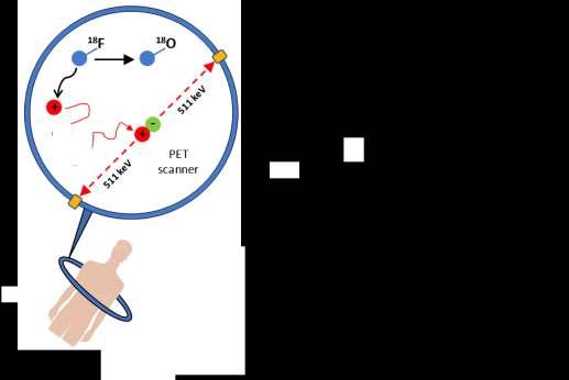
. 2. FDG spreads via blood stream till it enter organs. 3. Positron Emission 18 18 F!! 9 8 O +e + 4. Positron will travels a short distance (0.")
6 PET Mechanism Compounds with positron emitting Radioisotopes probes as molecular probes. (Isotopes of ) 9 Flurodeoxyglucose (FDG) is the most famous example. 1. FDG is injected into the body (stay for min). 2. FDG spreads via blood stream till it enter organs. 3. Positron Emission F!! 9 8 O +e + 4. Positron will travels a short distance (0.5 mm in water ) before it annihilates with an electron. e + +e " " 2γ Mass is converted into Energy (2 photons of 511 KeV) 5. An Electronic signal ( Resolving time 6-15 ns). 6. Reconstruction of images. 18 F 7/3/17 Universität Bonn 6


7 Image Reconstruction Radionuclide synthesis Detection process PET probes synthesis Probes absorption in tissues 7/3/17 Universität Bonn 7

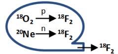
8 Radionuclide synthesis and On-site cyclotron 7/3/17 Universität Bonn 8
9 General Principles: What Isotope to use? (1) Radionuclide synthesis Life time: short enough to minimize the radiation exposure to the patient. long enough to allow regional distribution. Cheap. Non Toxic. Available in Daily life. Time Time Time Time Cost Time+ 7/3/17 Universität Bonn 9
 Isotopes: 18 F, 17")

10 Fluorine (Halogen element). Lies in Group 7 of periodic table (7 Electrons in the outer shell) Isotopes: 18 F, F F-18 preparation (on site Cyclotron) By firing Protons (12 MeV) onto a target material of O-18 p O!! 18 9 F Negatively charged H (proton with 2 electrons), 2e are stripped with carbon foil 7/3/17 Universität Bonn 10


11 General Principles: (2) PET probes synthesis The aim is to remove one Hydroxy group from Glucose Molecule and replace it with Fluorine atom + 18 F!! 9 Florodyoxy-Glucose Why Glucose? Normal body consumes energy in the form of glucose. Tumor has high rate of consumption (3) Probes absorption within tissues F!! 9 8 O +e + e + +e " " 2γ 7/3/17 Universität Bonn 11
 7/3/17 Universität")
12 Detection concept and Development The Siemens Biograph, a combined PET/CT scanner (Courtesy Siemens) 7/3/17 Universität Bonn 12

13 Mathematical Model Problem: If no refraction or diffraction: photon beams travel along straight lines that are not bent by the objects they pass through. Both give the same projection!!! Solution: Use Different orientations One object Two objects How can we reconstruct a known function from unknown one? starts with the results and then calculates the causes. Radon Transform Each slice represent a constructed line with (1-D projection) is the line integral of the image intensity F(x,y) Data Model parameters Inverse problem The collection of R at all angles is called the Radon Transform of image f(x,y). 7/3/17 Universität Bonn 13
14 Photon Detection To image the annihilation radiation one should profit from its unique properties: 1. Two Collinear photons (180 o ). 2. Simultaneously. 3. Energy of each photon 511 kev. Goal Measuring the total energy deposited by the photon when it traverses the detector ns x =ct Coincidence time window (t): the time interval at which two a pair of annihilated photons counted (X): The position of the tumor where the annihilation happened can be achieved. 7/3/17 Universität Bonn 14
.")
, The sensitive volume inside the detector cylinder that a patient can occupy.")
15 Time of flight To improve the quality of reconstruction: a coincidence measurement of the time difference (Time of Flight) using two detectors is included T.O.F=3.3ps If two photons arising from the same annihilation and an event is attributed to the line-of-response (LOR). With time-of-flight PET imaging The relative time difference (Δt) between the detection of the two annihilation photons is used to determine the most likely location (d) of the annihilation event along the LOR Field-of-view (FOV), The sensitive volume inside the detector cylinder that a patient can occupy. In human scanners is typically 70 cm in diameter and cm in axial length. 7/3/17 Universität Bonn 15


16 Coincidence processing Do we have a pure signal? The detected coincidence events (called coincidences) can be classified into: 1. True coincidences. 2. Background events. Accidental (or random) coincidences The two photons did not arise from the same annihilation event Scattered coincidences The two photons arise from the same annihilation event. because one photon has experienced Compton scatter within the patient and therefore has had a change of direction True coincidences. Accidental coincidences Scattered coincidences 7/3/17 Universität Bonn 16


17 PET Imaging system Scintillation detectors. The incident photon creates tens of thousands visible wavelength photons (about 1 ev energy each). The number of scintillation photons produced in the crystal is proportional to the energy deposited by the annihilation photon. Early PET scanners used large scintillation crystals and coupled one crystal to one PMT in a single slice. (Limits the spatial resolution) Increasing efficiency requires 1. Using many small crystals for higher Resolution. 2. Using More crystals and more PMTS for higher Sensitivity. 3. Applying septa between the transverse slices to reduce scatter from the patient Recently, The common used setup is the block detector What detector to use? Example Crystal/PMT= 1/4 7/3/17 Universität Bonn 17
18 Based on the following properties: 1. The stopping power Short (Depend on Density and Atomic number of material Z) (The inverse of the mean distance traveled by photons before depositing energy in the crystal). 2. The decay constant small (How long the scintillation flash lasts in the crystal?), Small decay constant allows high photons counting rates and lower background rates. 3. A good Energy resolution High The energy resolution depends on the light output and the intrinsic energy resolution of the crystal. 4. The light output High (The number of scintillation photons produced by each incident photon). 5. Cost Too much or too cheap? In most commercial PET scanners, the cost of the scintillator material represents 30 50% of the material cost of the scanner. 7/3/17 Universität Bonn 18
19 Scintillators used in PET Scanners Determines scanner sensitivity Determines energy and spatial resolution Determines scanner dead time and random coincidences Thalium-doped sodium iodide Bismuth germinate lutetium oxyorthosilicate lutetium yttrium orthosilicate Gadolinium orthosilicate High atomic number (Z) is preferred 1. Gives high stopping power 2. Higher Photoelectric than compton interaction facilities energy discrimination of scattered photons. 7/3/17 Universität Bonn 19
20 2D vs 3D crystal choice 2D 2D 3D 3D For 2D scanners, scintillators with low stopping power is favored. Decay time is less important For 3D scanners,, scintillators with high stopping power is favored. Decay time is important Resolution NaI(TI) detector of 6-7 mm using only 6 mm PMT with crystal : PMT= 100:1. 7/3/17 Universität Bonn 20


21 Detector configuration A full PET scanner is constructed as an assembly of block detectors in different designs (Rings or polygonal). A. Dual Detector Heads Gives 2D planner images (1/3 efficiency of the full ring) 40% lower in cost than D. Full reconstruction achieved by rotating the head to collect sufficient angular data to reconstruct. B. Half Ring better resolution than A. C. Hexagonal Ring Gives 2D and 3D planner images Lower Cost than D- Low counting rate D. Full Ring Gives 2D and 3D planner images To improve the resolution High number of small crystals required More Bending towards the source 7/3/17 Universität Bonn 21
22 2D versus 3D acquisitions Rings of detector elements may or may not be separated by thin annular rings or septa of photon absorptive material (tungsten), that provide collimation. 2D protocol With collimation 3D protocol Without collimation Lower sensitivity Improved contrast Easier to construct Many coincidences are blocked from reaching the detector. Many events out-of-plane contribute Brain imaging (small activity concentrations) Higher sensitivity Lower contrast Harder to construct Coincidences from all axial angles in the FOV. whole-body imaging (more activity concentration) 7/3/17 Universität Bonn 22
23 PET Performance and Resolution The overall spatial resolution is expressed as the Full-Width-Half-Maximum of the spread function 1. Range effect Maximum Energy 0 < E γ < E max Spectrum Energy Physical factors!! Positron Range 0 < R < R e Extrapolated range R RMS << R e (10 timesshorter) 7/3/17 Universität Bonn 23
24 PET Performance and Resolution MonteCarlo Simulation Degrading of 0.1 mm More dispersion Degrading of 0.5 mm 7/3/17 Universität Bonn 24
25 PET Performance and Resolution The overall spatial resolution is expressed as the Full-Width-Half-Maximum of the spread function 1. Range effect Maximum Energy 0 < E γ < E max Spectrum Energy Physical factors!! Extrapolated range Positron Range 0 < R < R e R RMS << R e (10 timesshorter) 2. Non Colinearity of the two photons The two annihilated photons are not exactly back to back Reason The positron has a small residual momentum and Kinetic Energy at the end of the range Δθ 0.25 o FWHM Range FWHM 180o & D= 80 cm 100 cm 7/3/17 Universität Bonn 25


26 PET Performance and Resolution Instrumentation factors Intrinsic detector resolution takes place. The depth-of-interaction-effect. For Small area detectors, resolution is determined by the detector width (W) Resolution increases from the middle to the other side of the detector W W 2 Interactions in the patient FWHM Intrinsic The observed photons in a straight line decreases exponentially with increasing length of the material traversed. The image quality degrades rapidly as the patient weight increases. TOF information is recommended for heavy patient 58 kg 89 kg 127 kg 7/3/17 Universität Bonn 26
27 PET Performance and Resolution Coincidence factor True Random Compton Spurious Coincidence Coincidence One Due Two A to of detected an the 511 annihilation photons ev coincidence photons losses and are some from a detected cascade of two energy separate within gamma and the annihilations ray time associated (scattered window with change or (6-12 unscattered) one of ns) direction the of the two falls two detected due within coincidence to the within interaction 511 detector. the ev the timing with energy an window electron of. in the coincidence human tissue. detector pair. 7/3/17 Universität Bonn 27
 than photons that travel through sparser parts of the body.")
28 Data Correction Aim: obtain clinically useful images and accurate quantitative information from PET studies. Attenuation correction Photons that encounter more or denser material on their path from the annihilation site to the detectors are more likely to be absorbed or scattered (i.e. attenuated) than photons that travel through sparser parts of the body. For example : lung tissue and skin exhibits lower attenuation Solution shows higher tracer uptake than muscle. Determine the attenuation through the patient for all LORs. How? By inserting a thin, hollow cylinder of a positron emitting activity around the patient (transmission scan) In PET/CT scanners, the acquired CT image is used for PET attenuation correction. Another possibility of PET/MRI (Julich institute of Neuroscience and biophyiscs ) CT (Computerized Axial Tomography): X-ray test for cross sectional images 7/3/17 Universität Bonn 28

29 PET/CT acquisition CT image PET image PET/CT image 7/3/17 Universität Bonn 29
30 Applications 1. Brain (patients who have memory disorders of suspected or proven brain tumors) 2. Heart (Determine blood flow to the heart muscle, determine effects of heart attack ) 3. Certain types of Cancer (Examine the effects of cancer therapy by characterizing biochemical changes in the cancer, spread of the cancer ) 4. Alzheimer s disease (there is no gross structural abnormality, but PET is able to show a biochemical change) 5. Some important body functions, to help doctors evaluate how well organs and tissues are functioning. (Blood flow, oxygen use, and sugar (glucose) metabolism) 7/3/17 Universität Bonn 30
31 Summary In this talk I gave an overview about PET.. 1. Basic Concepts of PET. 2. Detection process in PET. 3. Detector Configurations and different materials used. 4. CT/PET Images. 5. PET updates for reconstruction algorithms and attenuation correction. 6. The spatial resolution PET. 7. Clinical applications for PET 7/3/17 Universität Bonn 31
32 Thank you 7/3/17 Universität Bonn 32
33 References [1] G. Muehllehner and J. S Karp, Review Positron emission tomography, IOP Publishing Ltd, Physics in Medicine & Biology, Volume 51, Number 13(2006). [2] Molecular Imaging with Positron Emission Tomography, Michael E. Phelps, Department of Molecular and Medical Pharmacy, UCLA School of Medicine, California, (2002). [3] Pat Zanzonico, Positron Emission Tomography: A Review of Basic Principles, Scanner Design and Performance and Current Systems, Memorial Sloan-Kettering Cancer Center, New York,NY, (2004). [4] Cherry SR, Sorensen JA, Phelps ME. Physics in Nuclear Medicine. Orlando, FL: Grune & Stratton, Inc.; (2003). [5] Valk PE, Bailey DE, Townsend DW, Maisey MN. Positron Emission Tomography: Basic Science and Clinical Practice. London: Springer-Verlag; (2003). 7/3/17 Universität Bonn 33
Corso di laurea in Fisica A.A Fisica Medica 5 SPECT, PET
 Corso di laurea in Fisica A.A. 2007-2008 Fisica Medica 5 SPECT, PET Step 1: Inject Patient with Radioactive Drug Drug is labeled with positron (β + ) emitting radionuclide. Drug localizes
Introduction to Positron Emission Tomography
 Planar and SPECT Cameras Summary Introduction to Positron Emission Tomography, Ph.D. Nuclear Medicine Basic Science Lectures srbowen@uw.edu System components: Collimator Detector Electronics Collimator
Medical Imaging BMEN Spring 2016
 Name Medical Imaging BMEN 420-501 Spring 2016 Homework #4 and Nuclear Medicine Notes All questions are from the introductory Powerpoint (based on Chapter 7) and text Medical Imaging Signals and Systems,
Introduction to Emission Tomography
 Introduction to Emission Tomography Gamma Camera Planar Imaging Robert Miyaoka, PhD University of Washington Department of Radiology rmiyaoka@u.washington.edu Gamma Camera: - collimator - detector (crystal
Diagnostic imaging techniques. Krasznai Zoltán. University of Debrecen Medical and Health Science Centre Department of Biophysics and Cell Biology
 Diagnostic imaging techniques Krasznai Zoltán University of Debrecen Medical and Health Science Centre Department of Biophysics and Cell Biology 1. Computer tomography (CT) 2. Gamma camera 3. Single Photon
Cherenkov Radiation. Doctoral Thesis. Rok Dolenec. Supervisor: Prof. Dr. Samo Korpar
 Doctoral Thesis Time-of-Flight Time-of-Flight Positron Positron Emission Emission Tomography Tomography Using Using Cherenkov Cherenkov Radiation Radiation Rok Dolenec Supervisor: Prof. Dr. Samo Korpar
Positron. MillenniumVG. Emission Tomography Imaging with the. GE Medical Systems
 Positron Emission Tomography Imaging with the MillenniumVG GE Medical Systems Table of Contents Introduction... 3 PET Imaging With Gamma Cameras PET Overview... 4 Coincidence Detection on Gamma Cameras...
Emission Computed Tomography Notes
 Noll (24) ECT Notes: Page 1 Emission Computed Tomography Notes Introduction Emission computed tomography (ECT) is the CT applied to nuclear medicine. There are two varieties of ECT: 1. SPECT single-photon
Implementation and evaluation of a fully 3D OS-MLEM reconstruction algorithm accounting for the PSF of the PET imaging system
 Implementation and evaluation of a fully 3D OS-MLEM reconstruction algorithm accounting for the PSF of the PET imaging system 3 rd October 2008 11 th Topical Seminar on Innovative Particle and Radiation
BME I5000: Biomedical Imaging
 1 Lucas Parra, CCNY BME I5000: Biomedical Imaging Lecture 4 Computed Tomography Lucas C. Parra, parra@ccny.cuny.edu some slides inspired by lecture notes of Andreas H. Hilscher at Columbia University.
Review of PET Physics. Timothy Turkington, Ph.D. Radiology and Medical Physics Duke University Durham, North Carolina, USA
 Review of PET Physics Timothy Turkington, Ph.D. Radiology and Medical Physics Duke University Durham, North Carolina, USA Chart of Nuclides Z (protons) N (number of neutrons) Nuclear Data Evaluation Lab.
FRONT-END DATA PROCESSING OF NEW POSITRON EMIS- SION TOMOGRAPHY DEMONSTRATOR
 SOUDABEH MORADI FRONT-END DATA PROCESSING OF NEW POSITRON EMIS- SION TOMOGRAPHY DEMONSTRATOR Master of Science Thesis Examiners: Prof. Ulla Ruotsalainen MSc Defne Us Examiners and topic approved by the
UNIVERSITY OF SOUTHAMPTON
 UNIVERSITY OF SOUTHAMPTON PHYS2007W1 SEMESTER 2 EXAMINATION 2014-2015 MEDICAL PHYSICS Duration: 120 MINS (2 hours) This paper contains 10 questions. Answer all questions in Section A and only two questions
Fast Timing and TOF in PET Medical Imaging
 Fast Timing and TOF in PET Medical Imaging William W. Moses Lawrence Berkeley National Laboratory October 15, 2008 Outline: Time-of-Flight PET History Present Status Future This work was supported in part
SNIC Symposium, Stanford, California April The Hybrid Parallel Plates Gas Counter for Medical Imaging
 The Hybrid Parallel Plates Gas Counter for Medical Imaging F. Anulli, G. Bencivenni, C. D Ambrosio, D. Domenici, G. Felici, F. Murtas Laboratori Nazionali di Frascati - INFN, Via E. Fermi 40, I-00044 Frascati,
3-D PET Scatter Correction
 Investigation of Accelerated Monte Carlo Techniques for PET Simulation and 3-D PET Scatter Correction C.H. Holdsworth, Student Member, IEEE, C.S. Levin", Member, IEEE, T.H. Farquhar, Student Member, IEEE,
SUV Analysis of F-18 FDG PET Imaging in the Vicinity of the Bladder. Colleen Marie Allen. Graduate Program in Medical Physics Duke University
 SUV Analysis of F-18 FDG PET Imaging in the Vicinity of the Bladder by Colleen Marie Allen Graduate Program in Medical Physics Duke University Date: Approved: Timothy Turkington, Supervisor Terence Wong
218 IEEE TRANSACTIONS ON NUCLEAR SCIENCE, VOL. 44, NO. 2, APRIL 1997
 218 IEEE TRANSACTIONS ON NUCLEAR SCIENCE, VOL. 44, NO. 2, APRIL 1997 Compton Scatter and X-ray Crosstalk and the Use of Very Thin Intercrystal Septa in High-Resolution PET Detectors Craig S. Levin, Member,
Computational Medical Imaging Analysis
 Computational Medical Imaging Analysis Chapter 2: Image Acquisition Systems Jun Zhang Laboratory for Computational Medical Imaging & Data Analysis Department of Computer Science University of Kentucky
Image Acquisition Systems
 Image Acquisition Systems Goals and Terminology Conventional Radiography Axial Tomography Computer Axial Tomography (CAT) Magnetic Resonance Imaging (MRI) PET, SPECT Ultrasound Microscopy Imaging ITCS
Constructing System Matrices for SPECT Simulations and Reconstructions
 Constructing System Matrices for SPECT Simulations and Reconstructions Nirantha Balagopal April 28th, 2017 M.S. Report The University of Arizona College of Optical Sciences 1 Acknowledgement I would like
Tomographic Reconstruction
 Tomographic Reconstruction 3D Image Processing Torsten Möller Reading Gonzales + Woods, Chapter 5.11 2 Overview Physics History Reconstruction basic idea Radon transform Fourier-Slice theorem (Parallel-beam)
Nuclear Medicine Imaging
 Introduction to Medical Engineering (Medical Imaging) Suetens 5 Nuclear Medicine Imaging Ho Kyung Kim Pusan National University Introduction Use of radioactive isotopes for medical purposes since 1920
Introduction to Biomedical Imaging
 Alejandro Frangi, PhD Computational Imaging Lab Department of Information & Communication Technology Pompeu Fabra University www.cilab.upf.edu X-ray Projection Imaging Computed Tomography Digital X-ray
Goals of this presentation
 Transferring HEP Innovative concepts of electronics, trigger and DAQ Architecture for future large experiments to state of the art applications in medical imaging. P. Le Dû p.ledu@ipnl.in2p3.fr DEC_2010
In-vivo dose verification for particle therapy
 In-vivo dose verification for particle therapy D.R. Schaart, NCS Lustrum, 5-Oct-2012 HollandPTC 1 Protons vs. photons Photons Protons Dennis R. Schaart Delft University of Technology 2 Protons: the promise
Digital Image Processing
 Digital Image Processing SPECIAL TOPICS CT IMAGES Hamid R. Rabiee Fall 2015 What is an image? 2 Are images only about visual concepts? We ve already seen that there are other kinds of image. In this lecture
in PET Medical Imaging
 Fast Timing and TOF in PET Medical Imaging William W. Moses Lawrence Berkeley National Laboratory October 15, 2008 Outline: Time-of-Flight PET History Present Status Future This work was supported in part
REMOVAL OF THE EFFECT OF COMPTON SCATTERING IN 3-D WHOLE BODY POSITRON EMISSION TOMOGRAPHY BY MONTE CARLO
 REMOVAL OF THE EFFECT OF COMPTON SCATTERING IN 3-D WHOLE BODY POSITRON EMISSION TOMOGRAPHY BY MONTE CARLO Abstract C.S. Levin, Y-C Tai, E.J. Hoffman, M. Dahlbom, T.H. Farquhar UCLA School of Medicine Division
Workshop on Quantitative SPECT and PET Brain Studies January, 2013 PUCRS, Porto Alegre, Brasil Corrections in SPECT and PET
 Workshop on Quantitative SPECT and PET Brain Studies 14-16 January, 2013 PUCRS, Porto Alegre, Brasil Corrections in SPECT and PET Físico João Alfredo Borges, Me. Corrections in SPECT and PET SPECT and
Detection of Lesions in Positron Emission Tomography
 Detection of Lesions in Positron Emission Tomography Bachelor Thesis Nina L.F. Bezem Study: Physics and Astronomy Faculty of Science Supervised by: Dr. Andre Mischke Utrecht University, Institute for Subatomic
COUNT RATE AND SPATIAL RESOLUTION PERFORMANCE OF A 3-DIMENSIONAL DEDICATED POSITRON EMISSION TOMOGRAPHY (PET) SCANNER
 SCANNER") COUNT RATE AND SPATIAL RESOLUTION PERFORMANCE OF A 3-DIMENSIONAL DEDICATED POSITRON EMISSION TOMOGRAPHY (PET) SCANNER By RAMI RIMON ABU-AITA A THESIS PRESENTED TO THE GRADUATE SCHOOL OF THE UNIVERSITY
COUNT RATE AND SPATIAL RESOLUTION PERFORMANCE OF A 3-DIMENSIONAL DEDICATED POSITRON EMISSION TOMOGRAPHY (PET) SCANNER By RAMI RIMON ABU-AITA A THESIS PRESENTED TO THE GRADUATE SCHOOL OF THE UNIVERSITY
Radiology. Marta Anguiano Millán. Departamento de Física Atómica, Molecular y Nuclear Facultad de Ciencias. Universidad de Granada
 Departamento de Física Atómica, Molecular y Nuclear Facultad de Ciencias. Universidad de Granada Overview Introduction Overview Introduction Tecniques of imaging in Overview Introduction Tecniques of imaging
Improvement of contrast using reconstruction of 3D Image by PET /CT combination system
 Available online at www.pelagiaresearchlibrary.com Advances in Applied Science Research, 2013, 4(1):285-290 ISSN: 0976-8610 CODEN (USA): AASRFC Improvement of contrast using reconstruction of 3D Image
Continuation Format Page
 C.1 PET with submillimeter spatial resolution Figure 2 shows two views of the high resolution PET experimental setup used to acquire preliminary data [92]. The mechanics of the proposed system are similar
Medical Images Analysis and Processing
 Medical Images Analysis and Processing - 25642 Emad Course Introduction Course Information: Type: Graduated Credits: 3 Prerequisites: Digital Image Processing Course Introduction Reference(s): Insight
Characterization of a Time-of-Flight PET Scanner based on Lanthanum Bromide
 2005 IEEE Nuclear Science Symposium Conference Record M04-8 Characterization of a Time-of-Flight PET Scanner based on Lanthanum Bromide J. S. Karp, Senior Member, IEEE, A. Kuhn, Member, IEEE, A. E. Perkins,
Physical bases of X-ray diagnostics
 Physical bases of X-ray diagnostics Dr. István Voszka Possibilities of X-ray production (X-ray is produced, when charged particles of high velocity are stopped) X-ray tube: Relatively low accelerating
Detector simulations for in-beam PET with FLUKA. Francesco Pennazio Università di Torino and INFN, TORINO
 Detector simulations for in-beam PET with FLUKA Francesco Pennazio Università di Torino and INFN, TORINO francesco.pennazio@unito.it Outline Why MC simulations in HadronTherapy monitoring? The role of
Improving Positron Emission Tomography Imaging with Machine Learning David Fan-Chung Hsu CS 229 Fall
 Improving Positron Emission Tomography Imaging with Machine Learning David Fan-Chung Hsu (fcdh@stanford.edu), CS 229 Fall 2014-15 1. Introduction and Motivation High- resolution Positron Emission Tomography
Semi-Quantitative Metrics in Positron Emission Tomography. Michael Adams. Department of Biomedical Engineering Duke University.
 Semi-Quantitative Metrics in Positron Emission Tomography by Michael Adams Department of Biomedical Engineering Duke University Date: Approved: Timothy G. Turkington, Supervisor Adam P. Wax Terence Z.
Medical Imaging Modalities
 Image Science Introduction Medical Imaging Modalities Ho Kyung Kim hokyung@pusan.ac.kr Pusan National University Projection Radiography Routine diagnostic radiography Chest x rays, fluoroscopy, mammography,
NIH Public Access Author Manuscript J Nucl Med. Author manuscript; available in PMC 2010 February 9.
 NIH Public Access Author Manuscript Published in final edited form as: J Nucl Med. 2010 February ; 51(2): 237. doi:10.2967/jnumed.109.068098. An Assessment of the Impact of Incorporating Time-of-Flight
MEDICAL IMAGING 2nd Part Computed Tomography
 MEDICAL IMAGING 2nd Part Computed Tomography Introduction 2 In the last 30 years X-ray Computed Tomography development produced a great change in the role of diagnostic imaging in medicine. In convetional
Principles of PET Imaging. Positron Emission Tomography (PET) Fundamental Principles WHAT IS PET?
 Fundamental Principles WHAT IS PET?") Positron Emission Tomography (PET) Fundamental Principles Osama Mawlawi Ph.D Department of Imaging Physics MD Anderson Cancer Center Houston TX. WHAT IS PET? Functional imaging modality as compared to
Positron Emission Tomography (PET) Fundamental Principles Osama Mawlawi Ph.D Department of Imaging Physics MD Anderson Cancer Center Houston TX. WHAT IS PET? Functional imaging modality as compared to
8/2/2017. Disclosure. Philips Healthcare (Cleveland, OH) provided the precommercial
 provided the precommercial") 8//0 AAPM0 Scientific Symposium: Emerging and New Generation PET: Instrumentation, Technology, Characteristics and Clinical Practice Aug Wednesday 0:4am :pm Solid State Digital Photon Counting PET/CT Instrumentation
8//0 AAPM0 Scientific Symposium: Emerging and New Generation PET: Instrumentation, Technology, Characteristics and Clinical Practice Aug Wednesday 0:4am :pm Solid State Digital Photon Counting PET/CT Instrumentation
Evaluation of Centrally Located Sources in. Coincidence Timing Calibration for Time-of-Flight PET
 Evaluation of Centrally Located Sources in Coincidence Timing Calibration for Time-of-Flight PET by Richard Ryan Wargo Graduate Program in Medical Physics Duke University Date: Approved: Timothy G. Turkington,
CHAPTER 2 MEDICAL IMAGING WITH NON-IONIZING RADIATION
 CHAPTER 2 MEDICAL IMAGING WITH NON-IONIZING RADIATION 1 Ultrasound Imaging 1.1 Ultrasound Production and Detection Ultrasound is frequency vibration. To produce and detect ultrasound, we use crystals which
Extremely Fast Detector for 511 kev Gamma
 Extremely Fast Detector for 511 kev Gamma V. Sharyy, D. Yvon, G. Tauzin, E.Delagnes, Ph. Abbon, J P. Bard, M. Kebbiri, M. Alokhina, C. Canot IRFU, CEA D. Breton, J. Maalmi LAL,IN2P3 Journée 2015 du Labex
Validation of GEANT4 for Accurate Modeling of 111 In SPECT Acquisition
 Validation of GEANT4 for Accurate Modeling of 111 In SPECT Acquisition Bernd Schweizer, Andreas Goedicke Philips Technology Research Laboratories, Aachen, Germany bernd.schweizer@philips.com Abstract.
Investigations of a novel small animal PET scanner with depth of interaction using GATE and a newly developed data rebinning application
 University of Wollongong Thesis Collections University of Wollongong Thesis Collection University of Wollongong Year 2009 Investigations of a novel small animal PET scanner with depth of interaction using
ANALYSIS OF CT AND PET/SPECT IMAGES FOR DOSIMETRY CALCULATION
 2009 International Nuclear Atlantic Conference - INAC 2009 Rio de Janeiro,RJ, Brazil, September27 to October 2, 2009 ASSOCIAÇÃO BRASILEIRA DE ENERGIA NUCLEAR - ABEN ISBN: 978-85-99141-03-8 ANALYSIS OF
The great interest shown toward PET instrumentation is
 CONTINUING EDUCATION PET Instrumentation and Reconstruction Algorithms in Whole-Body Applications* Gabriele Tarantola, BE; Felicia Zito, MSc; and Paolo Gerundini, MD Department of Nuclear Medicine, Ospedale
MOHAMMAD MINHAZ AKRAM THE EFFECT OF SAMPLING IN HISTOGRAMMING AND ANALYTICAL RECONSTRUCTION OF 3D AX-PET DATA
 MOHAMMAD MINHAZ AKRAM THE EFFECT OF SAMPLING IN HISTOGRAMMING AND ANALYTICAL RECONSTRUCTION OF 3D AX-PET DATA Master of Science Thesis Examiners: Prof. Ulla Ruotsalainen M.Sc. Uygar Tuna Examiners and
Ch. 4 Physical Principles of CT
 Ch. 4 Physical Principles of CT CLRS 408: Intro to CT Department of Radiation Sciences Review: Why CT? Solution for radiography/tomography limitations Superimposition of structures Distinguishing between
A full-brain PET scanner based on the AX-PET concept: Monte Carlo performance study
 Universitat de València Master Thesis A full-brain PET scanner based on the AX-PET concept: Monte Carlo performance study Author: Gabriel Reynés-Llompart Principal Supervisor: Paola Solevi, PhD Co-Supervisor:
An educational tool for demonstrating the TOF-PET technique
 Nuclear Instruments and Methods in Physics Research A 471 (2001) 200 204 An educational tool for demonstrating the TOF-PET technique T.Bȧack a, *, J. Cederkȧall a, B. Cederwall a, A. Johnson a, A. Kerek
INVESTIGATION OF ACCURACY IN QUANTITATION OF 18 F-FDG CONCENTRATION OF PET/CT. A Thesis
 INVESTIGATION OF ACCURACY IN QUANTITATION OF 18 F-FDG CONCENTRATION OF PET/CT A Thesis Submitted to the Graduate Faculty of the Louisiana State University and Agricultural and Mechanical College in partial
Small Angle Gamma Ray Scatter: What Is The Impact On Image Quality
 ISPUB.COM The Internet Journal of Medical Technology Volume 4 Number 2 Small Angle Gamma Ray Scatter: What Is The Impact On Image Quality G CUrrie, J Wheat Citation G CUrrie, J Wheat.. The Internet Journal
CHAPTER 11 NUCLEAR MEDICINE IMAGING DEVICES
 CHAPTER 11 M.A. LODGE, E.C. FREY Russell H. Morgan Department of Radiology and Radiological Sciences, Johns Hopkins University, Baltimore, Maryland, United States of America 11.1. INTRODUCTION Imaging
664 IEEE TRANSACTIONS ON NUCLEAR SCIENCE, VOL. 52, NO. 3, JUNE 2005
 664 IEEE TRANSACTIONS ON NUCLEAR SCIENCE, VOL. 52, NO. 3, JUNE 2005 Attenuation Correction for the NIH ATLAS Small Animal PET Scanner Rutao Yao, Member, IEEE, Jürgen Seidel, Jeih-San Liow, Member, IEEE,
SPECT QA and QC. Bruce McBride St. Vincent s Hospital Sydney.
 SPECT QA and QC Bruce McBride St. Vincent s Hospital Sydney. SPECT QA and QC What is needed? Why? How often? Who says? QA and QC in Nuclear Medicine QA - collective term for all the efforts made to produce
The Effects of PET Reconstruction Parameters on Radiotherapy Response Assessment. and an Investigation of SUV peak Sampling Parameters.
 The Effects of PET Reconstruction Parameters on Radiotherapy Response Assessment and an Investigation of SUV peak Sampling Parameters by Leith Rankine Graduate Program in Medical Physics Duke University
Effects of system geometry and other physical factors on photon sensitivity of high-resolution
 Home Search Collections Journals About Contact us My IOPscience Effects of system geometry and other physical factors on photon sensitivity of high-resolution positron emission tomography This article
Evaluation of attenuation and scatter correction requirements in small animal PET and SPECT imaging
 University of Iowa Iowa Research Online Theses and Dissertations Summer 2010 Evaluation of attenuation and scatter correction requirements in small animal PET and SPECT imaging Arda Bekir Konik University
Joint CI-JAI advanced accelerator lecture series Imaging and detectors for medical physics Lecture 1: Medical imaging
 Joint CI-JAI advanced accelerator lecture series Imaging and detectors for medical physics Lecture 1: Medical imaging Dr Barbara Camanzi barbara.camanzi@stfc.ac.uk Course layout Day AM 09.30 11.00 PM 15.30
Fits you like no other
 Fits you like no other BrightView X and XCT specifications The new BrightView X system is a fully featured variableangle camera that is field-upgradeable to BrightView XCT without any increase in room
Time-of-Flight Technology
 Medical Review Time-of-Flight Technology Bing Bai, PhD Clinical Sciences Manager, PET/CT Canon Medical Systems INTRODUCTION Improving the care for every patient while providing a high standard care to
Computed Tomography January 2002 KTH A.K.
 CT A.K. Computed Tomography January KTH 1 Introduction X-ray was discovered (accidentally) by a German physicist, Wilhelm Konrad Röntgen in 1895. A few years later, in 191, Röntgen was awarded the first
Fits you like no other
 Fits you like no other Philips BrightView X and XCT specifications The new BrightView X system is a fully featured variableangle camera that is field-upgradeable to BrightView XCT without any increase
Motion Correction in PET Image. Reconstruction
 Motion Correction in PET Image Reconstruction Wenjia Bai Wolfson College Supervisors: Professor Sir Michael Brady FRS FREng Dr David Schottlander D.Phil. Transfer Report Michaelmas 2007 Abstract Positron
Shadow casting. What is the problem? Cone Beam Computed Tomography THE OBJECTIVES OF DIAGNOSTIC IMAGING IDEAL DIAGNOSTIC IMAGING STUDY LIMITATIONS
 Cone Beam Computed Tomography THE OBJECTIVES OF DIAGNOSTIC IMAGING Reveal pathology Reveal the anatomic truth Steven R. Singer, DDS srs2@columbia.edu IDEAL DIAGNOSTIC IMAGING STUDY Provides desired diagnostic
Acceleration of an image reconstruction algorithm for Positron Tomography using a Graphics Processing Unit
 Image Reconstruction Algorithm for PET using a GPU Thomas Felder 13 Mitglied der Helmholtz-Gemeinschaft Gesundheit Health Band Volume 13 ISBN 978-3-89336-566-1 Acceleration of an image reconstruction algorithm
Performance Evaluation of radionuclide imaging systems
 Performance Evaluation of radionuclide imaging systems Nicolas A. Karakatsanis STIR Users meeting IEEE Nuclear Science Symposium and Medical Imaging Conference 2009 Orlando, FL, USA Geant4 Application
Validation of PET/CT dataset for radiation treatment planning
 Louisiana State University LSU Digital Commons LSU Master's Theses Graduate School 2004 Validation of PET/CT dataset for radiation treatment planning Rajesh Manoharan Louisiana State University and Agricultural
Development of a Medical Image Database for Data Mining and Group Analysis Applications
 Available online at www.sciencedirect.com Procedia Technology 5 (2012 ) 912 921 CENTERIS 2012 - Conference on ENTERprise Information Systems / HCIST 2012 - International Conference on Health and Social
Study of a High-Resolution PET System Using a Silicon Detector Probe TESIS DOCTORAL UNIVERSITAT DE VALENCIA
 thesis 2015/5/29 15:50 page 1 #1 thesis 2015/5/29 15:50 page 2 #2 thesis 2015/5/29 15:50 page 1 #3 Study of a High-Resolution PET System Using a Silicon Detector Probe Karol Brzeziński TESIS DOCTORAL UNIVERSITAT
RICE UNIVERSITY. Optimization of Novel Developments in Positron Emission Tomography (PET) Imaging. Tingting Chang
 Imaging. Tingting Chang") RICE UNIVERSITY Optimization of Novel Developments in Positron Emission Tomography (PET) Imaging by Tingting Chang A THESIS SUBMITTED IN PARTIAL FULFILLMENT OF THE REQUIREMENTS FOR THE DEGREE Doctor of
RICE UNIVERSITY Optimization of Novel Developments in Positron Emission Tomography (PET) Imaging by Tingting Chang A THESIS SUBMITTED IN PARTIAL FULFILLMENT OF THE REQUIREMENTS FOR THE DEGREE Doctor of
Position and Energy Reconstruction from Scintillation Light in a Liquid Xenon Gamma Ray Detector designed for PET
 Position and Energy Reconstruction from Scintillation Light in a Liquid Xenon Gamma Ray Detector designed for PET by Andrew Gordon Wilson A THESIS SUBMITTED IN PARTIAL FULFILMENT OF THE REQUIREMENTS FOR
Inter-crystal scatter in positron emission tomography: Identification techniques and effects on reconstructed images for AX-PET demonstrator
 Máster oficial de Física Avanzada Itinerario de Física Nuclear y de Partículas Trabajo de fin de Máster Inter-crystal scatter in positron emission tomography: Identification techniques and effects on reconstructed
Introduction to Medical Image Analysis
 Introduction to Medical Image Analysis Rasmus R. Paulsen DTU Compute rapa@dtu.dk http://courses.compute.dtu.dk/02511 http://courses.compute.dtu.dk/02511 Plenty of slides adapted from Thomas Moeslunds lectures
Index. aliasing artifacts and noise in CT images, 200 measurement of projection data, nondiffracting
 Index Algebraic equations solution by Kaczmarz method, 278 Algebraic reconstruction techniques, 283-84 sequential, 289, 293 simultaneous, 285-92 Algebraic techniques reconstruction algorithms, 275-96 Algorithms
Quantitative Techniques for PET/CT: A Clinical Assessment of the Impact of PSF and TOF
 University of Tennessee, Knoxville Trace: Tennessee Research and Creative Exchange Doctoral Dissertations Graduate School 5-2013 Quantitative Techniques for PET/CT: A Clinical Assessment of the Impact
Computational Medical Imaging Analysis
 Computational Medical Imaging Analysis Chapter 1: Introduction to Imaging Science Jun Zhang Laboratory for Computational Medical Imaging & Data Analysis Department of Computer Science University of Kentucky
Enhanced material contrast by dual-energy microct imaging
 Enhanced material contrast by dual-energy microct imaging Method note Page 1 of 12 2 Method note: Dual-energy microct analysis 1. Introduction 1.1. The basis for dual energy imaging Micro-computed tomography
Moscow-Bavarian Joint Advanced Student School 2006 / Medical Imaging Principles of Computerized Tomographic Imaging and Cone-Beam Reconstruction
 Line Integrals Line integrals represent the integral of some parameter of the object along the line (e.g. attenuation of x-rays) Object: f(x,y) Line: x cosθ + y sinθ = t Line integral / Radon transform:
PURE. ViSION Edition PET/CT. Patient Comfort Put First.
 PURE ViSION Edition PET/CT Patient Comfort Put First. 2 System features that put patient comfort and safety first. Oncology patients deserve the highest levels of safety and comfort during scans. Our Celesteion
Scattering/Wave Terminology A few terms show up throughout the discussion of electron microscopy:
 1. Scattering and Diffraction Scattering/Wave Terology A few terms show up throughout the discussion of electron microscopy: First, what do we mean by the terms elastic and inelastic? These are both related
Image reconstruction for PET/CT scanners: past achievements and future challenges
 Review Image reconstruction for PET/CT scanners: past achievements and future challenges PET is a medical imaging modality with proven clinical value for disease diagnosis and treatment monitoring. The
Conflicts of Interest Nuclear Medicine and PET physics reviewer for the ACR Accreditation program
 James R Halama, PhD Loyola University Medical Center Conflicts of Interest Nuclear Medicine and PET physics reviewer for the ACR Accreditation program Learning Objectives 1. Be familiar with recommendations
Reconstruction in CT and relation to other imaging modalities
 Reconstruction in CT and relation to other imaging modalities Jørgen Arendt Jensen November 1, 2017 Center for Fast Ultrasound Imaging, Build 349 Department of Electrical Engineering Center for Fast Ultrasound
Performance Evaluation of the Philips Gemini PET/CT System
 Performance Evaluation of the Philips Gemini PET/CT System Rebecca Gregory, Mike Partridge, Maggie A. Flower Joint Department of Physics, Institute of Cancer Research, Royal Marsden HS Foundation Trust,
C a t p h a n / T h e P h a n t o m L a b o r a t o r y
 C a t p h a n 5 0 0 / 6 0 0 T h e P h a n t o m L a b o r a t o r y C a t p h a n 5 0 0 / 6 0 0 Internationally recognized for measuring the maximum obtainable performance of axial, spiral and multi-slice
AX-PET : A novel PET concept with G-APD readout
 AX-PET : A novel PET concept with G-APD readout Matthieu Heller CERN - PH/DT Marie Curie network MC-PAD Matthieu.heller@cern.ch On behalf of the AX-PET collaboration https://twiki.cern.ch/twiki/bin/view/axialpet
Lecture 6: Medical imaging and image-guided interventions
 ME 328: Medical Robotics Winter 2019 Lecture 6: Medical imaging and image-guided interventions Allison Okamura Stanford University Updates Assignment 3 Due this Thursday, Jan. 31 Note that this assignment
Basic Principles of Computed Axial Tomography
 Basic Principles of Computed Axial Tomography Michel M. Ter-Pogossian Computer tomography (CT) is a radiological imaging method which yields transverse tomographic images reflecting with high accuracy
Basics of treatment planning II
 Basics of treatment planning II Sastry Vedam PhD DABR Introduction to Medical Physics III: Therapy Spring 2015 Monte Carlo Methods 1 Monte Carlo! Most accurate at predicting dose distributions! Based on
Respiratory Motion Compensation in 3D. Positron Emission Tomography
 Respiratory Motion Compensation in 3D Positron Emission Tomography Submitted by Jianfeng He M. Sc., B. Sc A thesis submitted in total fulfilment of the requirement for the degree of Doctor of Philosophy
QIBA PET Amyloid BC March 11, Agenda
 QIBA PET Amyloid BC March 11, 2016 - Agenda 1. QIBA Round 6 Funding a. Deadlines b. What projects can be funded, what cannot c. Discussion of projects Mechanical phantom and DRO Paul & John? Any Profile
Principles of Computerized Tomographic Imaging
 Principles of Computerized Tomographic Imaging Parallel CT, Fanbeam CT, Helical CT and Multislice CT Marjolein van der Glas August 29, 2000 Abstract The total attenuation suffered by one beam of x-rays