Metal Artifact Reduction CT Techniques. Tobias Dietrich University Hospital Balgrist University of Zurich Switzerland
|
|
|
- Dwain Barnett
- 5 years ago
- Views:
Transcription


1 Metal Artifact Reduction CT Techniques R S S S Tobias Dietrich University Hospital Balgrist University of Zurich Switzerland N. 1 v o


2 Postoperative CT Metal Implants CT is accurate for assessment of hardware integrity, wear, fractures, heterotopic. Metal Implants degrade CT images cement extrusion next to nerve root ant iliopsoas Impingement loosening

3 Outline: Metal Artifact Reduction Basic Principles Iterative Reconstruction versus Filtered Back Projections specific Metal Artifact Reduction Software Edge Effects Dual-Energy Computed Tomography Periprosthetic Fx


4 Metal-induced Artifacts Metal Implants degrade CT images due to two main Artifact Components photon starvation due to absorption of x-ray photons => image noise beam hardening due to absorption of low-energy photons => dark streaks Additional Metal Artifact Components scattering, partial volume and edge gradient effects

5 Beam-Hardening Artifact Pessis et al. Virtual Monochromatic Spectral. RadioGraphics lower-energy photons are absorbed more rapidly than higher-energy photons - the detected x-ray beam contains the higher-energy portion of the spectrum, resulting in dark streaks next to metal structures
6 Basic Principles-Metal Artifact Reduction Basic Principles


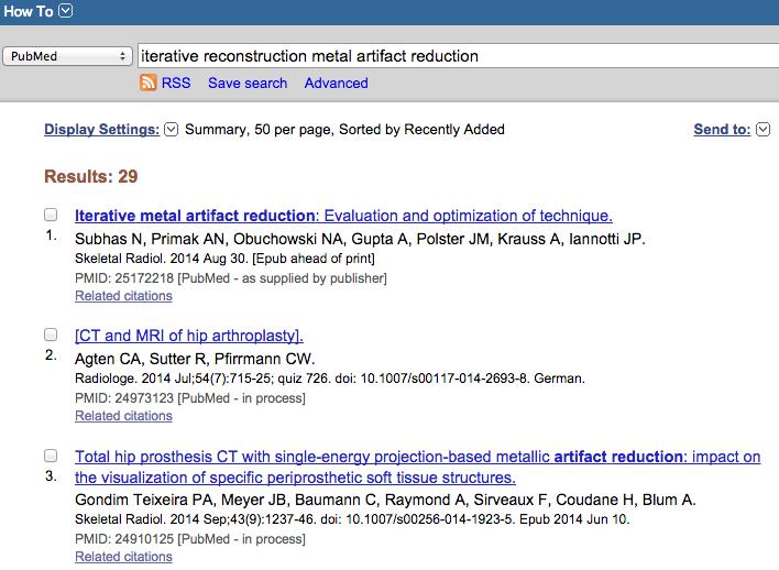
7 Hardware Composition Metal-induced Artifacts: Titanium < Cobalt-Chrome < Stainless-Steel related to Mass Attenuation Coefficient Titanium Screw 4.5 mm Stainless Steel Screw 4.5 mm Lee et al. Overcoming Artifacts Radiographics 2007


8 Patient Positioning X-ray beam should traverse smallest possible cross-sectional area of implant R S S S v o N kvp 150 mas 3 mm 100 kvp 150 mas 3 mm


9 Effect of Tube Voltage & Current 80 kvp 15 mas 140 kvp 350 mas Radiation Exposure: 185 x higher Hip Arthroplasty work-up at Balgrist: 140 kvp, 350 mas

10 Section Thickness 0.7 mm 2.0 mm 4.0 mm partial volume artifacts can best be avoided by acquiring thin sections thicker sections during image reconstruction reduces image noise and decreases metal-related artifacts Lee et al. Overcoming Artifacts Radiographics 2007


11 Critical Role: Kernel-Selection Bone Kernel Bone Kernel Soft Tissue Kernel Soft Tissue Kernel standard or smooth reconstruction filter is preferred compared to edge-enhancing algorithms
12 Iterative Reconstruction


13 Iterative Reconstruction FBP FBP idose 3 idose 6 idose 3 idose 6
 Metal Artifact")

14 Summary - Iterative Reconstruction Filtered Back Projection Iterative Frequency Split Normalized (IFS) Metal Artifact Reduction Iterative reconstruction has a high potential to reduce metal artifacts Morsbach et al. Reduction of Metal artifacts from Radiology 2013

15 Specific Metal Artifact Reduction Software (MAR)

16 Specific Metal Artifact Reduction Software (MAR) O-MAR (Orthopedic Metal Artifact Reduction, Philips): - first step is to create a metal only image - all pixels set to zero except for those pixels categorized as metal - repetitive loop where the output correction image is subtracted from the original input image Li H et al. Clinical evaluation of a commercial Med Phys. 2012


17 Specific Metal Artifact Reduction Software (O-MAR) R S S S FBP v o N. 1 O-MAR MAR software is valuable for soft tissue: improvement of anatomical visualization e.g. intrapelvic anatomy and lymphadenopathy Li H et al. Clinical evaluation of a commercial orthopedic metal artifact reduction tool for CT simulations in radiation therapy Med Phys. 2012


18 Metal Artifact Reduction Software Must Be Used with Caution Standard O-MAR


19 Metal Artifact Reduction Software Should Be Used with Caution Standard O-MAR O-MAR does not improve visualization of metal-to-bone interface O-MAR reduces metal artifacts in soft tissue Li H et al. Clinical evaluation of a commercial orthopedic metal artifact reduction tool for CT simulations in radiation therapy Med Phys. 2012


20 Edge Effects I-50 Kernel I-50 Kernel Edge effects: artifacts due to sharp changes in x-ray attenuation

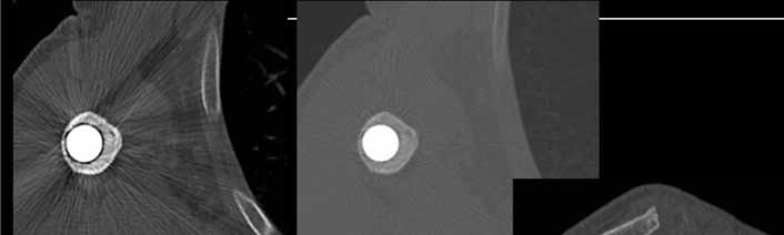
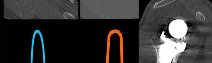
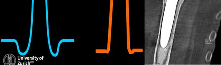
21 Edge Effects Kernel Optimiziation I-50 Kernel Advanced Kernel I-50 Kernel Advanced Kerne


22 Edge Effects Kernel Optimiziation I-50 Kernel Advanced Kernel

23 Dual Energy CT

24 Virtual Monochromatic Spectral Imaging versus Polychromatic Spectral Imaging x-ray tube: polychromatic x-ray beam photons with a range of energies, maximum energy expressed as kilovolt peak monochromatic = monoenergetic => virtual CT image reconstruction of x- ray photons at a single energy level Reduction of Beam-Hardening Effect


25 Dual Energy CT exclusively soft tissue kernel for sequential single-source DECT polychromatic monochromatic 142keV Dual-energy CT allows an efficient reduction of metal artifacts using high-energy monochromatic extrapolation Mangold et al. Single-Source Dual-Energy CT. Invest Radiol 2014

26 Dual Energy Bamberg et al. Metal artifact reduction by dual energy computed Eur Radiol kev 69 kev 88 kev 105 kev optimal kev kev Improve visualization of metal-to-bone interface higher energy


27 Dual Energy 140 kev 80 kev Pessis et al. Virtual Monochromatic RadioGraphics 2013 Improve visualization of metal-to-bone interface higher monochrom. energy Improve visualization of soft tissue lower monochromatic energy levels ( contrast & noise)
28 Summary Dual Energy dual-energy CT techniques can reduce metal artifact due to beamhardening reduction radiation exposure similar to standard polychromatic protocol financial investment and maintenance cost DECT scanners are not widely available, restricted application

29 Conclusion Protocol should be tailored Consider first basic principles to reduce Metal Artifacts Commercially available specific MAR algorithm are appropriate for soft tissue, not metal-to-bone interface Dual-energy CT can reduce metal related artifacts
30 Thank You R S S S N. 1 v o
Carestream s 2 nd Generation Metal Artifact Reduction Software (CMAR 2)
") Carestream s 2 nd Generation Metal Artifact Reduction Software (CMAR 2) Author: Levon Vogelsang Introduction Cone beam computed tomography (CBCT), or cone beam CT technology, offers considerable promise
Carestream s 2 nd Generation Metal Artifact Reduction Software (CMAR 2) Author: Levon Vogelsang Introduction Cone beam computed tomography (CBCT), or cone beam CT technology, offers considerable promise
8/7/2017. Disclosures. MECT Systems Overview and Quantitative Opportunities. Overview. Computed Tomography (CT) CT Numbers. Polyenergetic Acquisition
 CT Numbers. Polyenergetic Acquisition") Quantitative Multi-Energy Computed Tomography: Imaging and Therapy Advancements Disclosures MECT Systems Overview and Quantitative Opportunities The speaker receives research funding from GE Healthcare
Quantitative Multi-Energy Computed Tomography: Imaging and Therapy Advancements Disclosures MECT Systems Overview and Quantitative Opportunities The speaker receives research funding from GE Healthcare
Optimization of CT Simulation Imaging. Ingrid Reiser Dept. of Radiology The University of Chicago
 Optimization of CT Simulation Imaging Ingrid Reiser Dept. of Radiology The University of Chicago Optimization of CT imaging Goal: Achieve image quality that allows to perform the task at hand (diagnostic
3/27/2012 WHY SPECT / CT? SPECT / CT Basic Principles. Advantages of SPECT. Advantages of CT. Dr John C. Dickson, Principal Physicist UCLH
 3/27/212 Advantages of SPECT SPECT / CT Basic Principles Dr John C. Dickson, Principal Physicist UCLH Institute of Nuclear Medicine, University College London Hospitals and University College London john.dickson@uclh.nhs.uk
[PDR03] RECOMMENDED CT-SCAN PROTOCOLS
![[PDR03] RECOMMENDED CT-SCAN PROTOCOLS](/thumbs/72/66454100.jpg "[PDR03] RECOMMENDED CT-SCAN PROTOCOLS") SURGICAL & PROSTHETIC DESIGN [PDR03] RECOMMENDED CT-SCAN PROTOCOLS WORK-INSTRUCTIONS DOCUMENT (CUSTOMER) RECOMMENDED CT-SCAN PROTOCOLS [PDR03_V1]: LIVE 1 PRESCRIBING SURGEONS Patient-specific implants,
SURGICAL & PROSTHETIC DESIGN [PDR03] RECOMMENDED CT-SCAN PROTOCOLS WORK-INSTRUCTIONS DOCUMENT (CUSTOMER) RECOMMENDED CT-SCAN PROTOCOLS [PDR03_V1]: LIVE 1 PRESCRIBING SURGEONS Patient-specific implants,
ML reconstruction for CT
 ML reconstruction for CT derivation of MLTR rigid motion correction resolution modeling polychromatic ML model dual energy ML model Bruno De Man, Katrien Van Slambrouck, Maarten Depypere, Frederik Maes,
Low-Dose Dual-Energy CT for PET Attenuation Correction with Statistical Sinogram Restoration
 Low-Dose Dual-Energy CT for PET Attenuation Correction with Statistical Sinogram Restoration Joonki Noh, Jeffrey A. Fessler EECS Department, The University of Michigan Paul E. Kinahan Radiology Department,
Contrast Enhancement with Dual Energy CT for the Assessment of Atherosclerosis
 Contrast Enhancement with Dual Energy CT for the Assessment of Atherosclerosis Stefan C. Saur 1, Hatem Alkadhi 2, Luca Regazzoni 1, Simon Eugster 1, Gábor Székely 1, Philippe Cattin 1,3 1 Computer Vision
Evaluation of Spectrum Mismatching using Spectrum Binning Approach for Statistical Polychromatic Reconstruction in CT
 Evaluation of Spectrum Mismatching using Spectrum Binning Approach for Statistical Polychromatic Reconstruction in CT Qiao Yang 1,4, Meng Wu 2, Andreas Maier 1,3,4, Joachim Hornegger 1,3,4, Rebecca Fahrig
Fast iterative beam hardening correction based on frequency splitting in computed tomography
 Fast iterative beam hardening correction based on frequency splitting in computed tomography Qiao Yang a,b, Matthias Elter b, Ingo Schasiepen b, Nicole Maass b and Joachim Hornegger a,c a Pattern Recognition
Optimization of scanner parameters for dual energy micro-ct
 Optimization of scanner parameters for dual energy micro-ct E. PAUWELS* 1, J. DHAENE 1, A. DE MUYNCK 1 E., M. DIERICK 1, L. VAN HOOREBEKE 1 1 UGCT Dept. Physics and Astronomy, Ghent University, Proeftuinstraat
Ch. 4 Physical Principles of CT
 Ch. 4 Physical Principles of CT CLRS 408: Intro to CT Department of Radiation Sciences Review: Why CT? Solution for radiography/tomography limitations Superimposition of structures Distinguishing between
Empirical cupping correction: A first-order raw data precorrection for cone-beam computed tomography
 Empirical cupping correction: A first-order raw data precorrection for cone-beam computed tomography Marc Kachelrieß, a Katia Sourbelle, and Willi A. Kalender Institute of Medical Physics, University of
Artifacts in Spiral X-ray CT Scanners: Problems and Solutions
 Artifacts in Spiral X-ray CT Scanners: Problems and Solutions Mehran Yazdi, and Luc Beaulieu International Science Index, Electrical and Computer Engineering waset.org/publication/12966 Abstract Artifact
DUAL energy CT (DECT) is a modality where one and. Empirical Dual Energy Calibration (EDEC) for Cone-Beam Computed Tomography
 is a modality where one and. Empirical Dual Energy Calibration (EDEC) for Cone-Beam Computed Tomography") Empirical Dual Energy Calibration (EDEC) for Cone-Beam Computed Tomography Marc Kachelrieß, Member, IEEE, Timo Berkus, Philip Stenner, Willi A. Kalender Abstract Material selective imaging using dual energy
Empirical Dual Energy Calibration (EDEC) for Cone-Beam Computed Tomography Marc Kachelrieß, Member, IEEE, Timo Berkus, Philip Stenner, Willi A. Kalender Abstract Material selective imaging using dual energy
Superiorized polyenergetic reconstruction algorithm for reduction of metal artifacts in CT images
 1 Superiorized polyenergetic reconstruction algorithm for reduction of metal artifacts in CT images T. Humphries 1 and A. Gibali 2 Abstract Artifacts caused by metal objects such as dental fillings, hip
CT Reconstruction Using Spectral and Morphological Prior Knowledge: Application to Imaging the Prosthetic Knee
 CT Reconstruction Using Spectral and Morphological Prior Knowledge: Application to Imaging the Prosthetic Knee Wojciech Zbijewski, J. Webster Stayman, Abdullah Muhit, John Yorkston, John A. Carrino and
Metal Streak Artifacts in X-ray Computed Tomography: A Simulation Study
 Metal Streak Artifacts in X-ray Computed Tomography: A Simulation Study B. De Man, J. Nuyts, P. Dupont, G. Marchal and P. Suetens Medical Image Computing, ESAT-PSI, K.U.Leuven, B-3000 Leuven, Belgium Department
Frequency split metal artifact reduction (FSMAR) in computed tomography
 in computed tomography") The Johns Hopkins University Advanced Computer Integrated Surgery Group 4 Metal Artifact Removal in C-arm Cone-Beam CT Paper Seminar Critical Review of Frequency split metal artifact reduction (FSMAR)
The Johns Hopkins University Advanced Computer Integrated Surgery Group 4 Metal Artifact Removal in C-arm Cone-Beam CT Paper Seminar Critical Review of Frequency split metal artifact reduction (FSMAR)
Digital Image Processing
 Digital Image Processing SPECIAL TOPICS CT IMAGES Hamid R. Rabiee Fall 2015 What is an image? 2 Are images only about visual concepts? We ve already seen that there are other kinds of image. In this lecture
DUAL energy X-ray radiography [1] can be used to separate
![DUAL energy X-ray radiography [1] can be used to separate](/thumbs/91/106229592.jpg "DUAL energy X-ray radiography [1] can be used to separate") IEEE TRANSACTIONS ON NUCLEAR SCIENCE, VOL. 53, NO. 1, FEBRUARY 2006 133 A Scatter Correction Using Thickness Iteration in Dual-Energy Radiography S. K. Ahn, G. Cho, and H. Jeon Abstract In dual-energy
IEEE TRANSACTIONS ON NUCLEAR SCIENCE, VOL. 53, NO. 1, FEBRUARY 2006 133 A Scatter Correction Using Thickness Iteration in Dual-Energy Radiography S. K. Ahn, G. Cho, and H. Jeon Abstract In dual-energy
RADIOLOGY AND DIAGNOSTIC IMAGING
 Day 2 part 2 RADIOLOGY AND DIAGNOSTIC IMAGING Dr hab. Zbigniew Serafin, MD, PhD serafin@cm.umk.pl 2 3 4 5 CT technique CT technique 6 CT system Kanal K: RSNA/AAPM web module: CT Systems & CT Image Quality
Reconstruction of CT Images from Sparse-View Polyenergetic Data Using Total Variation Minimization
 1 Reconstruction of CT Images from Sparse-View Polyenergetic Data Using Total Variation Minimization T. Humphries and A. Faridani Abstract Recent work in CT image reconstruction has seen increasing interest
Digital Scatter Removal in Mammography to enable Patient Dose Reduction
 Digital Scatter Removal in Mammography to enable Patient Dose Reduction Mary Cocker Radiation Physics and Protection Oxford University Hospitals NHS Trust Chris Tromans, Mike Brady University of Oxford
FAST KVP-SWITCHING DUAL ENERGY CT FOR PET ATTENUATION CORRECTION
 2009 IEEE Nuclear Science Symposium Conference Record M03-7 FAST KVP-SWITCHING DUAL ENERGY CT FOR PET ATTENUATION CORRECTION Wonseok Huh, Jeffrey A. Fessler, Adam M. Alessio, and Paul E. Kinahan Department
Introduction to Biomedical Imaging
 Alejandro Frangi, PhD Computational Imaging Lab Department of Information & Communication Technology Pompeu Fabra University www.cilab.upf.edu X-ray Projection Imaging Computed Tomography Digital X-ray
Tomographic Reconstruction
 Tomographic Reconstruction 3D Image Processing Torsten Möller Reading Gonzales + Woods, Chapter 5.11 2 Overview Physics History Reconstruction basic idea Radon transform Fourier-Slice theorem (Parallel-beam)
Simplified statistical image reconstruction algorithm for polyenergetic X-ray CT. y i Poisson I i (E)e R } where, b i
e R } where, b i") implified statistical image reconstruction algorithm for polyenergetic X-ray C omesh rivastava, tudent member, IEEE, and Jeffrey A. Fessler, enior member, IEEE Abstract In X-ray computed tomography (C),
implified statistical image reconstruction algorithm for polyenergetic X-ray C omesh rivastava, tudent member, IEEE, and Jeffrey A. Fessler, enior member, IEEE Abstract In X-ray computed tomography (C),
DUE to beam polychromacity in CT and the energy dependence
 1 Empirical Water Precorrection for Cone-Beam Computed Tomography Katia Sourbelle, Marc Kachelrieß, Member, IEEE, and Willi A. Kalender Abstract We propose an algorithm to correct for the cupping artifact
Financial disclosure. Onboard imaging modality for IGRT
 Tetrahedron Beam Computed Tomography Based On Multi-Pixel X- Ray Source and Its Application in Image Guided Radiotherapy Tiezhi Zhang, Ph.D. Advanced X-ray imaging Lab Financial disclosure Patent royalty
Effects of the difference in tube voltage of the CT scanner on. dose calculation
 Effects of the difference in tube voltage of the CT scanner on dose calculation Dong Joo Rhee, Sung-woo Kim, Dong Hyeok Jeong Medical and Radiological Physics Laboratory, Dongnam Institute of Radiological
Computed Tomography. Principles, Design, Artifacts, and Recent Advances. Jiang Hsieh THIRD EDITION. SPIE PRESS Bellingham, Washington USA
 Computed Tomography Principles, Design, Artifacts, and Recent Advances THIRD EDITION Jiang Hsieh SPIE PRESS Bellingham, Washington USA Table of Contents Preface Nomenclature and Abbreviations xi xv 1 Introduction
Radiology. Marta Anguiano Millán. Departamento de Física Atómica, Molecular y Nuclear Facultad de Ciencias. Universidad de Granada
 Departamento de Física Atómica, Molecular y Nuclear Facultad de Ciencias. Universidad de Granada Overview Introduction Overview Introduction Tecniques of imaging in Overview Introduction Tecniques of imaging
Quantitative Attenuation Correction for PET/CT Using Iterative Reconstruction of Low-Dose Dual-Energy CT
 Quantitative Attenuation Correction for PET/CT Using Iterative Reconstruction of Low-Dose Dual-Energy CT Paul E. Kinahan, Senior Member, IEEE, Jeffrey A. Fessler, Senior Member, IEEE, Adam M. Alessio,
Scatter Correction Methods in Dimensional CT
 Scatter Correction Methods in Dimensional CT Matthias Baer 1,2, Michael Hammer 3, Michael Knaup 1, Ingomar Schmidt 3, Ralf Christoph 3, Marc Kachelrieß 2 1 Institute of Medical Physics, Friedrich-Alexander-University
A Comparative Study of Two Methods for the Correction of Beam Hardening Artifacts in X-ray Computed Tomography
 A Comparative Study of Two Methods for the Correction of Beam Hardening Artifacts in X-ray Computed Tomography by Maryam Khalid Alarfaj submitted to Oregon State University in partial fulfillment of the
CT issues in PET / CT scanning. ImPACT technology update no. 4
 abc David Platten Introduction ImPACT technology update no. 4 ImPACT October 2004 Since its introduction in the 1970s, X-ray computed tomography (CT) scanning has become a widespread three-dimensional
Enhanced material contrast by dual-energy microct imaging
 Enhanced material contrast by dual-energy microct imaging Method note Page 1 of 12 2 Method note: Dual-energy microct analysis 1. Introduction 1.1. The basis for dual energy imaging Micro-computed tomography
DICOM. Supplement 188 Multi-Energy CT Imaging. DICOM Working Group 21 Computed Tomography
 DICOM Supplement 188 Multi-Energy CT Imaging DICOM Working Group 21 Computed Tomography Rationale Short introduction of Multi Energy (ME) s Overview: Imaging techniques, including scanning, reconstruction,
Simulation of Mammograms & Tomosynthesis imaging with Cone Beam Breast CT images
 Simulation of Mammograms & Tomosynthesis imaging with Cone Beam Breast CT images Tao Han, Chris C. Shaw, Lingyun Chen, Chao-jen Lai, Xinming Liu, Tianpeng Wang Digital Imaging Research Laboratory (DIRL),
Spiral CT. Protocol Optimization & Quality Assurance. Ge Wang, Ph.D. Department of Radiology University of Iowa Iowa City, Iowa 52242, USA
 Spiral CT Protocol Optimization & Quality Assurance Ge Wang, Ph.D. Department of Radiology University of Iowa Iowa City, Iowa 52242, USA Spiral CT Protocol Optimization & Quality Assurance Protocol optimization
Background. Outline. Radiographic Tomosynthesis: Image Quality and Artifacts Reduction 1 / GE /
 Radiographic Tomosynthesis: Image Quality and Artifacts Reduction Baojun Li, Ph.D Department of Radiology Boston University Medical Center 2012 AAPM Annual Meeting Background Linear Trajectory Tomosynthesis
Dual-Energy CT Based Monochromatic Imaging
 Dual-Energy CT Review Yu et al. Monochromatic Imaging Based on Dual-Energy CT Dual-Energy CT Review Lifeng Yu 1 Shuai Leng Cynthia H. McCollough Yu L, Leng S, McCollough CH Keywords: CT, dual-energy CT,
As fl exible as your care requires
 As fl exible as your care requires Philips Ingenuity Flex 32 CT Built on Ingenuity The Philips Ingenuity Flex 32 helps you provide excellent care with outstanding flexibility, now and in the future. Built
Biomedical Imaging. Computed Tomography. Patrícia Figueiredo IST
 Biomedical Imaging Computed Tomography Patrícia Figueiredo IST 2013-2014 Overview Basic principles X ray attenuation projection Slice selection and line projections Projection reconstruction Instrumentation
Reduction of metal streak artifacts in x-ray computed tomography using a transmission maximum a posteriori algorithm
 Reduction of metal streak artifacts in x-ray computed tomography using a transmission maximum a posteriori algorithm B. De Man 1, Student Member, IEEE, J. Nuyts 2, Member, IEEE,. Dupont 2, G. Marchal 3,
CT NOISE POWER SPECTRUM FOR FILTERED BACKPROJECTION AND ITERATIVE RECONSTRUCTION
 CT NOISE POWER SPECTRUM FOR FILTERED BACKPROJECTION AND ITERATIVE RECONSTRUCTION Frank Dong, PhD, DABR Diagnostic Physicist, Imaging Institute Cleveland Clinic Foundation and Associate Professor of Radiology
8/2/2016. Measures the degradation/distortion of the acquired image (relative to an ideal image) using a quantitative figure-of-merit
 using a quantitative figure-of-merit") Ke Li Assistant Professor Department of Medical Physics and Department of Radiology School of Medicine and Public Health, University of Wisconsin-Madison This work is partially supported by an NIH Grant
Ke Li Assistant Professor Department of Medical Physics and Department of Radiology School of Medicine and Public Health, University of Wisconsin-Madison This work is partially supported by an NIH Grant
Artifact Mitigation in High Energy CT via Monte Carlo Simulation
 PIERS ONLINE, VOL. 7, NO. 8, 11 791 Artifact Mitigation in High Energy CT via Monte Carlo Simulation Xuemin Jin and Robert Y. Levine Spectral Sciences, Inc., USA Abstract The high energy (< 15 MeV) incident
CLASS HOURS: 4 CREDIT HOURS: 4 LABORATORY HOURS: 0
 Revised 10/10 COURSE SYLLABUS TM 220 COMPUTED TOMOGRAPHY PHYSICS CLASS HOURS: 4 CREDIT HOURS: 4 LABORATORY HOURS: 0 CATALOG COURSE DESCRIPTION: This course is one of a three course set in whole body Computed
Metal artifacts reduction in x-ray CT based on segmentation and forward-projection
 Bio-Medical Materials and Engineering 24 (2014) 3287 3293 DOI 10.3233/BME-141151 IOS Press 3287 Metal artifacts reduction in x-ray CT based on segmentation and forward-projection Shoukat Nawaz, Jian Fu
Streak artifacts arising from metal implants such as dental fillings, surgical clips, coils, wires, and orthopedic hardware may obscure important diag
 Note: This copy is for your personal, non-commercial use only. To order presentation-ready copies for distribution to your colleagues or clients, contact us at www.rsna.org/rsnarights. ORIGINAL RESEARCH
AIDR 3D Iterative Reconstruction:
 Iterative Reconstruction: Integrated, Automated and Adaptive Dose Reduction Erin Angel, PhD Manager, Clinical Sciences, CT Canon Medical Systems USA Iterative Reconstruction 1 Since the introduction of
Development of a multi-axis X-ray CT for highly accurate inspection of electronic devices
 Development of a multi-axis X-ray CT for highly accurate inspection of electronic devices Toru Kano 1, Michihiko Koseki 2 More info about this article: http://www.ndt.net/?id=20843 1 Tokyo University of
Incorporation of Prior Knowledge for Region of Change Imaging from Sparse Scan Data in Image-Guided Surgery
 Incorporation of Prior Knowledge for Region of Change Imaging from Sparse Scan Data in Image-Guided Surgery J. Lee a, J. W. Stayman b, Y. Otake c, S. Schafer b, W. Zbijewski b, A. J. Khanna b,d, J. L.
A Curvelet based Sinogram Correction Method for Metal Artifact Reduction
 A based Sinogram Correction Method for Metal Artifact Reduction Kiwan Jeon 1 and Hyoung Suk Park 1 More info about this article: http://www.ndt.net/?id=3715 1 National Institute for Mathematical Sciences,
Deep Scatter Estimation (DSE): Accurate Real-Time Scatter Estimation for X-Ray CT using a Deep Convolutional Neural Network
: Accurate Real-Time Scatter Estimation for X-Ray CT using a Deep Convolutional Neural Network") Deep Scatter Estimation (DSE): Accurate Real-Time Scatter Estimation for X-Ray CT using a Deep Convolutional Neural Network Joscha Maier 1,2, Stefan Sawall 1,2, and Marc Kachelrieß 1,2 1 German Cancer
Deep Scatter Estimation (DSE): Accurate Real-Time Scatter Estimation for X-Ray CT using a Deep Convolutional Neural Network Joscha Maier 1,2, Stefan Sawall 1,2, and Marc Kachelrieß 1,2 1 German Cancer
Image Acquisition Systems
 Image Acquisition Systems Goals and Terminology Conventional Radiography Axial Tomography Computer Axial Tomography (CAT) Magnetic Resonance Imaging (MRI) PET, SPECT Ultrasound Microscopy Imaging ITCS
Some reference material
 Some reference material Physics reference book on medical imaging: A good one is The Essential Physics of Medical Imaging, 3 rd Ed. by Bushberg et al. ($170! new). However, there are several similar books
A Radiometry Tolerant Method for Direct 3D/2D Registration of Computed Tomography Data to X-ray Images
 A Radiometry Tolerant Method for Direct 3D/2D Registration of Computed Tomography Data to X-ray Images Transfer Function Independent Registration Boris Peter Selby 1, Georgios Sakas 2, Stefan Walter 1,
Simulation of Beam Hardening in Industrial CT with x-ray and Monoenergetic Source by Monte Carlo Code
 2012, TextRoad Publication ISSN 2090-4304 Journal of Basic and Applied Scientific Research www.textroad.com Simulation of Beam Hardening in Industrial CT with x-ray and Monoenergetic Source by Monte Carlo
HIP IMPLANT METAL ARTIFACT REDUCTION FOR PELVIC CT SCANS
 ARTUR SOSSIN HIP IMPLANT METAL ARTIFACT REDUCTION FOR PELVIC CT SCANS Master s Thesis Examiners: Professor Hannu Eskola, Jarkko Ojala, Lic. Sci. (Tech.) Examiners and topic approved by the Faculty Council
Advanced Multi Material Decomposition of Dual Energy in Computed Tomography Image
 Advanced Multi Material Decomposition of Dual Energy in Computed Tomography Image A.Prema 1, M.Priyadharshini 2, S.Renuga 3, K.Radha 4 UG Students, Department of CSE, Muthayammal Engineering College, Rasipuram,
Empirical dual energy calibration EDEC for cone-beam computed tomography
 Empirical dual energy calibration EDEC for cone-beam computed tomography Philip Stenner, Timo Berkus, and Marc Kachelriess Institute of Medical Physics, University of Erlangen-Nürnberg, Henkestrasse 91,
Projection and Reconstruction-Based Noise Filtering Methods in Cone Beam CT
 Projection and Reconstruction-Based Noise Filtering Methods in Cone Beam CT Benedikt Lorch 1, Martin Berger 1,2, Joachim Hornegger 1,2, Andreas Maier 1,2 1 Pattern Recognition Lab, FAU Erlangen-Nürnberg
Scatter Correction for Dual source Cone beam CT Using the Pre patient Grid. Yingxuan Chen. Graduate Program in Medical Physics Duke University
 Scatter Correction for Dual source Cone beam CT Using the Pre patient Grid by Yingxuan Chen Graduate Program in Medical Physics Duke University Date: Approved: Lei Ren, Supervisor Fang Fang Yin, Chair
Reduction of Metal Artifacts in Computed Tomographies for the Planning and Simulation of Radiation Therapy
 Reduction of Metal Artifacts in Computed Tomographies for the Planning and Simulation of Radiation Therapy T. Rohlfing a, D. Zerfowski b, J. Beier a, P. Wust a, N. Hosten a, R. Felix a a Department of
MEDICAL EQUIPMENT: COMPUTED TOMOGRAPHY. Prof. Yasser Mostafa Kadah
 MEDICAL EQUIPMENT: COMPUTED TOMOGRAPHY Prof. Yasser Mostafa Kadah www.k-space.org Recommended Textbook X-Ray Computed Tomography in Biomedical Engineering, by Robert Cierniak, Springer, 211 Computed Tomography
Physical bases of X-ray diagnostics
 Physical bases of X-ray diagnostics Dr. István Voszka Possibilities of X-ray production (X-ray is produced, when charged particles of high velocity are stopped) X-ray tube: Relatively low accelerating
Acknowledgments. High Performance Cone-Beam CT of Acute Traumatic Brain Injury
 A. Sisniega et al. (presented at RSNA 214) High Performance Cone-Beam CT of Acute Traumatic Brain Injury A. Sisniega 1 W. Zbijewski 1, H. Dang 1, J. Xu 1 J. W. Stayman 1, J. Yorkston 2, N. Aygun 3 V. Koliatsos
Computer-Tomography II: Image reconstruction and applications
 Computer-Tomography II: Image reconstruction and applications Prof. Dr. U. Oelfke DKFZ Heidelberg Department of Medical Physics (E040) Im Neuenheimer Feld 280 69120 Heidelberg, Germany u.oelfke@dkfz.de
Fundamentals of CT imaging
 SECTION 1 Fundamentals of CT imaging I History In the early 1970s Sir Godfrey Hounsfield s research produced the first clinically useful CT scans. Original scanners took approximately 6 minutes to perform
CT imaging using energy-sensitive photon-counting detectors. Disclosure. Vision 20/20 paper
 CT imaging using energy-sensitive photon-counting detectors Katsuyuki Ken Taguchi, Ph.D. ktaguchi@jhmi.edu Division of Medical Imaging Physics Department of Radiology Johns Hopkins University No financial
A closer look at CT scanning
 Vet Times The website for the veterinary profession https://www.vettimes.co.uk A closer look at CT scanning Author : Charissa Lee, Natalie Webster Categories : General, Vets Date : April 3, 2017 A basic
Iterative Reconstructions: The Impact on Dose and Image Quality
 Shane Foley: IPR, Chicago 2016 Iterative Reconstructions: The Impact on Dose and Image Quality Shane Foley, PhD Radiography and Diagnostic Imaging School of Medicine University College Dublin. Radagrafaíocht
The Near Future in Cardiac CT Image Reconstruction
 SCCT 2010 The Near Future in Cardiac CT Image Reconstruction Marc Kachelrieß Institute of Medical Physics (IMP) Friedrich-Alexander Alexander-University Erlangen-Nürnberg rnberg www.imp.uni-erlangen.de
Spiral ASSR Std p = 1.0. Spiral EPBP Std. 256 slices (0/300) Kachelrieß et al., Med. Phys. 31(6): , 2004
 Kachelrieß et al., Med. Phys. 31(6): , 2004") Spiral ASSR Std p = 1.0 Spiral EPBP Std p = 1.0 Kachelrieß et al., Med. Phys. 31(6): 1623-1641, 2004 256 slices (0/300) Advantages of Cone-Beam Spiral CT Image quality nearly independent of pitch Increase
Spiral ASSR Std p = 1.0 Spiral EPBP Std p = 1.0 Kachelrieß et al., Med. Phys. 31(6): 1623-1641, 2004 256 slices (0/300) Advantages of Cone-Beam Spiral CT Image quality nearly independent of pitch Increase
An Iterative Approach to the Beam Hardening Correction in Cone Beam CT (Proceedings)
") Marquette University e-publications@marquette Biomedical Engineering Faculty Research and Publications Engineering, College of 1-1-1999 An Iterative Approach to the Beam Hardening Correction in Cone Beam
Marquette University e-publications@marquette Biomedical Engineering Faculty Research and Publications Engineering, College of 1-1-1999 An Iterative Approach to the Beam Hardening Correction in Cone Beam
MEDICAL IMAGING 2nd Part Computed Tomography
 MEDICAL IMAGING 2nd Part Computed Tomography Introduction 2 In the last 30 years X-ray Computed Tomography development produced a great change in the role of diagnostic imaging in medicine. In convetional
CALIBRATIONS FOR ANALYZING INDUSTRIAL SAMPLES ON MEDICAL CT SCANNERS
 CALIBRATIONS FOR ANALYZING INDUSTRIAL SAMPLES ON MEDICAL CT SCANNERS INTRODUCTION Patricia K. Hunt, Philip Engler, and William D. Friedman BP American Research and Development 4440 Warrensville Center
A new calibration-free beam hardening reduction method for industrial CT
 A new calibration-free beam hardening reduction method for industrial CT ECC 2 for industrial CT Tobias Würfl 1, Nicole Maaß 2, Frank Dennerlein 2, Andreas K. Maier 1 1 Pattern Recognition Lab, FAU Erlangen-Nürnberg;
Application of 450 kv Computed Tomography to Engine Blocks with Steel Liners
 Application of 450 kv Computed Tomography to Engine Blocks with Steel Liners Charles R. Smith, Kevin Holt BIR, Inc. Uwe Bischoff, Bernd Georgi, Ferdinand Hansen, Frank Jeltsch Volkswagen Commercial Vehicles
Corso di laurea in Fisica A.A Fisica Medica 4 TC
 Corso di laurea in Fisica A.A. 2007-2008 Fisica Medica 4 TC Computed Tomography Principles 1. Projection measurement 2. Scanner systems 3. Scanning modes Basic Tomographic Principle The internal structure
Shadow casting. What is the problem? Cone Beam Computed Tomography THE OBJECTIVES OF DIAGNOSTIC IMAGING IDEAL DIAGNOSTIC IMAGING STUDY LIMITATIONS
 Cone Beam Computed Tomography THE OBJECTIVES OF DIAGNOSTIC IMAGING Reveal pathology Reveal the anatomic truth Steven R. Singer, DDS srs2@columbia.edu IDEAL DIAGNOSTIC IMAGING STUDY Provides desired diagnostic
A New Approach for the Enhancement. of Dual-energy Computed Tomography Images. Kyung Kook Park
 A New Approach for the Enhancement of Dual-energy Computed Tomography Images by Kyung Kook Park A Dissertation Presented in Partial Fulfillment of the Requirements for the Degree Doctor of Philosophy Approved
Photon counting spectroscopic CT with dynamic beam attenuator
 Photon counting spectroscopic CT with dynamic beam attenuator Haluk Atak 1) and Polad M. Shikhaliev a) 1) Department of Nuclear Engineering, Hacettepe University, Ankara, Turkey Abstract Purpose: Photon
Cardiac Dual Energy CT: Technique
 RSNA 2013, VSCA51-01, Chicago, Dec. 5, 2013 Cardiac Radiology Series Cardiac Dual Energy CT: Technique Willi A. Kalender, Ph.D. Institute of Medical Physics University of Erlangen www.imp.uni-erlangen.de
Comparison of Scatter Correction Methods for CBCT. Author(s): Suri, Roland E.; Virshup, Gary; Kaissl, Wolfgang; Zurkirchen, Luis
: Suri, Roland E.; Virshup, Gary; Kaissl, Wolfgang; Zurkirchen, Luis") Research Collection Working Paper Comparison of Scatter Correction Methods for CBCT Author(s): Suri, Roland E.; Virshup, Gary; Kaissl, Wolfgang; Zurkirchen, Luis Publication Date: 2010 Permanent Link:
Research Collection Working Paper Comparison of Scatter Correction Methods for CBCT Author(s): Suri, Roland E.; Virshup, Gary; Kaissl, Wolfgang; Zurkirchen, Luis Publication Date: 2010 Permanent Link:
Impact of X-ray Scatter When Using CT-based Attenuation Correction in PET: A Monte Carlo Investigation
 26 IEEE Nuclear Science Symposium Conference Record M6-349 Impact of X-ray Scatter When Using CT-based Attenuation Correction in PET: A Monte Carlo Investigation Habib Zaidi, Senior Member, IEEE and Mohammad
CBCT Equivalent Source Generation Using HVL and Beam Profile Measurements. Johnny Little PSM - Medical Physics Graduate Student University of Arizona
 CBCT Equivalent Source Generation Using HVL and Beam Profile Measurements. Johnny Little PSM - Medical Physics Graduate Student University of Arizona Introduction CBCT has become a routine procedure for
PROVIDE A UNIFORM DOSE IN THE SMALL ANIMAL.
 Considerations in the Use of the RS 2000 X ray Irradiator for Biological Research (Primarily Small Animal, tissue, and cells) and the fallacy of the High KV spectrum. The performance goal for a small animal
Proton dose calculation algorithms and configuration data
 Proton dose calculation algorithms and configuration data Barbara Schaffner PTCOG 46 Educational workshop in Wanjie, 20. May 2007 VARIAN Medical Systems Agenda Broad beam algorithms Concept of pencil beam
BME I5000: Biomedical Imaging
 1 Lucas Parra, CCNY BME I5000: Biomedical Imaging Lecture 4 Computed Tomography Lucas C. Parra, parra@ccny.cuny.edu some slides inspired by lecture notes of Andreas H. Hilscher at Columbia University.
Cone-beam computed tomography with a flat-panel imager: Magnitude and effects of x-ray scatter
 Cone-beam computed tomography with a flat-panel imager: Magnitude and effects of x-ray scatter Jeffrey H. Siewerdsen a) and David A. Jaffray Department of Radiation Oncology, William Beaumont Hospital,
The effect of automatic exposure control with inappropriate scout image on radiation dose: a chest phantom experiment with three different CT machines
 The effect of automatic exposure control with inappropriate scout image on radiation dose: a chest phantom experiment with three different CT machines Poster No.: C-0891 Congress: ECR 2015 Type: Authors:
Joint ICTP-TWAS Workshop on Portable X-ray Analytical Instruments for Cultural Heritage. 29 April - 3 May, 2013
 2455-5 Joint ICTP-TWAS Workshop on Portable X-ray Analytical Instruments for Cultural Heritage 29 April - 3 May, 2013 Lecture NoteBasic principles of X-ray Computed Tomography Diego Dreossi Elettra, Trieste
arxiv: v1 [physics.med-ph] 18 Apr 2016
![arxiv: v1 [physics.med-ph] 18 Apr 2016](/thumbs/89/97491883.jpg "arxiv: v1 [physics.med-ph] 18 Apr 2016") Segmentation-free x-ray energy spectrum estimation for computed tomography arxiv:164.4986v1 [physics.med-ph] 18 Apr 216 Wei Zhao a, Qiude Zhang a, Tianye Niu b a Department of Biomedical Engineering, Huazhong
Segmentation-free x-ray energy spectrum estimation for computed tomography arxiv:164.4986v1 [physics.med-ph] 18 Apr 216 Wei Zhao a, Qiude Zhang a, Tianye Niu b a Department of Biomedical Engineering, Huazhong
Refinement of Imaging Processing of Scatter Correction and Beam Hardening Tools for Industrial Radiography and Cone Beam CT
 Digital Industrial Radiology and Computed Tomography (DIR 2015) 22-25 June 2015, Belgium, Ghent - www.ndt.net/app.dir2015 More Info at Open Access Database www.ndt.net/?id=18073 Refinement of Imaging Processing
DUAL-ENERGY CT IN PROTON THERAPY
 10/31/17 DUAL-ENERGY CT IN PROTON THERAPY Isabel Almeida, MAASTRO Clinic 7th NCS Lustrum Symposium 1 10/31/17 http://zonptc.bouwwebcam.nl https://www.youtube.com/watch?v=3vvvf5bqn7g Range uncertainties
CT vs. VolumeScope: image quality and dose comparison
 CT vs. VolumeScope: image quality and dose comparison V.N. Vasiliev *a, A.F. Gamaliy **b, M.Yu. Zaytsev b, K.V. Zaytseva ***b a Russian Sci. Center of Roentgenology & Radiology, 86, Profsoyuznaya, Moscow,