Symbia T Series System Specifications Answers for life.
|
|
|
- James Daniel
- 6 years ago
- Views:
Transcription

1 System Specifications Answers for life.
2 2

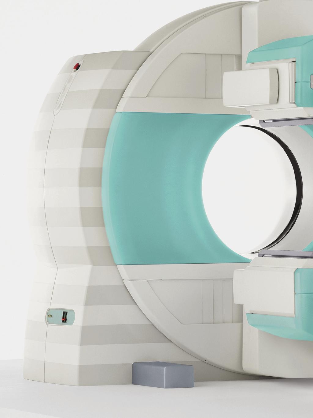
3 Diagnostic Excellence Today s healthcare institutions have increasingly diverse patient needs, greater budget limitations and the need for higher system utilization. As a result, greater efficiency and more accurate diagnostic solutions are becoming increasingly important in molecular imaging, where the dual mandate to reduce patient radiation exposure and increase throughput has a direct effect on patient wellbeing, clinical workflow and the bottom-line. This is especially true for cardiac studies the number one nuclear medicine procedure performed worldwide. Unclear images, insufficient lesion localization and low specificity are issues common to SPECT systems that require confirmation from other imaging modalities. In addition, remote access to patient data has become a key requirement in physician mobility. These challenges require a multi-purpose system that provides solutions on several levels. Symbia T Series is the first SPECT system with a diagnostic CT for precise anatomical localization and accurate attenuation correction, giving physicians the image quality and specificity they need for diagnostic excellence. The system delivers a combination of applications that reduce radiation exposure and increase throughput. For example, Siemens offers the world s only ultra-fast cardiac imaging solution on a general purpose system: IQ SPECT. The introduction of automated processes, such as Automated Quality Control and Automated Collimator Changer, saves time, reduces potential equipment damage and streamlines workflow. To accommodate diverse patient needs, the fully-integrated Symbia T Series enables SPECT, SPECT/CT and CT-only procedures. On-demand access to patient data with Symbia.net offers physicians image viewing and processing wherever they are. provides high-quality imaging for excellent patient care, and is a solution to the pressing clinical and business needs for healthcare institutions worldwide. 3
4 Features High-Definition SPECT CT Minimum Dose, Maximum Speed Key Highlight Features Reconstruction Frame-of-Reference Spect Reconstruction Matrix Size Advanced Reconstruction Detectors Rotational Uniformity SPECT frame-of-reference 128x128, 64x64 Flash3D iterative Yes Detectors Rotational Accuracy 0.1 Detector Caudal Tilt +16 /-16 CT Focal Spot Size Table Flex Reconstruction Workstation CT Continuous Scan Length CT Dose Modulation Flexible CT Voltage Settings CTDI Dose Values - Abdomen AC CTDI Dose Values - Cardiac AC CTDI Dose Values - Parathyroid AC LEHR Collimator cm SMARTZOOM Collimator cm (Recommended) Symbia T2: 0.8x0.7 mm/8, T6: 0.8x0.5 mm/7 and 0.8x0.7 mm/7, T16: 0.8x0.5 mm/7 and 0.8x0.7 mm/7 Rear bed support prevents flex 32-bit architecture Symbia T2: 166 cm, T6: 168 cm, T16: 186 cm Yes, 4D and fully automatic Yes, 80 kv, 110 kv, 130 kv 1.20 kv 1.56 kv or 1.00 kv or kv 1.80 kv 202 cpm/μci* 810 cpm/μci at 28 cm** (unique to Siemens) Full Automation Patient Detector Contouring Automated body contour Average Autocontour Distance 1.1 cm (0.45 in) Quality Control Yes, fully automated with 2 shielded sources embedded in the patient bed Collimator Exchange Fully automatic with integrated set of collimators Seamless Integration Electrocardiogram Support Fully integrated ECG Gantry Tunnel Length 89 cm System Footprint 373x630 cm (12 ft 3 inx20 ft 7 in) Entertainment System Yes, e.media Upgradability Yes, Symbia T2 to T6, or T16 and elevate option to Symbia Intevo xspect systems * Values measured in accordance with NEMA Standards Publication NU using 3/8" crystal. ** Values measured in accordance with NEMA Standards Publication NU using 3/8" crystal and a 5 cm diameter phantom. 4
5 SPECT Specifications Gantry Dimensions Height Width Depth Axis of Rotation (from Floor) Weight* Min./Max. Patient Opening (HE Coll) Min./Max. Patient Opening (LEHR Coll) Patient Positioning Monitor Tunnel Opening Tunnel Length Distance between SPECT and CT Field-of-View (FOV) SPECT Motions Average Autocontour Distance Max. Radial and Lateral Max. Lateral Position Left/Right Max. Clockwise (CW)/Counter-Clockwise (CCW) Rotation Detector 1 Ring Rotation Range 540 Rotational Uniformity Rotational Accuracy 0.1 Rotational Speed Center of Rotation 225 cm (7 ft 4.7 in) 231 cm (7 ft 7 in) 203 cm (6 ft 8 in) 104 cm (3 ft 5 in) 3,506 kg (7,714 lbs) 12 cm (4.7 in)/65.4 cm (25.7 in) 19.2 cm (7.6 in)/72.6 cm (28.6 in) 15 flat panel color LCD display 70 cm aperture (27.6 in) 89 cm ( 35 in) 136 cm (53.3 in) 1.1 cm (0.45 in) 72 cm/min (28.3 in/min) 37.5 cm (14.7 in)/10 cm (4 in) 405 /-135 Yes RPM Max. Caudal Tilt +16 / pixel (64x64 matrix) * Gantry weight: NM gantry 2,374 kg (5,224 lbs) + CT gantry 1,132 kg (2,490 lbs). 5
6 SPECT Specifications Patient Bed Width Length Weight without Integrated Collimator Changer (ICC)/ Automated Collimator Changer (ACC) Height Vertical Motion Range Vertical Speed Pallet Material Pallet Thickness Pallet Width Attenuation at 140 kev <10% Max. Patient Weight Max. Deflection of Patient Pallet Max. Scan Length in Whole-Body Mode Horizontal Motion Accuracy Min./Max. Horizontal Speed Optional Pallets 81.9 cm (32.2 in) cm (8 ft 1.6 in) 950 kg (2,096 lbs) cm (3 ft 8 in) cm (19-44 in) 72 cm/min (28 in/min), maximum Carbon fiber 15 mm (.6 in) 40.0 cm (15.8 in) 227 kg (500 lbs) <2.0 mm (<0.08 in) for 92 kg (200 lbs) patient 200 cm (6 ft 6.7 in) 0.7 mm (0.03 in) cm/min ( in/min) Pediatric Material Carbon fiber composite Thickness 0.6 cm (0.25 in) Width 25.4 cm (10 in) Length 145 cm (57 in) Weight 7.3 kg (16 lbs) Attenuation at 140 kev <10% Max. Patient Weight 27 kg (60 lbs) Scintimammography Material Carbon fiber composite Thickness 1.6 cm (0.63 in) Width 35.6 cm (14 in) Length cm (75 in) Weight 7.7 kg (17 lbs) Attenuation at 140 kev <10% Max. Patient Weight 135 kg (300 lbs) 6
7 SPECT Specifications Optional Pallets Radiotherapy Planning Material Carbon fiber composite Thickness 1.5 cm (0.6 in) Width 53 cm (20.9 in) Length cm (80.1 in) Weight 9 kg (20 lbs) Attenuation at 140 kev <10% Max. Patient Weight 227 kg (500 lbs)* Rear Pallet Support Width 26.3 cm (10.3 in) Length cm (3 ft 5.1 in) Weight kg (415.2 lbs) ECG Trigger Integration Internal (inside patient bed) or external Framing Modes Forward or forward/backward by thirds Buffered Beat Window Yes Bad Beat Rejection Yes Criteria for Framing Images Frames/R-R interval Beat Acceptance Window Automatic or manual selection Collimator Exchanger Cart Height cm (3 ft 3.9 in) Width 82.8 cm (2 ft 8.6 in) Depth cm (3 ft 11.4 in) Weight* kg (400 lbs) Detector Dimensions FOV 53.3x38.7 cm (21x15.25 in) Diagonal FOV 65.9 cm (25.9 in) Crystal Size 59.1x44.5 cm (23.25x17.5 in) Diagonal 73.9 cm (29.1 in) Thickness 9.5 mm (3/8 in) or 15.9 mm (5/8 in) * Without collimators. 7
8 SPECT Specifications Photomultiplier Tubes Total Number 59 Diameter cm (3 in) and cm (2.4-2 in) Type Bialkali high-efficiency box-type dynodes Array Hexagonal Detector Shielding Back 9.5 mm (0.375 in) Sides 12.7 mm (0.5 in) Min./Max. in Patient Direction* 27.9/36.4 mm (1.1/1.435 in) Brain Reach** 7.6 cm (3 in) Detector*** 3/8 5/8 Intrinsic Spatial Resolution FWHM in CFOV 3.8 mm 4.5 mm FWHM in UFOV 3.9 mm 4.6 mm FWTM in CFOV 7.5 mm 8.7 mm FWTM in UFOV 7.7 mm 8.9 mm Intrinsic Spatial Linearity Differential in CFOV 0.2 mm 0.2 mm Differential in UFOV 0.2 mm 0.2 mm Absolute in CFOV 0.4 mm 0.5 mm Absolute in UFOV 0.7 mm 1.0 mm Intrinsic Energy Resolution FWHM in CFOV 9.9% 9.9% Intrinsic Flood Field Uniformity (Uncorrected) Differential in CFOV 2.5% 2.5% Differential in UFOV 2.7% 2.7% Integral in CFOV 2.9% 2.9% Integral in UFOV 3.7% 3.7% Multiple Window Spatial Registration 0.6 mm 1.0 mm Intrinsic Count Rate Performance in Air Maximum Count Rate 310 kcps 310 kcps Intrinsic Spatial Resolution at 75 kcps FWHM in UFOV 4.1 mm 4.6 mm FWTM in UFOV 7.8 mm 8.9 mm * For any point on the pallet at maximum 183 cm (6 ft) from the detector while the detector is at 25.4 cm (10 in) radial position. ** Distance from the edge of the detector housing to the edge of the FOV. *** Values are determined at the manufacturer s facility using methods described in NEMA Standards Publications NU Performance measurements of Scintillation Cameras. The specialized phantoms and software required to reproduce these measurements are available from Siemens. 8
9 SPECT Specifications Detector* 3/8 5/8 Intrinsic Flood Field Uniformity at 75 kcps (Uncorrected) Differential in CFOV 2.5% 2.5% Differential in UFOV 2.7% 2.7% Integral in CFOV 2.9% 2.9% Integral in UFOV 3.7% 3.7% Detector with Collimator* 3/8 5/8 System Spatial Resolution without Scatter (LEHR at 10 cm) FWHM in CFOV 7.5 mm 7.8 mm FWTM in CFOV 13.6 mm 14.9 mm System Spatial Resolution with Scatter (LEHR at 10 cm) FWHM in CFOV 8.3 mm 8.9 mm FWTM in CFOV 18.6 mm 19.5 mm System Planar Sensitivity (LEHR at 10 cm) Absolute 202 cpm/µci 225 cpm/µci System Planar Sensitivity (ME at 10 cm) Absolute 111 In 430 cpm/µci 565 cpm/µci Detector with Collimator Tomographic Specifications* 3/8 5/8 Reconstructed Spatial Resolution without Scatter Filtered Back Projection at 15 cm Radius (LEHR) Central Transaxial 10.2 mm Central Axial 10.8 mm Peripheral Radial 9.8 mm Peripheral Tangential 8.4 mm Peripheral Axial 9.0 mm Reconstructed Spatial Resolution without Scatter Flash 3D Iterative Reconstruction at 15 cm Radius (LEHR) Central Transaxial 4.4 mm Central Axial 4.4 mm Peripheral Radial 4.0 mm Peripheral Tangential 3.9 mm Peripheral Axial 4.2 mm Reconstructed Spatial Resolution with Scatter (LEHR) Filtered Back Projection Center 10.7 mm 11.5 mm Radial 10.9 mm 12.0 mm Tangential 7.9 mm 8.8 mm * Values are determined at the manufacturer s facility using methods described in NEMA Standards Publications NU Performance measurements of Scintillation Cameras. 9
10 SPECT Specifications Detector with Collimator Tomographic Specifications* 3/8 5/8 Reconstructed Spatial Resolution with Scatter (LEHR) Flash 3D Iterative Reconstruction Center 5.8 mm Radial 5.0 mm Tangential 4.1 mm System Volume Sensitivity (LEHR) UFOV ±7% 12,000 (cts/sec)/(mbq/cm 2 ) Detector-Detector Sensitivity Variation (LEHR, 99m Tc) 5.0% Detector with Collimator Whole-Body 3/8 5/8 Scanning Specifications Whole-Body System Spatial Resolution without Scatter at 10 cm/min Scan Speed (LEHR at 10 cm) FWHM Perpendicular 7.5 mm FWHM Parallel 7.9 mm FWTM Perpendicular 14.0 mm FWTM Parallel 14.2 mm 10
11 SPECT Specifications Collimators LEHR LEAP LEUHR LEFB ME HE SMART- ZOOM Low Energy High Resolution Low Energy All Purpose Low Energy Ultra High Resolution Low Energy Fan Beam Medium Energy High Energy IQ SPECT Isotope 99m Tc 99m Tc 99m Tc 99m Tc 67 Ga 131 I 99m Tc Hole Shape Hex Hex Hex Hex Hex Hex Hex Number of Holes (x1000) Hole Length mm mm 35.8 mm 35 mm mm 59.7 mm mm Septal 0.16 mm 0.2 mm 0.13 mm 0.16 mm 1.14 mm 2 mm Thickness Hole Diameter Across the Flats 1.11 mm 1.45 mm 1.16 mm 1.53 mm 2.94 mm 4 mm 1.9 mm Sensitivity at 10 cm* 202 cpm/ µci 330 cpm/ µci 100 cpm/ µci 280 cpm/ µci 275 cpm/ µci 135 cpm/ µci 285 cpm/ µci** 810 cpm/µci at 28 cm** Geometric 6.4 mm 8.3 mm 4.6 mm 6.3 mm 10.8 mm 13.2 mm 6.95 mm Resolution at 10 cm System 7.5 mm 9.4 mm 6.0 mm 7.3 mm 12.5 mm 13.4 mm 7.4 mm*** Resolution at 10 cm* Septal 1.5% 1.9% 0.8% 1.0% 1.2% 3.5% N/A Penetration Exit Surface N/A N/A N/A 44.5 cm N/A N/A 52x60 cm Weight 22.1 kg (48.7 lbs) 22.6 kg (49.8 lbs) 28 kg (61.8 lbs) 28.4 kg (62.5 lbs) 63.5 kg (140.1 lbs) kg (275 lbs) 47.2 kg (104 lbs) * Values measured in accordance with NEMA Standards Publication NU using 3/8 crystal. ** Values measured using a 5 cm diameter phantom. *** Values measured with lines spaced 2 cm apart at the center of the collimator. 11
12 SPECT Specifications Pinhole Collimator* Isotope 99m Tc 123 I 131 I Hole Shape Round Round Round Number of Holes Cone Aperture 4 mm 6 mm 8 mm 4 mm 6 mm 8 mm 4 mm 6 mm 8 mm Cone Length mm mm mm Diameter at Base of Cone (approximate) 220 mm 220 mm 220 mm Sensitivity at 10 cm with 4 mm 123 cpm/μci 111 cpm/μci 67 cpm/μci Sensitivity at 10 cm with 6 mm 271 cpm/μci 243 cpm/μci 133 cpm/μci Sensitivity at 10 cm with 8 mm 478 cpm/μci 426 cpm/μci 221 cpm/μci Geometric Resolution at 10 cm with 4 mm 6.2 mm 6.3 mm 7.5 mm Geometric Resolution at 10 cm with 6 mm 9.3 mm 9.3 mm 10.6 mm Geometric Resolution at 10 cm with 8 mm 12.3 mm 12.4 mm 13.6 mm System Resolution at 10 cm with 4 mm 6.6 mm 6.6 mm 7.6 mm System Resolution at 10 cm with 6 mm 9.5 mm 9.5 mm 10.7 mm System Resolution at 10 cm with 8 mm 12.5 mm 12.5 mm 13.7 mm Weight 80.3 kg (177 lbs) 80.3 kg (177 lbs) 80.3 kg (177 lbs) * Sensitivity and System Resolution values measured in accordance with NEMA Standards Publication NU using 3/8 crystal. 12
13 CT System Hardware Gantry Dimensions Symbia T16 Symbia T6 Symbia T2, T* Aperture 70 cm 70 cm 70 cm Scan Field 50 cm 50 cm 50 cm Rotation Time 0.5 s 0.6 s 1.0 s 1.5 s 0.6 s 0.8 s 1.0 s 1.5 s Temporal Resolution (Min.)** 125 ms 150 ms 0.8 s 1.0 s 1.5 s Data Acquisition System Symbia T16 Symbia T6 Symbia T2, T* Max. Number of Slices/Rotation Number of Physical Detector Rows Number of Physical Detector Channels/Slice Number of Detector Elements 17,664 11,776 1,344 Total Channels Per Slice 1,472 1,472 1,344 Number of Projections Up to 1,250 (1 s/360 ) Sequence Acquisition Modes Spiral Acquisition Modes 4x0.6 mm 12x0.6 mm 16x0.6 mm 2x5 mm 12x1.2 mm 2x8 mm 16x1.2 mm 4x0.6 mm 16x0.6 mm 16x1.2 mm Up to 1,875 (1 s/360 ) 6x1 mm 6x2 mm 6x3 mm 2x5 mm 6x0.5 mm 1x1 mm 6x1 mm 6x2 mm 6x3 mm 2x5 mm Up to 1,500 (1.5 s/360 ) 2x1 mm 2x1.5 mm 2x4 mm 2x5 mm 1x2 mm 1x3 mm 1x5 mm 1x8 mm 1x10 mm 2x1 mm 2x1.5 mm 2x2.5 mm 2x4 mm 2x5 mm Tube Assembly Symbia T16 Symbia T6 Symbia T2, T* Tube DURA 422 MVHigh performance CT X-ray tube DURA 422 MVHigh performance CT X-ray tube DURA 352 MVHigh performance CT X-ray tube Tube Current mas mas mas Tube Voltage 80, 110, 130 kv 80, 110, 130 kv 80, 110, 130 kv Tube Anode Heat Storage Capacity 5.0 MHU 5.0 MHU 3.5 MHU Focal Spot Size According to IEC x0.5 mm/7 0.8x0.7 mm/7 0.8x0.5 mm/7 0.8x0.7 mm/7 0.8x0.7 mm/8 * Symbia T is does not have a fully functional, stand-alone diagnostic CT and is only commercially available in countries where the Symbia Intevo Excel license is pending. ** Requires syngo Heartview CT option. 13
14 CT System Hardware CARE Filter Symbia T16 Symbia T6 Symbia T2, T* CARE Filter Tube Equivalent to 5.5 mm Al at 140 kv Equivalent to 5.5 mm Al at 140 kv Equivalent to 5.5 mm Al at 140 kv CARE Filter Beam Limiting Device 0.5 mm Al 0.5 mm Al Equivalent to 0.25 mm Al (75 kv/hvl 1.8 mm Al) Generator Symbia T16 Symbia T6 Symbia T2, T* Max. Power 50 kw 50 kw 40 kw * Symbia T is does not have a fully functional, stand-alone diagnostic CT and is only commercially available in countries where the Symbia Intevo Excel license is pending. 14
15 CT System Software Topogram Symbia T16 Symbia T6 Symbia T2, T* Length (Max.) 184 cm (6 ft) 184 cm (6 ft) 184 cm (6 ft) Scan Times s s s Views a.p., p.a., lateral a.p., p.a., lateral a.p., p.a., lateral Sequence Acquisition Symbia T16 Symbia T6 Symbia T2, T* Reconstructed Slice Widths 0.6, 1.2, 2.4, 3.6, 4.8, 5.0, 8.0, 9.6, 10.0, 16.0***, 19.2 mm Scan Times Full Scan (360 ) 0.5***, 0.6, 1.0, 1.5 s (±5%) 1.0, 2.0, 3.0, 4.0, 5.0, 6.0, 9.0, 10.0, 12.0, 18.0 mm 0.6, 0.8,1.0, 1.5 s (±5%) 1.0, 1.5, 2.0, 2.5, 3.0, 4.0, 5.0, 8.0, 10.0 mm 0.8, 1.0, 1.5 s (±5%) Partial Scan Times (240 ) 0.35*, 0.42 s (±5%) 0.4, 0.53 s (±5%) 0.53, 0.67 s (±5%) Number of Uninterrupted Scans Per Range Number of Ranges in Autorange Standard Scan Cycle Time (±10%) 1.8 s at 0.6 s scan time, 2.1 s at 0.6 s scan time, 2.5 s at 1.0 s scan time 1.75 s at 0.5 s scan time** 2.4 s at 0.8 s scan time** Multislice Spiral Acquisition Symbia T16 Symbia T6 Symbia T2, T* Reconstructed Slice Widths 0.6, 0.75, 1.0, 1.5, 2.0, 3.0, 4.0, 5.0, 6.0, 8.0, 10.0 mm 0.63***,0.75***, 1.0, 1.25, 2.0, 2.5, 3.0, 4.0, 5.0, 6.0, 8.0, 10.0 mm 1.0, 1.25, 2.0, 3.0, 5.0, 6.0, 8.0, 10.0 mm Scan Times Full Scan (360 ) 0.5, 0.6, 1.0, 1.5 s 0.6, 0.8, 1.0, 1.5 s 0.8, 1.0, 1.5 s Reconstruction Increment mm mm mm Pitch Factor (with cone beam correction), (without cone beam correction), 0.33 (ECG - gated studies) Volume Pitch Spiral Scan Time Max. 100 s 100 s 100 s CT Scan Range cm cm cm Continuous Scan Length 186 cm (6 ft 1 in) 168 cm (5 ft 6 in) 166 cm (5 ft 4 in) Dynamic Multiscan Symbia T16 Symbia T6 Symbia T2, T* Dynamic Scan Cycle Time (±10%) 0.9 s at 0.6 s scan time, 0.75 s at 0.5 s scan time** 0.9 s at 0.6 s scan time, 1.2 s at 0.8 s scan time 1.2 s at 0.8 s scan time, 1.5 s at 1.0 s scan time * Symbia T is does not have a fully functional stand-alone diagnostic CT and is only commercially available in countries where the Symbia Intevo Excel license is pending. ** Requires high resolution option. *** Requires syngo Heartview CT option. 15
16 CT System Software Image Reconstruction Symbia T16 Symbia T6 Symbia T2, T* Real-Time Display** 512x x x512 Slice Thickness mm mm mm Scan Field 50 cm 50 cm 50 cm Recon Field 5-50 cm, 5-70 cm*** Recon Time up to 16 images/s 5-50 cm, 5-70 cm*** up to 8 images/s 5-50 cm, 5-70 cm*** up to 5 images/s Recon Matrix 512x x x512 HU Scale -1,024 to ,024 to ,024 to Extended HU Scale -10,240 to -10,240 to -10,240 to +30, ,710 Phantom CATPHAN (16 cm) Symbia T16 Symbia T6 Symbia T2, T* Object Size 3 mm 3 mm 3 mm Contrast Difference 3 HU 3 HU 3 HU Dose at Surface Technique 21.5 mgy**** at 102 mas 0.6 s,10 mm, 130 kv 19.7 mgy**** at 100 mas 0.6 s, 10 mm, 130 kv 19.7 mgy**** at 100 mas 0.8 s, 10 mm, 130 kv Phantom CATPHAN (20 cm) Symbia T16 Symbia T6 Symbia T2, T* Object Size 5 mm 5 mm 5 mm Contrast Difference 3 HU 3 HU 3 HU Dose at Surface 16.6 mgy**** at 100 mas 15.8 mgy**** at 90 mas 15.8 mgy**** at 90 mas Technique 0.6 s, 10 mm, 130 kv 0.6 s, 10 mm, 130 kv 0.8 s, 10 mm, 130 kv High Contrast Resolution Symbia T16 Symbia T6 Symbia T2, T* 0% MTF (±10%) 17.5 lp/cm, 0.29 mm 2% MTF (±10%) 15.6 lp/cm, 0.32 mm Technique: Tungsten Wire in Air 160 mas, 130 kv, 1 s, 2.4 mm 17.5 lp/cm, 0.29 mm 15.1 lp/cm, 0.32 mm 160 mas, 130 kv, 0.8 s, 1.0 mm 15.5 lp/cm, 0.32 mm 14 lp/cm, 0.36 mm 60 mas, 130 kv, 1.5 s, 1.0 mm Homogeneity Symbia T16 Symbia T6 Symbia T2, T* Cross Field Uniformity in a 20 cm Water Phantom Positioned Near the Center of Rotation Typical ±2 HU (Max. ±4 HU) Typical ±2 HU (Max. ±4 HU) Typical ±2 HU (Max. ±4 HU) * Symbia T is does not have a fully functional stand-alone diagnostic CT and is only commercially available in countries where the Symbia Intevo Excel license is pending. ** Requires syngo Heartview CT option. *** The reconstruction area outside the standard 50 cm FOV is for visualization purposes only and is not of diagnostic image quality. **** Air KERMA, measured on the surface of the phantom with max. deviation ±30%. 16
17 CT System Software Dose, CTD100 Values* Symbia T16 Symbia T6 Symbia T2, T** Phantom Ø 110 kv 130 kv 110 kv 130 kv 110 kv 130 kv 16 cm A B cm A B A is at the center and B is 1 cm below the surface. All values are in mgy/100 mas. * PMMA Phantom. Absorbed dose for reference material air. Max. deviation ±30%. Expected deviation ±15%. Slice >1mm. Please note that these specifications are CTDI100 values. ** Symbia T is does not have a fully functional stand-alone diagnostic CT and is only commercially available in countries where the Symbia Intevo Excel license is pending. 17
18 Minimum Room Size Room Size Ceiling Height Hung Ceiling Height 3.73 m (12 ft 3 in)x6.30 m (20 ft 7 in) 2.44 m (8 ft 0 in) 2.29 m (7 ft 5 in) Example layout. Please request site-specific plans for your project. 18
19 Installation Specifications Label Item Name Weight Heat Output G Gantry 2,369 kg (5,224 lbs) CT CT Components 1,129 kg (2,490 lbs) T R A LCB Imaging Table Rear PHS Acquisition Computers Line Connection Box 950 kg (2,096 lbs) kg (415.2 lbs) 3,413 BTU/h, 1.0 kw <3,413 BTU/h, <1.0 kw 2,389 BTU/h, 0.7 kw* 1,365 BTU/h, 0.4 kw Power Requirements SPECT Input Voltage Single-Phase 200/208/220/230/240 VAC ~ 50/60 Hz CT Input Voltage Three-Phase 380/400/420/440/460/480 VAC ~ 50/60 Hz Electrical Supply For Symbia T2: 46.2 kva** For Symbia T6, T16: 72.2 kva** Environment Floor Loading 5.1 kg/sq cm (72 lbs/sq in) maximum under the gantry Ambient Operating Temperature C (64-86 F) Allowable Temperature Change 4.4 C (8 F) per hour Humidity 20-80% non-condensing Allowable Humidity Change 5%/hour Heat Dissipation 10,580 BTU/h, 3.1 kw*** Maximum Altitude 2,438 m (8,000 ft) * Symbia T and T2 have has a lower heat dissipation value of 1,365 BTU/h, 0.4 kw. ** Maximum power consumption during CT operation. *** In idle mode, values higher during CT operation mode. 19
20 Global Siemens Headquarters Siemens AG Wittelsbacherplatz Muenchen Germany Global Siemens Healthcare Headquarters Siemens AG Healthcare Sector Henkestrasse Erlangen Germany Telephone: Global Business Unit Address Siemens Medical Solutions USA, Inc. Molecular Imaging 2501 N. Barrington Road Hoffman Estates, IL USA Telephone: Legal Manufacturer Siemens Medical Solutions USA, Inc. Molecular Imaging 2501 N. Barrington Road Hoffman Estates, IL USA Telephone: MI-1262.KF.JV PDF ONLY , Siemens AG Siemens Molecular Imaging reserves the right to modify the design and specifications contained herein without prior notice. Please contact your local Sales representative for the most current information. Some options and functionality will not be available immediately on product release. Where certain options and functionality are not available on delivery, these will be delivered as part of subsequent software or hardware releases. Please confirm availability and timing with your representative. Trademarks and service marks used in this material are property of Siemens Medical Solutions USA or Siemens AG. All other company, brand, product and service names may be trademarks or registered trademarks of their respective holders.
Symbia E and S System Specifications Answers for life.
 www.siemens.com/mi Symbia E and S System Specifications Answers for life. Symbia E: Work with confidence. What does it mean to work with confidence? It is clarity not only image clarity but the clarity
Fits you like no other
 Fits you like no other BrightView X and XCT specifications The new BrightView X system is a fully featured variableangle camera that is field-upgradeable to BrightView XCT without any increase in room
Fits you like no other
 Fits you like no other Philips BrightView X and XCT specifications The new BrightView X system is a fully featured variableangle camera that is field-upgradeable to BrightView XCT without any increase
Mediso AnyScan. Single-Head and Dual-Head SPECT
 Mediso AnyScan S Single-Head and Dual-Head SPECT ANYSCAN S (SINGLE-HEAD & DUAL-HEAD SPECT) Low Cost of Ownership Associated Imaging Services is the exclusive U.S. source in the Midwest for Mediso Medical
Mediso AnyScan S Single-Head and Dual-Head SPECT
 Mediso AnyScan S Single-Head and Dual-Head SPECT ANYSCAN S (SINGLE-HEAD & DUAL-HEAD SPECT) Low Cost of Ownership Small Footprint Absolute Imaging Solutions tm is the exclusive U.S. source for Mediso Medical
Philips SPECT/CT Systems
 Philips SPECT/CT Systems Ling Shao, PhD Director, Imaging Physics & System Analysis Nuclear Medicine, Philips Healthcare June 14, 2008 *Presented SNM08 Categorical Seminar - Quantitative SPECT and PET
BEIJING HAMAMATSU PHOTON TECHNIQUES INC. Photon is our business BHP6602 Smart γ Camera Special NM Device for Details
 BHP6602 Smart γ Camera Special NM Device for Details 1 / 6 Over View The BHP6602 is a smart γ camera just required less than eight square feet of floor space and provided DICOM compatibility. The redesigned
Conflicts of Interest Nuclear Medicine and PET physics reviewer for the ACR Accreditation program
 James R Halama, PhD Loyola University Medical Center Conflicts of Interest Nuclear Medicine and PET physics reviewer for the ACR Accreditation program Learning Objectives 1. Be familiar with recommendations
James R Halama, PhD Loyola University Medical Center
 James R Halama, PhD Loyola University Medical Center Conflicts of Interest Nuclear Medicine and PET physics reviewer for the ACR Accreditation program Learning Objectives Be familiar with the tests recommended
PRODUCT DATA. Advanced 128
 PRODUCT DATA Advanced 128 Supria 16 Slice CT Puts You On The Path of High Quality, Cost-Effective CT Scanning Solid Capabilities Are Built Into the Supria Addressing the challenges of controlling healthcare
Solid Capabilities Are Built Into the Supria Plus. Putting You On The Path of High Quality, Cost-Effective CT Scanning
 Specification Data Putting You On The Path of High Quality, Cost-Effective CT Scanning Solid Capabilities Are Built Into the Supria Plus Addressing the challenges of controlling healthcare organization
Design and performance characteristics of a Cone Beam CT system for Leksell Gamma Knife Icon
 Design and performance characteristics of a Cone Beam CT system for Leksell Gamma Knife Icon WHITE PAPER Introduction Introducing an image guidance system based on Cone Beam CT (CBCT) and a mask immobilization
O-ARM IMAGING SYSTEM TECHNICAL SPECIFICATION GUIDE
 IMAGING SYSTEM TECHNICAL SPECIFICATION GUIDE SYSTEM FEATURES CATEGORY PHYSICAL DIMENSIONS IMAGING MODALITY PERFORMANCE SPECIFICATION Length Width Height Weight Gantry Opening Bore Diameter Single Plane
PURE. ViSION Edition PET/CT. Patient Comfort Put First.
 PURE ViSION Edition PET/CT Patient Comfort Put First. 2 System features that put patient comfort and safety first. Oncology patients deserve the highest levels of safety and comfort during scans. Our Celesteion
SPECT QA and QC. Bruce McBride St. Vincent s Hospital Sydney.
 SPECT QA and QC Bruce McBride St. Vincent s Hospital Sydney. SPECT QA and QC What is needed? Why? How often? Who says? QA and QC in Nuclear Medicine QA - collective term for all the efforts made to produce
3/27/2012 WHY SPECT / CT? SPECT / CT Basic Principles. Advantages of SPECT. Advantages of CT. Dr John C. Dickson, Principal Physicist UCLH
 3/27/212 Advantages of SPECT SPECT / CT Basic Principles Dr John C. Dickson, Principal Physicist UCLH Institute of Nuclear Medicine, University College London Hospitals and University College London john.dickson@uclh.nhs.uk
Fujifilm DR Solution. FDR AcSelerate. The new pinnacle in diagnostic imaging from Fujifilm ISS. CsI. Dynamic Visualization. Technology.
 Fujifilm DR Solution FDR AcSelerate The new pinnacle in diagnostic imaging from Fujifilm CsI Scintillator ISS Technology Dynamic Visualization Welcome to the X-ray room of the future! A streamlined solution
Spiral CT. Protocol Optimization & Quality Assurance. Ge Wang, Ph.D. Department of Radiology University of Iowa Iowa City, Iowa 52242, USA
 Spiral CT Protocol Optimization & Quality Assurance Ge Wang, Ph.D. Department of Radiology University of Iowa Iowa City, Iowa 52242, USA Spiral CT Protocol Optimization & Quality Assurance Protocol optimization
DX-D 300. Flexible Direct Radiography System
 DX-D 300 Flexible Direct Radiography System MUSICA² processing provides outstanding contrast detail and consistently excellent image quality Universal, flexible and affordable modality combines a single
Inveon User Notes. Inveon Scanners and Inveon Acquisition Workplace 2.0
 Inveon User Notes Inveon Scanners and Inveon Acquisition Workplace 2.0 Legal Notice 2014 by Siemens Medical Solutions USA, Inc. All rights reserved. Siemens reserves the right to modify the design and
MEDICAL EQUIPMENT: COMPUTED TOMOGRAPHY. Prof. Yasser Mostafa Kadah
 MEDICAL EQUIPMENT: COMPUTED TOMOGRAPHY Prof. Yasser Mostafa Kadah www.k-space.org Recommended Textbook X-Ray Computed Tomography in Biomedical Engineering, by Robert Cierniak, Springer, 211 Computed Tomography
ISO ISO ISO OHSAS ISO
 ISO 9001 ISO 13485 ISO 14001 OHSAS 18001 ISO 27001 Pro-NM Performance 08-101 - standard version 08-103 - version with the PET Lid Phantom for NM and PET systems performance evaluation (collimator, artifacts,
SCENARIA 64-Slice CT Putting Advanced CT Within Reach.
 SCENARIA 64-Slice CT Putting Advanced CT Within Reach. Advancing the technology of Computed Tomography for over 30 years, Hitachi is a recognized innovator of lower dose*, high diagnostic value CT solutions.
Computer-Tomography II: Image reconstruction and applications
 Computer-Tomography II: Image reconstruction and applications Prof. Dr. U. Oelfke DKFZ Heidelberg Department of Medical Physics (E040) Im Neuenheimer Feld 280 69120 Heidelberg, Germany u.oelfke@dkfz.de
DX-D 300 FLEXIBLE DIRECT RADIOGRAPHY SYSTEM DX-D 300 DR SYSTEM
 DX-D 300 FLEXIBLE DIRECT RADIOGRAPHY SYSTEM The DX-D 300 DR system unites excellent image quality with the ultimate convenience. It offers top-of-the-line technology: a fullymotorized positioner enabling
Corso di laurea in Fisica A.A Fisica Medica 4 TC
 Corso di laurea in Fisica A.A. 2007-2008 Fisica Medica 4 TC Computed Tomography Principles 1. Projection measurement 2. Scanner systems 3. Scanning modes Basic Tomographic Principle The internal structure
CT Basics Principles of Spiral CT Dose. Always Thinking Ahead.
 1 CT Basics Principles of Spiral CT Dose 2 Who invented CT? 1963 - Alan Cormack developed a mathematical method of reconstructing images from x-ray projections Sir Godfrey Hounsfield worked for the Central
Introduction to Positron Emission Tomography
 Planar and SPECT Cameras Summary Introduction to Positron Emission Tomography, Ph.D. Nuclear Medicine Basic Science Lectures srbowen@uw.edu System components: Collimator Detector Electronics Collimator
ImPACT. Information Leaflet No. 1: CT Scanner Acceptance Testing
 ImPACT Information Leaflet No. 1: CT Scanner Acceptance Testing Version 1.02, 18/05/01 CONTENTS: 1. SCOPE OF LEAFLET 2. GENERAL PRINCIPLES OF ACCEPTANCE AND COMMISSIONING 2.1 PHANTOMS 2.2 EXPOSURE AND
DX-D 800 DX-D 800 DYNAMIC, 3-IN-1 DIRECT RADIOGRAPHY SYSTEM OFFERING REAL TIME IMAGES FOR FLUOROSCOPY, GENERAL RADIOGRAPHY AND DIRECT EXPOSURES.
 D I R E C T R A D I O G R A P H Y S Y S T E M DX-D 800 The DX-D 800, part of Agfa HealthCare s family of Direct Radiography (DR) solutions, provides enhanced flexibility in diagnostics through its direct
Equipment Specification
 MULTISLICE CT SCANNER ( 16 Slices ) Merk : Hitachi Japan Model : SUPRIA 5 MHU Price : Rp 6.512.360.215,27 No Equipment Specification 1 2 3 Scanner Gantry Object for scanning : Whole body including head
DX-D 100 with WITH ITS EXCELLENT IMAGE QUALITY AND
 M O B I L E D R S O L U T I O N DX-D 100 with wireless detector Patients who most need imaging exams may lack the mobility necessary to move to the X-ray room or to position themselves properly for optimum
DX-D 300. Flexible Direct Radiography System
 DX-D 300 Flexible Direct Radiography System MUSICA 2 processing provides superior contrast detail and exam-independent, consistent image quality Cesium Iodide DR detector technology offers potential for
DX-D 100 with wireless detector
 M O B I L E D R S O L U T I O N DX-D 100 with wireless detector Patients who most need imaging exams may lack the mobility necessary to move to the X-ray room or to position themselves properly for optimum
SCENARIA Hitachi Innovation at Work for You.
 PRODUCT DATA SCENARIA Hitachi Innovation at Work for You. Continuing its commitment to the SCENARIA scalable CT platform, Hitachi introduces cutting-edge 64/128-slice technology with the SCENARIA SE. The
DX-D 300 FLEXIBLE DIRECT RADIOGRAPHY SYSTEM DX-D 300
 DX-D 300 FLEXIBLE DIRECT RADIOGRAPHY SYSTEM MUSICA processing provides excellent contrast detail and exam-independent, consistent image quality Cesium Iodide DR detector technology offers potential for
Optimisation of Toshiba Aquilion ONE Volume Imaging
 Optimisation of Toshiba Aquilion ONE Volume Imaging Jane Edwards, RPRSG Royal Free London NHS Foundation Trust Dr Mufudzi Maviki, Plymouth Hospitals NHS Trust Background In 2011/12 Radiology at RFH was
Z-MOTION. Universal Digital Radiographic System Z-MOTION. Control-X Medical CONTROL-X MEDICAL
 Control-X Medical Z-MOTION Compact design, low ceiling height requirement Motorized and manual movement capability Wide motion / SID range Best-in-class image quality Flexible connectivity to PACS systems
FLEXIBLE DIRECT RADIOGRAPHY SYSTEM DX-D 300
 FLEXIBLE DIRECT RADIOGRAPHY SYSTEM DX-D 300 The DX-D 300 DR system unites excellent image quality with complete convenience. It offers top-of-the-line technology, a single detector and a fully-motorized
Introduction to Biomedical Imaging
 Alejandro Frangi, PhD Computational Imaging Lab Department of Information & Communication Technology Pompeu Fabra University www.cilab.upf.edu X-ray Projection Imaging Computed Tomography Digital X-ray
Image Acquisition Systems
 Image Acquisition Systems Goals and Terminology Conventional Radiography Axial Tomography Computer Axial Tomography (CAT) Magnetic Resonance Imaging (MRI) PET, SPECT Ultrasound Microscopy Imaging ITCS
Financial disclosure. Onboard imaging modality for IGRT
 Tetrahedron Beam Computed Tomography Based On Multi-Pixel X- Ray Source and Its Application in Image Guided Radiotherapy Tiezhi Zhang, Ph.D. Advanced X-ray imaging Lab Financial disclosure Patent royalty
DX-D 100 WITH WIRELESS DETECTOR
 DX-D 100 WITH WIRELESS DETECTOR MOBILE DR SOLUTION WITH ITS EXCELLENT IMAGE QUALITY AND FLEXIBLE HANDLING, THE MOBILE DX-D 100 WITH WIRELESS DETECTOR OFFERS FAST IMAGING THAT CAN BE VALIDATED IMMEDIATELY.
MAMMOMAT 1000 / 3000 Nova. Accessories. Accessories (optional) Compression plate with low edge 18 cm x 24 cm #
 Compression plate with low edge 18 cm x 24 cm #") MAMMOMAT 1000 / 3000 Nova Accessories Accessories (optional) Compression plate with low edge 18 cm x 24 cm # 44 94 378 Standard compression plate with 40 mm high front edge. Compression plate with high
MAMMOMAT 1000 / 3000 Nova Accessories Accessories (optional) Compression plate with low edge 18 cm x 24 cm # 44 94 378 Standard compression plate with 40 mm high front edge. Compression plate with high
Optimization of CT Simulation Imaging. Ingrid Reiser Dept. of Radiology The University of Chicago
 Optimization of CT Simulation Imaging Ingrid Reiser Dept. of Radiology The University of Chicago Optimization of CT imaging Goal: Achieve image quality that allows to perform the task at hand (diagnostic
Annexure XII SPECIFICATIONS FOR A NEW STATE OF ART 16 SLICE ALL PURPOSE C. T. SCANNER
 Annexure XII SPECIFICATIONS FOR A NEW STATE OF ART 16 SLICE ALL PURPOSE C. T. SCANNER A) Scanner Design X-Ray generator and tube: 1. Scanner: Whole body spiral CT scanner (16 slices) of latest technology.
Radiology. Marta Anguiano Millán. Departamento de Física Atómica, Molecular y Nuclear Facultad de Ciencias. Universidad de Granada
 Departamento de Física Atómica, Molecular y Nuclear Facultad de Ciencias. Universidad de Granada Overview Introduction Overview Introduction Tecniques of imaging in Overview Introduction Tecniques of imaging
Medical Imaging BMEN Spring 2016
 Name Medical Imaging BMEN 420-501 Spring 2016 Homework #4 and Nuclear Medicine Notes All questions are from the introductory Powerpoint (based on Chapter 7) and text Medical Imaging Signals and Systems,
S. Guru Prasad, Ph.D., DABR
 PURPOSE S. Guru Prasad, Ph.D., DABR Director of Medical Physics IAEA Consultant NorthShore University Health System and University of Chicago, Pritzker School of Medicine Current TPS utilize more information
DX-D WITH DX-D 40/45 DETECTOR FAMILY MOBILE DR SOLUTION
 DX-D 100 + WITH DX-D 40/45 DETECTOR FAMILY MOBILE DR SOLUTION WITH ITS EXCELLENT IMAGE QUALITY AND FLEXIBLE HANDLING, THE MOBILE DX-D 100 + WITH WIRELESS DETECTOR OFFERS FAST IMAGING THAT CAN BE VALIDATED
Ch. 4 Physical Principles of CT
 Ch. 4 Physical Principles of CT CLRS 408: Intro to CT Department of Radiation Sciences Review: Why CT? Solution for radiography/tomography limitations Superimposition of structures Distinguishing between
Cardiac Dual Energy CT: Technique
 RSNA 2013, VSCA51-01, Chicago, Dec. 5, 2013 Cardiac Radiology Series Cardiac Dual Energy CT: Technique Willi A. Kalender, Ph.D. Institute of Medical Physics University of Erlangen www.imp.uni-erlangen.de
MEDICAL IMAGING 2nd Part Computed Tomography
 MEDICAL IMAGING 2nd Part Computed Tomography Introduction 2 In the last 30 years X-ray Computed Tomography development produced a great change in the role of diagnostic imaging in medicine. In convetional
CLASS HOURS: 4 CREDIT HOURS: 4 LABORATORY HOURS: 0
 Revised 10/10 COURSE SYLLABUS TM 220 COMPUTED TOMOGRAPHY PHYSICS CLASS HOURS: 4 CREDIT HOURS: 4 LABORATORY HOURS: 0 CATALOG COURSE DESCRIPTION: This course is one of a three course set in whole body Computed
Corso di laurea in Fisica A.A Fisica Medica 5 SPECT, PET
 Corso di laurea in Fisica A.A. 2007-2008 Fisica Medica 5 SPECT, PET Step 1: Inject Patient with Radioactive Drug Drug is labeled with positron (β + ) emitting radionuclide. Drug localizes
Shadow casting. What is the problem? Cone Beam Computed Tomography THE OBJECTIVES OF DIAGNOSTIC IMAGING IDEAL DIAGNOSTIC IMAGING STUDY LIMITATIONS
 Cone Beam Computed Tomography THE OBJECTIVES OF DIAGNOSTIC IMAGING Reveal pathology Reveal the anatomic truth Steven R. Singer, DDS srs2@columbia.edu IDEAL DIAGNOSTIC IMAGING STUDY Provides desired diagnostic
8/2/2017. Disclosure. Philips Healthcare (Cleveland, OH) provided the precommercial
 provided the precommercial") 8//0 AAPM0 Scientific Symposium: Emerging and New Generation PET: Instrumentation, Technology, Characteristics and Clinical Practice Aug Wednesday 0:4am :pm Solid State Digital Photon Counting PET/CT Instrumentation
8//0 AAPM0 Scientific Symposium: Emerging and New Generation PET: Instrumentation, Technology, Characteristics and Clinical Practice Aug Wednesday 0:4am :pm Solid State Digital Photon Counting PET/CT Instrumentation
Computed Tomography. Principles, Design, Artifacts, and Recent Advances. Jiang Hsieh THIRD EDITION. SPIE PRESS Bellingham, Washington USA
 Computed Tomography Principles, Design, Artifacts, and Recent Advances THIRD EDITION Jiang Hsieh SPIE PRESS Bellingham, Washington USA Table of Contents Preface Nomenclature and Abbreviations xi xv 1 Introduction
Data. ModuLeaf Mini Multileaf Collimator Precision Beam Shaping for Advanced Radiotherapy
 Data ModuLeaf Mini Multileaf Collimator Precision Beam Shaping for Advanced Radiotherapy ModuLeaf Mini Multileaf Collimator Precision Beam Shaping for Advanced Radiotherapy The ModuLeaf Mini Multileaf
Computer-Tomography I: Principles, History, Technology
 Computer-Tomography I: Principles, History, Technology Prof. Dr. U. Oelfke DKFZ Heidelberg Department of Medical Physics (E040) Im Neuenheimer Feld 280 69120 Heidelberg, Germany u.oelfke@dkfz.de History
Introduction to Emission Tomography
 Introduction to Emission Tomography Gamma Camera Planar Imaging Robert Miyaoka, PhD University of Washington Department of Radiology rmiyaoka@u.washington.edu Gamma Camera: - collimator - detector (crystal
RADIOLOGY AND DIAGNOSTIC IMAGING
 Day 2 part 2 RADIOLOGY AND DIAGNOSTIC IMAGING Dr hab. Zbigniew Serafin, MD, PhD serafin@cm.umk.pl 2 3 4 5 CT technique CT technique 6 CT system Kanal K: RSNA/AAPM web module: CT Systems & CT Image Quality
CHAPTER 11 NUCLEAR MEDICINE IMAGING DEVICES
 CHAPTER 11 M.A. LODGE, E.C. FREY Russell H. Morgan Department of Radiology and Radiological Sciences, Johns Hopkins University, Baltimore, Maryland, United States of America 11.1. INTRODUCTION Imaging
Biomedical Imaging. Computed Tomography. Patrícia Figueiredo IST
 Biomedical Imaging Computed Tomography Patrícia Figueiredo IST 2013-2014 Overview Basic principles X ray attenuation projection Slice selection and line projections Projection reconstruction Instrumentation
AIDR 3D Iterative Reconstruction:
 Iterative Reconstruction: Integrated, Automated and Adaptive Dose Reduction Erin Angel, PhD Manager, Clinical Sciences, CT Canon Medical Systems USA Iterative Reconstruction 1 Since the introduction of
DX-D 600 DX-D 600 HIGH-PRODUCTIVITY, EXCELLENT IMAGE QUALITY, DIRECT RADIOGRAPHY SYSTEM WITH STATE-OF-THE-ART DESIGN IN THREE CONFIGURATION OPTIONS.
 D i r e c t r a d i o g r a p h y s y s t e m DX-D 600 HIGH-PRODUCTIVITY, EXCELLENT IMAGE QUALITY, DIRECT RADIOGRAPHY SYSTEM WITH STATE-OF-THE-ART DESIGN IN THREE CONFIGURATION OPTIONS. Two-detector, high-productivity,
Dosimetry and Medical Radiation Physics section Division of Human Health. Quality Controls in Nuclear Medicine. IAEA-NMQC Toolkit
 Dosimetry and Medical Radiation Physics section Division of Human Health Quality Controls in Nuclear Medicine IAEA-NMQC Toolkit For Fiji Application Version 1.00 User s Manual Version 1.00 October, 2017
Technical aspects of SPECT and SPECT-CT. John Buscombe
 Technical aspects of SPECT and SPECT-CT John Buscombe What does the clinician need to know? For SPECT What factors affect SPECT How those factors should be sought Looking for artefacts For SPECT-CT Issues
Experience Boundless Performance
 Experience Boundless Performance About Samsung Samsung Electronics Co., Ltd. inspires the world and shapes the future with transformative ideas and technologies, redefining the worlds of TVs, smartphones,
Implementation and evaluation of a fully 3D OS-MLEM reconstruction algorithm accounting for the PSF of the PET imaging system
 Implementation and evaluation of a fully 3D OS-MLEM reconstruction algorithm accounting for the PSF of the PET imaging system 3 rd October 2008 11 th Topical Seminar on Innovative Particle and Radiation
GPU implementation for rapid iterative image reconstruction algorithm
 GPU implementation for rapid iterative image reconstruction algorithm and its applications in nuclear medicine Jakub Pietrzak Krzysztof Kacperski Department of Medical Physics, Maria Skłodowska-Curie Memorial
xorantech.com Suite of DR Products
 xorantech.com Suite of DR Products 2 / xorantech.com Xoran provides unsurpassed, white-glove customer service and high quality, reliable products that are user- and patient-friendly Xoran is the pioneer
Recognition and Measurement of Small Defects in ICT Testing
 19 th World Conference on Non-Destructive Testing 2016 Recognition and Measurement of Small Defects in ICT Testing Guo ZHIMIN, Ni PEIJUN, Zhang WEIGUO, Qi ZICHENG Inner Mongolia Metallic Materials Research
INTERNATIONAL STANDARD
 INTERNATIONAL STANDARD IEC 60601-2-44 2001 AMENDMENT 1 2002-09 Amendment 1 Medical electrical equipment Part 2-44: Particular requirements for the safety of X-ray equipment for computed tomography Amendement
Quantitative capabilities of four state-of-the-art SPECT-CT cameras
 Seret et al. EJNMMI Research 2012, 2:45 ORIGINAL RESEARCH Open Access Quantitative capabilities of four state-of-the-art SPECT-CT cameras Alain Seret 1,2*, Daniel Nguyen 1 and Claire Bernard 3 Abstract
AAPM Standard of Practice: CT Protocol Review Physicist
 AAPM Standard of Practice: CT Protocol Review Physicist Dianna Cody, Ph.D., DABR, FAAPM U.T.M.D. Anderson Cancer Center September 11, 2014 2014 Texas Radiation Regulatory Conference Goals Understand purpose
Technological Advances and Challenges: Experience with Time-Of-Flight PET Combined with 3T MRI. Floris Jansen, GE Healthcare July, 2015
 Technological Advances and Challenges: Experience with Time-Of-Flight PET Combined with 3T MRI Floris Jansen, GE Healthcare July, 2015 PET/MR 101 : challenges Thermal Workflow & Apps RF interactions?!!
Evaluation of AutoQA Lite TM Image Quality Measurement Software
 Evaluation of AutoQA Lite TM Image Quality Measurement Software Andrew J Reilly Imaging Physicist Oncology Physics Edinburgh Cancer Centre Western General Hospital EDINBURGH EH4 2XU Phone: 0131 537 1161
DMS 803 FEATURES OPTIONS GONIOMETER SYSTEM FOR COMPLETE DISPLAY CHARACTERIZATION
 GONIOMETER SYSTEM FOR COMPLETE DISPLAY CHARACTERIZATION Turnkey solution with motorized scanning for detailed electro-optical display characterization for quality control, research and development. FEATURES
A study on image quality provided by a kilovoltage cone-beam computed tomography
 JOURNAL OF APPLIED CLINICAL MEDICAL PHYSICS, VOLUME 14, NUMBER 1, 2013 A study on image quality provided by a kilovoltage cone-beam computed tomography Julia Garayoa a and Pablo Castro Servicio de Radiofísica,
Position accuracy analysis of the stereotactic reference defined by the CBCT on Leksell Gamma Knife Icon
 Position accuracy analysis of the stereotactic reference defined by the CBCT on Leksell Gamma Knife Icon WHITE PAPER Introduction An image guidance system based on Cone Beam CT (CBCT) is included in Leksell
The smaller, simpler, safer alternative to radioisotope irradiators. Precision s MultiRad160 is ideal for irradiating larger cell cultures and/or
 The smaller, simpler, safer alternative to radioisotope irradiators. Precision s MultiRad160 is ideal for irradiating larger cell cultures and/or superficial small animal irradiation. STOP Off On The MultiRad160
Digital Imaging and Communications in Medicine (DICOM) Supplement 116: 3D X-Ray Storage SOP Class
 Supplement 116: 3D X-Ray Storage SOP Class") 2 4 6 Digital Imaging and Communications in Medicine (DICOM) 8 Supplement 116: 3D X-Ray Storage SOP Class 10 12 14 16 18 20 22 Prepared by: 24 DICOM Standards Committee, Working Groups 02, Projection Imaging
2 4 6 Digital Imaging and Communications in Medicine (DICOM) 8 Supplement 116: 3D X-Ray Storage SOP Class 10 12 14 16 18 20 22 Prepared by: 24 DICOM Standards Committee, Working Groups 02, Projection Imaging
Multi-slice CT Image Reconstruction Jiang Hsieh, Ph.D.
 Multi-slice CT Image Reconstruction Jiang Hsieh, Ph.D. Applied Science Laboratory, GE Healthcare Technologies 1 Image Generation Reconstruction of images from projections. textbook reconstruction advanced
CT Protocol Review: Practical Tips for the Imaging Physicist Physicist
 CT Protocol Review: Practical Tips for the Imaging Physicist Physicist Dianna Cody, Ph.D., DABR, FAAPM U.T.M.D. Anderson Cancer Center August 8, 2013 AAPM Annual Meeting Goals Understand purpose and importance
A closer look at CT scanning
 Vet Times The website for the veterinary profession https://www.vettimes.co.uk A closer look at CT scanning Author : Charissa Lee, Natalie Webster Categories : General, Vets Date : April 3, 2017 A basic
MarShaft. MarShaft SCOPE 250 plus
 + MarShaft MarShaft SCOPE 250 plus Flexible shaft measuring machine for measuring small, rotationally symmetrical workpieces such as turned parts Use in production Fast and easy operation Maximum measuring
BME I5000: Biomedical Imaging
 1 Lucas Parra, CCNY BME I5000: Biomedical Imaging Lecture 4 Computed Tomography Lucas C. Parra, parra@ccny.cuny.edu some slides inspired by lecture notes of Andreas H. Hilscher at Columbia University.
If it matters to you, it matters to us
 If it matters to you, it matters to us Philips clinical innovations in nuclear medicine Innovation with insight We understand that clinical innovations are only as valuable as the day-to-day difference
QIBA PET Amyloid BC March 11, Agenda
 QIBA PET Amyloid BC March 11, 2016 - Agenda 1. QIBA Round 6 Funding a. Deadlines b. What projects can be funded, what cannot c. Discussion of projects Mechanical phantom and DRO Paul & John? Any Profile
UNCOMPROMISING QUALITY
 ION CHAMBERS UNCOMPROMISING QUALITY Designed with over 30 years of scientific integrity for a broad range of dosimetry measurements in diverse radiation beams Farmer-type Chambers For absolute dosimetry
Constructing System Matrices for SPECT Simulations and Reconstructions
 Constructing System Matrices for SPECT Simulations and Reconstructions Nirantha Balagopal April 28th, 2017 M.S. Report The University of Arizona College of Optical Sciences 1 Acknowledgement I would like
Slide 1. Technical Aspects of Quality Control in Magnetic Resonance Imaging. Slide 2. Annual Compliance Testing. of MRI Systems.
 Slide 1 Technical Aspects of Quality Control in Magnetic Resonance Imaging Slide 2 Compliance Testing of MRI Systems, Ph.D. Department of Radiology Henry Ford Hospital, Detroit, MI Slide 3 Compliance Testing
Lower Dose, Compact Design
 Lower Dose, Compact Design Advanced Dose Reduction Capabilities XR-29 Smart Dose Compliant Enlarged 75cm Aperture Fast Workflow Compact Design, 200 sq. ft. Minimum Scan Room Innovative CT Design for Positive
Review of PET Physics. Timothy Turkington, Ph.D. Radiology and Medical Physics Duke University Durham, North Carolina, USA
 Review of PET Physics Timothy Turkington, Ph.D. Radiology and Medical Physics Duke University Durham, North Carolina, USA Chart of Nuclides Z (protons) N (number of neutrons) Nuclear Data Evaluation Lab.
Routine excellence. Philips Ingenuity Core specifications
 Routine excellence Philips Ingenuity Core specifications Table of contents 1 Introduction 3 2 User interface 4 2.1 ipatient key benefits 4 2.2 ExamCards 4 2.3 ScanRuler 4 3 DoseWise 5 3.1 DoseRight Index
Diagnostic imaging techniques. Krasznai Zoltán. University of Debrecen Medical and Health Science Centre Department of Biophysics and Cell Biology
 Diagnostic imaging techniques Krasznai Zoltán University of Debrecen Medical and Health Science Centre Department of Biophysics and Cell Biology 1. Computer tomography (CT) 2. Gamma camera 3. Single Photon
C a t p h a n / T h e P h a n t o m L a b o r a t o r y
 C a t p h a n 5 0 0 / 6 0 0 T h e P h a n t o m L a b o r a t o r y C a t p h a n 5 0 0 / 6 0 0 Internationally recognized for measuring the maximum obtainable performance of axial, spiral and multi-slice
SPECT/CT Basics, Technology Updates, Quality Assurance, and Applications
 SPECT/CT Basics, Technology Updates, Quality Assurance, and Applications S. Cheenu Kappadath, PhD Department of Imaging Physics University of Texas M D Anderson Cancer Center, Houston, Texas skappadath@mdanderson.org
Complete 3D measurement solution
 Complete 3D measurement solution Complete access The S neox Five Axis 3D optical profiler combines a high-accuracy rotational module with the advanced inspection and analysis capabilities of the S neox