Deviceless respiratory motion correction in PET imaging exploring the potential of novel data driven strategies
|
|
|
- Clinton McKinney
- 5 years ago
- Views:
Transcription


1 g Deviceless respiratory motion correction in PET imaging exploring the potential of novel data driven strategies Presented by Adam Kesner, Ph.D., DABR Assistant Professor, Division of Radiological Sciences, Department of Radiology, University of Colorado School of Medicine AAPM Spring clinical meeting, 2015

2 Introduction Respiratory and cardiac motion are inherent problems in medical imaging Limits scan qaulity Resolution Quantification Lesion detectability AC artifacts Simulation
3 16 Evolution of PET Spatial Resolution FWHM (mm) 10 8 Respiratory motion (thorax)


4 Introduction Considered the resolution limiting factor in thorax imaging Gating is a potential solution Image s borrowed from Philips Healthcare website
5 Introduction State of respiratory gating technology in nuclear medicine: o o o 10+ years of research Major vendors sell integrated systems Many clinics own necessary equipment Respiratory gating rarely used in routine imaging (my) question: why is respiratory motion correction failing its transition into the clinic? (my) answer: cost / benefit (my) solution: stick around for the talk!


6 Introduction Cost / benefit of gating o Most respiratory gating is implemented using hardware based respiratory tracking equipment o Negatives of such equipment include Patient discomfort Prone to setup error Slower throughput Increased costs (hardware, training) Increased radiation dose o Overall gating represents a change towards complexity when considered for use in routine scanning
7 Cost / benefit of gating Introduction o There is a fundamental tradeoff when gating Improved resolution comes with the loss of image statistics, o Benefit uncertain Simulation
8 Introduction Gating comes with a cost and has uncertain benefit If we can bring the cost of gating to nil, and the benefit to guaranteed that could change the equation. We propose to do this with 2 independent / integrateable steps o Both based on utilizing information currently unused
9 Presentation overview Software driven motion control 1. Software driven gating analogus to hardware 2. Gating+ method for signal optimization 3. Recovery of continuous motion Automated workflow method for decoupling data from gates it was created with 4. Summary/implications
10 Software driven gating Section I
11 Respiratory gating in PET Hardware driven gating is the field standard In recent years several software-based methods have been presented to extract respiratory signal to be used for gating Software driven methods appeal o Ease of use o Operator independent o None of the errors in the application of hardware o If integrated properly, their implementation would be a software add-in, and require no change to current clinical protocols
12 Introduction The idea behind software based algorithms: There is a lot of information in list mode data not being utilized - signal from respiratory motion The challenge: How to sort out signal from the noise?

13 IVF method (2009) Image Voxel Fluctuation method for extracting respiratory signal from data o Use the fluctuating signals per voxel over time ~2*10 7 voxels in scan o Signal extracted from each voxel evaluated o Global respiratory signal is created as a combination of many individual voxel contributions Different than traditional image based methods of following structural movement o fully automated

14 voxel Trigger Amplitude Time

15 Trigger Mean signal in delineated area of sinogram Amplitude Time

16 Acquisition of respiratory signal Patient receives scan (SRF method)


17 Acquisition of respiratory signal (SRF method) Data is transferred to computer PET listmode output
18 Acquisition of respiratory signal (SRF method) Raw list-mode data is binned into short time sinogram frames (representing consecutive 500 ms acquisitions).
19 Acquisition of respiratory signal (SRF method) These sinograms are collapsed in the ρ and θ dimensions ~0.04% memory
20 Acquisition of respiratory signal (SRF method) A one dimensional blurring kernel is applied along the z axis of the resized sinogram data to improve statistics.
 relative to its signal in non-respiratory frequencies. 1 13 17 4 22 31 18 15 19")
21 Acquisition of respiratory signal (SRF method) The time activity curves for each of the small sinograms voxels is analyzed and scored by the ratio of its signal in respiratory frequencies (periodicity 2-9 seconds) relative to its signal in non-respiratory frequencies

22 Acquisition of respiratory signal (SRF method) Using the order of highest to lowest score (from step 4) the time activity curves of each of the voxels in the small sinograms are strategically combined using the below steps

23 Acquisition of respiratory signal (SRF method) Final trace output to be used for sorting data Patient Respiratory Trace Amplitude Amplitude Time Time

24 Acquisition of respiratory signal Summary (SRF method)
25 Results SRF method Comparison of hardware based and software based signals
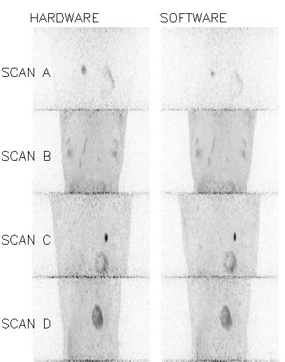
26 Results


27 Results summary in population 22 FDG Human scans 0.9 Pearson Correlation Coefficient IVF SRF GSG GSGe Scan
28 Discussion Advantages of fully automated data based algorithms for acquiring respiratory signal from PET data: o Machine independent o Operator independent o Can be written into software package Can be used with existing scans or scanners o Fast, we project they can be run in real time cost -> virtually nil o Require no extra efforts or deviation from routine clinical procedures o If not used alone, can be used as a second check for vendor hardware systems o If not used in the clinic, can be used in research for large population studies
29 Gated signal optimization: Gating+ Section II
30 Introduction Separating available statistics into phase-bins results in decreased image quality less statistics per bin Simulation
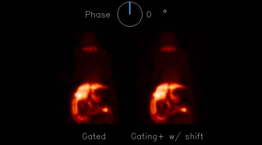
 It appears")


31 Introduction A center with gating equipment has a choice Raw scan data Ungated image Dependable (time tested) It appears utilization of extra motion information comes with uncertain risk Gated images Improved resolution, inferior contrast *Benefits condition specific Non-linear image morphing correction Improved resolution and contrast, uncertain accuracy *Benefits condition specific
32 Methods Non-linear image morphing has been proposed for recombining gated data We present an alternative strategy for utilizing the additional information provided by motion characterization - gating+ o Basic precept: Movement of signal in space is expressed in intensity fluctuations in individual voxels over the gated frames o Our methods are based on isolating the fluctuations in voxels, and modulating them according to their reliability

33 Methods A gated scan can provide two sets of information per voxel Correctly gated Randomly gated Triggers Voxel Fluctuations = motion + noise Voxel Fluctuations = noise Simulations
34 Methods Information in voxel signals can characterize voxel Sample Voxel x,y,z FFT -> Frequency amplitudes will deviate from mean FFT -> Uniform amplitudes of all frequencies
35 Methods Filter non-supported frequencies Sample Voxel x,y,z FFT -1 ->

36 Methods By looking at the effective signal to noise ratio at every voxel, we can selectively accept fluctuation information in voxels that benefit from gating, and filter fluctuation information in voxels that do not, thus optimizing information at every voxel. Voxel at liver lung boundary benefits from gating -> preserve fluctuations Voxel signal in background tissue is degraded from gating > dampen fluctuations
 and noise (randomly gated scan) through frequency amplitude analysis 3.")
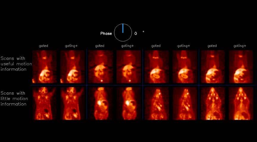
37 Gating+ protocol: Methods 1. Look at activity over gates in every voxel x,y,z for volume 2. Characterize real fluctuations (correctly gated scan) and noise (randomly gated scan) through frequency amplitude analysis 3. Accept only those frequencies which are supported by statistics Method verification o o Simulations Population gated small animal PET Rats, 16 gates, n=85, acquired with various tracers using Siemens Inveon μpet Acquired using data driven gating methods (no hardware)

38 Results Simulation Relative count statistics Ungated Gated Lesion/background ratio = 3 Upper diaphragm/background = 1.5 Lower diaphragm/background = 3.0 Gating+ Motion Map Relative Counts ungated gated gating+ ungated gated gating+ ungated gated gating+ ungated gated gating+ ungated gated gating+ Max 76% 174% 76% 58% 112% 87% 58% 94% 90% 55% 79% 79% 55% 76% 75% Volume (70% max) 29% 4% 29% 154% 4% 29% 171% 46% 71% 188% 96% 92% 183% 100% 96% SUV (mean/bckgrnd) 63% 187% 63% 49% 112% 77% 49% 73% 72% 47% 66% 67% 47% 65% 65% FWHM 130% 27% 130% 177% 50% 85% 169% 102% 104% 216% 105% 95% 189% 100% 101%
")
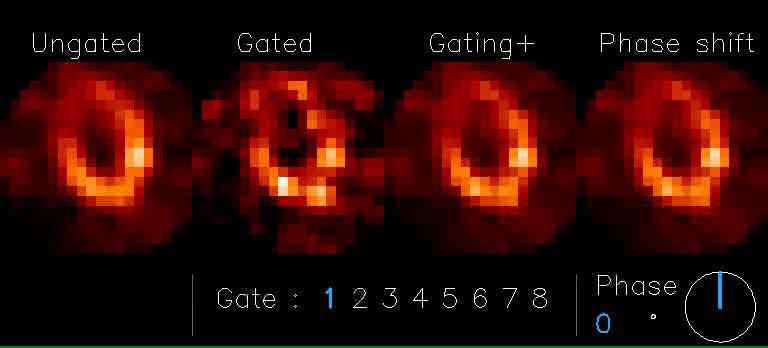
39 Results Example PET slice ( 18 F-DMDPA)
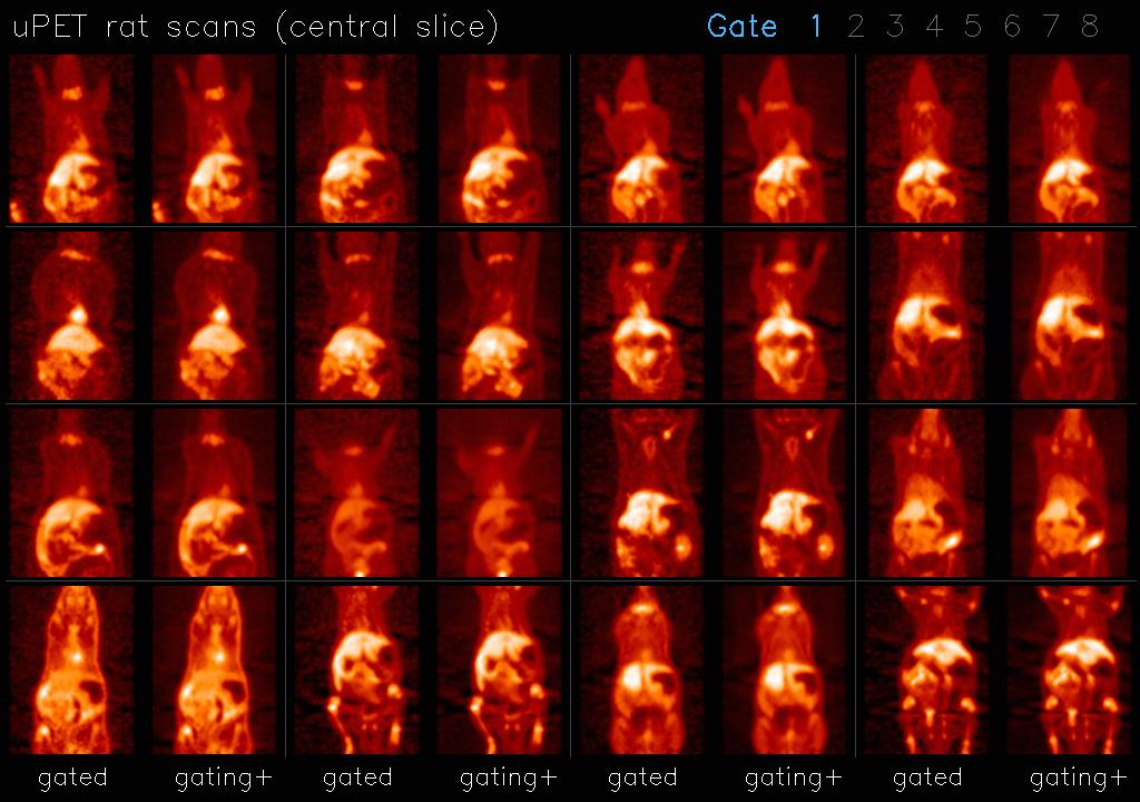
40 Results
41 Our gating+ gate combination algorithm offers an alternative approach to optimizing information acquired in a gated scans All correction comes down to (simple) 1dimensional equation Characterizable/reproducible Fast o ~20 sec processing for gating+ (µpet volume * 16 gates) Accuracy: o o All corrected voxels in simulation have a higher probability of being closer to truth than uncorrected voxels Corrected image is derived from a selective use of raw information No offset vectors created from assumptions 100% Fully automated Discussion A 4D non-linear image morphing algorithms B 1D signal optimization algorithm

42 Discussion Algorithm works with effective signal o o Irrespective of reconstruction algorithm, smoothing, etc Irrespective of quality of signal Areas not benefiting from gating, or entire scans not benefiting from gating, will be returned to their ungated embodiment Algorithm utilizes available information and optimizes its transformation into Cartesian space. o Does not preclude the use of non-linear morphing algorithms Potential applications: o o o o Support use of routine gating thorax imaging Respiratory, Cardiac imaging Human, small animal PET, SPECT, CT (low dose 4D CT), MRI
43 Motion-gate information decoupling Section III
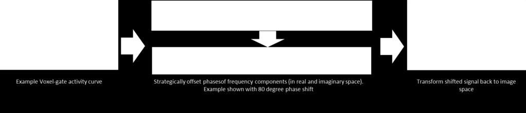
44 Introduction When information is optimized in frequency space, during the gating+ processing, there is an opportunity to shift the phase of the signal by rotating the frequency components in real and imaginary space. This allows a user to extract a voxel value at any and all phases of the cycle. o Values adhere to the optimized frequency information By repeating process for all voxels, can reconstruct phase shifted images o ~0.02 seconds processing per slice With this process, we can reconstruct continuous motion image (CMI) sequences
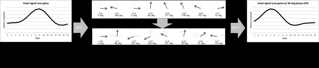
45 Voxel time-activity curve phase shifting example

46 Phase shifted curve validation We generated 10 6 simulations of true gateactivity o Signals < Nyquist frequency Gated (step function) values were derived from the true curves, CMI values were derived from gated curves In 100% of the simulations the CMI curves correlated better with truth than the respective gated curves. Example randomly generated voxel activity vs gate curve simulation.
47 Combined data driven workflow results Data driven gating + Gating+ + Phase shifted CMI frame images
48 Workflow results All images created using standard, non-gated, small animal PET rat acquisitions Animations created with 90 frames/cycle, displayed with 30 frames/second
49 Movie 1
50 Movie 2

51 Movie 3
52 Results quantified Note all numbers are average values of all appropriate scans Number of frames (360ᵒ/n) Average kidney Average VOI uptake value, averaged [relative values] over all gates Measurements Gating+ Gating with Gated Ungated + phase shift Kidney VOI n=102 % SD Average uptake across gates 2.69% 1.47% 1.47% Max displacement [mm] Average over all scans (SD over all scans) 1.50 (0.56) 1.10 (0.75) 1.10 (0.75) Liver profile n=34 Liver boundary displacement [mm] Shoulder VOI n=84 % SD in VOI [relative values] Average over all gates Global COM n=84 Max COM displacement [mm] Average over all scans (SD over all scans) 0.71 (0.46) 0.43 (0.41) 0.43 (0.41)

53 Can it all work in Humans?

54 Human work

55 Human work

56 Human work
57 Presentation summary There is information in PET data that is not being utilized We present data driven gating, signal optimization, and information decoupling strategies o o o Can be used separately or combined in an automated workflow. Can be implemented in clinical setting with minimal impact Can be used with minimal risk of degradation of care Our strategies can reframe the boundaries of motion control o o o o # of gates vs noise paradigm Characterization of motion control strategies Risks of using motion correction Visualization of motion Further validation needed we provided proof of principles and small population measurements Still room for improvement o Not seen limits in accuracy or speed o As technology advances (sensitivity and resolution), so will potential of such algorithms Still areas of application to explore

58
59 Additional material
60 Methods Sinogram Slice Axial Image Slice Image Volume Sinogram Slice Projection Sinogram Volume Projection Histogram of Counts in 500ms Image Histogram of Counts in 500ms Sinogram E E E Counts Counts

61 IVF method (2009) We can then grow a motion amplitude curve through sequential combination of information in voxels Software trace Hardware trace
 Human Scan (Standard Clinical FDG")
62 IVF method (2009) Ungated Gated Volume Projection Slice Slice (zoomed) Human Scan (Standard Clinical FDG scan)
63 IVF method (2009) IVF method employs a fully automated software algorithm o o o Image based Require no external equipment or hardware Come at the cost of processing Mostly due to image reconstruction
Gating, enhanced gating, and beyond: information utilization strategies for motion management, applied to preclinical PET
 Kesner et al. EJNMMI Research 2013, 3:29 ORIGINAL RESEARCH Open Access Gating, enhanced gating, and beyond: information utilization strategies for motion management, applied to preclinical PET Adam Leon
UvA-DARE (Digital Academic Repository) Motion compensation for 4D PET/CT Kruis, M.F. Link to publication
 Motion compensation for 4D PET/CT Kruis, M.F. Link to publication") UvA-DARE (Digital Academic Repository) Motion compensation for 4D PET/CT Kruis, M.F. Link to publication Citation for published version (APA): Kruis, M. F. (2014). Motion compensation for 4D PET/CT General
UvA-DARE (Digital Academic Repository) Motion compensation for 4D PET/CT Kruis, M.F. Link to publication Citation for published version (APA): Kruis, M. F. (2014). Motion compensation for 4D PET/CT General
CT NOISE POWER SPECTRUM FOR FILTERED BACKPROJECTION AND ITERATIVE RECONSTRUCTION
 CT NOISE POWER SPECTRUM FOR FILTERED BACKPROJECTION AND ITERATIVE RECONSTRUCTION Frank Dong, PhD, DABR Diagnostic Physicist, Imaging Institute Cleveland Clinic Foundation and Associate Professor of Radiology
Brilliance CT Big Bore.
 1 2 2 There are two methods of RCCT acquisition in widespread clinical use: cine axial and helical. In RCCT with cine axial acquisition, repeat CT images are taken each couch position while recording respiration.
8/3/2017. Contour Assessment for Quality Assurance and Data Mining. Objective. Outline. Tom Purdie, PhD, MCCPM
 Contour Assessment for Quality Assurance and Data Mining Tom Purdie, PhD, MCCPM Objective Understand the state-of-the-art in contour assessment for quality assurance including data mining-based techniques
4DM Packages. 4DM Packages & License Types. Information to help you order the appropriate licenses for your site.
 4DM Packages 4DM Packages & License Types. Information to help you order the appropriate licenses for your site. Nuclear Cardiac Quantification, Review, and Reporting Select Your 4DM Package and corresponding
3/27/2012 WHY SPECT / CT? SPECT / CT Basic Principles. Advantages of SPECT. Advantages of CT. Dr John C. Dickson, Principal Physicist UCLH
 3/27/212 Advantages of SPECT SPECT / CT Basic Principles Dr John C. Dickson, Principal Physicist UCLH Institute of Nuclear Medicine, University College London Hospitals and University College London john.dickson@uclh.nhs.uk
Image Acquisition Systems
 Image Acquisition Systems Goals and Terminology Conventional Radiography Axial Tomography Computer Axial Tomography (CAT) Magnetic Resonance Imaging (MRI) PET, SPECT Ultrasound Microscopy Imaging ITCS
Classification of Subject Motion for Improved Reconstruction of Dynamic Magnetic Resonance Imaging
 1 CS 9 Final Project Classification of Subject Motion for Improved Reconstruction of Dynamic Magnetic Resonance Imaging Feiyu Chen Department of Electrical Engineering ABSTRACT Subject motion is a significant
QIBA PET Amyloid BC March 11, Agenda
 QIBA PET Amyloid BC March 11, 2016 - Agenda 1. QIBA Round 6 Funding a. Deadlines b. What projects can be funded, what cannot c. Discussion of projects Mechanical phantom and DRO Paul & John? Any Profile
Philips SPECT/CT Systems
 Philips SPECT/CT Systems Ling Shao, PhD Director, Imaging Physics & System Analysis Nuclear Medicine, Philips Healthcare June 14, 2008 *Presented SNM08 Categorical Seminar - Quantitative SPECT and PET
If it matters to you, it matters to us
 If it matters to you, it matters to us Philips clinical innovations in nuclear medicine Innovation with insight We understand that clinical innovations are only as valuable as the day-to-day difference
REAL-TIME ADAPTIVITY IN HEAD-AND-NECK AND LUNG CANCER RADIOTHERAPY IN A GPU ENVIRONMENT
 REAL-TIME ADAPTIVITY IN HEAD-AND-NECK AND LUNG CANCER RADIOTHERAPY IN A GPU ENVIRONMENT Anand P Santhanam Assistant Professor, Department of Radiation Oncology OUTLINE Adaptive radiotherapy for head and
Image Quality Assessment and Quality Assurance of Advanced Imaging Systems for IGRT. AAPM Penn-Ohio Chapter Sep 25, 2015 Soyoung Lee, PhD
 Image Quality Assessment and Quality Assurance of Advanced Imaging Systems for IGRT AAPM Penn-Ohio Chapter Sep 25, 2015 Soyoung Lee, PhD 1 Outline q Introduction q Imaging performances in 4D-CBCT Image
Respiratory Motion Compensation for Simultaneous PET/MR Based on Strongly Undersampled Radial MR Data
 Respiratory Motion Compensation for Simultaneous PET/MR Based on Strongly Undersampled Radial MR Data Christopher M Rank 1, Thorsten Heußer 1, Andreas Wetscherek 1, and Marc Kachelrieß 1 1 German Cancer
NumaStore Preclinical FAQ
 NumaStore Preclinical FAQ 1. What is NumaStore Preclinical? 2. How does NumaStore Preclinical work with Inveon? 3. What data types does NumaStore Preclinical support? 4. How much storage space do I need
AAPM Standard of Practice: CT Protocol Review Physicist
 AAPM Standard of Practice: CT Protocol Review Physicist Dianna Cody, Ph.D., DABR, FAAPM U.T.M.D. Anderson Cancer Center September 11, 2014 2014 Texas Radiation Regulatory Conference Goals Understand purpose
Introduction to Positron Emission Tomography
 Planar and SPECT Cameras Summary Introduction to Positron Emission Tomography, Ph.D. Nuclear Medicine Basic Science Lectures srbowen@uw.edu System components: Collimator Detector Electronics Collimator
FMRI Pre-Processing and Model- Based Statistics
 FMRI Pre-Processing and Model- Based Statistics Brief intro to FMRI experiments and analysis FMRI pre-stats image processing Simple Single-Subject Statistics Multi-Level FMRI Analysis Advanced FMRI Analysis
Diffusion Mapping with FireVoxel Quick Start Guide
 Diffusion Mapping with FireVoxel Quick Start Guide Medical image analysis tool developed by Artem Mikheev and Henry Rusinek Radiology Department, NYU School of Medicine Original version prepared by Jinyu
S. Guru Prasad, Ph.D., DABR
 PURPOSE S. Guru Prasad, Ph.D., DABR Director of Medical Physics IAEA Consultant NorthShore University Health System and University of Chicago, Pritzker School of Medicine Current TPS utilize more information
Slide 1. Technical Aspects of Quality Control in Magnetic Resonance Imaging. Slide 2. Annual Compliance Testing. of MRI Systems.
 Slide 1 Technical Aspects of Quality Control in Magnetic Resonance Imaging Slide 2 Compliance Testing of MRI Systems, Ph.D. Department of Radiology Henry Ford Hospital, Detroit, MI Slide 3 Compliance Testing
Tomographic Reconstruction
 Tomographic Reconstruction 3D Image Processing Torsten Möller Reading Gonzales + Woods, Chapter 5.11 2 Overview Physics History Reconstruction basic idea Radon transform Fourier-Slice theorem (Parallel-beam)
SPECT QA and QC. Bruce McBride St. Vincent s Hospital Sydney.
 SPECT QA and QC Bruce McBride St. Vincent s Hospital Sydney. SPECT QA and QC What is needed? Why? How often? Who says? QA and QC in Nuclear Medicine QA - collective term for all the efforts made to produce
REMOVAL OF THE EFFECT OF COMPTON SCATTERING IN 3-D WHOLE BODY POSITRON EMISSION TOMOGRAPHY BY MONTE CARLO
 REMOVAL OF THE EFFECT OF COMPTON SCATTERING IN 3-D WHOLE BODY POSITRON EMISSION TOMOGRAPHY BY MONTE CARLO Abstract C.S. Levin, Y-C Tai, E.J. Hoffman, M. Dahlbom, T.H. Farquhar UCLA School of Medicine Division
Computational Medical Imaging Analysis Chapter 4: Image Visualization
 Computational Medical Imaging Analysis Chapter 4: Image Visualization Jun Zhang Laboratory for Computational Medical Imaging & Data Analysis Department of Computer Science University of Kentucky Lexington,
Gadgetron implementation for PET/MR motion correction
 implementation for motion correction Thomas Küstner School of Biomedical Engineering & Imaging Sciences, King s College London, St. Thomas Hospital, London, UK Dept. of Diagnostic and Interventional Radiology,
Basic fmri Design and Analysis. Preprocessing
 Basic fmri Design and Analysis Preprocessing fmri Preprocessing Slice timing correction Geometric distortion correction Head motion correction Temporal filtering Intensity normalization Spatial filtering
UvA-DARE (Digital Academic Repository) Motion compensation for 4D PET/CT Kruis, M.F. Link to publication
 Motion compensation for 4D PET/CT Kruis, M.F. Link to publication") UvA-DARE (Digital Academic Repository) Motion compensation for 4D PET/CT Kruis, M.F. Link to publication Citation for published version (APA): Kruis, M. F. (2014). Motion compensation for 4D PET/CT General
UvA-DARE (Digital Academic Repository) Motion compensation for 4D PET/CT Kruis, M.F. Link to publication Citation for published version (APA): Kruis, M. F. (2014). Motion compensation for 4D PET/CT General
Implementation and evaluation of a fully 3D OS-MLEM reconstruction algorithm accounting for the PSF of the PET imaging system
 Implementation and evaluation of a fully 3D OS-MLEM reconstruction algorithm accounting for the PSF of the PET imaging system 3 rd October 2008 11 th Topical Seminar on Innovative Particle and Radiation
PURE. ViSION Edition PET/CT. Patient Comfort Put First.
 PURE ViSION Edition PET/CT Patient Comfort Put First. 2 System features that put patient comfort and safety first. Oncology patients deserve the highest levels of safety and comfort during scans. Our Celesteion
8/2/2016. Measures the degradation/distortion of the acquired image (relative to an ideal image) using a quantitative figure-of-merit
 using a quantitative figure-of-merit") Ke Li Assistant Professor Department of Medical Physics and Department of Radiology School of Medicine and Public Health, University of Wisconsin-Madison This work is partially supported by an NIH Grant
Ke Li Assistant Professor Department of Medical Physics and Department of Radiology School of Medicine and Public Health, University of Wisconsin-Madison This work is partially supported by an NIH Grant
CT Protocol Review: Practical Tips for the Imaging Physicist Physicist
 CT Protocol Review: Practical Tips for the Imaging Physicist Physicist Dianna Cody, Ph.D., DABR, FAAPM U.T.M.D. Anderson Cancer Center August 8, 2013 AAPM Annual Meeting Goals Understand purpose and importance
Corridor4DM Feature List
 Software Services Features Corridor4DM Feature List 4DM v2013 FDG PET Standard Uptake Value (SUV) Calculation Auto-Selection of Report Template Motion Correction of SPECT datasets New Normals Databases
Automated segmentation methods for liver analysis in oncology applications
 University of Szeged Department of Image Processing and Computer Graphics Automated segmentation methods for liver analysis in oncology applications Ph. D. Thesis László Ruskó Thesis Advisor Dr. Antal
Retrospective Spiral Respiratory Correlated Imaging with Varian RPM
 Retrospective Spiral Respiratory Correlated Imaging with Varian RPM This is a Quick Step Guide for Retrospective Spiral Respiratory Correlated Imaging (4D CT) using the Varian RPM device v1.7 with the
Performance Evaluation of radionuclide imaging systems
 Performance Evaluation of radionuclide imaging systems Nicolas A. Karakatsanis STIR Users meeting IEEE Nuclear Science Symposium and Medical Imaging Conference 2009 Orlando, FL, USA Geant4 Application
Digital Image Processing
 Digital Image Processing SPECIAL TOPICS CT IMAGES Hamid R. Rabiee Fall 2015 What is an image? 2 Are images only about visual concepts? We ve already seen that there are other kinds of image. In this lecture
RADIOMICS: potential role in the clinics and challenges
 27 giugno 2018 Dipartimento di Fisica Università degli Studi di Milano RADIOMICS: potential role in the clinics and challenges Dr. Francesca Botta Medical Physicist Istituto Europeo di Oncologia (Milano)
Improvement of contrast using reconstruction of 3D Image by PET /CT combination system
 Available online at www.pelagiaresearchlibrary.com Advances in Applied Science Research, 2013, 4(1):285-290 ISSN: 0976-8610 CODEN (USA): AASRFC Improvement of contrast using reconstruction of 3D Image
Review of PET Physics. Timothy Turkington, Ph.D. Radiology and Medical Physics Duke University Durham, North Carolina, USA
 Review of PET Physics Timothy Turkington, Ph.D. Radiology and Medical Physics Duke University Durham, North Carolina, USA Chart of Nuclides Z (protons) N (number of neutrons) Nuclear Data Evaluation Lab.
Basics of treatment planning II
 Basics of treatment planning II Sastry Vedam PhD DABR Introduction to Medical Physics III: Therapy Spring 2015 Dose calculation algorithms! Correction based! Model based 1 Dose calculation algorithms!
Advancements in molecular medicine
 Advancements in molecular medicine Philips Ingenuity TF PET/MR attenuation correction Z. Hu, 1 S. Renisch, 2 B. Schweizer, 3 T. Blaffert, 2 N. Ojha, 1 T. Guo, 1 J. Tang, 1 C. Tung, 1 J. Kaste, 1 V. Schulz,
Multi-slice CT Image Reconstruction Jiang Hsieh, Ph.D.
 Multi-slice CT Image Reconstruction Jiang Hsieh, Ph.D. Applied Science Laboratory, GE Healthcare Technologies 1 Image Generation Reconstruction of images from projections. textbook reconstruction advanced
2005 IEEE Nuclear Science Symposium Conference Record M10-2
 25 IEEE Nuclear Science Symposium Conference Record M1-2 Estimating 3D Respiratory Motion from Orbiting Views Rongping Zeng 1, Jeffrey A. Fessler 1, and James M. Balter 2 rzeng@eecs.umich.edu, fessler@eecs.umich.edu,
Modeling and Incorporation of System Response Functions in 3D Whole Body PET
 Modeling and Incorporation of System Response Functions in 3D Whole Body PET Adam M. Alessio, Member IEEE, Paul E. Kinahan, Senior Member IEEE, and Thomas K. Lewellen, Senior Member IEEE University of
Functional MRI in Clinical Research and Practice Preprocessing
 Functional MRI in Clinical Research and Practice Preprocessing fmri Preprocessing Slice timing correction Geometric distortion correction Head motion correction Temporal filtering Intensity normalization
Helical 4D CT pitch management for the Brilliance CT Big Bore in clinical practice
 JOURNAL OF APPLIED CLINICAL MEDICAL PHYSICS, VOLUME 16, NUMBER 3, 2015 Helical 4D CT pitch management for the Brilliance CT Big Bore in clinical practice Guido Hilgers, a Tonnis Nuver, André Minken Department
Joint CI-JAI advanced accelerator lecture series Imaging and detectors for medical physics Lecture 1: Medical imaging
 Joint CI-JAI advanced accelerator lecture series Imaging and detectors for medical physics Lecture 1: Medical imaging Dr Barbara Camanzi barbara.camanzi@stfc.ac.uk Course layout Day AM 09.30 11.00 PM 15.30
Respiratory Motion Estimation using a 3D Diaphragm Model
 Respiratory Motion Estimation using a 3D Diaphragm Model Marco Bögel 1,2, Christian Riess 1,2, Andreas Maier 1, Joachim Hornegger 1, Rebecca Fahrig 2 1 Pattern Recognition Lab, FAU Erlangen-Nürnberg 2
Machine Learning for Medical Image Analysis. A. Criminisi
 Machine Learning for Medical Image Analysis A. Criminisi Overview Introduction to machine learning Decision forests Applications in medical image analysis Anatomy localization in CT Scans Spine Detection
SPM8 for Basic and Clinical Investigators. Preprocessing
 SPM8 for Basic and Clinical Investigators Preprocessing fmri Preprocessing Slice timing correction Geometric distortion correction Head motion correction Temporal filtering Intensity normalization Spatial
SPM8 for Basic and Clinical Investigators. Preprocessing. fmri Preprocessing
 SPM8 for Basic and Clinical Investigators Preprocessing fmri Preprocessing Slice timing correction Geometric distortion correction Head motion correction Temporal filtering Intensity normalization Spatial
Robust Lung Ventilation Assessment
 Fifth International Workshop on Pulmonary Image Analysis -75- Robust Lung Ventilation Assessment Sven Kabus 1, Tobias Klinder 1, Tokihiro Yamamoto 2, Paul J. Keall 3, Billy W. Loo, Jr. 4, and Cristian
Cardiac Dual Energy CT: Technique
 RSNA 2013, VSCA51-01, Chicago, Dec. 5, 2013 Cardiac Radiology Series Cardiac Dual Energy CT: Technique Willi A. Kalender, Ph.D. Institute of Medical Physics University of Erlangen www.imp.uni-erlangen.de
Agenda : Lung Density Breakout Session
 Agenda : Lung Density Breakout Session 1. : Mathew Fuld and Bernice Hoppel 2. Automatic Exposure Control (AEC) Evaluation : Sean Fain Round 4 Project 3. Dose reduction effects on emphysema metrics : Philip
Motion Artifacts and Suppression in MRI At a Glance
 Motion Artifacts and Suppression in MRI At a Glance Xiaodong Zhong, PhD MR R&D Collaborations Siemens Healthcare MRI Motion Artifacts and Suppression At a Glance Outline Background Physics Common Motion
Background. Outline. Radiographic Tomosynthesis: Image Quality and Artifacts Reduction 1 / GE /
 Radiographic Tomosynthesis: Image Quality and Artifacts Reduction Baojun Li, Ph.D Department of Radiology Boston University Medical Center 2012 AAPM Annual Meeting Background Linear Trajectory Tomosynthesis
Respiratory Motion Compensation for C-arm CT Liver Imaging
 Respiratory Motion Compensation for C-arm CT Liver Imaging Aline Sindel 1, Marco Bögel 1,2, Andreas Maier 1,2, Rebecca Fahrig 3, Joachim Hornegger 1,2, Arnd Dörfler 4 1 Pattern Recognition Lab, FAU Erlangen-Nürnberg
Clinical Prospects and Technological Challenges for Multimodality Imaging Applications in Radiotherapy Treatment Planning
 Clinical Prospects and Technological Challenges for Multimodality Imaging Applications in Radiotherapy Treatment Planning Issam El Naqa, PhD Assistant Professor Department of Radiation Oncology Washington
multimodality image processing workstation Visualizing your SPECT-CT-PET-MRI images
 multimodality image processing workstation Visualizing your SPECT-CT-PET-MRI images FUSION FUSION is a new visualization and evaluation software developed by Mediso built on state of the art technology,
CT Basics Principles of Spiral CT Dose. Always Thinking Ahead.
 1 CT Basics Principles of Spiral CT Dose 2 Who invented CT? 1963 - Alan Cormack developed a mathematical method of reconstructing images from x-ray projections Sir Godfrey Hounsfield worked for the Central
Introduction to Emission Tomography
 Introduction to Emission Tomography Gamma Camera Planar Imaging Robert Miyaoka, PhD University of Washington Department of Radiology rmiyaoka@u.washington.edu Gamma Camera: - collimator - detector (crystal
AIDR 3D Iterative Reconstruction:
 Iterative Reconstruction: Integrated, Automated and Adaptive Dose Reduction Erin Angel, PhD Manager, Clinical Sciences, CT Canon Medical Systems USA Iterative Reconstruction 1 Since the introduction of
Optimisation of Toshiba Aquilion ONE Volume Imaging
 Optimisation of Toshiba Aquilion ONE Volume Imaging Jane Edwards, RPRSG Royal Free London NHS Foundation Trust Dr Mufudzi Maviki, Plymouth Hospitals NHS Trust Background In 2011/12 Radiology at RFH was
Optimization of CT Simulation Imaging. Ingrid Reiser Dept. of Radiology The University of Chicago
 Optimization of CT Simulation Imaging Ingrid Reiser Dept. of Radiology The University of Chicago Optimization of CT imaging Goal: Achieve image quality that allows to perform the task at hand (diagnostic
7/31/2011. Learning Objective. Video Positioning. 3D Surface Imaging by VisionRT
 CLINICAL COMMISSIONING AND ACCEPTANCE TESTING OF A 3D SURFACE MATCHING SYSTEM Hania Al-Hallaq, Ph.D. Assistant Professor Radiation Oncology The University of Chicago Learning Objective Describe acceptance
Motion artifact detection in four-dimensional computed tomography images
 Motion artifact detection in four-dimensional computed tomography images G Bouilhol 1,, M Ayadi, R Pinho, S Rit 1, and D Sarrut 1, 1 University of Lyon, CREATIS; CNRS UMR 5; Inserm U144; INSA-Lyon; University
Venus Explorer Processing Technical specifications
 24, rue du champ de l Alouette F 75013 Paris, France Venus Explorer Processing Technical specifications Venus Explorer Processing Server software Web base application sever; tomcat Apache technology. Include
As fl exible as your care requires
 As fl exible as your care requires Philips Ingenuity Flex 32 CT Built on Ingenuity The Philips Ingenuity Flex 32 helps you provide excellent care with outstanding flexibility, now and in the future. Built
Micro-CT Methodology Hasan Alsaid, PhD
 Micro-CT Methodology Hasan Alsaid, PhD Preclinical & Translational Imaging LAS, PTS, GlaxoSmithKline 20 April 2015 Provide basic understanding of technical aspects of the micro-ct Statement: All procedures
Computer-Tomography II: Image reconstruction and applications
 Computer-Tomography II: Image reconstruction and applications Prof. Dr. U. Oelfke DKFZ Heidelberg Department of Medical Physics (E040) Im Neuenheimer Feld 280 69120 Heidelberg, Germany u.oelfke@dkfz.de
Image Guidance and Beam Level Imaging in Digital Linacs
 Image Guidance and Beam Level Imaging in Digital Linacs Ruijiang Li, Ph.D. Department of Radiation Oncology Stanford University School of Medicine 2014 AAPM Therapy Educational Course Disclosure Research
Diagnostic quality of time-averaged ECG-gated CT data
 Diagnostic quality of time-averaged ECG-gated CT data Almar Klein a, Luuk J. Oostveen b, Marcel J.W. Greuter c, Yvonne Hoogeveen b, Leo J. Schultze Kool b, Cornelis H. Slump a and W. Klaas Jan Renema b
A closer look at CT scanning
 Vet Times The website for the veterinary profession https://www.vettimes.co.uk A closer look at CT scanning Author : Charissa Lee, Natalie Webster Categories : General, Vets Date : April 3, 2017 A basic
Module 4. K-Space Symmetry. Review. K-Space Review. K-Space Symmetry. Partial or Fractional Echo. Half or Partial Fourier HASTE
 MRES 7005 - Fast Imaging Techniques Module 4 K-Space Symmetry Review K-Space Review K-Space Symmetry Partial or Fractional Echo Half or Partial Fourier HASTE Conditions for successful reconstruction Interpolation
Data-driven optimal binning for respiratory motion management in PET
 Data-driven optimal binning for respiratory motion management in PET Adam L. Kesner a) Department of Medical Physics, Memorial Sloan Kettering Cancer Center, New York, NY, USA Joseph G. Meier Department
Chapter 3 Set Redundancy in Magnetic Resonance Brain Images
 16 Chapter 3 Set Redundancy in Magnetic Resonance Brain Images 3.1 MRI (magnetic resonance imaging) MRI is a technique of measuring physical structure within the human anatomy. Our proposed research focuses
A method and algorithm for Tomographic Imaging of highly porous specimen using Low Frequency Acoustic/Ultrasonic signals
 More Info at Open Access Database www.ndt.net/?id=15210 A method and algorithm for Tomographic Imaging of highly porous specimen using Low Frequency Acoustic/Ultrasonic signals Subodh P S 1,a, Reghunathan
EJNMMI Physics. Anna Turco 1*, Johan Nuyts 1, Olivier Gheysens 1,2, Jürgen Duchenne 3, Jens-Uwe Voigt 3,4,PietClaus 3 and Kathleen Vunckx 1
 Turco et al. EJNMMI Physics (2016) 3:9 DOI 10.1186/s40658-016-0145-4 EJNMMI Physics ORIGINAL RESEARCH Open Access Lesion quantification and detection in myocardial 18 F-FDG PET using edge-preserving priors
Suitability of a new alignment correction method for industrial CT
 Suitability of a new alignment correction method for industrial CT Matthias Elter 1, Nicole Maass 1, Peter Koch 2 1 Siemens AG, Healthcare Sector, Erlangen, Germany, e-mail: matthias.elter@siemens.com,
A Non-Linear Image Registration Scheme for Real-Time Liver Ultrasound Tracking using Normalized Gradient Fields
 A Non-Linear Image Registration Scheme for Real-Time Liver Ultrasound Tracking using Normalized Gradient Fields Lars König, Till Kipshagen and Jan Rühaak Fraunhofer MEVIS Project Group Image Registration,
Workshop on Quantitative SPECT and PET Brain Studies January, 2013 PUCRS, Porto Alegre, Brasil Corrections in SPECT and PET
 Workshop on Quantitative SPECT and PET Brain Studies 14-16 January, 2013 PUCRS, Porto Alegre, Brasil Corrections in SPECT and PET Físico João Alfredo Borges, Me. Corrections in SPECT and PET SPECT and
Development and Optimization of Fourdimensional Magnetic Resonance Imaging (4D-MRI) for Radiation Therapy
 for Radiation Therapy") Development and Optimization of Fourdimensional Magnetic Resonance Imaging (4D-MRI) for Radiation Therapy by Yilin Liu Medical Physics Graduate Program Duke University Date: Approved: Jing Cai, co-advisor
Development and Optimization of Fourdimensional Magnetic Resonance Imaging (4D-MRI) for Radiation Therapy by Yilin Liu Medical Physics Graduate Program Duke University Date: Approved: Jing Cai, co-advisor
TomoTherapy Related Projects. An image guidance alternative on Tomo Low dose MVCT reconstruction Patient Quality Assurance using Sinogram
 TomoTherapy Related Projects An image guidance alternative on Tomo Low dose MVCT reconstruction Patient Quality Assurance using Sinogram Development of A Novel Image Guidance Alternative for Patient Localization
CLASS HOURS: 4 CREDIT HOURS: 4 LABORATORY HOURS: 0
 Revised 10/10 COURSE SYLLABUS TM 220 COMPUTED TOMOGRAPHY PHYSICS CLASS HOURS: 4 CREDIT HOURS: 4 LABORATORY HOURS: 0 CATALOG COURSE DESCRIPTION: This course is one of a three course set in whole body Computed
White Pixel Artifact. Caused by a noise spike during acquisition Spike in K-space <--> sinusoid in image space
 White Pixel Artifact Caused by a noise spike during acquisition Spike in K-space sinusoid in image space Susceptibility Artifacts Off-resonance artifacts caused by adjacent regions with different
An Efficient Technique For Multi-Phase Model Based Iterative Reconstruction
 1 An Efficient Technique For Multi-Phase Model Based Iterative Reconstruction Shiyu Xu, Debashish Pal and Jean-Baptiste Thibault Abstract Multi-phase scan is a fundamental CT acquisition technology used
The Near Future in Cardiac CT Image Reconstruction
 SCCT 2010 The Near Future in Cardiac CT Image Reconstruction Marc Kachelrieß Institute of Medical Physics (IMP) Friedrich-Alexander Alexander-University Erlangen-Nürnberg rnberg www.imp.uni-erlangen.de
Magnetic Resonance Elastography (MRE) of Liver Disease
 of Liver Disease") Magnetic Resonance Elastography (MRE) of Liver Disease Authored by: Jennifer Dolan Fox, PhD VirtualScopics Inc. jennifer_fox@virtualscopics.com 1-585-249-6231 1. Overview of MRE Imaging MRE is a magnetic
Magnetic Resonance Elastography (MRE) of Liver Disease Authored by: Jennifer Dolan Fox, PhD VirtualScopics Inc. jennifer_fox@virtualscopics.com 1-585-249-6231 1. Overview of MRE Imaging MRE is a magnetic
White Paper. EQ PET: Achieving NEMAreferenced. Technologies. Matthew Kelly, PhD, Siemens Healthcare
 White Paper EQ PET: Achieving NEMAreferenced SUV Across Technologies Matthew Kelly, PhD, Siemens Healthcare Table of Contents Introduction 1 Case Study 1 Cross-Scanner Response Assessment 2 Clinical Example
Nonrigid Registration using Free-Form Deformations
 Nonrigid Registration using Free-Form Deformations Hongchang Peng April 20th Paper Presented: Rueckert et al., TMI 1999: Nonrigid registration using freeform deformations: Application to breast MR images
Medical Imaging BMEN Spring 2016
 Name Medical Imaging BMEN 420-501 Spring 2016 Homework #4 and Nuclear Medicine Notes All questions are from the introductory Powerpoint (based on Chapter 7) and text Medical Imaging Signals and Systems,
EPI Data Are Acquired Serially. EPI Data Are Acquired Serially 10/23/2011. Functional Connectivity Preprocessing. fmri Preprocessing
 Functional Connectivity Preprocessing Geometric distortion Head motion Geometric distortion Head motion EPI Data Are Acquired Serially EPI Data Are Acquired Serially descending 1 EPI Data Are Acquired
8/2/2017. Disclosure. Philips Healthcare (Cleveland, OH) provided the precommercial
 provided the precommercial") 8//0 AAPM0 Scientific Symposium: Emerging and New Generation PET: Instrumentation, Technology, Characteristics and Clinical Practice Aug Wednesday 0:4am :pm Solid State Digital Photon Counting PET/CT Instrumentation
8//0 AAPM0 Scientific Symposium: Emerging and New Generation PET: Instrumentation, Technology, Characteristics and Clinical Practice Aug Wednesday 0:4am :pm Solid State Digital Photon Counting PET/CT Instrumentation
Artefakt-resistente Bewegungsschätzung für die bewegungskompensierte CT
 Artefakt-resistente Bewegungsschätzung für die bewegungskompensierte CT Marcus Brehm 1,2, Thorsten Heußer 1, Pascal Paysan 3, Markus Oehlhafen 3, and Marc Kachelrieß 1,2 1 German Cancer Research Center
Quantitative imaging for clinical dosimetry
 Quantitative imaging for clinical dosimetry Irène Buvat Laboratoire d Imagerie Fonctionnelle U678 INSERM - UPMC CHU Pitié-Salpêtrière, Paris buvat@imed.jussieu.fr http://www.guillemet.org/irene Methodology
Separate CT-Reconstruction for Orientation and Position Adaptive Wavelet Denoising
 Separate CT-Reconstruction for Orientation and Position Adaptive Wavelet Denoising Anja Borsdorf 1,, Rainer Raupach, Joachim Hornegger 1 1 Chair for Pattern Recognition, Friedrich-Alexander-University
Q.Clear. Steve Ross, Ph.D.
 Steve Ross, Ph.D. Accurate quantitation (SUV - Standardized Uptake Value) is becoming more important as clinicians seek to utilize PET imaging for more than just diagnosing and staging disease, but also
664 IEEE TRANSACTIONS ON NUCLEAR SCIENCE, VOL. 52, NO. 3, JUNE 2005
 664 IEEE TRANSACTIONS ON NUCLEAR SCIENCE, VOL. 52, NO. 3, JUNE 2005 Attenuation Correction for the NIH ATLAS Small Animal PET Scanner Rutao Yao, Member, IEEE, Jürgen Seidel, Jeih-San Liow, Member, IEEE,