SPECT/CT Basics, Technology Updates, Quality Assurance, and Applications
|
|
|
- Beryl Ward
- 5 years ago
- Views:
Transcription

1 SPECT/CT Basics, Technology Updates, Quality Assurance, and Applications S. Cheenu Kappadath, PhD Department of Imaging Physics University of Texas M D Anderson Cancer Center, Houston, Texas skappadath@mdanderson.org
2 Educational Objectives 1. Understand the underlying principles of SPECT/CT image acquisition, processing and reconstruction 2. Understand current and future clinical applications of SPECT/CT imaging 3. Familiarization with commercially-available SPECT/CT systems
3 Outline Review of SPECT principles Iterative SPECT reconstruction Hybrid SPECT/CT imaging SPECT/CT quality assurance Commercial SPECT/CT systems SPECT/CT clinical applications
4 SPECT Basics Single Photon Emission Computed Tomography Radio-pharmaceutical administration injected, ingested, or inhaled Bio-distribution of pharmaceutical uptake time Decay of radionuclide from within the patient the source of information Gamma camera detects gamma rays and images (tomography) the radio-pharmaceutical distribution within the patient SPECT Used for visualization of functional information based on the specific radio-pharmaceutical uptake mechanism
5 SPECT Hardware Anatomy of a Gamma camera 1. Collimator 2. Scintillation Detector 3. Photomultiplier Tubes 4. Position Circuitry 5. Data Analysis Computer U of British Columbia
6 SPECT Back-Projection Model g(s, ) = f(x,y) along an in-plane line integral Bruyant, P. P., J Nucl Med 2002; 43:
7 Crystal Thickness Thinner crystals spatial resolution interactions occur at a better defined depth multiple interactions less likely less light spread interaction likelihood for higher energy s Thicker crystals sensitivity interaction likelihood (esp. for higher E s) likelihood of multiple interactions greater light spread spatial resolution
8 Spatial Resolution Intrinsic Spatial and Energy Resolution # of scintillation photons, N Gamma-ray energy, E Spatial Resolution = 100 /N 1/ N 1/ E Energy Resolution = 100 FWHM/E 1/ E B L e H Collimator Resolution R g D( Le H B) L e L e System Resolution R R R s i g D


9 SPECT Acquisitions SPECT acquires 2-D projections of a 3-D volume Yale School of Medicine SPECT in the year 2000: Basic principles, JNMT 24:233, 2000
 Anterior View P( + ) horizontally flipped Posterior")
10 Radon transform angular symmetry violated in SPECT P( ) Anterior View P( + ) horizontally flipped Posterior
11 Radon transform angular symmetry violated in SPECT Why? Due to Differential Attenuation L a i b I( i ) b I( i ) = I 0 e - a (L)dL c I( i + ) = I 0 e - a (L)dL I 0 c Other mediating factors: I( i + ) distance-dependent resolution depth-dependent scatter


12 SPECT Acquisitions SPECT projections acquired over 360 Exception: Cardiac SPECT acquired over


13 SPECT images have isotropic voxel size 2-D filter of projections 3-D post-reconstruction filter transverse sagittal coronal No volume smoothing Butterworth: 0.6 Nyquist, 10 th order
14 SPECT Filtered Back- Projection FBP based on ideal Radon inversion formula assumes a linear, shift-invariant system and angular symmetry of projections SPECT imaging systems are neither angularly symmetric nor shift-invariant SPECT projection data affected by attenuation, scatter, and spatial resolution that are all depth-or distance-dependent Thus, FBP reconstruction cannot adequately model the physics of SPECT
15 Conventional SPECT Corrections Attenuation: Chang post-processing algorithm I(x,y) = SPECT image w/o AC I(x,y, i ) = I AC (x,y).e - L(x,y, i ) I AC (x,y) = I(x,y) / {(1/M). i e - L(x,y, i ) }; i = 1, M I(x,y, i ) L(x,y, i ) i I AC (x,y) Scatter: Energy window subtraction Energy Spectrum of Sm-153 P(x,y) = projections w/ scatter P LE (x,y) = projection at lower energy P HE (x,y) = projection at higher energy P SC (x,y) = P(x,y) k L.P LE (x,y) k H.P HE (x,y) Counts Lower Scatter Window Photo- Peak Window Upper Scatter Window STD in acrylic STD in air STD in acrylic with TEW Scatter Correction Photon Energy [kev]
16 Outline Review of SPECT principles Iterative SPECT reconstruction Hybrid SPECT/CT imaging SPECT/CT quality assurance Commercial SPECT/CT systems SPECT/CT clinical applications
17 SPECT Iterative Reconstruction Maximum Likelihood-Expectation Maximization (ML-EM) Accounts for the statistical nature of SPECT imaging Incorporates the system response p(b,d) the probability that a photon emitted from an object voxel b is detected by projection pixel d p(b,d) captures 1. Depth-dependent resolution 2. Position-dependent scatter 3. Depth-dependent attenuation voxel b detector d Use a measured attenuation map along with models of scatter and camera resolution to perform a far more accurate reconstruction
18 SPECT Iterative Recon: Attenuation Modeling along a line integral g(s, ) = f(x,y) * p attn (x,y,s, ) p attn (x,y,s, ) = probability due to attenuation p attn (x,y,s, ) = exp(- ab (x,y ) x,y )) b a
19 SPECT Iterative Recon: System Resolution Modeling Distance-dependent collimator beam R s = R 2 i + R 2 c r Pencil Beam (FBP) Intrinsic Detector Resolution R i Fan Beam (2D - iterative) Cone Beam (3D iterative)
20 SPECT Iterative Recon: Resolution Modeling 2D: g(s, ) = f(x,y) * p res (x,y,s, ) 3D: g(s, ) = f(x,y,z) * p res (x,y,z,s, ) p res = probability due to resolution fan of acceptance (2D fan beam model) cone of acceptance (3D cone beam model)
21 SPECT Imaging: Scatter Scatter compensation occurs before attenuation the photopeak window contains scatter attenuation accounts for the removal of photopeak photons Scatter contribution estimated as a weighted sum of one or more adjacent energy window images, Ci(x,y, ) S(x,y, ) = i k i C i (x,y, ) Subtract scatter prior to reconstruction P corr (x,y, ) P(x,y, ) - S(x,y, ) Incorporate scatter into forward projection P(x,y, ) P corr (x,y, ) + S(x,y, ) SC techniques: DEW TEW ESSE
22 SPECT Iterative Reconstruction True projection intensity = sum of true voxel intensities weighted by detection probabilities Forward Projection B y( d) ( b) p( b, d) b 1 True voxel intensity = sum of true detector intensities weighted by detection probabilities Back Projection D ( b) y( d) p( b, d) d 1
23 Iterative Reconstruction Flow Diagram D [ k ] ( b) B d 1 [ k 1] b ' 1 ( b) D d 1 yd ( ) pbd (, ) [ k ] pbd (, ) ( b') p( b', d) In clinical practice, the stopping criteria is number of iterations (a time constraint) instead of a convergence criteria.
24 Ordered Subset EM (OSEM) Each OSEM iteration is a ML-EM iteration using an ordered subset of n (out of N) projections (eg: 4/36 views - 9 subsets, start with 0,90,180,270 views) The next OSEM iteration starts with the result of the previous OSEM iterations but uses a different ordered subset of n projections (next set uses 10,100,190,280 views) rate of convergence by using an ordered subset of all N projections for each iteration m OSEM iterations with n subsets each m n ML-EM iterations using all N each time

25 OSEM Iterative SPECT Reconstruction: Attenuation and Scatter Correction Un-Corrected Corrected Note the hot-rim artifact
 2-D pre-filter: Butterworth, fc = 0.6 Nyquist, order = 10 3-D OSEM w/ cone beam modeling (m=25,n=10) 3-D Gaussian Post-Filter (7.8 mm FWHM)")
26 OSEM Iterative SPECT Reconstruction: Collimator Resolution Modeling 99m Tc Bone Scan (osteosarcoma), LEHR Collimator Standard Filtered Backprojection 2-D OSEM w/ fan beam modeling (m=12,n=10) 2-D pre-filter: Butterworth, fc = 0.6 Nyquist, order = 10 3-D OSEM w/ cone beam modeling (m=25,n=10) 3-D Gaussian Post-Filter (7.8 mm FWHM)
27 Outline Review of SPECT principles Iterative SPECT reconstruction Hybrid SPECT/CT imaging SPECT/CT quality assurance Commercial SPECT/CT systems SPECT/CT clinical applications
28 SPECT/CT Hybrid Imaging: Why? Non-uniform attenuation maps required Previous methods used constant maps that work for brain but are problematic for thorax and pelvis radioactive source-based transmission CT time penalty Functional-anatomical overlay (image fusion) Improve localization of uptake regions Increase confidence in interpretation
29 CT-based AC for SPECT/CT CT CT noise reduced CT AC μ map Smooth, re bin CT to match SPECT Register CT w/ SPECT Apply bi linear transform on pixel by pixel basis Reconstructed SPECT Transition Matrix a ijk Other factors: SPECT projections Scatter estimates Collimator response
30 CT-based values 0.3 (cm 2 /g) Material attenuation versus Energy Air Muscle Bone Photoelectric effect dominant CT Energy (kev) Compton scatter dominant m = k CT-HU (simple but not accurate) Compton Scatter probability proportional to e - density Photoelectric effect probability proportional to (Z/E) 3 Attenuation mismatch between PE and CS with energy for high Z
31 CT-based values - HU-to-cm -1 conversion - not linearly related - piece-wise linear - bi- or tri-modal - Effective energy differences - CT (~ kev) - SPECT (nuclide dependent) eg: 140 kev for Tc-99m CT Number-to-Tc-99m value Function value (cm-1) CT Number (HU) 1000
 3-D OSEM w/ resolution modeling 3-D OSEM w/ resolution and")
32 SPECT/CT Hybrid Imaging: Iterative Reconstruction FBP w/ Butterworth 0.4/5 99m Tc EC-DG (NSCLC) 3-D OSEM w/ resolution modeling 3-D OSEM w/ resolution and attenuation modeling
33 Outline Review of SPECT principles Iterative SPECT reconstruction Hybrid SPECT/CT imaging SPECT/CT quality assurance Commercial SPECT/CT systems SPECT/CT clinical applications
34 SPECT/CT QA/QC Planar (AAPM Reports 6 and 9; NEMA NU ) Inherently includes all planar gamma camera QA Energy/Spatial resolution, uniformity, deadtime, sensitivity, rotational uniformity, opposed-head registration, etc. SPECT (AAPM Report 22 and 52) Uniformity and Contrast Resolution SPECT/CT (AAPM TG 177: Jim Halama) NM-CT registration CT-HU to linear attenuation ( ) transformation

35 NM-CT Registration Use Co-57 button sources w/ SPECT phantom


36 CT-HU to -map transformation Use an electron density phantom CIRS Inc. CT image: -790 to 235 HU
37 Outline Review of SPECT principles Iterative SPECT reconstruction Hybrid SPECT/CT imaging SPECT/CT quality assurance Commercial SPECT/CT systems SPECT/CT clinical applications

 Philips BrightView")
38 Commercial SPECT/CT systems Siemens SymbiaT (1-, 2-, 6, 16-slice CT) GE Hawkeye (1- or 4-slice CT) Philips BrightView (Flat-panel CT)
39 GE Millennium VG Hawkeye NM CT 3/8 and 1 NaI(Tl) crystals 16 simultaneous energy windows Slip-ring gantry Body-contouring based on infrared-based transmitters Co-planar, dental tube, 4-slice 20 mm beam no additional real estate needed Resolution: 3.5 or 1.75 mm (transaxial); 5 or 10 mm (axial) Time-averaged: 23 s per rotation (slow-scan) kvp: ; ma: 1 2.5
40 Phillips BrightView XCT NM CT 3/8 and ¾ NaI(Tl) crystals Energy-independent flood calibration (up to 300 kev) 15 simultaneous energy windows Body-contouring based on tissue impedance Co-planar, flat-panel detector, 14 cm axial FOV no additional real estate needed High-resolution: 0.33 mm isotropic voxels Time-averaged: 12 s or 24 s per rotation (slow-scan) kvp: 120; ma: 5 80
41 NM CT Siemens - SymbiaT 3/8 and 5/8 NaI(Tl) crystals Energy-independent flood calibration (up to 300 kev) 6 simultaneous energy windows Body-contouring based on infrared-based transmitters Diagnostic CT scanner kvp: 80/110/130; ma: (T16) & (T6) Scan time: 0.5, 0.6, 1, 1,5 s per rotation 1-, 2-, 6-, and 16-slice CT scanners
42 Outline Review of SPECT principles Iterative SPECT reconstruction Hybrid SPECT/CT imaging SPECT/CT quality assurance Commercial SPECT/CT systems SPECT/CT clinical applications
43 Clinical SPECT/CT Imaging Stress/Rest Myocardial Perfusion Imaging Stress: 99m Tc-sestaMIBI or 99m Tc-Tetrafosmin Tetrafosmin Rest: 99m Tc-labeled agents or 201 Tl-chloride Tc-MDP: : bone diseases, bone metasteses Tc-sestaMIBI: : parathyroid adenomas Tc-sulphur colloid: : liver/spleen, lymphoscintigraphy In-Pentetreotide: neuroendocrine cancers In-ProstaScint: : prostate cancer 131 I-MIBG: pheochromocytoma, neuroblastoma 131 I-NaI: : thyroid cancer 99m Tc 99m Tc 99m Tc 111 In 111 In 123 I/
44 Clinical SPECT/CT Imaging 99m Tc-CEA CEA: : colorectal cancer 99m Tc-RBCs RBCs: hemangioma 99m Tc-HMPAO HMPAO, -ECD:: brain perfusion 111 In-WBC WBC: : infection 67 Ga-citrate citrate: : inflammation, lymphoma 201 Tl-chloride chloride: : tumor perfusion
45 Clinical Benefits of SPECT/CT Visualization, diagnosis and interpretation of primary and metastatic diseases higher sensitivity and contrast than Planar imaging CT scan increases confidence in interpretation of SPECT examination Surgical planning and IMRT treatment planning 90 Y-microspheres radio-embolotherapy (selective internal RT or micro-brachytherapy) Internal radio-pharmaceutical therapy planning
46 SPECT/CT: Limitations Patient motion between SPECT and CT scans respiratory and cardiac motion during SPECT acquisitions Contrast CT contrast introduces electron density-material mismatch map algorithms do not yet account for contrast CT Absolute quantification (Bq/ml) not yet fully developed radionuclide-dependent acquisition/reconstruction technique-dependent calibration techniques not yet standardized
47 SPECT/CT: Future Applications Whole body SPECT/CT (analogous to PET/CT) Quantification of absolute activity (like PET) Compensation for CT contrast in map Compensation for respiratory, cardiac motion SPECT/CT-based 3-D dosimetry/treatment planning
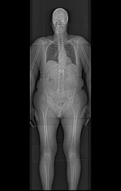

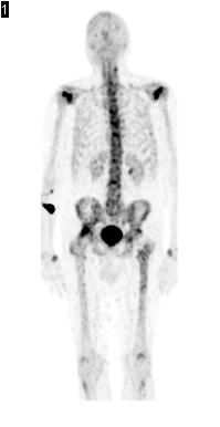
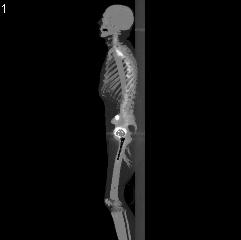
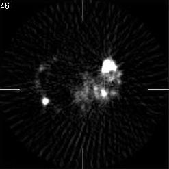
48 Future: Whole-body Bone SPECT/CT Tc-99m MDP Bone Imaging S. Cheenu Kappadath, PhD AAPM Anaheim, CA

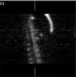
49 99m-Tc MDP SPECT/CT: Fused Coronal views

50 Future: Multi-nuclide SPECT/CT Maximum Intensity Projection (MIP) of a dual-isotope (Tc-99m and I-123) SPECT/CT mouse study. Published by the Molecular Imaging Center for Excellence newsletter, SNM publication Volume 2, 2008
Educational Objectives SPECT/CT: Basics, Quality Assurance, and Clinical Applications
 Educational Objectives SPECT/CT: Basics, Quality Assurance, and Clinical Applications 1. To understand the physics principles underlying SPECT/CT image acquisition and reconstruction 2. To understand the
Conflicts of Interest Nuclear Medicine and PET physics reviewer for the ACR Accreditation program
 James R Halama, PhD Loyola University Medical Center Conflicts of Interest Nuclear Medicine and PET physics reviewer for the ACR Accreditation program Learning Objectives 1. Be familiar with recommendations
Philips SPECT/CT Systems
 Philips SPECT/CT Systems Ling Shao, PhD Director, Imaging Physics & System Analysis Nuclear Medicine, Philips Healthcare June 14, 2008 *Presented SNM08 Categorical Seminar - Quantitative SPECT and PET
James R Halama, PhD Loyola University Medical Center
 James R Halama, PhD Loyola University Medical Center Conflicts of Interest Nuclear Medicine and PET physics reviewer for the ACR Accreditation program Learning Objectives Be familiar with the tests recommended
3/27/2012 WHY SPECT / CT? SPECT / CT Basic Principles. Advantages of SPECT. Advantages of CT. Dr John C. Dickson, Principal Physicist UCLH
 3/27/212 Advantages of SPECT SPECT / CT Basic Principles Dr John C. Dickson, Principal Physicist UCLH Institute of Nuclear Medicine, University College London Hospitals and University College London john.dickson@uclh.nhs.uk
Introduction to Positron Emission Tomography
 Planar and SPECT Cameras Summary Introduction to Positron Emission Tomography, Ph.D. Nuclear Medicine Basic Science Lectures srbowen@uw.edu System components: Collimator Detector Electronics Collimator
Medical Imaging BMEN Spring 2016
 Name Medical Imaging BMEN 420-501 Spring 2016 Homework #4 and Nuclear Medicine Notes All questions are from the introductory Powerpoint (based on Chapter 7) and text Medical Imaging Signals and Systems,
Introduction to Emission Tomography
 Introduction to Emission Tomography Gamma Camera Planar Imaging Robert Miyaoka, PhD University of Washington Department of Radiology rmiyaoka@u.washington.edu Gamma Camera: - collimator - detector (crystal
Fits you like no other
 Fits you like no other BrightView X and XCT specifications The new BrightView X system is a fully featured variableangle camera that is field-upgradeable to BrightView XCT without any increase in room
Workshop on Quantitative SPECT and PET Brain Studies January, 2013 PUCRS, Porto Alegre, Brasil Corrections in SPECT and PET
 Workshop on Quantitative SPECT and PET Brain Studies 14-16 January, 2013 PUCRS, Porto Alegre, Brasil Corrections in SPECT and PET Físico João Alfredo Borges, Me. Corrections in SPECT and PET SPECT and
Fits you like no other
 Fits you like no other Philips BrightView X and XCT specifications The new BrightView X system is a fully featured variableangle camera that is field-upgradeable to BrightView XCT without any increase
SPECT QA and QC. Bruce McBride St. Vincent s Hospital Sydney.
 SPECT QA and QC Bruce McBride St. Vincent s Hospital Sydney. SPECT QA and QC What is needed? Why? How often? Who says? QA and QC in Nuclear Medicine QA - collective term for all the efforts made to produce
BME I5000: Biomedical Imaging
 1 Lucas Parra, CCNY BME I5000: Biomedical Imaging Lecture 4 Computed Tomography Lucas C. Parra, parra@ccny.cuny.edu some slides inspired by lecture notes of Andreas H. Hilscher at Columbia University.
Introduction to Biomedical Imaging
 Alejandro Frangi, PhD Computational Imaging Lab Department of Information & Communication Technology Pompeu Fabra University www.cilab.upf.edu X-ray Projection Imaging Computed Tomography Digital X-ray
Diagnostic imaging techniques. Krasznai Zoltán. University of Debrecen Medical and Health Science Centre Department of Biophysics and Cell Biology
 Diagnostic imaging techniques Krasznai Zoltán University of Debrecen Medical and Health Science Centre Department of Biophysics and Cell Biology 1. Computer tomography (CT) 2. Gamma camera 3. Single Photon
Assessment of OSEM & FBP Reconstruction Techniques in Single Photon Emission Computed Tomography Using SPECT Phantom as Applied on Bone Scintigraphy
 Assessment of OSEM & FBP Reconstruction Techniques in Single Photon Emission Computed Tomography Using SPECT Phantom as Applied on Bone Scintigraphy Physics Department, Faculty of Applied Science,Umm Al-Qura
Quantitative capabilities of four state-of-the-art SPECT-CT cameras
 Seret et al. EJNMMI Research 2012, 2:45 ORIGINAL RESEARCH Open Access Quantitative capabilities of four state-of-the-art SPECT-CT cameras Alain Seret 1,2*, Daniel Nguyen 1 and Claire Bernard 3 Abstract
Ch. 4 Physical Principles of CT
 Ch. 4 Physical Principles of CT CLRS 408: Intro to CT Department of Radiation Sciences Review: Why CT? Solution for radiography/tomography limitations Superimposition of structures Distinguishing between
Implementation and evaluation of a fully 3D OS-MLEM reconstruction algorithm accounting for the PSF of the PET imaging system
 Implementation and evaluation of a fully 3D OS-MLEM reconstruction algorithm accounting for the PSF of the PET imaging system 3 rd October 2008 11 th Topical Seminar on Innovative Particle and Radiation
Review of PET Physics. Timothy Turkington, Ph.D. Radiology and Medical Physics Duke University Durham, North Carolina, USA
 Review of PET Physics Timothy Turkington, Ph.D. Radiology and Medical Physics Duke University Durham, North Carolina, USA Chart of Nuclides Z (protons) N (number of neutrons) Nuclear Data Evaluation Lab.
Validation of GEANT4 for Accurate Modeling of 111 In SPECT Acquisition
 Validation of GEANT4 for Accurate Modeling of 111 In SPECT Acquisition Bernd Schweizer, Andreas Goedicke Philips Technology Research Laboratories, Aachen, Germany bernd.schweizer@philips.com Abstract.
Corso di laurea in Fisica A.A Fisica Medica 5 SPECT, PET
 Corso di laurea in Fisica A.A. 2007-2008 Fisica Medica 5 SPECT, PET Step 1: Inject Patient with Radioactive Drug Drug is labeled with positron (β + ) emitting radionuclide. Drug localizes
ML reconstruction for CT
 ML reconstruction for CT derivation of MLTR rigid motion correction resolution modeling polychromatic ML model dual energy ML model Bruno De Man, Katrien Van Slambrouck, Maarten Depypere, Frederik Maes,
Symbia E and S System Specifications Answers for life.
 www.siemens.com/mi Symbia E and S System Specifications Answers for life. Symbia E: Work with confidence. What does it mean to work with confidence? It is clarity not only image clarity but the clarity
Shadow casting. What is the problem? Cone Beam Computed Tomography THE OBJECTIVES OF DIAGNOSTIC IMAGING IDEAL DIAGNOSTIC IMAGING STUDY LIMITATIONS
 Cone Beam Computed Tomography THE OBJECTIVES OF DIAGNOSTIC IMAGING Reveal pathology Reveal the anatomic truth Steven R. Singer, DDS srs2@columbia.edu IDEAL DIAGNOSTIC IMAGING STUDY Provides desired diagnostic
Spiral CT. Protocol Optimization & Quality Assurance. Ge Wang, Ph.D. Department of Radiology University of Iowa Iowa City, Iowa 52242, USA
 Spiral CT Protocol Optimization & Quality Assurance Ge Wang, Ph.D. Department of Radiology University of Iowa Iowa City, Iowa 52242, USA Spiral CT Protocol Optimization & Quality Assurance Protocol optimization
Optimization of CT Simulation Imaging. Ingrid Reiser Dept. of Radiology The University of Chicago
 Optimization of CT Simulation Imaging Ingrid Reiser Dept. of Radiology The University of Chicago Optimization of CT imaging Goal: Achieve image quality that allows to perform the task at hand (diagnostic
Biomedical Imaging. Computed Tomography. Patrícia Figueiredo IST
 Biomedical Imaging Computed Tomography Patrícia Figueiredo IST 2013-2014 Overview Basic principles X ray attenuation projection Slice selection and line projections Projection reconstruction Instrumentation
SPECT reconstruction
 Regional Training Workshop Advanced Image Processing of SPECT Studies Tygerberg Hospital, 19-23 April 2004 SPECT reconstruction Martin Šámal Charles University Prague, Czech Republic samal@cesnet.cz Tomography
Positron. MillenniumVG. Emission Tomography Imaging with the. GE Medical Systems
 Positron Emission Tomography Imaging with the MillenniumVG GE Medical Systems Table of Contents Introduction... 3 PET Imaging With Gamma Cameras PET Overview... 4 Coincidence Detection on Gamma Cameras...
GPU implementation for rapid iterative image reconstruction algorithm
 GPU implementation for rapid iterative image reconstruction algorithm and its applications in nuclear medicine Jakub Pietrzak Krzysztof Kacperski Department of Medical Physics, Maria Skłodowska-Curie Memorial
Reconstruction from Projections
 Reconstruction from Projections M.C. Villa Uriol Computational Imaging Lab email: cruz.villa@upf.edu web: http://www.cilab.upf.edu Based on SPECT reconstruction Martin Šámal Charles University Prague,
Multi-slice CT Image Reconstruction Jiang Hsieh, Ph.D.
 Multi-slice CT Image Reconstruction Jiang Hsieh, Ph.D. Applied Science Laboratory, GE Healthcare Technologies 1 Image Generation Reconstruction of images from projections. textbook reconstruction advanced
CT Basics Principles of Spiral CT Dose. Always Thinking Ahead.
 1 CT Basics Principles of Spiral CT Dose 2 Who invented CT? 1963 - Alan Cormack developed a mathematical method of reconstructing images from x-ray projections Sir Godfrey Hounsfield worked for the Central
V/Q Imaging Protocols
 Elizabeth ailey ppsc(mrs) M Department of Nuclear Medicine Royal North Shore Hospital, Sydney, ustralia email: ebailey@nsccahs.health.nsw.gov.au The V/Q scan has traditionally used planar imaging techniques.
4DM Packages. 4DM Packages & License Types. Information to help you order the appropriate licenses for your site.
 4DM Packages 4DM Packages & License Types. Information to help you order the appropriate licenses for your site. Nuclear Cardiac Quantification, Review, and Reporting Select Your 4DM Package and corresponding
Image Acquisition Systems
 Image Acquisition Systems Goals and Terminology Conventional Radiography Axial Tomography Computer Axial Tomography (CAT) Magnetic Resonance Imaging (MRI) PET, SPECT Ultrasound Microscopy Imaging ITCS
REMOVAL OF THE EFFECT OF COMPTON SCATTERING IN 3-D WHOLE BODY POSITRON EMISSION TOMOGRAPHY BY MONTE CARLO
 REMOVAL OF THE EFFECT OF COMPTON SCATTERING IN 3-D WHOLE BODY POSITRON EMISSION TOMOGRAPHY BY MONTE CARLO Abstract C.S. Levin, Y-C Tai, E.J. Hoffman, M. Dahlbom, T.H. Farquhar UCLA School of Medicine Division
PURE. ViSION Edition PET/CT. Patient Comfort Put First.
 PURE ViSION Edition PET/CT Patient Comfort Put First. 2 System features that put patient comfort and safety first. Oncology patients deserve the highest levels of safety and comfort during scans. Our Celesteion
Corso di laurea in Fisica A.A Fisica Medica 4 TC
 Corso di laurea in Fisica A.A. 2007-2008 Fisica Medica 4 TC Computed Tomography Principles 1. Projection measurement 2. Scanner systems 3. Scanning modes Basic Tomographic Principle The internal structure
Constructing System Matrices for SPECT Simulations and Reconstructions
 Constructing System Matrices for SPECT Simulations and Reconstructions Nirantha Balagopal April 28th, 2017 M.S. Report The University of Arizona College of Optical Sciences 1 Acknowledgement I would like
SPECT: Physics Principles and Equipment Design
 SPECT: Physics Principles and Equipment Design Eric C. Frey, Ph.D., Professor Division of Medical Imaging Physics Russell H. Morgan Department of Radiology and Radiological Science Disclosures Johns Hopkins
MEDICAL IMAGING 2nd Part Computed Tomography
 MEDICAL IMAGING 2nd Part Computed Tomography Introduction 2 In the last 30 years X-ray Computed Tomography development produced a great change in the role of diagnostic imaging in medicine. In convetional
Digital Image Processing
 Digital Image Processing SPECIAL TOPICS CT IMAGES Hamid R. Rabiee Fall 2015 What is an image? 2 Are images only about visual concepts? We ve already seen that there are other kinds of image. In this lecture
If it matters to you, it matters to us
 If it matters to you, it matters to us Philips clinical innovations in nuclear medicine Innovation with insight We understand that clinical innovations are only as valuable as the day-to-day difference
DICOM. Supplement 188 Multi-Energy CT Imaging. DICOM Working Group 21 Computed Tomography
 DICOM Supplement 188 Multi-Energy CT Imaging DICOM Working Group 21 Computed Tomography Rationale Short introduction of Multi Energy (ME) s Overview: Imaging techniques, including scanning, reconstruction,
Spiral ASSR Std p = 1.0. Spiral EPBP Std. 256 slices (0/300) Kachelrieß et al., Med. Phys. 31(6): , 2004
 Kachelrieß et al., Med. Phys. 31(6): , 2004") Spiral ASSR Std p = 1.0 Spiral EPBP Std p = 1.0 Kachelrieß et al., Med. Phys. 31(6): 1623-1641, 2004 256 slices (0/300) Advantages of Cone-Beam Spiral CT Image quality nearly independent of pitch Increase
Spiral ASSR Std p = 1.0 Spiral EPBP Std p = 1.0 Kachelrieß et al., Med. Phys. 31(6): 1623-1641, 2004 256 slices (0/300) Advantages of Cone-Beam Spiral CT Image quality nearly independent of pitch Increase
DUE to beam polychromacity in CT and the energy dependence
 1 Empirical Water Precorrection for Cone-Beam Computed Tomography Katia Sourbelle, Marc Kachelrieß, Member, IEEE, and Willi A. Kalender Abstract We propose an algorithm to correct for the cupping artifact
MEDICAL IMAGING 2nd Part Computed Tomography
 MEDICAL IMAGING 2nd Part Computed Tomography Introduction 2 In the last 30 years X-ray Computed Tomography development produced a great change in the role of diagnostic imaging in medicine. In convetional
8/2/2017. Disclosure. Philips Healthcare (Cleveland, OH) provided the precommercial
 provided the precommercial") 8//0 AAPM0 Scientific Symposium: Emerging and New Generation PET: Instrumentation, Technology, Characteristics and Clinical Practice Aug Wednesday 0:4am :pm Solid State Digital Photon Counting PET/CT Instrumentation
8//0 AAPM0 Scientific Symposium: Emerging and New Generation PET: Instrumentation, Technology, Characteristics and Clinical Practice Aug Wednesday 0:4am :pm Solid State Digital Photon Counting PET/CT Instrumentation
Monte-Carlo-Based Scatter Correction for Quantitative SPECT Reconstruction
 Monte-Carlo-Based Scatter Correction for Quantitative SPECT Reconstruction Realization and Evaluation Rolf Bippus 1, Andreas Goedicke 1, Henrik Botterweck 2 1 Philips Research Laboratories, Aachen 2 Fachhochschule
Nuclear Medicine Imaging
 Introduction to Medical Engineering (Medical Imaging) Suetens 5 Nuclear Medicine Imaging Ho Kyung Kim Pusan National University Introduction Use of radioactive isotopes for medical purposes since 1920
Fast Timing and TOF in PET Medical Imaging
 Fast Timing and TOF in PET Medical Imaging William W. Moses Lawrence Berkeley National Laboratory October 15, 2008 Outline: Time-of-Flight PET History Present Status Future This work was supported in part
664 IEEE TRANSACTIONS ON NUCLEAR SCIENCE, VOL. 52, NO. 3, JUNE 2005
 664 IEEE TRANSACTIONS ON NUCLEAR SCIENCE, VOL. 52, NO. 3, JUNE 2005 Attenuation Correction for the NIH ATLAS Small Animal PET Scanner Rutao Yao, Member, IEEE, Jürgen Seidel, Jeih-San Liow, Member, IEEE,
Concepts, Applications, and Requirements for Quantitative SPECT/CT. Conflict of Interest Disclosure
 Concepts, Applications, and Requirements for Quantitative SPECT/CT Eric C. Frey, Ph.D. (efrey@jhmi.edu) Division of Medical Imaging Physics Russell H. Morgan Department of Radiology and Radiological Science
Positron Emission Tomography
 Physics 656 Seminar on Physical Fundamentals of Medical Imaging Positron Emission Tomography Ahmed Qamesh Outline What is PET? PET mechanism Radionuclide and its synthesis Detection concept and Development
Iterative SPECT reconstruction with 3D detector response
 Iterative SPECT reconstruction with 3D detector response Jeffrey A. Fessler and Anastasia Yendiki COMMUNICATIONS & SIGNAL PROCESSING LABORATORY Department of Electrical Engineering and Computer Science
Medical Image Processing: Image Reconstruction and 3D Renderings
 Medical Image Processing: Image Reconstruction and 3D Renderings 김보형 서울대학교컴퓨터공학부 Computer Graphics and Image Processing Lab. 2011. 3. 23 1 Computer Graphics & Image Processing Computer Graphics : Create,
Moscow-Bavarian Joint Advanced Student School 2006 / Medical Imaging Principles of Computerized Tomographic Imaging and Cone-Beam Reconstruction
 Line Integrals Line integrals represent the integral of some parameter of the object along the line (e.g. attenuation of x-rays) Object: f(x,y) Line: x cosθ + y sinθ = t Line integral / Radon transform:
Symbia T Series System Specifications Answers for life.
 www.siemens.com/mi System Specifications Answers for life. 2 Diagnostic Excellence Today s healthcare institutions have increasingly diverse patient needs, greater budget limitations and the need for higher
Master of Science Thesis. Activity quantification of planar gamma camera images
 Master of Science Thesis Activity quantification of planar gamma camera images Marie Sydoff Supervisor: Sigrid Leide-Svegborn The work has been performed at Department of Radiation Physics Malmö University
Nuclear Medicine Imaging Systems
 Nuclear Medicine Imaging Systems Position estimation based on 2-D centroid calculation called Anger logic or Anger camera PMT signal Posi/on op/cal photons Scin/lla/on gamma-ray Collimator Analogies with
Cherenkov Radiation. Doctoral Thesis. Rok Dolenec. Supervisor: Prof. Dr. Samo Korpar
 Doctoral Thesis Time-of-Flight Time-of-Flight Positron Positron Emission Emission Tomography Tomography Using Using Cherenkov Cherenkov Radiation Radiation Rok Dolenec Supervisor: Prof. Dr. Samo Korpar
Technical aspects of SPECT and SPECT-CT. John Buscombe
 Technical aspects of SPECT and SPECT-CT John Buscombe What does the clinician need to know? For SPECT What factors affect SPECT How those factors should be sought Looking for artefacts For SPECT-CT Issues
Computer-Tomography II: Image reconstruction and applications
 Computer-Tomography II: Image reconstruction and applications Prof. Dr. U. Oelfke DKFZ Heidelberg Department of Medical Physics (E040) Im Neuenheimer Feld 280 69120 Heidelberg, Germany u.oelfke@dkfz.de
8/7/2017. Disclosures. MECT Systems Overview and Quantitative Opportunities. Overview. Computed Tomography (CT) CT Numbers. Polyenergetic Acquisition
 CT Numbers. Polyenergetic Acquisition") Quantitative Multi-Energy Computed Tomography: Imaging and Therapy Advancements Disclosures MECT Systems Overview and Quantitative Opportunities The speaker receives research funding from GE Healthcare
Quantitative Multi-Energy Computed Tomography: Imaging and Therapy Advancements Disclosures MECT Systems Overview and Quantitative Opportunities The speaker receives research funding from GE Healthcare
Radon Transform and Filtered Backprojection
 Radon Transform and Filtered Backprojection Jørgen Arendt Jensen October 13, 2016 Center for Fast Ultrasound Imaging, Build 349 Department of Electrical Engineering Center for Fast Ultrasound Imaging Department
Quantitative imaging for clinical dosimetry
 Quantitative imaging for clinical dosimetry Irène Buvat Laboratoire d Imagerie Fonctionnelle U678 INSERM - UPMC CHU Pitié-Salpêtrière, Paris buvat@imed.jussieu.fr http://www.guillemet.org/irene Methodology
Introduc)on to PET Image Reconstruc)on. Tomographic Imaging. Projec)on Imaging. Types of imaging systems
on to PET Image Reconstruc)on. Tomographic Imaging. Projec)on Imaging. Types of imaging systems") Introduc)on to PET Image Reconstruc)on Adam Alessio http://faculty.washington.edu/aalessio/ Nuclear Medicine Lectures Imaging Research Laboratory Division of Nuclear Medicine University of Washington Fall
Introduc)on to PET Image Reconstruc)on Adam Alessio http://faculty.washington.edu/aalessio/ Nuclear Medicine Lectures Imaging Research Laboratory Division of Nuclear Medicine University of Washington Fall
A Comparison of the Uniformity Requirements for SPECT Image Reconstruction Using FBP and OSEM Techniques
 IMAGING A Comparison of the Uniformity Requirements for SPECT Image Reconstruction Using FBP and OSEM Techniques Lai K. Leong, Randall L. Kruger, and Michael K. O Connor Section of Nuclear Medicine, Department
Mediso AnyScan S Single-Head and Dual-Head SPECT
 Mediso AnyScan S Single-Head and Dual-Head SPECT ANYSCAN S (SINGLE-HEAD & DUAL-HEAD SPECT) Low Cost of Ownership Small Footprint Absolute Imaging Solutions tm is the exclusive U.S. source for Mediso Medical
Mediso AnyScan. Single-Head and Dual-Head SPECT
 Mediso AnyScan S Single-Head and Dual-Head SPECT ANYSCAN S (SINGLE-HEAD & DUAL-HEAD SPECT) Low Cost of Ownership Associated Imaging Services is the exclusive U.S. source in the Midwest for Mediso Medical
Advanced Scatter Correction for Quantitative Cardiac SPECT
 Advanced Scatter Correction for Quantitative Cardiac SPECT Frederik J. Beekman PhD Molecular Image Science Laboratory University Medical Center Utrecht The Netherlands Outline What is scatter? Scatter
The Near Future in Cardiac CT Image Reconstruction
 SCCT 2010 The Near Future in Cardiac CT Image Reconstruction Marc Kachelrieß Institute of Medical Physics (IMP) Friedrich-Alexander Alexander-University Erlangen-Nürnberg rnberg www.imp.uni-erlangen.de
Index. aliasing artifacts and noise in CT images, 200 measurement of projection data, nondiffracting
 Index Algebraic equations solution by Kaczmarz method, 278 Algebraic reconstruction techniques, 283-84 sequential, 289, 293 simultaneous, 285-92 Algebraic techniques reconstruction algorithms, 275-96 Algorithms
Inveon User Notes. Inveon Scanners and Inveon Acquisition Workplace 2.0
 Inveon User Notes Inveon Scanners and Inveon Acquisition Workplace 2.0 Legal Notice 2014 by Siemens Medical Solutions USA, Inc. All rights reserved. Siemens reserves the right to modify the design and
Tomographic Reconstruction
 Tomographic Reconstruction 3D Image Processing Torsten Möller Reading Gonzales + Woods, Chapter 5.11 2 Overview Physics History Reconstruction basic idea Radon transform Fourier-Slice theorem (Parallel-beam)
Radiology. Marta Anguiano Millán. Departamento de Física Atómica, Molecular y Nuclear Facultad de Ciencias. Universidad de Granada
 Departamento de Física Atómica, Molecular y Nuclear Facultad de Ciencias. Universidad de Granada Overview Introduction Overview Introduction Tecniques of imaging in Overview Introduction Tecniques of imaging
Validation of PET/CT dataset for radiation treatment planning
 Louisiana State University LSU Digital Commons LSU Master's Theses Graduate School 2004 Validation of PET/CT dataset for radiation treatment planning Rajesh Manoharan Louisiana State University and Agricultural
Enhancement Image Quality of CT Using Single Slice Spiral Technique
 Enhancement Image Quality of CT Using Single Slice Spiral Technique Doaa. N. Al Sheack 1 and Dr.Mohammed H. Ali Al Hayani 2 1 2 Electronic and Communications Engineering Department College of Engineering,
Emission Computed Tomography Notes
 Noll (24) ECT Notes: Page 1 Emission Computed Tomography Notes Introduction Emission computed tomography (ECT) is the CT applied to nuclear medicine. There are two varieties of ECT: 1. SPECT single-photon
Evaluation of Spectrum Mismatching using Spectrum Binning Approach for Statistical Polychromatic Reconstruction in CT
 Evaluation of Spectrum Mismatching using Spectrum Binning Approach for Statistical Polychromatic Reconstruction in CT Qiao Yang 1,4, Meng Wu 2, Andreas Maier 1,3,4, Joachim Hornegger 1,3,4, Rebecca Fahrig
Automated Image Analysis Software for Quality Assurance of a Radiotherapy CT Simulator
 Automated Image Analysis Software for Quality Assurance of a Radiotherapy CT Simulator Andrew J Reilly Imaging Physicist Oncology Physics Edinburgh Cancer Centre Western General Hospital EDINBURGH EH4
Performance Evaluation of radionuclide imaging systems
 Performance Evaluation of radionuclide imaging systems Nicolas A. Karakatsanis STIR Users meeting IEEE Nuclear Science Symposium and Medical Imaging Conference 2009 Orlando, FL, USA Geant4 Application
Iterative and analytical reconstruction algorithms for varying-focal-length cone-beam
 Home Search Collections Journals About Contact us My IOPscience Iterative and analytical reconstruction algorithms for varying-focal-length cone-beam projections This content has been downloaded from IOPscience.
Effects of the difference in tube voltage of the CT scanner on. dose calculation
 Effects of the difference in tube voltage of the CT scanner on dose calculation Dong Joo Rhee, Sung-woo Kim, Dong Hyeok Jeong Medical and Radiological Physics Laboratory, Dongnam Institute of Radiological
Respiratory Motion Compensation for Simultaneous PET/MR Based on Strongly Undersampled Radial MR Data
 Respiratory Motion Compensation for Simultaneous PET/MR Based on Strongly Undersampled Radial MR Data Christopher M Rank 1, Thorsten Heußer 1, Andreas Wetscherek 1, and Marc Kachelrieß 1 1 German Cancer
QIBA PET Amyloid BC March 11, Agenda
 QIBA PET Amyloid BC March 11, 2016 - Agenda 1. QIBA Round 6 Funding a. Deadlines b. What projects can be funded, what cannot c. Discussion of projects Mechanical phantom and DRO Paul & John? Any Profile
COMPARATIVE STUDIES OF DIFFERENT SYSTEM MODELS FOR ITERATIVE CT IMAGE RECONSTRUCTION
 COMPARATIVE STUDIES OF DIFFERENT SYSTEM MODELS FOR ITERATIVE CT IMAGE RECONSTRUCTION BY CHUANG MIAO A Thesis Submitted to the Graduate Faculty of WAKE FOREST UNIVERSITY GRADUATE SCHOOL OF ARTS AND SCIENCES
3-D Monte Carlo-based Scatter Compensation in Quantitative I-131 SPECT Reconstruction
 3-D Monte Carlo-based Scatter Compensation in Quantitative I-131 SPECT Reconstruction Yuni K. Dewaraja, Member, IEEE, Michael Ljungberg, Member, IEEE, and Jeffrey A. Fessler, Member, IEEE Abstract- we
Outline. What is Positron Emission Tomography? (PET) Positron Emission Tomography I: Image Reconstruction Strategies
 Positron Emission Tomography I: Image Reconstruction Strategies") CE: PET Physics and Technology II 2005 AAPM Meeting, Seattle WA Positron Emission Tomography I: Image Reconstruction Strategies Craig S. Levin, Ph.D. Department of Radiology and Molecular Imaging Program
CE: PET Physics and Technology II 2005 AAPM Meeting, Seattle WA Positron Emission Tomography I: Image Reconstruction Strategies Craig S. Levin, Ph.D. Department of Radiology and Molecular Imaging Program
MEDICAL EQUIPMENT: COMPUTED TOMOGRAPHY. Prof. Yasser Mostafa Kadah
 MEDICAL EQUIPMENT: COMPUTED TOMOGRAPHY Prof. Yasser Mostafa Kadah www.k-space.org Recommended Textbook X-Ray Computed Tomography in Biomedical Engineering, by Robert Cierniak, Springer, 211 Computed Tomography
Metal Artifact Reduction CT Techniques. Tobias Dietrich University Hospital Balgrist University of Zurich Switzerland
 Metal Artifact Reduction CT Techniques R S S S Tobias Dietrich University Hospital Balgrist University of Zurich Switzerland N. 1 v o 4 1 0 2. Postoperative CT Metal Implants CT is accurate for assessment
RADIOLOGY AND DIAGNOSTIC IMAGING
 Day 2 part 2 RADIOLOGY AND DIAGNOSTIC IMAGING Dr hab. Zbigniew Serafin, MD, PhD serafin@cm.umk.pl 2 3 4 5 CT technique CT technique 6 CT system Kanal K: RSNA/AAPM web module: CT Systems & CT Image Quality
Medical Image Analysis
 Computer assisted Image Analysis VT04 29 april 2004 Medical Image Analysis Lecture 10 (part 1) Xavier Tizon Medical Image Processing Medical imaging modalities XRay,, CT Ultrasound MRI PET, SPECT Generic
The Emory Reconstruction Toolbox Version 1.0
 The Emory Reconstruction Toolbox Version 1.0 Operating Instructions Revision 02 (April, 2008) Operating Instructions The Emory Reconstruction Toolbox Application Copyrights, Trademarks, Restrictions
USING cone-beam geometry with pinhole collimation,
 IEEE TRANSACTIONS ON NUCLEAR SCIENCE, VOL. 56, NO. 3, JUNE 2009 687 A Backprojection-Based Parameter Estimation Technique for Skew-Slit Collimation Jacob A. Piatt, Student Member, IEEE, and Gengsheng L.
ISO ISO ISO OHSAS ISO
 ISO 9001 ISO 13485 ISO 14001 OHSAS 18001 ISO 27001 Pro-NM Performance 08-101 - standard version 08-103 - version with the PET Lid Phantom for NM and PET systems performance evaluation (collimator, artifacts,
A closer look at CT scanning
 Vet Times The website for the veterinary profession https://www.vettimes.co.uk A closer look at CT scanning Author : Charissa Lee, Natalie Webster Categories : General, Vets Date : April 3, 2017 A basic
(RMSE). Reconstructions showed that modeling the incremental blur improved the resolution of the attenuation map and quantitative accuracy.
. Reconstructions showed that modeling the incremental blur improved the resolution of the attenuation map and quantitative accuracy.") Modeling the Distance-Dependent Blurring in Transmission Imaging in the Ordered-Subset Transmission (OSTR) Algorithm by Using an Unmatched Projector/Backprojector Pair B. Feng, Member, IEEE, M. A. King,
Modeling the Distance-Dependent Blurring in Transmission Imaging in the Ordered-Subset Transmission (OSTR) Algorithm by Using an Unmatched Projector/Backprojector Pair B. Feng, Member, IEEE, M. A. King,