Nature Methods: doi: /nmeth Supplementary Figure 1
|
|
|
- Ralph Ramsey
- 5 years ago
- Views:
Transcription

1 Supplementary Figure 1 Schematic demonstrating the preferred orientation sampling problem in single-particle cryo-em. Specimens sticking to the air-water interface or a substrate support of a cryo-em grid in a single preferred orientation always adopt random in-plane orientations. Tilting of the specimen stage therefore leads to (a) a precession of views around the axis of preferred orientation in a conical manner (precession indicated in green with grey arrows, axis of preferred orientation is indicated by a red arrow). (b) Projections of the object in (a) are displayed for 30 and 60 tilts (theta angle), both at a φ angle increment of 15. Whereas the 30 tilted projections resemble more the top views of the object, the 60 tilted projections resemble more the side views of the object. (c) In reciprocal space, each Fourier slice inserted into the reconstruction is perpendicular to the direction of its real-space projection image. Therefore, a large missing cone is formed from a reconstruction using 30 tilted images, and a much smaller missing cone is present from a reconstruction using 60 tilted images. No missing cone would be present for (hypothetically) 90 tilted images, although the X/Y plane would be characterized by 1D lines rather than a complete 2D slice. The practical implications of this diagram and anisotropic sampling are demonstrated in Supplementary Figure 2.

2 Supplementary Figure 2 3D reconstructions of a synthetic, absolutely preferred dataset of an HA trimer presumed to be tilted at various angles.
3 (a) Euler angle distribution profiles of the synthetic projections. (b) The resulting 3D reconstructions. The red arrow shows the direction of preferred orientation. The grey arrows show the angle at which the synthetic projection images were generated at the various tilt angles. A synthetic dataset of equally sampled HA trimer is also shown for comparison in the last row. (c) Slice through the 3D FSC volumes describing the 3D reconstructions along the X/Z plane. The 3D FSC was thresholded at 0.143, while sphericity was calculated by thresholding the 3D FSC at 0.5 and applying a Gaussian filter of 2 pixels. Due to C3 symmetry along the Z-axis of the reconstruction, only the X/Z slice of the 3D FSC is shown. Missing cones are apparent within the 30, and especially the 60 3D FSCs. (d) Plots of the global FSC after reconstruction (solid blue line), the spread of the directional resolutions defined by plus and minus one standard deviation from the mean of the directional resolutions (green area encompassed by green dotted lines) and the histogram of one hundred directional resolutions evenly sampled over the 3D FSC (yellow bars). Labels on the left Y-axis refer to global FSC curves, while labels on the right Y-axis refer to directional FSC histograms. The grey dotted line indicates when FSC =

4
5 Supplementary Figure 3 HA trimer per-tilt analysis. HA trimer cryo-em data was collected (a) untilted, or at tilts of (b) 10, (c) 20, (d) 30, (e) 40, and (f) 50. Representative 2D class averages for each tilt angle are shown as insert. (g) The average frame-to-frame shift for each tilt angle is shown for both the first 15 frames and also for the entire range of 100 frames (insert). The exact cause of the slightly higher beam-induced movement within the 0 dataset is unclear and was not observed for the ribosome dataset (Supplementary Figure 6g).

6
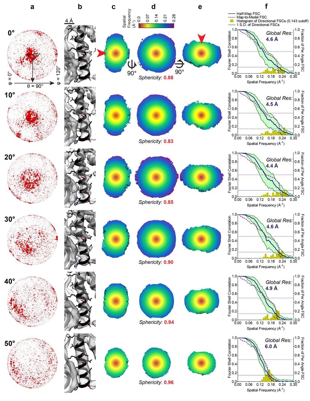
7 Supplementary Figure 4 Tilting improves resolution and directional isotropy in a direct comparison of HA trimer reconstructions without and with tilting. (a) Euler angle distribution of the particles. (b) 3D reconstruction superimposed onto a projection of the HA trimer crystal structure (pink). Loss of axial density at typical display thresholds is clearly evident within reconstructions from 0, 10, and 20 tilted images. (c) Close-up of a particular region within the reconstruction. Circled region indicates false positive density resulting from elongation of the reconstruction along the Z-axis, which progressively disappears within reconstructions from higher tilts characterized by improved directional resolution isotropy. (d) Slice through the 3D FSC describing (top, solid blue outline) half-map, resolution evaluating internal consistency and (bottom, dotted purple outline) map-to-model resolution evaluating external consistency, along with their corresponding sphericity values (e). 3D FSC sphericity values. The slight dip in 3D FSC sphericity at 10 is caused by greater improvements in directional resolution perpendicular to the preferred orientation axis that are not met with a concomitant increase in Z resolution. (f) Graph showing the global half-map resolution (solid blue), map-to-model resolution (dotted purple), and spread of directional resolution (refer to Supplementary Figure 2 for detailed graph description).
8
9 Supplementary Figure 5 HA particle titration experiment. HA trimer reconstructions using subsets of particles from (a) untilted and (b) 40 tilted images. Random subsets of particles from the 130,000 total particles, in multiples of 13,000, were selected for each titration point and refined independently. The half map resolution (using threshold) and map-to-model resolution (using 0.5 threshold) against the structure of HA (PDB 3WHE) is shown for each titration point. The Euler angle distribution of the reconstructions at each titration point for the (c) dataset from 0 images and (d) 40 tilted images show that the apparent side views in the untilted reconstruction (green arrows) only appear when more particles are added to the reconstruction, indicating overfitting.
10
11 Supplementary Figure 6 Per-tilt analysis of L17-depleted 50S ribosomal assembly intermediates (LSU bl17dep ). Cryo-EM data was collected (a) untilted and at tilts of (b) 10, (c) 20, (d) 30, (e) 40 and (f) 50. The average frame-to-frame shift for each tilt angle is shown for both the first 15 frames (g) and also for the entire range of 50 frames (insert). (h) The 4 super-classes of L17-depleted 50S ribosomal assembly intermediates are shown.

12
13 Supplementary Figure 7 LSU bl17dep Class B. For each tilt angle, the figure shows (a) Euler angle distributions of the particles, (b,c,d) 3D FSC at orthogonal views, and (e) global half-map and map-to-model FSC plots with the spread of directional resolutions defined by the green area and 3D FSC values overlaid as histograms. Direction of preferred orientation is indicated by the red arrow.

14
15 Supplementary Figure 8 LSU bl17dep Class C. For each tilt angle, the figure shows (a) Euler angle distributions of the particles, (b) an alpha helix density from ul29 protein, (c,d,e) 3D FSC at orthogonal views, and (f) global half-map and map-to-model FSC plots with the spread of directional resolutions defined by the green area and 3D FSC values overlaid as histograms. Direction of preferred orientation is indicated by the red arrow.

16
17 Supplementary Figure 9 LSU bl17dep Class D. For each tilt angle, the figure shows (a) Euler angle distributions of the particles, (b) an alpha helix density from ul29 protein, (c,d,e) 3D FSC at orthogonal views, and (f) global half-map and map-to-model FSC plots with the spread of directional resolutions defined by the green area and 3D FSC values overlaid as histograms. Direction of preferred orientation is indicated by the red arrow.
18
19 Supplementary Figure 10 LSU bl17dep Class E. For each tilt angle, the figure shows (a) Euler angle distributions of the particles, (b) an alpha helix density from ul29 protein, (c,d,e) 3D FSC at orthogonal views, and (f) global half-map and map-to-model FSC plots with the spread of directional resolutions defined by the green area and 3D FSC values overlaid as histograms. Direction of preferred orientation is indicated by the red arrow.
 resolution, (b) half-map 3D FSC sphericity, and (c) map-to-model 3D FSC sphericity are plotted with respect to the dataset from untilted images and")
20 Supplementary Figure 11 Tilting improves resolution and angular isotropy for all four super-classes of L17-depleted 50S ribosomal intermediates. For the four super-classes, B-E, the changes in (a) resolution, (b) half-map 3D FSC sphericity, and (c) map-to-model 3D FSC sphericity are plotted with respect to the dataset from untilted images and as a function of tilt angle (see Supplementary Figures 7-10 for all raw data). Map densities of (d) beta sheets and (e) alpha helices from reconstructions at various tilts for class E are also shown to illustrate the effects of change in resolution and resolution anisotropy (the two are interrelated). Beta sheets are from ul22 and the alpha helices are from ul29. Side chain densities are marked by red asterisks, while beta sheet separation is indicated by a green arrow. The direction of preferred orientation is indicated by the red double-headed arrow. As expected, map isotropy steadily improves with increasing angular tilt, which also affects global resolution and can accordingly facilitate interpretation of structural features. For example, smearing of beta-strands (d) and alpha-helices (e) parallel to the direction of preferred orientation is ameliorated with tilts, which can be especially important at borderline resolutions for interpreting atomic models.
 3D reconstruction from a preferred oriented LSU bl17dep ribosomal intermediate dataset, where particles were combined from all super-classes but only using data from 50 tilted images.")
21 Supplementary Figure 12 Tilt angles up to 50 provide near-atomic resolution single-particle reconstructions. (a) 3D reconstruction from a preferred oriented LSU bl17dep ribosomal intermediate dataset, where particles were combined from all super-classes but only using data from 50 tilted images. The sample heterogeneity from combining different super-classes is reflected in the local resolution colored onto the reconstruction. Whereas the peripheral density changes, the homogeneous core components are much better resolved. (b) Global half-map 3D FSC and (c) map-to-model 3D FSC. The high tilt angle used for collection results in nearly spherical 3D FSCs, with sphericity of 0.98 and 0.92 respectively. (d) The ten best resolved ribosomal proteins have resolutions of between 4.2 Å and 4.8 Å. (e-g) Map density of (e) bl21, (f) bl20 and (g) ul29 show side chain density and separation of beta strands.

22
23 Supplementary Figure 13 Workflow for collection of single particle data at tilts. Data can be collected at either one or multiple tilt angles. Other than the need to tilt the stage and perform per-particle CTF estimation, the other processing steps are similar to a conventional single particle cryo-em workflow. In order to analyze the degree of directional anisotropy, 3D FSC is performed at the end (isosurface shown) and its results are visualized in Chimera - when rotating the map, the map s color and associated directional FSC line component changes on the fly, enabling immediate assessment of resolution anisotropy.
24 Supplementary Note 1 Previous efforts to overcome preferred specimen orientation in single-particle cryo-em Given the plethora of samples affected to various extents by preferred specimen orientation, efforts have been made to solve this problem, with and without the implementation of tilts. Experimentally, one can collect the same field of view at multiple tilts and rely upon the differences in the goniometer tilt angle to define relative differences in orientations, as exemplified by the random conical 1, 2 or orthogonal tilt 3 reconstruction schemes in their explicit implementations. The drawback of these approaches is that multiple exposures of the same area generates beam-induced movement that cannot be explicitly accounted for by the absolute difference in relative tilt 4, which in principle limits the achievable resolution. Variations on tilted single-particle 5 or tomographic reconstruction methods 6 have also generally been limited to low resolutions. One recent tomographic reconstruction has reached near-atomic resolution, but required ~600k asymmetric units for the reconstruction and benefitted from isotropic orientation distribution during sub-tomogram selection on virus surfaces 7. The grids themselves can also be treated to induce additional orientations, for example by using a continuous layer of carbon as a substrate 8, coating with self-assembled monolayers 9, treating the carbon support in the presence of N-amylamine 10, or mechanically deforming the grid 11. Additives like poly-l-lysine 12 or detergents 13 can sometimes increase the range of orientations the molecules adopt. None of these techniques are generally applicable, and most have drawbacks, such as reduced particle contrast (e.g. poly-l-lysine or support films) or the requirement of significantly higher specimen concentrations (e.g. detergents). Computationally, 3D reconstruction procedures can partially compensate for and up-weight under-represented views 14, but they are not able to recover missing information if it is absent. A brute force approach is to simply collect more particles to obtain sufficient quantities of low abundance views 15.
25 Supplementary Note 2 Preferred specimen orientation leads to uneven coverage in reciprocal space Preferred particle orientation on a cryo-em grid arises from specimen interaction with the air-waterinterface 16, 17. In a typical single-particle cryo-em data collection in the absence of any tilts, the axis of preferred orientation is approximately parallel to the axis of the electron beam. Since individual particles adopt random in-plane orientations with respect to the electron beam, physically tilting the stage induces a precession of angular sampling around each preferred orientation axis, of which there may be several in any individual dataset. Thus, for each subset of preferred angular orientations, tilting the specimen stage fills reciprocal space with Fourier slices that are distributed in a conical fashion around the axis of preferred orientation (Supplementary Fig. 1).
26 Supplementary Note 3 Evaluation of directional resolution and density isotropy within synthetic single-particle data To demonstrate how different angular samplings affect the directional resolution of a 3D reconstruction, we used a synthetic dataset that simulates the effects of imaging under an electron microscope at defined orientations relative to the electron beam. The HA envelope trimer (PDB 3WHE) was selected as the biological sample for the synthetic dataset, as it exhibits a highly preferred orientation when vitrified on an EM grid (see also Supplementary Note 4). Four synthetic datasets were generated (Supplementary Fig. 2), with the specimen assumed to be preferentially oriented along its long axis and tilted at 30, 60, 90, or uniformly in all directions (Supplementary Fig. 2a-b). The density map improves monotonically with increasing tilt angle; notably, at 60 tilt angle, the map becomes visually very similar to the more isotropic reconstructions. The 3D FSC effectively incorporates a series of 1D directional FSC curves computed over distinct angles and compiled into a 3D volume, which then fully describes directional anisotropy. Throughout this manuscript, the 3D FSC is displayed as a slice through the volume at a nominal threshold (0.143 here), which provides a quantitative evaluation of resolution and helps to show the degree of deformation in the directions along the displayed plane. A simple calculation of 3D FSC sphericity (ranging from 0 to 1) approximates density isotropy (Supplemental Fig. 2c). For example, the dataset from 30 images is characterized by a dramatic loss of resolution in the Z direction, parallel to the electron beam, resulting in a pancake-shaped 3D FSC (sphericity 0.63). This effect is smaller, albeit still present for the reconstruction from 60 tilts (sphericity 0.84). A reconstruction from 90 tilts (sphericity 0.99) or from uniformly distributed projection orientations (sphericity 1.00) produces an isotropic 3D FSC. Distinct directional resolution values obtained at a nominal cutoff threshold can also be plotted as a histogram onto a conventional 2D FSC plot and compared with the global resolution (Supplemental Fig. 2d). As expected, improvements in directional resolution isotropy parallel improvements in global resolution. In sum, this analysis shows that, under ideal conditions, improvement in angular distribution and density isotropy results in: 1) a reconstruction with higher resolution features, 2) a 3D FSC that becomes more spherical, 3) reduced spread of directional resolutions, and 4) overall improvement in individual directional resolution components.
27 Supplementary Note 4 Rationale for selecting influenza hemagglutinin (HA) trimer as test sample To evaluate how tilts affect structure determination of samples exhibiting a highly preferred orientation, we utilized the soluble portion of the small, 150 kda influenza hemagglutinin (HA) trimer. The HA trimer was selected as a test sample for several reasons: 1) there are numerous highresolution crystal structures present in the public domain; 2) it is clinically relevant 18 ; 3) its molecular weight is low for cryo-em structure determination; 4) most importantly, it exhibits a single, highly preferred top orientation on vitrified cryo-em grids (Supplementary Fig. 3). The HA trimer therefore presents many challenges to conventional EM structure determination it has been solved at low resolution using electron tomography and negative stain, but thus far there has not been a highresolution single particle cryo-em reconstruction.
28 References 1. Radermacher, M. Three-dimensional reconstruction of single particles from random and nonrandom tilt series. J Electron Microsc Tech 9, (1988). 2. Radermacher, M., Wagenknecht, T., Verschoor, A. & Frank, J. Three-dimensional reconstruction from a single-exposure, random conical tilt series applied to the 50S ribosomal subunit of Escherichia coli. J Microsc 146, (1987). 3. Leschziner, A.E. & Nogales, E. The orthogonal tilt reconstruction method: an approach to generating single-class volumes with no missing cone for ab initio reconstruction of asymmetric particles. Journal of structural biology 153, (2006). 4. Henderson, R. et al. Tilt-pair analysis of images from a range of different specimens in singleparticle electron cryomicroscopy. J Mol Biol 413, (2011). 5. Yip, C.K., Murata, K., Walz, T., Sabatini, D.M. & Kang, S.A. Structure of the human mtor complex I and its implications for rapamycin inhibition. Mol Cell 38, (2010). 6. Bartesaghi, A., Lecumberry, F., Sapiro, G. & Subramaniam, S. Protein secondary structure determination by constrained single-particle cryo-electron tomography. Structure 20, (2012). 7. Schur, F.K. et al. An atomic model of HIV-1 capsid-sp1 reveals structures regulating assembly and maturation. Science 353, (2016). 8. Frank, J., Penczek, P., Grassucci, R. & Srivastava, S. Three-dimensional reconstruction of the 70S Escherichia coli ribosome in ice: the distribution of ribosomal RNA. J Cell Biol 115, (1991). 9. Meyerson, J.R. et al. Self-assembled monolayers improve protein distribution on holey carbon cryo-em supports. Sci Rep 4, 7084 (2014). 10. Nguyen, T.H. et al. The architecture of the spliceosomal U4/U6.U5 tri-snrnp. Nature 523, (2015). 11. Liu, Y., Meng, X. & Liu, Z. Deformed grids for single-particle cryo-electron microscopy of specimens exhibiting a preferred orientation. Journal of structural biology 182, (2013). 12. Chowdhury, S., Ketcham, S.A., Schroer, T.A. & Lander, G.C. Structural organization of the dynein-dynactin complex bound to microtubules. Nat Struct Mol Biol 22, (2015). 13. Lyumkis, D. et al. Cryo-EM structure of a fully glycosylated soluble cleaved HIV-1 envelope trimer. Science 342, (2013). 14. Penczek, P.A. Fundamentals of three-dimensional reconstruction from projections. Methods Enzymol 482, 1-33 (2010). 15. Urnavicius, L. et al. The structure of the dynactin complex and its interaction with dynein. Science 347, (2015). 16. Glaeser, R.M. How good can cryo-em become? Nature methods 13, (2016). 17. Glaeser, R.M. & Han, B.-G. Opinion: hazards faced by macromolecules when confined to thin aqueous films. Biophysics Reports, 1-7 (2016). 18. Skehel, J.J. & Wiley, D.C. Receptor binding and membrane fusion in virus entry: the influenza hemagglutinin. Annu Rev Biochem 69, (2000).
Initial Model Generation
 Initial Model Generation Workshop on Advanced Topics in EM Structure Determination The Scripps Research Institute La Jolla, November 2007 The issue: Structures of the IP3 receptor as determined by single
Structural Basis for RNA Processing by the Human. RISC-Loading Complex
 Supplementary Information Structural Basis for RNA Processing by the Human RISC-Loading Complex Hong-Wei Wang, Cameron Noland, Bunpote Siridechadilok, David W. Taylor, Enbo Ma, Karin Felderer, Jennifer
Lecture 8. Conical tilt reconstruction
 Lecture 8 Conical tilt reconstruction Central section theorem Euler angles Conical tilt series Missing cone artefact Early D studies and negative staining problems Perspectives and new trends Tilt series
Supplementary Information. Structure of a RSC nucleosome Complex and Insights into Chromatin Remodeling
 Supplementary Information Structure of a RSC nucleosome Complex and Insights into Chromatin Remodeling Yuriy Chaban 1, Chukwudi Ezeokonkwo 1, Wen-Hsiang Chung 1, Fan Zhang 1, Roger D. Kornberg 2, Barbara
Random Conical tilt 3D reconstruction
 Sali A, Glaeser R, Earnest T, Baumeister W. (00) From Words to literature in structural proteomics. Nature (98): 1-. Random Conical tilt D reconstruction Central section theorem Euler angles Principle
Random Conical tilt 3D reconstruction
 Random Conical tilt 3D reconstruction Central section theorem Euler angles Principle of conical tilt series Missing cone artefact Multivariate statistical analysis Early 3D studies and negative staining
Cryo-electron microscopy Cryo-EM. Garry Taylor
 Cryo-electron microscopy Cryo-EM Garry Taylor www.st-andrews.ac.uk/~glt2/bl3301 Electron has a wavelength de Broglie relationship: m v = h / λ or λ = h / mv Accelerate e - in a field of potential V, it
II: Single particle cryoem - an averaging technique
 II: Single particle cryoem - an averaging technique Radiation damage limits the total electron dose that can be used to image biological sample. Thus, images of frozen hydrated macromolecules are very
THREE-DIMENSIONA L ELECTRON MICROSCOP Y OF MACROMOLECULAR ASSEMBLIE S. Visualization of Biological Molecules in Their Native Stat e.
 THREE-DIMENSIONA L ELECTRON MICROSCOP Y OF MACROMOLECULAR ASSEMBLIE S Visualization of Biological Molecules in Their Native Stat e Joachim Frank CHAPTER 1 Introduction 1 1 The Electron Microscope and
Structural Information obtained
 Structural Information obtained from Electron Microscopy Christiane Schaffitzel, 09.05.2013 Including slides from John Briggs, Bettina Boettcher, Nicolas Boisset, Andy Hoenger, Michael Schatz, and more
BMB/Bi/Ch 173 Winter 2017
 BMB/Bi/Ch 173 Winter 2017 Homework Set 3.2 Assigned 1/26/2017, Due 1/31/17 by 10:30am TA - Sara Weaver sjweaver [a] Caltech.edu Office hours Broad 3rd floor kitchen - Friday 1/27 1:30pm-2:30pm, Monday
Single-particle electron microscopy (cryo-electron microscopy) CS/CME/BioE/Biophys/BMI 279 Nov. 16 and 28, 2017 Ron Dror
 CS/CME/BioE/Biophys/BMI 279 Nov. 16 and 28, 2017 Ron Dror") Single-particle electron microscopy (cryo-electron microscopy) CS/CME/BioE/Biophys/BMI 279 Nov. 16 and 28, 2017 Ron Dror 1 Last month s Nobel Prize in Chemistry Awarded to Jacques Dubochet, Joachim Frank
Single-particle electron microscopy (cryo-electron microscopy) CS/CME/BioE/Biophys/BMI 279 Nov. 16 and 28, 2017 Ron Dror 1 Last month s Nobel Prize in Chemistry Awarded to Jacques Dubochet, Joachim Frank
Three-dimensional structure and flexibility of a membrane-coating module of the nuclear pore complex
 CORRECTION NOTICE Nat. Struct. Mol. Biol. advance online publication, doi:10.1038/nsmb.1618 (7 June 2009) Three-dimensional structure and flexibility of a membrane-coating module of the nuclear pore complex
Determination of the image orientation
 Lecture E. Orlova Determination of the image orientation Images of molecules in vitreous ice Images: shadow, projection Euler angles Methods of orientation determination: Common lines in Fourier space
Single-particle electron microscopy (cryo-electron microscopy) CS/CME/BioE/Biophys/BMI 279 Nov. 16 and 28, 2017 Ron Dror
 CS/CME/BioE/Biophys/BMI 279 Nov. 16 and 28, 2017 Ron Dror") Single-particle electron microscopy (cryo-electron microscopy) CS/CME/BioE/Biophys/BMI 279 Nov. 16 and 28, 2017 Ron Dror 1 Last month s Nobel Prize in Chemistry Awarded to Jacques Dubochet, Joachim Frank
Single-particle electron microscopy (cryo-electron microscopy) CS/CME/BioE/Biophys/BMI 279 Nov. 16 and 28, 2017 Ron Dror 1 Last month s Nobel Prize in Chemistry Awarded to Jacques Dubochet, Joachim Frank
Single Particle Reconstruction Techniques
 T H E U N I V E R S I T Y of T E X A S S C H O O L O F H E A L T H I N F O R M A T I O N S C I E N C E S A T H O U S T O N Single Particle Reconstruction Techniques For students of HI 6001-125 Computational
III: Single particle cryoem - practical approaches
 III: Single particle cryoem - practical approaches Single particle EM analysis can be performed at both 2D and 3D. Single particle EM (both negative stain and cryo) is to extract structural information
Supplementary Materials
 Supplementary Materials Microtubule doublets isolated from sea urchin sperm were studied by cryoelectron tomography, producing a density map in which we can identify a number of nontubulin components,
3DEM > GENERAL METHODS. SINGLE PARTICLE RECONSTRUCTION (SPA): Introduction
: Introduction") 3DEM > GENERAL METHODS SINGLE PARTICLE RECONSTRUCTION (SPA): Introduction SINGLE PARTICLE RECONSTRUCTION > INTRODUCTION General principle of single particle reconstruction o 2D projection images of a 3D
3DEM > GENERAL METHODS SINGLE PARTICLE RECONSTRUCTION (SPA): Introduction SINGLE PARTICLE RECONSTRUCTION > INTRODUCTION General principle of single particle reconstruction o 2D projection images of a 3D
Image Alignment and Application of 2D Fast Rotational Matching in Single Particle cryo-electron Microscopy. Yao Cong. Baylor College of Medicine
 Image Alignment and Application of 2D Fast Rotational Matching in Single Particle cryo-electron Microscopy Yao Cong Baylor College of Medicine cryo-em & single particle analysis Single-particle electron
3. Image formation, Fourier analysis and CTF theory. Paula da Fonseca
 3. Image formation, Fourier analysis and CTF theory Paula da Fonseca EM course 2017 - Agenda - Overview of: Introduction to Fourier analysis o o o o Sine waves Fourier transform (simple examples of 1D
II: Single particle cryoem - an averaging technique
 II: Single particle cryoem - an averaging technique Radiation damage limits the total electron dose that can be used to image biological sample. Thus, images of frozen hydrated macromolecules are very
Crystallography & Cryo-electron microscopy
 Crystallography & Cryo-electron microscopy Methods in Molecular Biophysics, Spring 2010 Sample preparation Symmetries and diffraction Single-particle reconstruction Image manipulation Basic idea of diffraction:
Structure of a Biologically Active Estrogen Receptor-Coactivator Complex on DNA
 Molecular Cell Supplemental Information Structure of a Biologically Active Estrogen Receptor-Coactivator Complex on DNA Ping Yi, Zhao Wang, Qin Feng, Grigore D. Pintilie, Charles E. Foulds, Rainer B. Lanz,
Cover Page. The handle holds various files of this Leiden University dissertation
 Cover Page The handle http://hdl.handle.net/1887/48877 holds various files of this Leiden University dissertation Author: Li, Y. Title: A new method to reconstruct the structure from crystal images Issue
Lecture 3: Geometric and Signal 3D Processing (and some Visualization)
") Lecture 3: Geometric and Signal 3D Processing (and some Visualization) Chandrajit Bajaj Algorithms & Tools Structure elucidation: filtering, contrast enhancement, segmentation, skeletonization, subunit
Lecture 3: Geometric and Signal 3D Processing (and some Visualization) Chandrajit Bajaj Algorithms & Tools Structure elucidation: filtering, contrast enhancement, segmentation, skeletonization, subunit
Transmission Electron Microscopy 3D Construction by IPET Method TCBG GPU Workshop
 Transmission Electron Microscopy 3D Construction by IPET Method TCBG GPU Workshop Xing Zhang 8/4/2013 Molecular Foundry Lawrence Berkeley National Laboratory Department of Applied Physics Xi an Jiaotong
3D Reconstruction in EM
 3D Reconstruction in EM C.O.S. Sorzano Biocomputing Unit, CNB-CSIC Instruct Image Processing Center Contents Initial volume Angular assignment 3D Reconstruction 3D Classification Resolution Amplitude correction
Chimera EM Map Tutorial: RNA Polymerase II
 Chimera EM Map Tutorial: RNA Polymerase II May 1, 2007 This tutorial focuses on display of volume data from single particle EM reconstructions. We'll look at maps of two conformations of human RNA polymerase
An easy-to-follow tutorial for anyone new to cistem
 Page 1 of 17 An easy-to-follow tutorial for anyone new to cistem Introduction Welcome to cistem! cistem is user-friendly software to process cryo-em images and obtain high-resolution 3D reconstructions
NCCR TransCure Single Particle Cryo-EM Workshop 2018 at C-CINA
 Page 1 of 21 An easy-to-follow tutorial for anyone new to cistem NCCR TransCure Single Particle Cryo-EM Workshop 2018 at C-CINA Disclaimer This tutorial is based on the original document written by the
ksa MOS Ultra-Scan Performance Test Data
 ksa MOS Ultra-Scan Performance Test Data Introduction: ksa MOS Ultra Scan 200mm Patterned Silicon Wafers The ksa MOS Ultra Scan is a flexible, highresolution scanning curvature and tilt-measurement system.
Likelihood-based optimization of cryo-em data. Sjors Scheres National Center for Biotechnology CSIC Madrid, Spain
 Likelihood-based optimization of cryo-em data Sjors Scheres National Center for Biotechnology CSIC Madrid, Spain Life based on molecular machines DNA replication Protein synthesis Dynein motion Molecular
SUPPLEMENTARY INFORMATION
 SUPPLEMENTARY INFORMATION doi:10.1038/nature10934 Supplementary Methods Mathematical implementation of the EST method. The EST method begins with padding each projection with zeros (that is, embedding
A First Introduction to Scientific Visualization Geoffrey Gray
 Visual Molecular Dynamics A First Introduction to Scientific Visualization Geoffrey Gray VMD on CIRCE: On the lower bottom left of your screen, click on the window start-up menu. In the search box type
X-RAY crystallography [1], [2] and nuclear magnetic. Computational Approaches for Automatic Structural Analysis of Large Bio-molecular Complexes
![X-RAY crystallography [1], [2] and nuclear magnetic. Computational Approaches for Automatic Structural Analysis of Large Bio-molecular Complexes](/thumbs/84/89164539.jpg "X-RAY crystallography [1], [2] and nuclear magnetic. Computational Approaches for Automatic Structural Analysis of Large Bio-molecular Complexes") IEEE/ACM TRANSACTIONS ON COMPUTATIONAL BIOLOGY AND BIOINFORMATICS 1 Computational Approaches for Automatic Structural Analysis of Large Bio-molecular Complexes Zeyun Yu, Student Member, IEEE, Chandrajit
IEEE/ACM TRANSACTIONS ON COMPUTATIONAL BIOLOGY AND BIOINFORMATICS 1 Computational Approaches for Automatic Structural Analysis of Large Bio-molecular Complexes Zeyun Yu, Student Member, IEEE, Chandrajit
Flexible Fitting of Atomic Models into Cryo-EM Density Maps Guided by
 Biophysical Journal, Volume 112 Supplemental Information Flexible Fitting of Atomic Models into Cryo-EM Density Maps Guided by Helix Correspondences Hang Dou, Derek W. Burrows, Matthew L. Baker, and Tao
Nature Structural & Molecular Biology: doi: /nsmb.2467
 Supplementary Figure 1. Cryo-electron tomography and GRAFIX optimization of COPII cages. a) Tomogram of Sec13-31 cages viewed along the air/water interface. The tomogram shows a large amount of disordered
Journal of Structural Biology 160 (2007) 11 27
 11 27") Journal of Structural Biology 160 (2007) 11 27 Journal of Structural Biology www.elsevier.com/locate/yjsbi Averaging tens to hundreds of icosahedral particle images to resolve protein secondary structure
Journal of Structural Biology 160 (2007) 11 27 Journal of Structural Biology www.elsevier.com/locate/yjsbi Averaging tens to hundreds of icosahedral particle images to resolve protein secondary structure
EMBO Practical Course on Image Processing for Cryo EM 4 14 September Practical 8: Fitting atomic structures into EM maps
 EMBO Practical Course on Image Processing for Cryo EM 4 14 September 2005 Practical 8: Fitting atomic structures into EM maps The best way to interpret the EM density map is to build a hybrid structure,
STEM electron tomography in the Scanning Electron Microscope
 Journal of Physics: Conference Series PAPER OPEN ACCESS STEM electron tomography in the Scanning Electron Microscope To cite this article: M Ferroni et al 2015 J. Phys.: Conf. Ser. 644 012012 Recent citations
Supporting information SI 0: TEM & STEM acquisition parameters, in 2D and 3D
 Electronic Supplementary Material (ESI) for Nanoscale. This journal is The Royal Society of Chemistry 2015 Supporting information SI 0: TEM & STEM acquisition parameters, in 2D and 3D Electron tomography
Introduction to Cryo Electron Microscopy/Tomography
 Introduction to Cryo Electron Microscopy/Tomography Ricardo Miguel Sánchez Loayza Max Planck Institute for Biophysics - Kudryashev Group Goethe Universität - Visual Sensorics and Information Processing
Assembly dynamics of microtubules at molecular resolution
 Supplementary Information with: Assembly dynamics of microtubules at molecular resolution Jacob W.J. Kerssemakers 1,2, E. Laura Munteanu 1, Liedewij Laan 1, Tim L. Noetzel 2, Marcel E. Janson 1,3, and
Segger Tutorial. NCMI Workshop on Single Particle Reconstruction, Validation and Analysis. March 14-17, 2011
 NCMI Workshop on Single Particle Reconstruction, Validation and Analysis March 14-17, 2011 Segger Tutorial Segmentation of Density Maps and Rigid Body Docking of Structures Greg Pintilie Baylor College
Supporting Information
 Supporting Information Schweitzer et al. 10.1073/pnas.1608050113 SI Materials and Methods 26S Proteasome Purification and Characterization. Fresh human blood from a healthy donor was collected under medical
Refinement into cryo-em maps. Garib Murshudov MRC-LMB, Cambridge, UK
 Refinement into cryo-em maps Garib Murshudov MRC-LMB, Cambridge, UK Contents About REFMAC Fit into EM maps Effect of oversharpening About REFMAC Refmac is a program for refinement of atomic models into
Science Olympiad Protein Modeling Event Guide to Scoring Zinc Finger Motif
 Science Olympiad Protein Modeling Event Guide to Scoring Zinc Finger Motif Once you have folded the zinc finger motif (chain C, residues 4-31 of 1ZAA.pdb), use this guide in conjunction with the rubric
Crystal Quality Analysis Group
 Crystal Quality Analysis Group Contents Contents 1. Overview...1 2. Measurement principles...3 2.1 Considerations related to orientation and diffraction conditions... 3 2.2 Rocking curve measurement...
Quantifying Three-Dimensional Deformations of Migrating Fibroblasts
 45 Chapter 4 Quantifying Three-Dimensional Deformations of Migrating Fibroblasts This chapter presents the full-field displacements and tractions of 3T3 fibroblast cells during migration on polyacrylamide
2D Image Alignment and Classification
 Structural Biology from Cells to Atoms Optical microscopy D Image Alignment and Classification Yao Cong cryotomography cryomicroscopy 8nm 00 nm 50nm 1nm 5nm Crystallography 0.1nm 0.35nm 0.6nm 0.3nm Shanghai
SUPPLEMENTARY INFORMATION
 doi:10.1038/nature12009 Supplementary Figure 1. Experimental tilt series of 104 projections with a tilt range of ±72.6 and equal slope increments, acquired from a Pt nanoparticle using HAADF- STEM (energy:
Scattering/Wave Terminology A few terms show up throughout the discussion of electron microscopy:
 1. Scattering and Diffraction Scattering/Wave Terology A few terms show up throughout the discussion of electron microscopy: First, what do we mean by the terms elastic and inelastic? These are both related
The Electron Microscopy Data Bank and OME
 The Electron Microscopy Data Bank and OME Rich data, quality assessment, and cloud computing Christoph Best European Bioinformatics Institute, Cambridge, UK Transmossion Electron Microscope ADVANTAGES
Intrinsic Classification of Single Particle Images by Spectral Clustering
 Intrinsic Classification of Single Particle Images by Spectral Clustering Yutaka Ueno 1, Masaki Kawata 2 and Shinji Umeyama 1 1 Neuroscience Research Institute, 2 Grid Technology Research Center, National
Vision in the Small: Reconstructing the Structure of Protein Macromolecules from Cryo-Electron Micrographs
 Vision in the Small: Reconstructing the Structure of Protein Macromolecules from Cryo-Electron Micrographs Satya Mallick Sameer Agarwal David Kriegman Serge Belongie Computer Science and Engineering University
Image Acquisition Systems
 Image Acquisition Systems Goals and Terminology Conventional Radiography Axial Tomography Computer Axial Tomography (CAT) Magnetic Resonance Imaging (MRI) PET, SPECT Ultrasound Microscopy Imaging ITCS
Tilt-Pair Analysis of Images from a Range of Different Specimens in Single-Particle Electron Cryomicroscopy
 doi:10.1016/j.jmb.2011.09.008 J. Mol. Biol. (2011) 413, 1028 1046 Contents lists available at www.sciencedirect.com Journal of Molecular Biology journal homepage: http://ees.elsevier.com.jmb Tilt-Pair
BIOINFORMATICS ORIGINAL PAPER doi: /bioinformatics/btu404
 Vol. 30 no. 20 2014, pages 2891 2898 BIOINFORMATICS ORIGINAL PAPER doi:10.1093/bioinformatics/btu404 Structural bioinformatics Advance Access publication June 27, 2014 Efficient initial volume determination
Batch Processing of Tomograms with IMOD
 Batch Processing of Tomograms with IMOD Batch Processing Can Be Useful for a Wide Range of Tilt Series Routine plastic section tilt series can be fully reconstructed automatically with 80-95% success rate
SUPPLEMENTARY FILE S1: 3D AIRWAY TUBE RECONSTRUCTION AND CELL-BASED MECHANICAL MODEL. RELATED TO FIGURE 1, FIGURE 7, AND STAR METHODS.
 SUPPLEMENTARY FILE S1: 3D AIRWAY TUBE RECONSTRUCTION AND CELL-BASED MECHANICAL MODEL. RELATED TO FIGURE 1, FIGURE 7, AND STAR METHODS. 1. 3D AIRWAY TUBE RECONSTRUCTION. RELATED TO FIGURE 1 AND STAR METHODS
Supporting Information. Super Resolution Imaging of Nanoparticles Cellular Uptake and Trafficking
 Supporting Information Super Resolution Imaging of Nanoparticles Cellular Uptake and Trafficking Daan van der Zwaag 1,2, Nane Vanparijs 3, Sjors Wijnands 1,4, Riet De Rycke 5, Bruno G. De Geest 2* and
All rights reserved Image Science Software GmbH Berlin, Germany
 All rights reserved Image Science Software GmbH Berlin, Germany Version: June 2010 Ribosome pictures kindly provided by Prof van Heel, Imperial College, London IMAGIC-4D: Introduction: Whereas IMAGIC programs
Chapter 1 Introduction
 Chapter 1 Introduction 1.1 Structural biology, cryo-em and image processing Structural biology is a branch of life science which focuses on the structures of biological macromolecules, investigating what
A NEW APPROACH FOR 3D SEGMENTATION OF CELLULAR TOMOGRAMS OBTAINED USING THREE-DIMENSIONAL ELECTRON MICROSCOPY
 A NEW APPROACH FOR 3D SEGMENTATION OF CELLULAR TOMOGRAMS OBTAINED USING THREE-DIMENSIONAL ELECTRON MICROSCOPY A. Bartesaghi and G. Sapiro University of Minnesota Electrical and Computer Engineering Department
DETECTION AND ROBUST ESTIMATION OF CYLINDER FEATURES IN POINT CLOUDS INTRODUCTION
 DETECTION AND ROBUST ESTIMATION OF CYLINDER FEATURES IN POINT CLOUDS Yun-Ting Su James Bethel Geomatics Engineering School of Civil Engineering Purdue University 550 Stadium Mall Drive, West Lafayette,
Tomography and Subtomogram Averaging
 Tomography and Subtomogram Averaging John Briggs EM course 2017 What do we need to get a 3D structure? Sample preparation methods A transmission electron microscope Different views of our object of interest
generator graphical interface for generating the IHRSR script
 IHRSR Programs generator graphical interface for generating the IHRSR script This program (generator) is invoked in the directory where you will want the IHRSR script written. The output file created by
Independent Resolution Test of
 Independent Resolution Test of as conducted and published by Dr. Adam Puche, PhD University of Maryland June 2005 as presented by (formerly Thales Optem Inc.) www.qioptiqimaging.com Independent Resolution
arxiv: v3 [q-bio.qm] 1 Oct 2015
![arxiv: v3 [q-bio.qm] 1 Oct 2015](/thumbs/81/84375529.jpg "arxiv: v3 [q-bio.qm] 1 Oct 2015") Alignment of cryo-em movies of individual particles by optimization of image translations John L. Rubinstein 1,2,3,* and Marcus A. Brubaker 4 arxiv:1409.6789v3 q-bio.qm] 1 Oct 2015 1 Molecular Structure
Alignment of cryo-em movies of individual particles by optimization of image translations John L. Rubinstein 1,2,3,* and Marcus A. Brubaker 4 arxiv:1409.6789v3 q-bio.qm] 1 Oct 2015 1 Molecular Structure
3D Reconstruction of Helical Specimens. Hernando Sosa Albert Einstein College of Medicine
 3D Reconstruction of Helical Specimens Hernando Sosa Albert Einstein College of Medicine Many Biological Specimens have helical symmetry -DNA -a-helix -Viruses (TMV) -Actin filaments -Myosin filaments
Alignment and Other Challenges in Reconstructing Cryotomograms with IMOD
 Alignment and Other Challenges in Reconstructing Cryotomograms with IMOD Challenges in Cryotomography Alignment, alignment, alignment It can be hard to get fiducials onto/in the sample The low SNR makes
From cryo-em images of protein complexes to high-resolution 3D structures
 From cryo-em images of protein complexes to high-resolution 3D structures Rouslan Efremov www.cryo-em.be 2018/01/23 Gent What you should do in order for us to make more rapid progress is to make the electron
Locating ego-centers in depth for hippocampal place cells
 204 5th Joint Symposium on Neural Computation Proceedings UCSD (1998) Locating ego-centers in depth for hippocampal place cells Kechen Zhang,' Terrence J. Sejeowski112 & Bruce L. ~cnau~hton~ 'Howard Hughes
Anisotropic model building with well control Chaoguang Zhou*, Zijian Liu, N. D. Whitmore, and Samuel Brown, PGS
 Anisotropic model building with well control Chaoguang Zhou*, Zijian Liu, N. D. Whitmore, and Samuel Brown, PGS Summary Anisotropic depth model building using surface seismic data alone is non-unique and
Principles of cryo-em single-particle image processing
 Microscopy, 2016, 57 67 doi: 10.1093/jmicro/dfv370 Advance Access Publication Date: 24 December 2015 Review Principles of cryo-em single-particle image processing Fred J. Sigworth 1,2, * 1 Department of
Supplementary Information
 Supplementary Information Interferometric scattering microscopy with polarization-selective dual detection scheme: Capturing the orientational information of anisotropic nanometric objects Il-Buem Lee
Nature Methods doi: /nmeth Supplementary Figure 1
 Supplementary Figure 1 A doming model describes the motion of frozen hydrated samples induced by the high-energy electron beam. (a) Traces of the projected motion measured at three different tilt angles
Points Lines Connected points X-Y Scatter. X-Y Matrix Star Plot Histogram Box Plot. Bar Group Bar Stacked H-Bar Grouped H-Bar Stacked
 Plotting Menu: QCExpert Plotting Module graphs offers various tools for visualization of uni- and multivariate data. Settings and options in different types of graphs allow for modifications and customizations
Apex 3/D8 Venture Quick Guide
 Apex 3/D8 Venture Quick Guide Login Sample Login Enter in Username (group name) and Password Create New Sample Sample New Enter in sample name, be sure to check white board or cards to establish next number
Depositing small-angle scattering data and models to the Small-Angle Scattering Biological Data Bank (SASBDB).
.") Depositing small-angle scattering data and models to the Small-Angle Scattering Biological Data Bank (SASBDB). Introduction. The following guide provides a basic outline of the minimum requirements necessary
Depositing small-angle scattering data and models to the Small-Angle Scattering Biological Data Bank (SASBDB). Introduction. The following guide provides a basic outline of the minimum requirements necessary
Novel evaluation method of low contrast resolution performance of dimensional X-ray CT
 More Info at Open Access Database www.ndt.net/?id=18754 Novel evaluation method of low contrast resolution performance of dimensional X-ray CT Makoto Abe 1, Hiroyuki Fujimoto 1, Osamu Sato 1, Kazuya Matsuzaki
Simultaneous surface texture classification and illumination tilt angle prediction
 Simultaneous surface texture classification and illumination tilt angle prediction X. Lladó, A. Oliver, M. Petrou, J. Freixenet, and J. Martí Computer Vision and Robotics Group - IIiA. University of Girona
CHEM-E5225 :Electron Microscopy Imaging I
 CHEM-E5225 :Electron Microscopy Imaging I 2018.11 Yanling Ge Outline Amplitude Contrast Phase Contrast Images Thickness and Bending Effects Amplitude Contrast Amplitude phase TEM STEM Incoherent elastic
T H E J O U R N A L O F C E L L B I O L O G Y
 Supplemental material Courtois et al., http://www.jcb.org/cgi/content/full/jcb.201202135/dc1 T H E J O U R N A L O F C E L L B I O L O G Y Figure S1. Measurement of distribution and size of MTOCs in the
Seismic physical modeling II: VVAZ and AVAZ effects observed on reflections from isolated HTI targets
 Seismic physical modeling II: VVAZ and AVAZ effects observed on reflections from isolated HTI targets Joe Wong ABSTRACT Seismic physical modeling was used to investigate VVAZ and AVAZ phenomena associated
Live-cell 3D super-resolution imaging in thick biological samples
 Nature Methods Live-cell 3D super-resolution imaging in thick biological samples Francesca Cella Zanacchi, Zeno Lavagnino, Michela Perrone Donnorso, Alessio Del Bue, Lauria Furia, Mario Faretta & Alberto
EMBO Practical Course on Image Processing for Cryo EM 1-11 September 2015
 EMBO Practical Course on Image Processing for Cryo EM 1-11 September 2015 Practical 4: Optional part for experienced IMOD users - Reconstructing a cryo tomogram and sub-tomogram averaging of GroEL IMOD
Image Processing in Single Particle Analysis
 Image Processing in Single Particle Analysis EM Meeting 04/06/14 Single Particle Analysis P. Thuman-Commike 2001 Single Particle Analysis Key steps Particle Selection Alignment- Centering Classification
DNA Tutorial for Arcimboldo
 BORGES ARCIMBOLDO DNA Tutorial for Arcimboldo Aims of the tutorial This tutorial shows how to launch BORGES ARCIMBOLDO in the particular case of DNA binding protein libraries (Pröpper et al., 2014). In
(Refer Slide Time: 00:10)
") Fundamentals of optical and scanning electron microscopy Dr. S. Sankaran Department of Metallurgical and Materials Engineering Indian Institute of Technology, Madras Module 02 Unit-4 Phase contrast, Polarized
Fundamentals of optical and scanning electron microscopy Dr. S. Sankaran Department of Metallurgical and Materials Engineering Indian Institute of Technology, Madras Module 02 Unit-4 Phase contrast, Polarized
CryoEM Skeleton Length Estimation using a Decimated Curve
 CryoEM Skeleton Length Estimation using a Decimated Curve Andrew McKnight, Kamal Al Nasr, Dong Si, Andrey Chernikov, Nikos Chrisochoides, Jing He Abstract - Cryo-electron Microscopy (cryoem) is an important
Summary. Introduction
 Chris Davison*, Andrew Ratcliffe, Sergio Grion (CGGeritas), Rodney Johnston, Carlos Duque, Jeremy Neep, Musa Maharramov (BP). Summary Azimuthal velocity models for HTI (Horizontal Transverse Isotropy)
Supplementary Materials for
 www.advances.sciencemag.org/cgi/content/full/1/3/e1400199/dc1 Supplementary Materials for Life and death of a single catalytic cracking particle Florian Meirer, Sam Kalirai, Darius Morris, Santosh Soparawalla,
Planes Intersecting Cones: Static Hypertext Version
 Page 1 of 12 Planes Intersecting Cones: Static Hypertext Version On this page, we develop some of the details of the plane-slicing-cone picture discussed in the introduction. The relationship between the
Quantifying the local resolution of cryo-em density maps
 BRIEF COMMUNICATIONS npg 2014 Nature America, Inc. All rights reserved. Quantifying the local resolution of cryo-em density maps Alp Kucukelbir 1, Fred J Sigworth 1,2 & Hemant D Tagare 1,3 We propose a
CHARACTERISATION OF WAVINESS DEFECTS IN INDUSTRIAL COMPOSITE SAMPLES
 1 Introduction The comparatively poor uniaxial compressive strength of unidirectional (UD) composites is due to localised fibre misalignment affecting the plastic microbuckling mechanism. Established theories
Determination of the aperture of the LHCb VELO RF foil
 LHCb-PUB-214-12 April 1, 214 Determination of the aperture of the LHCb VELO RF foil M. Ferro-Luzzi 1, T. Latham 2, C. Wallace 2. 1 CERN, Geneva, Switzerland 2 University of Warwick, United Kingdom LHCb-PUB-214-12
2010 SEG SEG Denver 2010 Annual Meeting
 Localized anisotropic tomography with checkshot : Gulf of Mexico case study Andrey Bakulin*, Yangjun (Kevin) Liu, Olga Zdraveva, WesternGeco/Schlumberger Summary Borehole information must be used to build
Automatic Partiicle Tracking Software USE ER MANUAL Update: May 2015
 Automatic Particle Tracking Software USER MANUAL Update: May 2015 File Menu The micrograph below shows the panel displayed when a movie is opened, including a playback menu where most of the parameters