PURE. ViSION Edition PET/CT. Patient Comfort Put First.
|
|
|
- Sharlene Wright
- 6 years ago
- Views:
Transcription

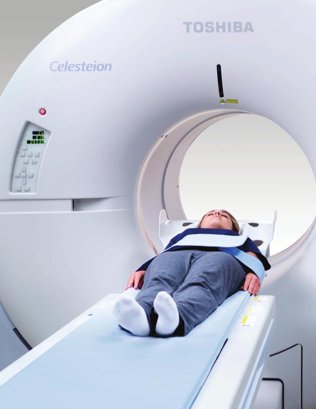
1 PURE ViSION Edition PET/CT Patient Comfort Put First.
2 2

3 System features that put patient comfort and safety first. Oncology patients deserve the highest levels of safety and comfort during scans. Our Celesteion PURE ViSION Edition PET/CT system delivers. This innovative shared PET/CT system includes extraordinary features such as a 90 cm wide CT bore and 70 cm true field of view for maximum comfort, access and positioning. Whether doing PET/CT, CT simulation or diagnostic CT, Celesteion s advanced and cost-effective technology helps allow for ease of use and efficient exams, improving clinicians ability to diagnose and treat oncology patients. 3
.")
4 Improved Accuracy and Increased Comfort Celesteion s unique patient-centered design delivers an enhanced patient comfort and care, without compromising on quality. 70 cm True Field of View With oncology patients, accuracy is everything. To meet this need, Celesteion offers a true 70 cm PET and CT field of view (FOV). This allows clinicians to overcome the challenges of a small 50 cm FOV, delivering unique access for better image quality and more accurate treatment plans. 90 cm Wide CT Bore Celesteion s wide bore is built for maximum patient positioning, comfort and peace of mind. The industry-leading 90 cm CT bore and 88 cm PET bore creates a feeling of openness and allows patients to be positioned for optimal treatment planning and setup. By making patients feel more at ease, clinicians can have more confidence in their images and diagnoses. 70 cm FOV 50 cm FOV 4

 Single Energy Metal Artifact Reduction (SEMAR ) NEMA XR-29")
5 Versatile Couch Our unique couch design makes it easy and safe to position your patients for a wide range of clinical needs. With the widest couch in the industry, the ability to lower the couch to almost 30 cm from the floor and the unique swift movement from the CT position into the PET position, makes the Celesteion PET/CT a versatile, multi-use scanner. CT 90 cm CT bore 70 cm true FOV 85 cm EFOV 47 cm couch 0.5 mm x 16 PURE ViSION detector 32 slice conexact reconstruction 1800 mm scan range Adaptive Iterative Dose Reduction (AIDR 3D) Single Energy Metal Artifact Reduction (SEMAR ) NEMA XR-29 compliant PET 88 cm PET bore 70 cm FOV 196 mm A-FOV 1,792 mm scan range 394 ps typical Time of Flight (TOF) imaging Point Spread Function (PSF) reconstruction 5

6 SURE FLiGHT PET Detector Technology Celesteion s PET detector is designed specifically for a large-bore PET, creating the ideal conditions for accuracy. This advanced technology allows for: Optimal use of LU-type scintillator materials Unique module and scalable detector design of mixed PMT sizes Provides optimal timing performance Delivers high count rate performance with optimal scintillator and a unique mixed PMT design Photosensors Light-guide layer improves performance High-performance 3D, Lu-based scintallator 6


7 PURE ViSION CT Detector Technology The PURE ViSION CT detector is designed to optimize patient care and acquire high-quality CT images. The 16 row 0.5 mm elements balance image quality, speed and patient dose, delivering isotropic images in all planes. Clinicians and patients alike benefit from: More efficient use of X-rays 0.5 mm slice resolution 40 percent better light output 7

8 SURE FLiGHT Reconstruction Technology Celesteion s unique imaging means better outcomes for patients. TOF and PSF create the optimal conditions for accurate and reliable scanning. Time of Flight Our TOF technology delivers 394 ps typical timing resolution, allowing for better visualization of tumors. This is especially useful for large patients, where the IQ deteriorates with non-tof scanners. 8

9 Point Spread Function PSF reconstruction generates sharper images and higher contrast, allowing better visualization and more accurate quantification of small tumors in routine clinical studies. Non TOF TOF TOF TOF + PSF 9


10 Better Images, Every Time SEMAR SEMAR utilizes a sophisticated reconstruction technique to reduce artifacts caused by metal and improve visualization of the implant, supporting bone and adjacent soft tissues. Original SEMAR 10

11 Respiratory Gating By synchronizing exposure to the patient s breath, gating creates detailed images for lung cancer patients. With these detailed images, clinicians have the information they need for treatment planning. Respiratory Waveform INSPIRATION PHASES EXPIRATION 11

12 AIDR 3D Our AIDR 3D is designed to lower radiation and maximize image quality. Without AIDR 3D With AIDR 3D In clinical practice, the use of AIDR 3D may reduce CT patient dose depending on the clinical task, patient size, anatomical location and clinical practice. A consultation with a radiologist and physicist should be made to determine the appropriate dose to obtain diagnostic image quality for the particular clinical task. 12

13 18 F-FDG Brain Imaging 13


14 18 F-NaF Bone Skeletal Imaging High-BMI with routine dose protocol 14
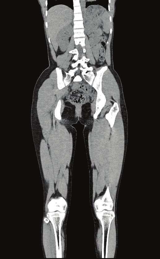
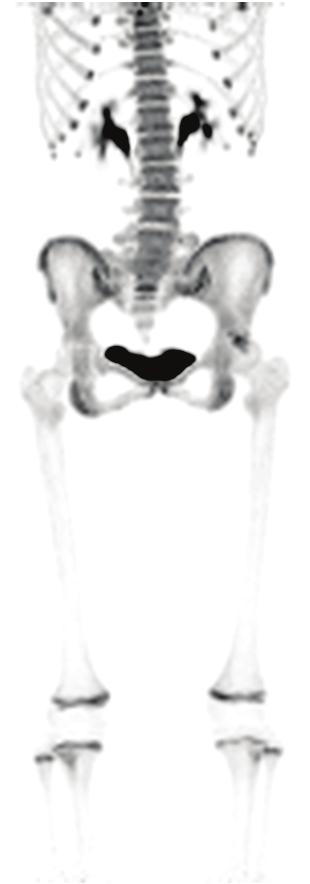
15 Average-BMI with low dose protocol 15


16 18 F-FDG Whole-Body Imaging High-BMI with routine dose protocol 16


17 High-BMI with low dose protocol 17

18 An innovative shared system that performs both CT and PET scans, Celesteion puts patient comfort and safety first. With the widest bore in the industry and a unique FOV, this advanced scanner is a cost-effective way to increase ease, efficiency and accuracy for oncology patients. 18
 Crystal Material Lu-based Rotation Rate 0.5 sec Scan Gantry Aperture Transaxial FOV 88 cm 70 cm Scan PURE ViSION CT Detector Slice Thickness 16-row (3.2 cm) 0.5 mm Axial FOV 19.")
19 Celesteion PET/CT Performance Specifications PET Performance CT Performance Number of Crystals Gantry Aperture 90 cm Crystal Size 4 mm x 4 mm Scan FOV 70 cm (85 cm Ext. FOV) Crystal Material Lu-based Rotation Rate 0.5 sec Scan Gantry Aperture Transaxial FOV 88 cm 70 cm Scan PURE ViSION CT Detector Slice Thickness 16-row (3.2 cm) 0.5 mm Axial FOV 19.6 cm Number of Slices 32 with conexact System Energy Resolution 11.2% typical Tube Current Modulation SURE Exposure 3D Count-rate TOF Timing Resolution Effective System Sensitivity 394 ps typical 13.5 cps/kbq typical (D=20 cm)* 23.7 cps/kbq typical (D=35 cm) X-ray Tube Maximum Output X-ray Tube Heat Capacity Dose Reduction Functions 72 kw 7.5 MHU AIDR 3D Spatial Resolution Effective Peak Noise Equivalent Count Rate (NECR) NEMA-2012 Resolution 1 cm Radial: FWHM@10cm 172 kcps (D=20 cm)** 302 kcps (D=35 cm) 5.1 mm 1.98 mm Dose Dose Management XR-29 Compliant Dose Check (NEMA XR-25) DICOM SC Exposure Summary DICOM SR Compliant Dose Report PSF Reconstruction Spatial Resolution*** Radial: FWHM@20cm Radial: FWHM@30cm Tangential: FWHM@10cm Tangential: FWHM@20cm Tangential: FWHM@30cm 1.96 mm 2.05 mm 2.04 mm 2.02 mm 2.08 mm Image Quality Reconstruction Method Image Noise (Standard Deviation) Spatial Cut Off Cone Beam Fan Beam SEMAR (Single Energy Metal Artifact Reduction) < 0.7% 18 lp/cm**** Axial: FWHM@10cm 2.01 mm High-Contrast Resolution 0.35 mm Axial: FWHM@20cm Axial: FWHM@30cm 1.98 mm 1.93 mm Low-Contrast Detectability 2 0.3% at 14.4 mgy 3 0.3% at 7.2 mgy NOTE: * Calculated based on TOF sensitivity gain=(snr gain) 2 =D/Δx, D: patient size, Δx: TOF spatial uncertainty. (Δx=cΔt/2, c=speed of light=3x10 10 cm/sec, Δt=TOF FWHM =394 ps=3.94x10⁻¹⁰ sec, Δx =5.91 cm. NEMA sensitivity =4.0 cps/kbq, For D=20 cm, (SNR gain) 2 =20/5.91=3.4, effective sensitivity =4.0x3.4=13.5 cps/kbq For D=35 cm, (SNR gain) 2 =35/5.91=5.9, effective sensitivity =4.0x5.9=23.7 cps/kbq) System energy resolution, TOF timing resolution, effective system sensitivity and PSF reconstruction spatial resolution are all typical values. **Effective Peak NECR: based on NEMA (non-tof) Peak NECR and TOF SNR gain calculated above. (NEMA Peak NECR 51 kcps, effective peak NECR=NEMA peak NECR x (TOF sensitivity gain) (51x3.4)=172 kcps with D=20 cm and (51x5.9)=302 kcps with D=35 cm) ***Option **** For reference WARNING: Any reference to X-ray exposure, intravenous contrast dosage, and other medication is intended as a reference guideline only. The guidelines in this document do not substitute for the judgment of a healthcare provider. Each scan requires medical judgment by the healthcare provider about exposing the patient to ionizing radiation. Use the As Low As Reasonably Achievable (ALARA) radiation dose principle to balance factors such as the patient s condition, size and age; region to be imaged; and diagnostic task. Disclaimer: In clinical practice, the use of the AIDR 3D feature may reduce CT patient dose depending on the clinical task, patient size, anatomical location and clinical practice. A consultation with a radiologist and a physicist should be made to determine the appropriate dose to obtain diagnostic image quality for the particular clinical task. Due to local regulatory processes, some of the products included in this brochure may not be available in each country. Please contact your sales representative for the most current information. 19
20 Follow us: +Toshiba Medical Toshiba America Medical Toshiba Medical TOSHIBA AMERICA MEDICAL SYSTEMS, INC Michelle Drive, Tustin CA Toshiba Medical Systems All rights reserved. Design and specifications are subject to change without notice. Celesteion, SEMAR, conexact, SURE Exposure, SURE FLiGHT and Made for Life are trademarks of Toshiba Medical Systems Corporation. Google+ logo and YouTube logo are trademarks of Google Inc. TWITTER, TWEET, RETWEET and the Twitter logo are trademarks of Twitter, Inc. or its affiliates. LinkedIn, the LinkedIn logo, the IN logo and InMail are registered trademarks or trademarks of LinkedIn Corporation and its affiliates in the United States and/or other countries. CTBR12689US MCANM0038EB
AIDR 3D Iterative Reconstruction:
 Iterative Reconstruction: Integrated, Automated and Adaptive Dose Reduction Erin Angel, PhD Manager, Clinical Sciences, CT Canon Medical Systems USA Iterative Reconstruction 1 Since the introduction of
Technological Advances and Challenges: Experience with Time-Of-Flight PET Combined with 3T MRI. Floris Jansen, GE Healthcare July, 2015
 Technological Advances and Challenges: Experience with Time-Of-Flight PET Combined with 3T MRI Floris Jansen, GE Healthcare July, 2015 PET/MR 101 : challenges Thermal Workflow & Apps RF interactions?!!
8/2/2017. Disclosure. Philips Healthcare (Cleveland, OH) provided the precommercial
 provided the precommercial") 8//0 AAPM0 Scientific Symposium: Emerging and New Generation PET: Instrumentation, Technology, Characteristics and Clinical Practice Aug Wednesday 0:4am :pm Solid State Digital Photon Counting PET/CT Instrumentation
8//0 AAPM0 Scientific Symposium: Emerging and New Generation PET: Instrumentation, Technology, Characteristics and Clinical Practice Aug Wednesday 0:4am :pm Solid State Digital Photon Counting PET/CT Instrumentation
7/11/2015. Imaging Equipment Specification and Selection in Radiation Oncology Department. What Therapy Physicists Need to Know
 Imaging Equipment Specification and Selection in Radiation Oncology Department Chiaho Hua St. Jude Children s Research Hospital Yue Cao University of Michigan, Ann Arbor Margie Hunt Memorial Sloan Kettering
Optimisation of Toshiba Aquilion ONE Volume Imaging
 Optimisation of Toshiba Aquilion ONE Volume Imaging Jane Edwards, RPRSG Royal Free London NHS Foundation Trust Dr Mufudzi Maviki, Plymouth Hospitals NHS Trust Background In 2011/12 Radiology at RFH was
PRODUCT DATA. Advanced 128
 PRODUCT DATA Advanced 128 Supria 16 Slice CT Puts You On The Path of High Quality, Cost-Effective CT Scanning Solid Capabilities Are Built Into the Supria Addressing the challenges of controlling healthcare
Implementation and evaluation of a fully 3D OS-MLEM reconstruction algorithm accounting for the PSF of the PET imaging system
 Implementation and evaluation of a fully 3D OS-MLEM reconstruction algorithm accounting for the PSF of the PET imaging system 3 rd October 2008 11 th Topical Seminar on Innovative Particle and Radiation
Solid Capabilities Are Built Into the Supria Plus. Putting You On The Path of High Quality, Cost-Effective CT Scanning
 Specification Data Putting You On The Path of High Quality, Cost-Effective CT Scanning Solid Capabilities Are Built Into the Supria Plus Addressing the challenges of controlling healthcare organization
Time-of-Flight Technology
 Medical Review Time-of-Flight Technology Bing Bai, PhD Clinical Sciences Manager, PET/CT Canon Medical Systems INTRODUCTION Improving the care for every patient while providing a high standard care to
Design and performance characteristics of a Cone Beam CT system for Leksell Gamma Knife Icon
 Design and performance characteristics of a Cone Beam CT system for Leksell Gamma Knife Icon WHITE PAPER Introduction Introducing an image guidance system based on Cone Beam CT (CBCT) and a mask immobilization
INSPIRED BY YOUR PATIENTS.
 Hitachi healthcare americas HHA INSPIRED BY YOUR PATIENTS. Inspired by your patients 1 HHA Hitachi healthcare americas Hitachi healthcare americas HHA WHAT WE ARE about... Since our founding in 1910, Hitachi
Philips SPECT/CT Systems
 Philips SPECT/CT Systems Ling Shao, PhD Director, Imaging Physics & System Analysis Nuclear Medicine, Philips Healthcare June 14, 2008 *Presented SNM08 Categorical Seminar - Quantitative SPECT and PET
Computer-Tomography II: Image reconstruction and applications
 Computer-Tomography II: Image reconstruction and applications Prof. Dr. U. Oelfke DKFZ Heidelberg Department of Medical Physics (E040) Im Neuenheimer Feld 280 69120 Heidelberg, Germany u.oelfke@dkfz.de
Optimization of CT Simulation Imaging. Ingrid Reiser Dept. of Radiology The University of Chicago
 Optimization of CT Simulation Imaging Ingrid Reiser Dept. of Radiology The University of Chicago Optimization of CT imaging Goal: Achieve image quality that allows to perform the task at hand (diagnostic
Fits you like no other
 Fits you like no other BrightView X and XCT specifications The new BrightView X system is a fully featured variableangle camera that is field-upgradeable to BrightView XCT without any increase in room
Shadow casting. What is the problem? Cone Beam Computed Tomography THE OBJECTIVES OF DIAGNOSTIC IMAGING IDEAL DIAGNOSTIC IMAGING STUDY LIMITATIONS
 Cone Beam Computed Tomography THE OBJECTIVES OF DIAGNOSTIC IMAGING Reveal pathology Reveal the anatomic truth Steven R. Singer, DDS srs2@columbia.edu IDEAL DIAGNOSTIC IMAGING STUDY Provides desired diagnostic
Lower Dose with Confidence
 Lower Dose with Confidence The Next Generation SCENARIA CT is Here. 64 or 128 Slice Capability The SCENARIA SE delivers lower dose technologies*, enhanced patient access, fast workflow speed and added
CT Protocol Review: Practical Tips for the Imaging Physicist Physicist
 CT Protocol Review: Practical Tips for the Imaging Physicist Physicist Dianna Cody, Ph.D., DABR, FAAPM U.T.M.D. Anderson Cancer Center August 8, 2013 AAPM Annual Meeting Goals Understand purpose and importance
Spiral CT. Protocol Optimization & Quality Assurance. Ge Wang, Ph.D. Department of Radiology University of Iowa Iowa City, Iowa 52242, USA
 Spiral CT Protocol Optimization & Quality Assurance Ge Wang, Ph.D. Department of Radiology University of Iowa Iowa City, Iowa 52242, USA Spiral CT Protocol Optimization & Quality Assurance Protocol optimization
Corso di laurea in Fisica A.A Fisica Medica 5 SPECT, PET
 Corso di laurea in Fisica A.A. 2007-2008 Fisica Medica 5 SPECT, PET Step 1: Inject Patient with Radioactive Drug Drug is labeled with positron (β + ) emitting radionuclide. Drug localizes
MEDICAL EQUIPMENT: COMPUTED TOMOGRAPHY. Prof. Yasser Mostafa Kadah
 MEDICAL EQUIPMENT: COMPUTED TOMOGRAPHY Prof. Yasser Mostafa Kadah www.k-space.org Recommended Textbook X-Ray Computed Tomography in Biomedical Engineering, by Robert Cierniak, Springer, 211 Computed Tomography
CT Basics Principles of Spiral CT Dose. Always Thinking Ahead.
 1 CT Basics Principles of Spiral CT Dose 2 Who invented CT? 1963 - Alan Cormack developed a mathematical method of reconstructing images from x-ray projections Sir Godfrey Hounsfield worked for the Central
Symbia T Series System Specifications Answers for life.
 www.siemens.com/mi System Specifications Answers for life. 2 Diagnostic Excellence Today s healthcare institutions have increasingly diverse patient needs, greater budget limitations and the need for higher
Fits you like no other
 Fits you like no other Philips BrightView X and XCT specifications The new BrightView X system is a fully featured variableangle camera that is field-upgradeable to BrightView XCT without any increase
SCENARIA 64-Slice CT Putting Advanced CT Within Reach.
 SCENARIA 64-Slice CT Putting Advanced CT Within Reach. Advancing the technology of Computed Tomography for over 30 years, Hitachi is a recognized innovator of lower dose*, high diagnostic value CT solutions.
Lower Dose, Compact Design
 Lower Dose, Compact Design Advanced Dose Reduction Capabilities XR-29 Smart Dose Compliant Enlarged 75cm Aperture Fast Workflow Compact Design, 200 sq. ft. Minimum Scan Room Innovative CT Design for Positive
AAPM Standard of Practice: CT Protocol Review Physicist
 AAPM Standard of Practice: CT Protocol Review Physicist Dianna Cody, Ph.D., DABR, FAAPM U.T.M.D. Anderson Cancer Center September 11, 2014 2014 Texas Radiation Regulatory Conference Goals Understand purpose
Image Acquisition Systems
 Image Acquisition Systems Goals and Terminology Conventional Radiography Axial Tomography Computer Axial Tomography (CAT) Magnetic Resonance Imaging (MRI) PET, SPECT Ultrasound Microscopy Imaging ITCS
Brilliance CT Big Bore.
 1 2 2 There are two methods of RCCT acquisition in widespread clinical use: cine axial and helical. In RCCT with cine axial acquisition, repeat CT images are taken each couch position while recording respiration.
Hitachi Medical Systems America, Inc. Hitachi Medical Corporation
 Advanced 128 Advanced 128 128 Slice Capability Enlarged 75cm Aperture Thin Gantry Profile 88cm Intelli IP Advanced, Iterative Reconstruction IntelliCenter Auto-Lateral Shifting Table, up to 16cm Range
Introduction to Positron Emission Tomography
 Planar and SPECT Cameras Summary Introduction to Positron Emission Tomography, Ph.D. Nuclear Medicine Basic Science Lectures srbowen@uw.edu System components: Collimator Detector Electronics Collimator
Position accuracy analysis of the stereotactic reference defined by the CBCT on Leksell Gamma Knife Icon
 Position accuracy analysis of the stereotactic reference defined by the CBCT on Leksell Gamma Knife Icon WHITE PAPER Introduction An image guidance system based on Cone Beam CT (CBCT) is included in Leksell
SCENARIA Hitachi Innovation at Work for You.
 PRODUCT DATA SCENARIA Hitachi Innovation at Work for You. Continuing its commitment to the SCENARIA scalable CT platform, Hitachi introduces cutting-edge 64/128-slice technology with the SCENARIA SE. The
CT NOISE POWER SPECTRUM FOR FILTERED BACKPROJECTION AND ITERATIVE RECONSTRUCTION
 CT NOISE POWER SPECTRUM FOR FILTERED BACKPROJECTION AND ITERATIVE RECONSTRUCTION Frank Dong, PhD, DABR Diagnostic Physicist, Imaging Institute Cleveland Clinic Foundation and Associate Professor of Radiology
Tomographic Reconstruction
 Tomographic Reconstruction 3D Image Processing Torsten Möller Reading Gonzales + Woods, Chapter 5.11 2 Overview Physics History Reconstruction basic idea Radon transform Fourier-Slice theorem (Parallel-beam)
If it matters to you, it matters to us
 If it matters to you, it matters to us Philips clinical innovations in nuclear medicine Innovation with insight We understand that clinical innovations are only as valuable as the day-to-day difference
Whole Body Submillimeter Scan
 128 Whole Body Submillimeter Scan The real value of 64ch/128slice CT systems does not come from the ability to perfom cardiac scans but the capability to scan all parts of the body in high definition submillimeter
Experience Boundless Performance
 Experience Boundless Performance About Samsung Samsung Electronics Co., Ltd. inspires the world and shapes the future with transformative ideas and technologies, redefining the worlds of TVs, smartphones,
Superior Imaging on the Go
 Superior Imaging on the Go About Samsung Samsung Electronics Co., Ltd. is a global leader in technology, opening new possibilities for people everywhere. Through relentless innovation and discovery, we
NIH Public Access Author Manuscript J Nucl Med. Author manuscript; available in PMC 2010 February 9.
 NIH Public Access Author Manuscript Published in final edited form as: J Nucl Med. 2010 February ; 51(2): 237. doi:10.2967/jnumed.109.068098. An Assessment of the Impact of Incorporating Time-of-Flight
3/27/2012 WHY SPECT / CT? SPECT / CT Basic Principles. Advantages of SPECT. Advantages of CT. Dr John C. Dickson, Principal Physicist UCLH
 3/27/212 Advantages of SPECT SPECT / CT Basic Principles Dr John C. Dickson, Principal Physicist UCLH Institute of Nuclear Medicine, University College London Hospitals and University College London john.dickson@uclh.nhs.uk
Symbia E and S System Specifications Answers for life.
 www.siemens.com/mi Symbia E and S System Specifications Answers for life. Symbia E: Work with confidence. What does it mean to work with confidence? It is clarity not only image clarity but the clarity
Ch. 4 Physical Principles of CT
 Ch. 4 Physical Principles of CT CLRS 408: Intro to CT Department of Radiation Sciences Review: Why CT? Solution for radiography/tomography limitations Superimposition of structures Distinguishing between
Superior Imaging on the Go
 Superior Imaging on the Go About Samsung Samsung Electronics Co., Ltd. is a global leader in technology, opening new possibilities for people everywhere. Through relentless innovation and discovery, we
7/13/2015 EVALUATION OF NONLINEAR RECONSTRUCTION METHODS. Outline. This is a decades-old challenge
 EVALUATION OF NONLINEAR RECONSTRUCTION METHODS Kyle J. Myers, Ph.D. Director, Division of Imaging, Diagnostics, and Software Reliability Office of Science and Engineering Laboratories, CDRH, FDA 2 Outline
Enhancement Image Quality of CT Using Single Slice Spiral Technique
 Enhancement Image Quality of CT Using Single Slice Spiral Technique Doaa. N. Al Sheack 1 and Dr.Mohammed H. Ali Al Hayani 2 1 2 Electronic and Communications Engineering Department College of Engineering,
DIRECT RADIOGRAPHY. automated. Delivering the fully. digital X-ray room. DX-D 600: Direct Radiography from Agfa HealthCare
 DIRECT RADIOGRAPHY automated Delivering the fully digital X-ray room DX-D 600: Direct Radiography from Agfa HealthCare A high performance X-ray room The DX-D 600 s range of innovative, integrated features
As fl exible as your care requires
 As fl exible as your care requires Philips Ingenuity Flex 32 CT Built on Ingenuity The Philips Ingenuity Flex 32 helps you provide excellent care with outstanding flexibility, now and in the future. Built
Image Quality Assessment and Quality Assurance of Advanced Imaging Systems for IGRT. AAPM Penn-Ohio Chapter Sep 25, 2015 Soyoung Lee, PhD
 Image Quality Assessment and Quality Assurance of Advanced Imaging Systems for IGRT AAPM Penn-Ohio Chapter Sep 25, 2015 Soyoung Lee, PhD 1 Outline q Introduction q Imaging performances in 4D-CBCT Image
BME I5000: Biomedical Imaging
 1 Lucas Parra, CCNY BME I5000: Biomedical Imaging Lecture 4 Computed Tomography Lucas C. Parra, parra@ccny.cuny.edu some slides inspired by lecture notes of Andreas H. Hilscher at Columbia University.
Annexure XII SPECIFICATIONS FOR A NEW STATE OF ART 16 SLICE ALL PURPOSE C. T. SCANNER
 Annexure XII SPECIFICATIONS FOR A NEW STATE OF ART 16 SLICE ALL PURPOSE C. T. SCANNER A) Scanner Design X-Ray generator and tube: 1. Scanner: Whole body spiral CT scanner (16 slices) of latest technology.
Improvement of contrast using reconstruction of 3D Image by PET /CT combination system
 Available online at www.pelagiaresearchlibrary.com Advances in Applied Science Research, 2013, 4(1):285-290 ISSN: 0976-8610 CODEN (USA): AASRFC Improvement of contrast using reconstruction of 3D Image
Multi-slice CT Image Reconstruction Jiang Hsieh, Ph.D.
 Multi-slice CT Image Reconstruction Jiang Hsieh, Ph.D. Applied Science Laboratory, GE Healthcare Technologies 1 Image Generation Reconstruction of images from projections. textbook reconstruction advanced
MUSICA makes the difference
 MUSICA makes the difference At busy Logan Hospital, MUSICA and the DR 600 are keeping the imaging workflow smooth and efficient, while meeting the quality and dose reduction needs for patients and staff
Characterization of a Time-of-Flight PET Scanner based on Lanthanum Bromide
 2005 IEEE Nuclear Science Symposium Conference Record M04-8 Characterization of a Time-of-Flight PET Scanner based on Lanthanum Bromide J. S. Karp, Senior Member, IEEE, A. Kuhn, Member, IEEE, A. E. Perkins,
Some reference material
 Some reference material Physics reference book on medical imaging: A good one is The Essential Physics of Medical Imaging, 3 rd Ed. by Bushberg et al. ($170! new). However, there are several similar books
James R Halama, PhD Loyola University Medical Center
 James R Halama, PhD Loyola University Medical Center Conflicts of Interest Nuclear Medicine and PET physics reviewer for the ACR Accreditation program Learning Objectives Be familiar with the tests recommended
Conflicts of Interest Nuclear Medicine and PET physics reviewer for the ACR Accreditation program
 James R Halama, PhD Loyola University Medical Center Conflicts of Interest Nuclear Medicine and PET physics reviewer for the ACR Accreditation program Learning Objectives 1. Be familiar with recommendations
Virtual Phantoms for IGRT QA
 TM Virtual Phantoms for IGRT QA Why ImSimQA? ImSimQA was developed to overcome the limitations of physical phantoms for testing modern medical imaging and radiation therapy software systems, when there
Flat Panel Detectors. P-Series ISS. Dynamic Visualization SmartSwitch. Wireless. Portable. Technology
 Flat Panel Detectors P-Series Portable Wireless ISS Technology DV Dynamic Visualization SmartSwitch Introducing P-Series Flat Panel Detectors DR solutions made especially for private practices like yours.
[PDR03] RECOMMENDED CT-SCAN PROTOCOLS
![[PDR03] RECOMMENDED CT-SCAN PROTOCOLS](/thumbs/72/66454100.jpg "[PDR03] RECOMMENDED CT-SCAN PROTOCOLS") SURGICAL & PROSTHETIC DESIGN [PDR03] RECOMMENDED CT-SCAN PROTOCOLS WORK-INSTRUCTIONS DOCUMENT (CUSTOMER) RECOMMENDED CT-SCAN PROTOCOLS [PDR03_V1]: LIVE 1 PRESCRIBING SURGEONS Patient-specific implants,
SURGICAL & PROSTHETIC DESIGN [PDR03] RECOMMENDED CT-SCAN PROTOCOLS WORK-INSTRUCTIONS DOCUMENT (CUSTOMER) RECOMMENDED CT-SCAN PROTOCOLS [PDR03_V1]: LIVE 1 PRESCRIBING SURGEONS Patient-specific implants,
Toshiba Aquilion 4 3d Manual READ ONLINE
 Toshiba Aquilion 4 3d Manual READ ONLINE TOSHIBA AQUILION VISION: Head. EP No: 4. Boost 3D: OFF: OSR *** Filter: OFF. SureStart - Manual trigger as soon as contrast is seen: Scan mode: TOSHIBA AQUILION
Deviceless respiratory motion correction in PET imaging exploring the potential of novel data driven strategies
 g Deviceless respiratory motion correction in PET imaging exploring the potential of novel data driven strategies Presented by Adam Kesner, Ph.D., DABR Assistant Professor, Division of Radiological Sciences,
MEDICAL IMAGING 2nd Part Computed Tomography
 MEDICAL IMAGING 2nd Part Computed Tomography Introduction 2 In the last 30 years X-ray Computed Tomography development produced a great change in the role of diagnostic imaging in medicine. In convetional
8/2/2016. Measures the degradation/distortion of the acquired image (relative to an ideal image) using a quantitative figure-of-merit
 using a quantitative figure-of-merit") Ke Li Assistant Professor Department of Medical Physics and Department of Radiology School of Medicine and Public Health, University of Wisconsin-Madison This work is partially supported by an NIH Grant
Ke Li Assistant Professor Department of Medical Physics and Department of Radiology School of Medicine and Public Health, University of Wisconsin-Madison This work is partially supported by an NIH Grant
Digital Image Processing
 Digital Image Processing SPECIAL TOPICS CT IMAGES Hamid R. Rabiee Fall 2015 What is an image? 2 Are images only about visual concepts? We ve already seen that there are other kinds of image. In this lecture
A dedicated tool for PET scanner simulations using FLUKA
 A dedicated tool for PET scanner simulations using FLUKA P. G. Ortega FLUKA meeting June 2013 1 Need for in-vivo treatment monitoring Particles: The good thing is that they stop... Tumour Normal tissue/organ
Medical Imaging BMEN Spring 2016
 Name Medical Imaging BMEN 420-501 Spring 2016 Homework #4 and Nuclear Medicine Notes All questions are from the introductory Powerpoint (based on Chapter 7) and text Medical Imaging Signals and Systems,
Q.Clear. Steve Ross, Ph.D.
 Steve Ross, Ph.D. Accurate quantitation (SUV - Standardized Uptake Value) is becoming more important as clinicians seek to utilize PET imaging for more than just diagnosing and staging disease, but also
The Near Future in Cardiac CT Image Reconstruction
 SCCT 2010 The Near Future in Cardiac CT Image Reconstruction Marc Kachelrieß Institute of Medical Physics (IMP) Friedrich-Alexander Alexander-University Erlangen-Nürnberg rnberg www.imp.uni-erlangen.de
UvA-DARE (Digital Academic Repository) Motion compensation for 4D PET/CT Kruis, M.F. Link to publication
 Motion compensation for 4D PET/CT Kruis, M.F. Link to publication") UvA-DARE (Digital Academic Repository) Motion compensation for 4D PET/CT Kruis, M.F. Link to publication Citation for published version (APA): Kruis, M. F. (2014). Motion compensation for 4D PET/CT General
UvA-DARE (Digital Academic Repository) Motion compensation for 4D PET/CT Kruis, M.F. Link to publication Citation for published version (APA): Kruis, M. F. (2014). Motion compensation for 4D PET/CT General
Helical 4D CT pitch management for the Brilliance CT Big Bore in clinical practice
 JOURNAL OF APPLIED CLINICAL MEDICAL PHYSICS, VOLUME 16, NUMBER 3, 2015 Helical 4D CT pitch management for the Brilliance CT Big Bore in clinical practice Guido Hilgers, a Tonnis Nuver, André Minken Department
ThE ultimate, INTuITIVE Mr INTErFAcE
 ThE ultimate, INTuITIVE Mr INTErFAcE Empowering you to do more The revolutionary Toshiba M-power user interface takes Mr performance and flexibility to levels higher than ever before. M-power is able to
Comprehensive treatment planning for brachytherapy. Advanced planning made easy
 Comprehensive treatment planning for brachytherapy Advanced planning made easy Oncentra Brachy offers a variety of smart tools that facilitate many of the repetitive tasks for you. In contemporary brachytherapy,
DX-D 300. Flexible Direct Radiography System
 DX-D 300 Flexible Direct Radiography System MUSICA² processing provides outstanding contrast detail and consistently excellent image quality Universal, flexible and affordable modality combines a single
CT vs. VolumeScope: image quality and dose comparison
 CT vs. VolumeScope: image quality and dose comparison V.N. Vasiliev *a, A.F. Gamaliy **b, M.Yu. Zaytsev b, K.V. Zaytseva ***b a Russian Sci. Center of Roentgenology & Radiology, 86, Profsoyuznaya, Moscow,
Fast Timing and TOF in PET Medical Imaging
 Fast Timing and TOF in PET Medical Imaging William W. Moses Lawrence Berkeley National Laboratory October 15, 2008 Outline: Time-of-Flight PET History Present Status Future This work was supported in part
New Technology in Radiation Oncology. James E. Gaiser, Ph.D. DABR Physics and Computer Planning Charlotte, NC
 New Technology in Radiation Oncology James E. Gaiser, Ph.D. DABR Physics and Computer Planning Charlotte, NC Technology s s everywhere From the imaging chain To the planning system To the linac To QA..it..it
Biomet PMI. Patient-Matched Implants. CT Protocols
 CT Protocols One Surgeon. One Patient. Over 1 million times per year, Biomet helps one surgeon provide personalized care to one patient. The science and art of medical care is to provide the right solution
DX-D 300 FLEXIBLE DIRECT RADIOGRAPHY SYSTEM DX-D 300 DR SYSTEM
 DX-D 300 FLEXIBLE DIRECT RADIOGRAPHY SYSTEM The DX-D 300 DR system unites excellent image quality with the ultimate convenience. It offers top-of-the-line technology: a fullymotorized positioner enabling
ImPACT. Information Leaflet No. 1: CT Scanner Acceptance Testing
 ImPACT Information Leaflet No. 1: CT Scanner Acceptance Testing Version 1.02, 18/05/01 CONTENTS: 1. SCOPE OF LEAFLET 2. GENERAL PRINCIPLES OF ACCEPTANCE AND COMMISSIONING 2.1 PHANTOMS 2.2 EXPOSURE AND
digital imaging A world of dedicated to animal healthcare Fast, affordable, easy-to-use solutions that offer high-quality digital imaging
 Digital radiography small animals A world of digital imaging dedicated to animal healthcare Fast, affordable, easy-to-use solutions that offer high-quality digital imaging 2 Digital radiography small animals
QIBA Profile: CT Tumor Volume Change for Advanced Disease (CTV-AD)
") 5 QIBA Profile: CT Tumor Volume Change for Advanced Disease (CTV-AD) Stage: Consensus When referencing this document, please use the following format: QIBA CT Volumetry Technical Committee. CT Tumor Volume
5 QIBA Profile: CT Tumor Volume Change for Advanced Disease (CTV-AD) Stage: Consensus When referencing this document, please use the following format: QIBA CT Volumetry Technical Committee. CT Tumor Volume
Agenda : Lung Density Breakout Session
 Agenda : Lung Density Breakout Session 1. : Mathew Fuld and Bernice Hoppel 2. Automatic Exposure Control (AEC) Evaluation : Sean Fain Round 4 Project 3. Dose reduction effects on emphysema metrics : Philip
DIRECT RADIOGRAPHY. flexible. Delivering. imaging excellence. DX-D Direct Radiography from Agfa HealthCare
 DIRECT RADIOGRAPHY flexible Delivering imaging excellence DX-D 300 - Direct Radiography from Agfa HealthCare versatile a and compact solution for all General Radiographic exams Maximum flexibility. The
Equipment Specification
 MULTISLICE CT SCANNER ( 16 Slices ) Merk : Hitachi Japan Model : SUPRIA 5 MHU Price : Rp 6.512.360.215,27 No Equipment Specification 1 2 3 Scanner Gantry Object for scanning : Whole body including head
7/31/2011. Learning Objective. Video Positioning. 3D Surface Imaging by VisionRT
 CLINICAL COMMISSIONING AND ACCEPTANCE TESTING OF A 3D SURFACE MATCHING SYSTEM Hania Al-Hallaq, Ph.D. Assistant Professor Radiation Oncology The University of Chicago Learning Objective Describe acceptance
MEDICAL IMAGING 2nd Part Computed Tomography
 MEDICAL IMAGING 2nd Part Computed Tomography Introduction 2 In the last 30 years X-ray Computed Tomography development produced a great change in the role of diagnostic imaging in medicine. In convetional
Carestream s 2 nd Generation Metal Artifact Reduction Software (CMAR 2)
") Carestream s 2 nd Generation Metal Artifact Reduction Software (CMAR 2) Author: Levon Vogelsang Introduction Cone beam computed tomography (CBCT), or cone beam CT technology, offers considerable promise
Carestream s 2 nd Generation Metal Artifact Reduction Software (CMAR 2) Author: Levon Vogelsang Introduction Cone beam computed tomography (CBCT), or cone beam CT technology, offers considerable promise
CT: Physics Principles & Equipment Design
 CT: Physics Principles & Equipment Design James Kofler, Ph.D Radiology Mayo Clinic Rochester, MN June 27, 2012 Disclosures Nothing to disclose Learning Objectives Understand fundamental concepts of - CT
Radiology. Marta Anguiano Millán. Departamento de Física Atómica, Molecular y Nuclear Facultad de Ciencias. Universidad de Granada
 Departamento de Física Atómica, Molecular y Nuclear Facultad de Ciencias. Universidad de Granada Overview Introduction Overview Introduction Tecniques of imaging in Overview Introduction Tecniques of imaging
RADIOLOGY AND DIAGNOSTIC IMAGING
 Day 2 part 2 RADIOLOGY AND DIAGNOSTIC IMAGING Dr hab. Zbigniew Serafin, MD, PhD serafin@cm.umk.pl 2 3 4 5 CT technique CT technique 6 CT system Kanal K: RSNA/AAPM web module: CT Systems & CT Image Quality
Financial disclosure. Onboard imaging modality for IGRT
 Tetrahedron Beam Computed Tomography Based On Multi-Pixel X- Ray Source and Its Application in Image Guided Radiotherapy Tiezhi Zhang, Ph.D. Advanced X-ray imaging Lab Financial disclosure Patent royalty
Computed Tomography. Principles, Design, Artifacts, and Recent Advances. Jiang Hsieh THIRD EDITION. SPIE PRESS Bellingham, Washington USA
 Computed Tomography Principles, Design, Artifacts, and Recent Advances THIRD EDITION Jiang Hsieh SPIE PRESS Bellingham, Washington USA Table of Contents Preface Nomenclature and Abbreviations xi xv 1 Introduction
Automated Image Analysis Software for Quality Assurance of a Radiotherapy CT Simulator
 Automated Image Analysis Software for Quality Assurance of a Radiotherapy CT Simulator Andrew J Reilly Imaging Physicist Oncology Physics Edinburgh Cancer Centre Western General Hospital EDINBURGH EH4
DX-D 300. Flexible Direct Radiography System
 DX-D 300 Flexible Direct Radiography System MUSICA 2 processing provides superior contrast detail and exam-independent, consistent image quality Cesium Iodide DR detector technology offers potential for
QIBA Profile: CT Tumor Volume Change for Advanced Disease (CTV-AD)
") 5 QIBA Profile: CT Tumor Volume Change for Advanced Disease (CTV-AD) Stage 3: Technically Confirmed June 22, 2018 When referencing this document, please use the following format: QIBA CT Volumetry Technical
5 QIBA Profile: CT Tumor Volume Change for Advanced Disease (CTV-AD) Stage 3: Technically Confirmed June 22, 2018 When referencing this document, please use the following format: QIBA CT Volumetry Technical
Mediso AnyScan. Single-Head and Dual-Head SPECT
 Mediso AnyScan S Single-Head and Dual-Head SPECT ANYSCAN S (SINGLE-HEAD & DUAL-HEAD SPECT) Low Cost of Ownership Associated Imaging Services is the exclusive U.S. source in the Midwest for Mediso Medical
The great interest shown toward PET instrumentation is
 CONTINUING EDUCATION PET Instrumentation and Reconstruction Algorithms in Whole-Body Applications* Gabriele Tarantola, BE; Felicia Zito, MSc; and Paolo Gerundini, MD Department of Nuclear Medicine, Ospedale
In the short span since the introduction of the first
 Performance Characteristics of a Newly Developed PET/CT Scanner Using NEMA Standards in 2D and 3D Modes Osama Mawlawi, PhD 1 ; Donald A. Podoloff, MD 2 ; Steve Kohlmyer, MS 3 ; John J. Williams 3 ; Charles