Venus Explorer Processing Technical specifications
|
|
|
- Jodie Ross
- 5 years ago
- Views:
Transcription
1 24, rue du champ de l Alouette F Paris, France Venus Explorer Processing Technical specifications Venus Explorer Processing Server software Web base application sever; tomcat Apache technology. Include Dicom server and MySQL database management. Venus Explorer Processing Server Venus Explorer Processing software Venus Explorer Processing is a null client installation and a navigator is used to access to the applications. Venus Explorer Processing Client licences are floating (concurrent); allow the software to be loaded on multiple PC s for concurrent user login. Some third party options (i.e. Cedars) are either fixed (node-locked) or floating (concurrent). Fixed Client licence is node-locked to a certain IP address on the network. Floating Client licence provides access from any Windows PC on the network (with compatible graphic board & memory) and concurrent access is only limited by the number of Venus Explorer Processing Client licences purchased. Venus Explorer Processing - run on any hardware (PC, MAC, ); the process is done an application server; client access through a web navigator. 24 rue du champ de l Alouette Paris - France - RCS PARIS B B04764 Tel: Fax:
2 Venus Processing NUCLEAR MEDICINE, PET/SPECT/CT/MR MULTIMODALITY IMAGING SOLUTIONS CONSISTENCY Uniform reading and processing environment for all your SPECT and PET cameras. With Venus Explorer Processing, you are no longer bound to a particular camera workstation. Venus Explorer Processing gives you complete functionality to process or review all NM scans regardless of camera vendor, using a suitable PC with secure access to the department server. CLINICALLY SUPERIOR Delivers the full spectrum of clinical NM and multi-modality applications Dedicated to deliver advance co-registration fusion, multimodality viewing and quantification specialized tools. Delivers a unique PET/SPECT/CT/MR multi-modality viewer completely operator configurable with advanced Tumor tracking feature. nicesoft endless continues its cooperation with leading worldwide medical institutions in order to provide you with the most complete, state of the art, while innovative multimodality clinical software library able to meet your needs. CUSTOMIZABLE, FLEXIBLE & SECURE Easy to used interactivity: Venus Explorer Processing scales its functionality to the expert level of the user based on logon credentials. Venus Explorer Processing provides the full flexibility to expert users and limits the available functionality to the occasional user, keeping the system simple enough for those users and avoiding misinterpretations due to the operator error. Use processing and display scripts and supply a powerful user preference mechanism to control them. Venus Explorer Processing uses processing and display scripts and supply a user preference mechanism to control them; permits a customization of display pages through a of lay-out editor which permits to change, resize, delete or create display objects. Easy to develop and maintain through modularization. Incorporate mechanism to create graphic layout. Highly display-centric; enhance 3-D displays, cine and curves. Numerous user interactions to display. Message and dialogue displays. Multiple monitor configuration: possible use of multi-screens, each screen being able to display a different page. Multiple resolutions for the monitors handled Unique use of screen - all formats. 2
3 Monitors Venus Explorer Processing workstations can be configured with different size monitors, accepting any screen resolution, in particular 16x9 formats, and multiple screen configurations; can come with single, dual, triple, quad or other multi-monitor configurations (up to 8 monitors) in order to best fit with your needs. Venus Explorer Processing software library comes with multi-monitor synchronization for easy review of multiple modalities or serial studies. Facilitates port to other vendors environnent. Venus Explorer Processing Offers the possibility of Floating Client software licenses. Venus Explorer Processing Client license allows access on the Venus Explorer Processing server & Venus Explorer Processing applications. Venus Explorer Processing Client licences are floating (concurrent). Some third party options (i.e. Cedars) are either fixed (node-locked) or floating (concurrent). SPECIALIZED Venus Explorer Processing specializes in customized, turnkey Nuclear Medicine, SPECT/PET/CT/MR and multimodality imaging solutions including seamless connectivity, uniform processing platform, multi-format archiving & remote reading distribution. CUSTOMER FOCUSED All Venus Explorer Processing software can be tailored to meet specific Nuclear Medicine and SPECT/PET/CT/ MR department needs. NICESOFT features personalized onsite and remote customer support to assist & support you quickly & efficiently. EFFICIENT ARCHITECTURE The Venus Explorer Processing Client/Server architecture provides an efficient infrastructure for data and application management. Allows remote-access The Venus Explorer Processing data architecture is very much like a PACS system as it deploys a redundant central database for all NM data. Venus Explorer Processing is fully DICOM compliant and integrates non-dicom compliant sources through an optional system. Use sophisticated DICOM library; include Cardio (EKG, etc.) Expandable: Venus Explorer Processing is the first system of its kind that provides you completely expendable application delivery architecture no matter what the future may hold. COST EFFECTIVE Client runs on standard PC hardware. Applications reside on the server side and are very fast downloaded to the client when needed. Processing is performed on the client. Each clinical application can be licensed separately which means you only pay for the software you really need. Also, it allows you to start with Venus Explorer Processing with a minimum investment and add more users and capabilities over time. 3
.")
. Venus Explorer Processing architecture utilizes the available power of OpenGL to the fullest extend.")
4 PACS COMPATIBLE Designed as the NM complement of modern PACS systems with integration support for legacy cameras and data formats. OPEN Allows for tight integration of common third party applications (e-g: cardiology - Cedars / Emory / 4DMSPECT Michigan). BLAZINGLY FAST The Venus Explorer Processing display engine capitalizes on the extreme processing power available in modern graphics cards: the utilization of GPU for display and computations maximizes performance while reducing CPU loads (processing on client stations). Venus Explorer Processing architecture utilizes the available power of OpenGL to the fullest extend. The result is immediately obvious as the display interactions on large datasets and 3D renderings are blazingly fast. Offers an instant access to data loaded from PACS & server. NON OBSOLESCENCE Fully protected against future changes in instrumentation. VENDOR NEUTRAL Venus Explorer Processing is a vendor-neutral multimodality processing & multimodality platform for Nuclear Medicine which maintains data integrity across all vendor platforms, providing you with all freedom of choice for new cameras & PET/CT scanners purchases. QUALITY DRIVEN All Venus Explorer Processing software library is CE Mark (Europe). Venus Explorer Processing complies with HIPAA, EU privacy laws. NICESOFT is ISO & CMRD certified. VALIDATED Venus Explorer Processing software library & applications are validated & tested and have been used clinically for part of the software library since

5 General Clinical Applications Venus Processing Functional specifications General NM Viewer Generic viewer for NM images in static, dynamic, wholebody, or planar gated format and secondary captures from all modalities. Display functions: window leveling, color scale selection, zoom, pan, magnify, colorize Measurement tool Independent or synchronized window leveling SPECT-CT (multimodality viewing) Clinical application for review and analysis of SPECT/CT studies. Standardized orthogonal slice displays Display functions: triangulation, window leveling, color scale selection, slice alignment, cine controls, zoom, pan, magnify, colorize Measurement tool User definable CT window presets Distance weighted maximum intensity projections (MIP) Fusion display mode based on variable alpha blending and/or variable thresholding Manual registration adjustment (translations, rotations) 3D region analysis based on iso-contouring Study comparison (follow-up) 5


6 User definable hotkeys for commonly used functions Dedicated Lesion, Linking, and Registration Managers User specific preferences based on logon credentials Compatible with all SPECT and hybrid SPECT/CT scanner models Customized layouts through display scripts Volume rendering option Volume rendering generation. 6

7 PET-CT-Onco (multimodality viewing) Clinical application for review and analysis of PET/CT studies. Standardized orthogonal slice displays Display functions: triangulation, window leveling, color scale selection, slice alignment, cine controls, zoom, pan, magnify, colorize Measurement tool Automatic window saturation function for PET User definable CT window presets SUV computation Distance weighted maximum intensity projections (MIP) Fusion display mode based on variable alpha blending and/or variable thresholding Manual registration adjustment (translations, rotations) Tumor-tracking analysis using auto 3D lesion contouring Study comparison (follow-up) with automatic rigid CT/CT registration User definable hotkeys for commonly used functions Dedicated Lesion, Linking, and Registration Managers Export of to RT systems User specific preferences based on logon credentials Compatible with all PET and hybrid PET/CT scanner models Customized layouts through display scripts 7

8 Bones Clinical application for review and analysis of bone scintigraphy studies. Combined display of Static, Dynamic, WB, and SPECT images Display functions: triangulation, window leveling, color scale selection, slice alignment, cine controls, zoom, pan, magnify, colorize Measurement tool Bladder masking tool Region of interest analysis (ROI) 3-phase bone analysis Distance weighted maximum intensity projections (MIP) User definable hotkeys for commonly used functions User specific preferences based on logon credentials Customized layouts through display scripts 8

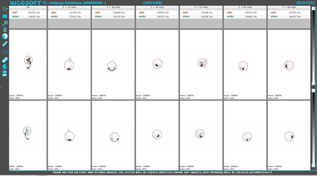
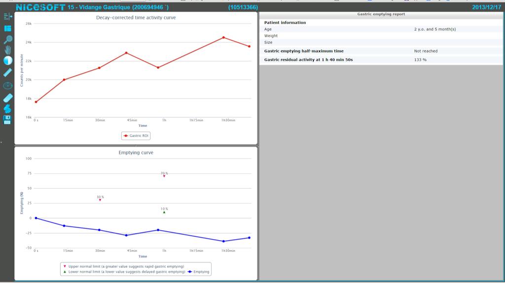
9 Gastric Reflux Clinical application for review and analysis of gastric reflux stimulation studies. Manual ROI definition Time activity curve analysis Compatible with dynamically acquired datasets User specific preferences based on logon credentials Customized layouts through display scripts Gastric Emptying Clinical application for review and analysis of gastric emptying studies. Analysis of stomach function Separate analysis of solid and liquid phase Compatible with dynamic or multiple static data Analysis of dynamic esophageal function Customized layouts through display scripts 9
10 10

 parathyroid studies Dual phase (Sestamibi) parathyroid")
11 Sentinel Node Display of two static images with a specialized gray scale optimized for the high maximum counts typical in sentinel node imaging. Thyroid and Parathyroid Clinical application for review and analysis of thyroid uptake, thyroid and parathyroid studies. Uptake calculation based on pre/post injection dose image Relative R/L lobe uptake Either technetium or iodine studies Fusion of marker and anterior thyroid images Subtraction analysis of dual isotope (Tc, Tl) parathyroid studies Dual phase (Sestamibi) parathyroid studies 11


12 Renal Clinical applications for review and analysis of renography studies. Analysis of kidney function Auto region of interest with manual override Time activity curve analysis Split function Measurement tool Compatible with Tc-99m-DTPA, Tc99m-MAG3 Multi-phase analysis Separate applications for handling of Anterior, Transplant and Single Kidney scenarios. 12

13 VenusCardiac Clinical application for reconstruction, processing and display of gated and ungated tomographic cardiac data. Access to optional Cedars MoCo motion correction module for the automatic and manual correction of SPECT acquisition motion artifacts. Pattern matching and segmentation algorithms are used in conjunction to minimize motion error metrics over the set of acquired projections; the resulting motion corrected projections are then presented to the operator for validation or modification. Reorientation, zooming and masking of reconstructed data. Slice review displays Direct access to quantification with third party applications such as Cedars Sinai QGS-QPS etc. MUGA Clinical application for review and analysis of planar gated blood pool studies. Automatic segmentation of left ventricle (ED, ES, and background ROI, with manual override) Unique Fourier based filtering technique for improved visualization of wall motion Display functions: window leveling, color scale selection, slice alignment, cine controls, zoom, pan, magnify Amplitude/Phase analysis 16
 Customized layouts through display scripts Planar Lung Analysis Clinical application for review and analysis of planar lung studies.")
14 Pseudo3D cine display (topographic dynamic mapping of counts) Multi-study comparison (multiple exercise levels, or follow-up) Right ventricle processing options (RVEF, RV volumes) Compatible with most acquisition protocols (matrix size, #frames/cycle) Customized layouts through display scripts Planar Lung Analysis Clinical application for review and analysis of planar lung studies. Analysis of Ventilation, Perfusion, or Ventilation + Perfusion Static image display of all acquired orientations Display functions: window leveling, color scale selection, zoom, pan, magnify, colorize Measurement tool Automatic 2-segmental quantification of anterior and posterior images ; Generation of geometric mean images User definable hotkeys for commonly used functions User specific preferences based on logon credentials 17


15 LungSPECT-CT An application for reconstruction, display and linked triangulation of ventilation and perfusion SPECT studies. Includes creation of reprojection images and MIRegistration of perfusion and ventilation series. 18

16 Utilities MIRegister A clinical application that provides direct access to the Mutual Image Information Registration utilities. Automatic process & manual adjustments 19
multimodality image processing workstation Visualizing your SPECT-CT-PET-MRI images
 multimodality image processing workstation Visualizing your SPECT-CT-PET-MRI images FUSION FUSION is a new visualization and evaluation software developed by Mediso built on state of the art technology,
4DM Packages. 4DM Packages & License Types. Information to help you order the appropriate licenses for your site.
 4DM Packages 4DM Packages & License Types. Information to help you order the appropriate licenses for your site. Nuclear Cardiac Quantification, Review, and Reporting Select Your 4DM Package and corresponding
Corridor4DM Feature List
 Software Services Features Corridor4DM Feature List 4DM v2013 FDG PET Standard Uptake Value (SUV) Calculation Auto-Selection of Report Template Motion Correction of SPECT datasets New Normals Databases
XD 3.6 Release Notes
 XD 3.6 Release Notes 1 Introduction 1.1 Purpose The purpose of these Release Notes is to: 1.2 Scope Communicate any known limitations for Mirada s XD release. Provide information on certain product features.
Integrating Modalities to Enterprise PACS through Thinking Systems
 Integrating Modalities to Enterprise PACS through 1 The Need For most enterprise PACS implementations, traditional CT, MR, CR, DR and general Ultrasound are of primary concerns. Not much attention has
VIEW PRO 4D. four dimensions in one workstation
 VIEW PRO 4D THE complete 3D/4D workstation for automatic image processing four dimensions in one workstation VERSiON 1.0 iq-view PRO 4D FROM HEAD TO TOE IN 4D iq-view PRO 4D is the perfect solution for
VieW 3D. 3D Post-Processing WorKstation THE THIRD DIMENSION. Version 3.1
 VieW 3D 3D Post-Processing WorKstation THE THIRD DIMENSION Version 3.1 iq-view 3D THE FULLY-FEATURED 3D MEDICAL IMAGING SOLUTION FOR RADIOLOGISTS iq-view 3D contains all components of iq-view with the
Philips SPECT/CT Systems
 Philips SPECT/CT Systems Ling Shao, PhD Director, Imaging Physics & System Analysis Nuclear Medicine, Philips Healthcare June 14, 2008 *Presented SNM08 Categorical Seminar - Quantitative SPECT and PET
Medical Image Analysis
 Computer assisted Image Analysis VT04 29 april 2004 Medical Image Analysis Lecture 10 (part 1) Xavier Tizon Medical Image Processing Medical imaging modalities XRay,, CT Ultrasound MRI PET, SPECT Generic
RTx Release Notes. Communicate any known limitations for the RTx release. Provide information on certain product features.
 RTx Release Notes 1 Introduction 1.1 Purpose The purpose of these Release Notes is to: 1.2 Scope Communicate any known limitations for the RTx release. Provide information on certain product features.
Technical aspects of SPECT and SPECT-CT. John Buscombe
 Technical aspects of SPECT and SPECT-CT John Buscombe What does the clinician need to know? For SPECT What factors affect SPECT How those factors should be sought Looking for artefacts For SPECT-CT Issues
A Closer Look at SERVER-SIDE RENDERING. Technology Overview
 A Closer Look at SERVER-SIDE RENDERING Technology Overview Driven by server-based rendering, Synapse 5 is the fastest PACS in the medical industry, offering subsecond image delivery and diagnostic quality.
Deviceless respiratory motion correction in PET imaging exploring the potential of novel data driven strategies
 g Deviceless respiratory motion correction in PET imaging exploring the potential of novel data driven strategies Presented by Adam Kesner, Ph.D., DABR Assistant Professor, Division of Radiological Sciences,
NumaStore Preclinical FAQ
 NumaStore Preclinical FAQ 1. What is NumaStore Preclinical? 2. How does NumaStore Preclinical work with Inveon? 3. What data types does NumaStore Preclinical support? 4. How much storage space do I need
Help Guide. mm Copyright Mirada Medical Ltd, Mirada Medical RTx 1
 Help Guide mm3237-1.6-1 Copyright Mirada Medical Ltd, 2000-2014. Mirada Medical RTx 1 Contents Help Guide... 1 Contents... 2 Introduction to RTx... 4 Regulatory Statement... 6 Notes... 15 Data Supported...
Living Image. Release Notes Version Purpose. 2. New Features. FLIT Algorithm Improvements. Single-Click FLIT and DLIT Reconstructions
 Living Image Release Notes Version 4.5 1. Purpose This document provides a brief overview of new features and improvements in Living Image 4.5. This release serves as an update for Living Image 4.4 on
CHILI /Workstation. Reporting multi modal digital images. Product Specification
 CHILI /Workstation Reporting multi modal digital images Product Specification CHILI /Workstation CHILI/Workstation is a diagnostic PACS workstation with additional functions for teleradiology. CHILI/Workstation
Interoperability Issues in Image Registration and ROI Generation
 1 DICOM 2005 International Conference, Budapest, Hungary Interoperability Issues in Image Registration and ROI Generation Todd Kantchev PhD, Siemens Molecular Imaging, Oxford, UK 2 Scope The following
ADVANCING CANCER TREATMENT
 The RayPlan treatment planning system makes proven, innovative RayStation technology accessible to clinics that need a cost-effective and streamlined solution. Fast, efficient and straightforward to use,
ADVANCING CANCER TREATMENT
 3 ADVANCING CANCER TREATMENT SUPPORTING CLINICS WORLDWIDE RaySearch is advancing cancer treatment through pioneering software. We believe software has un limited potential, and that it is now the driving
Mediso AnyScan. Single-Head and Dual-Head SPECT
 Mediso AnyScan S Single-Head and Dual-Head SPECT ANYSCAN S (SINGLE-HEAD & DUAL-HEAD SPECT) Low Cost of Ownership Associated Imaging Services is the exclusive U.S. source in the Midwest for Mediso Medical
FusionXD 2.0 DICOM Conformance Statement
 FusionXD 2.0 DICOM Conformance Statement 1 Introduction 1.1 Revision History See the Revision History in the beginning of this document 1.2 References Reference REF1 Document Digital Imaging and Communications
TOP DOG IN VETERINARY PACS
 WEBX WEB-BASED VETERINARY PACS TOP DOG IN VETERINARY PACS VERSION 6.3.8 Specialists in Medical Digital Imaging Solutions VET-WEBX VET-WEBX - THE PERFECT TOOL FOR VETERINARY FREEDOM VET-WEBX is a PACS especially
RT_Image v0.2β User s Guide
 RT_Image v0.2β User s Guide RT_Image is a three-dimensional image display and analysis suite developed in IDL (ITT, Boulder, CO). It offers a range of flexible tools for the visualization and quantitation
FusionXD 2.1 DICOM Conformance Statement
 FusionXD 2.1 DICOM Conformance Statement Contents 1 Introduction... 3 1.1 References... 3 1.2 Integration and Features... 3 1.3 Definitions, Acronyms and Abbreviations... 3 2 Services... 4 3 Networking...
Using Pinnacle 16 Deformable Image registration in a re-treat scenario
 Introduction Using Pinnacle 16 Deformable Image registration in a re-treat scenario This short Hands On exercise will introduce how the Deformable Image Registration (DIR) tools in Pinnacle can be used
Mediso AnyScan S Single-Head and Dual-Head SPECT
 Mediso AnyScan S Single-Head and Dual-Head SPECT ANYSCAN S (SINGLE-HEAD & DUAL-HEAD SPECT) Low Cost of Ownership Small Footprint Absolute Imaging Solutions tm is the exclusive U.S. source for Mediso Medical
PMOD Features dedicated to Oncology Research
 While brain research using dynamic data has always been a main target of PMOD s developments, many scientists working with static oncology data have also found ways to leverage PMOD s unique functionality.
If it matters to you, it matters to us
 If it matters to you, it matters to us Philips clinical innovations in nuclear medicine Innovation with insight We understand that clinical innovations are only as valuable as the day-to-day difference
The Advanced Neuro MR Processing & Visualization Solution
 The Advanced Neuro MR Processing & Visualization Solution Optimized Workflow Automated Processing Provides Answers Faster and More Efficiently The DynaSuite Neuro Workstation is a high performance advanced
HYBRID VIEWER. HERMES Data Analysis Applications. Hand book CD500.3_P31V 2.0C-2.1C. Template: T
 HERMES Data Analysis Applications HYBRID VIEWER Hand book Document Revision Date: Document Number: Version: 2012-12-19 2.0C-2.1C Canada Health Template: T-0211-12 E-mail: info@hermesmedical.com Website:
 Software solutions Nucline all modality acquisition software Nucline is an easy to use and intuitive interface for high throughput workflow across all the nanoscan Family systems - there is no need to
Segment CT Instructions For Use - English
 Segment CT Instructions For Use - English February 28, 2018 Software platform v2.2 R6239 MEDVISO AB http://www.medviso.com Griffelvägen 3 SE-224 67 Lund Sweden Tel: +46-76-183 6442 ii Contents 1 Terms
YOUR PACS, YOUR FREEDOM
 WEB THIN CLIENT PACS FOR STORAGE, ARCHIVING AND TELERADIOLOGY YOUR PACS, YOUR FREEDOM VERSION 6.6.2 iq-web THE CAREFREE, ALL-INCLUSIVE PACS iq-web is a complete, easy-to-use and affordable PACS for storing,
Conflicts of Interest Nuclear Medicine and PET physics reviewer for the ACR Accreditation program
 James R Halama, PhD Loyola University Medical Center Conflicts of Interest Nuclear Medicine and PET physics reviewer for the ACR Accreditation program Learning Objectives 1. Be familiar with recommendations
Medical Image Viewer Guide
 Cloud Medical Image Management Medical Image Viewer Guide March 2016 Table of Contents Indications for Use 3 Browser Requirements 3 User Interface Overview 4-5 Study Page 6 Main Toolbar 7 Sub-Toolbars
James R Halama, PhD Loyola University Medical Center
 James R Halama, PhD Loyola University Medical Center Conflicts of Interest Nuclear Medicine and PET physics reviewer for the ACR Accreditation program Learning Objectives Be familiar with the tests recommended
VALIDATION OF DIR. Raj Varadhan, PhD, DABMP Minneapolis Radiation Oncology
 VALIDATION OF DIR Raj Varadhan, PhD, DABMP Minneapolis Radiation Oncology Overview Basics: Registration Framework, Theory Discuss Validation techniques Using Synthetic CT data & Phantoms What metrics to
VIEW. a short tutorial. quick start
 VIEW a short tutorial quick start VERSiON 3.0 Contents Inhalt Introduction........................................................... 3 Import of DICOM media....................................................
Arbitrary cut planes Slab control with slab thickness Projection plane adjustment Box cropping Mandible detection MPR cross-section linked views
 Provides unparalleled quality and performance no need to sacrifice one for the other Enables high fidelity viewing with interactive deep supersampling Requires no GPUs or hardware accelerators a true,
Virtual Phantoms for IGRT QA
 TM Virtual Phantoms for IGRT QA Why ImSimQA? ImSimQA was developed to overcome the limitations of physical phantoms for testing modern medical imaging and radiation therapy software systems, when there
Biomedical Image Analysis based on Computational Registration Methods. João Manuel R. S. Tavares
 Biomedical Image Analysis based on Computational Registration Methods João Manuel R. S. Tavares tavares@fe.up.pt, www.fe.up.pt/~tavares Outline 1. Introduction 2. Methods a) Spatial Registration of (2D
3D Slicer Overview. Andras Lasso, PhD PerkLab, Queen s University
 3D Slicer Overview Andras Lasso, PhD PerkLab, Queen s University Right tool for the job Technological prototype Research tool Clinical tool Can it be done? Jalopnik.com Innovative, not robust, usually
WEBX. One For all. Specialists in Medical. Digital Imaging Solutions
 WEBX the DICOM server for storage, teleradiology and image distribution One For all VERSiON 6.2 Specialists in Medical Digital Imaging Solutions iq-webx iq-webx - a fast DICOM archive and a web-based viewing
TomoCon 3.0 User s Manual
 TomoCon 3.0 User s Manual TatraMed TomoCon 3.0 Workstation TomoCon 3.0 Viewer TomoCon 3.0 Lite Version 3.0.14 TomoCon PACS 3 TatraMed spol. s r.o., Líščie údolie 7, 841 04 Bratislava, Slovak Republic Tel.:
Clinical Prospects and Technological Challenges for Multimodality Imaging Applications in Radiotherapy Treatment Planning
 Clinical Prospects and Technological Challenges for Multimodality Imaging Applications in Radiotherapy Treatment Planning Issam El Naqa, PhD Assistant Professor Department of Radiation Oncology Washington
Medical Image Processing: Image Reconstruction and 3D Renderings
 Medical Image Processing: Image Reconstruction and 3D Renderings 김보형 서울대학교컴퓨터공학부 Computer Graphics and Image Processing Lab. 2011. 3. 23 1 Computer Graphics & Image Processing Computer Graphics : Create,
Inveon User Notes. Inveon Scanners and Inveon Acquisition Workplace 2.0
 Inveon User Notes Inveon Scanners and Inveon Acquisition Workplace 2.0 Legal Notice 2014 by Siemens Medical Solutions USA, Inc. All rights reserved. Siemens reserves the right to modify the design and
V/Q Imaging Protocols
 Elizabeth ailey ppsc(mrs) M Department of Nuclear Medicine Royal North Shore Hospital, Sydney, ustralia email: ebailey@nsccahs.health.nsw.gov.au The V/Q scan has traditionally used planar imaging techniques.
The Emory Reconstruction Toolbox Version 1.0
 The Emory Reconstruction Toolbox Version 1.0 Operating Instructions Revision 02 (April, 2008) Operating Instructions The Emory Reconstruction Toolbox Application Copyrights, Trademarks, Restrictions
Voxar 3D ColonMetrix. Reference Guide
 Voxar 3D ColonMetrix Reference Guide The software described in this document is furnished under a license, and may be used or copied only according to the terms of such license. Toshiba means, Toshiba
CHILI /Web. Intelligent image distribution inside and outside the hospital. Product Specification
 CHILI /Web Intelligent image distribution inside and outside the hospital Product Specification CHILI /Web CHILI/Web is an image distribution and teleradiology solution based on a proven and extensible
VIEW/PRO VET EDITION THE RADIOLOGY READING STATION FOR VETERINARIANS FOR VET EYES ONLY
 VIEW/PRO VET EDITION THE RADIOLOGY READING STATION FOR VETERINARIANS FOR VET EYES ONLY VERSION 3.1 iq-view/pro VET EDITION VETERINARY READING AT YOUR FINGERTIPS The key factor for vets who frequently read
UvA-DARE (Digital Academic Repository) Motion compensation for 4D PET/CT Kruis, M.F. Link to publication
 Motion compensation for 4D PET/CT Kruis, M.F. Link to publication") UvA-DARE (Digital Academic Repository) Motion compensation for 4D PET/CT Kruis, M.F. Link to publication Citation for published version (APA): Kruis, M. F. (2014). Motion compensation for 4D PET/CT General
UvA-DARE (Digital Academic Repository) Motion compensation for 4D PET/CT Kruis, M.F. Link to publication Citation for published version (APA): Kruis, M. F. (2014). Motion compensation for 4D PET/CT General
Cerner SkyVue Cardiology Remote Review with NVIDIA and VMware Horizon
 Cerner SkyVue Cardiology Remote Review with NVIDIA and VMware Horizon Stuart Jackson Sr. Technology Architect Agenda Meet Cerner Cardiology Solution Overview Workflow Challenges Workflow Requirements Testing
Ossa 3D User Manual. ios App v1.0.0
 Ossa 3D User Manual ios App v1.0.0 CONTENTS Description 3 Getting Started User Interface 4 Control Gestures 4 View Modes 5 Save Project 5 Settings Menu In-App Purchases 5 Enable Passcode 5 Page Tools View
Zero Foot- Print Browser * Mobile Device Viewing Of Medical Images
 Zero Foot- Print Browser * Mobile Device Viewing Of Medical Images WEBWORKS, an integral component of Roentgen Works, is a 100% browserbased viewer for medical images, reports and related files. It enables
SCENARIA Hitachi Innovation at Work for You.
 PRODUCT DATA SCENARIA Hitachi Innovation at Work for You. Continuing its commitment to the SCENARIA scalable CT platform, Hitachi introduces cutting-edge 64/128-slice technology with the SCENARIA SE. The
Chapter 32 3-D and 4-D imaging in Obstetrics and Gynecology
 Objectives Define common terms related to 3D/4D ultrasound Chapter 32 3-D and 4-D imaging in Obstetrics and Gynecology Bridgette Lunsford Describe how 3D and 4D imaging differs from 2D ultrasound Identify
Discovery LS 1.0 Conformance Statement For DICOM V3.0
 g GE Medical Systems Technical Publications DIRECTION 2343444GSP Revision 0 Discovery LS 1.0 Conformance Statement For DICOM V3.0 GE Medical Systems GE Medical Systems: telex 3797371 P.O. Box 414, Milwaukee,
syngo.mr Neuro 3D: Your All-In-One Post Processing, Visualization and Reporting Engine for BOLD Functional and Diffusion Tensor MR Imaging Datasets
 syngo.mr Neuro 3D: Your All-In-One Post Processing, Visualization and Reporting Engine for BOLD Functional and Diffusion Tensor MR Imaging Datasets Julien Gervais; Lisa Chuah Siemens Healthcare, Magnetic
3/27/2012 WHY SPECT / CT? SPECT / CT Basic Principles. Advantages of SPECT. Advantages of CT. Dr John C. Dickson, Principal Physicist UCLH
 3/27/212 Advantages of SPECT SPECT / CT Basic Principles Dr John C. Dickson, Principal Physicist UCLH Institute of Nuclear Medicine, University College London Hospitals and University College London john.dickson@uclh.nhs.uk
WEB THIN CLIENT PACS FOR STORAGE, ARCHIVING AND TELERADIOLOGY YOUR CENPACS, YOUR FREEDOM.
 WEB THIN CLIENT PACS FOR STORAGE, ARCHIVING AND TELERADIOLOGY YOUR CENPACS, YOUR FREEDOM. THE CAREFREE, ALL-INCLUSIVE CENPACS CENPACS is a complete, easy-to-use and affordable PACS for storing, distributing
ClinicalExpress Operator s Guide. version 6.1 for general radiography and mammography
 ClinicalExpress Operator s Guide version 6.1 for general radiography and mammography Copyright by VIDAR Systems Corporation. All rights reserved. No part of this publication may be reproduced, stored in
UNLOCKING. Possibilities
 UNLOCKING Possibilities In the early 1980 s members of the radiology community envisioned a future practice built around the concept of a Picture Archiving and Communication System (PACS). During the past
11/18/ CPT Preauthorization Groupings Effective January 1, Computerized Tomography (CT) Abdomen 6. CPT Description SEGR CT01
 Abdomen 6. CPT Description SEGR CT01") Computerized Tomography (CT) 6 & 101 5 Upper Extremity 11 Lower Extremity 12 Head 3 Orbit 1 Sinus 2 Neck 4 7 Cervical Spine 8 Thoracic Spine 9 Lumbar Spine 10 Colon 13 CPT Description SEGR 74150 74160
Computerized Tomography (CT) 6 & 101 5 Upper Extremity 11 Lower Extremity 12 Head 3 Orbit 1 Sinus 2 Neck 4 7 Cervical Spine 8 Thoracic Spine 9 Lumbar Spine 10 Colon 13 CPT Description SEGR 74150 74160
SCENARIA 64-Slice CT Putting Advanced CT Within Reach.
 SCENARIA 64-Slice CT Putting Advanced CT Within Reach. Advancing the technology of Computed Tomography for over 30 years, Hitachi is a recognized innovator of lower dose*, high diagnostic value CT solutions.
Medical Image Registration
 Medical Image Registration Submitted by NAREN BALRAJ SINGH SB ID# 105299299 Introduction Medical images are increasingly being used within healthcare for diagnosis, planning treatment, guiding treatment
PRODUCT DATA. Advanced 128
 PRODUCT DATA Advanced 128 Supria 16 Slice CT Puts You On The Path of High Quality, Cost-Effective CT Scanning Solid Capabilities Are Built Into the Supria Addressing the challenges of controlling healthcare
Good Morning! Thank you for joining us
 Good Morning! Thank you for joining us Deformable Registration, Contour Propagation and Dose Mapping: 101 and 201 Marc Kessler, PhD, FAAPM The University of Michigan Conflict of Interest I receive direct
8/3/2016. Image Guidance Technologies. Introduction. Outline
 8/3/26 Session: Image Guidance Technologies and Management Strategies Image Guidance Technologies Jenghwa Chang, Ph.D.,2 Department of Radiation Medicine, Northwell Health 2 Hofstra Northwell School of
USER MANUAL VP PLANNING
 USER MANUAL VP PLANNING 1.0 Mobile application ios Visible Patient SAS RCS Strasbourg TI 794 458 125 1 place de l'ho pital 67000 Strasbourg, FRANCE Share capital: 58330 Tel.: 33 (0)3 88 11 90 00 Fax: 33
VIEW READINGS MADE SIMPLE
 VIEW THE radiology reading station READINGS MADE SIMPLE the creation of individually customizable hanging protocols. As a result, studies and images can be loaded automatically as you wish to read them.
Implementation of Advanced Image Guided Radiation Therapy
 Image Acquisition Course Outline Principles, characteristics& applications of the available modalities Image Processing in the T x room Image guided treatment delivery What can / can t we do in the room
better images mean better results
 better images mean better results A better way for YOU and YOUR patient brought to you by Advanced Neuro analysis with access to studies wherever you need it Advanced Neuro from Invivo Advancements in
Medical Image Processing in Nuclear Medicine. Andrew Todd-Pokropek University College London INSERM U494 Paris. Aims and Objectives
 Medical Image Processing in Nuclear Medicine Andrew Todd-Pokropek University College London INSERM U494 Paris Aims and Objectives The aim of this module is to provide an understanding of the special nature
English. Instructions for Use. NM Application Suite. Extended Brilliance Workspace NM 1.0 PHI
 English Instructions for Use NM Application Suite Extended Brilliance Workspace NM 1.0 PHI NM Application Suite INSTRUCTIONS FOR USE Release 1.0 English Philips Healthcare 4535 604 78081 Rev A PAI Warranty
Advanced Visual Medicine: Techniques for Visual Exploration & Analysis
 Advanced Visual Medicine: Techniques for Visual Exploration & Analysis Interactive Visualization of Multimodal Volume Data for Neurosurgical Planning Felix Ritter, MeVis Research Bremen Multimodal Neurosurgical
NumaStore. 10 Northern Blvd. Unit 12, Amherst, NH Phone: (603) , Fax: (603)
 , Fax: (603)") Numa Products and Services for Nuclear Medicine NumaStore Image Management Solution Receives, Stores, Organizes, and Distributes Nuclear Medicine Data 10 Northern Blvd. Unit 12, Amherst, NH 03031 Phone:
Numa Products and Services for Nuclear Medicine NumaStore Image Management Solution Receives, Stores, Organizes, and Distributes Nuclear Medicine Data 10 Northern Blvd. Unit 12, Amherst, NH 03031 Phone:
iplan RT Image Advanced Contouring Workstation - Driving Physician Collaboration
 iplan RT Image Advanced Contouring Workstation - Driving Physician Collaboration The iplan Contouring Workstation offers unique and innovative capabilities for faster contouring and consistent segmentation
SISCOM (Subtraction Ictal SPECT CO-registered to MRI)
") SISCOM (Subtraction Ictal SPECT CO-registered to MRI) Introduction A method for advanced imaging of epilepsy patients has been developed with Analyze at the Mayo Foundation which uses a combination of
SISCOM (Subtraction Ictal SPECT CO-registered to MRI) Introduction A method for advanced imaging of epilepsy patients has been developed with Analyze at the Mayo Foundation which uses a combination of
Is deformable image registration a solved problem?
 Is deformable image registration a solved problem? Marcel van Herk On behalf of the imaging group of the RT department of NKI/AVL Amsterdam, the Netherlands DIR 1 Image registration Find translation.deformation
VIEW BY RADIOLOGISTS FOR RADIOLOGISTS
 VIEW THE radiology reading station BY RADIOLOGISTS FOR RADIOLOGISTS VERSiON 2.7 iq-view/pro Radiology reading at your fingertips The key factor for physicians who frequently read medical imaging studies
Import Organ Atlas to co-register with Optical 3D imaging (DLIT or FLIT):
:") Importing Organ Atlas Import Organ Atlas to co-register with Optical 3D imaging (DLIT or FLIT): 1. Select Import -> Organ Atlas in the File menu 2. Click on Add Organ Files 3. Select ALL organ files from
Importing Organ Atlas Import Organ Atlas to co-register with Optical 3D imaging (DLIT or FLIT): 1. Select Import -> Organ Atlas in the File menu 2. Click on Add Organ Files 3. Select ALL organ files from
Workshop on Quantitative SPECT and PET Brain Studies January, 2013 PUCRS, Porto Alegre, Brasil Corrections in SPECT and PET
 Workshop on Quantitative SPECT and PET Brain Studies 14-16 January, 2013 PUCRS, Porto Alegre, Brasil Corrections in SPECT and PET Físico João Alfredo Borges, Me. Corrections in SPECT and PET SPECT and
QMass 8.1. Quick Start Manual. June 13, v2.0
 QMass 8.1 Quick Start Manual June 13, 2017 9.10.250.81.3 v2.0 Medis medical imaging systems bv Schuttersveld 9, 2316 XG Leiden, the Netherlands http://www.medis.nl Medis medical imaging systems bv Medis
fmri/dti analysis using Dynasuite
 fmri/dti analysis using Dynasuite Contents 1 Logging in 2 Finding patient session 3 Viewing and adjusting images 4 Checking brain segmentation 5 Checking image registration 6 Seeing fmri results 7 Saving
Mathematical methods and simulations tools useful in medical radiation physics
 Mathematical methods and simulations tools useful in medical radiation physics Michael Ljungberg, professor Department of Medical Radiation Physics Lund University SE-221 85 Lund, Sweden Major topic 1:
Solid Capabilities Are Built Into the Supria Plus. Putting You On The Path of High Quality, Cost-Effective CT Scanning
 Specification Data Putting You On The Path of High Quality, Cost-Effective CT Scanning Solid Capabilities Are Built Into the Supria Plus Addressing the challenges of controlling healthcare organization
SIGMI. ISL & CGV Joint Research Proposal ~Image Fusion~
 SIGMI ISL & CGV Joint Research Proposal ~Image Fusion~ Introduction Research Diagram What CGV Lab is interested in What ISL is interested in Research Plan Research Diagram Medical Imaging and Application
n o r d i c B r a i n E x Tutorial DSC Module
 m a k i n g f u n c t i o n a l M R I e a s y n o r d i c B r a i n E x Tutorial DSC Module Please note that this tutorial is for the latest released nordicbrainex. If you are using an older version please
A rib-specific multimodal registration algorithm for fused unfolded rib visualization using PET/CT
 A rib-specific multimodal registration algorithm for fused unfolded rib visualization using PET/CT Jens N. Kaftan a, Marcin Kopaczka a, Andreas Wimmer b, Günther Platsch a, and Jérôme Declerck a a Siemens
Oncentra Brachy. Anatomy-based treatment planning for HDR/PDR brachytherapy
 Oncentra Brachy Anatomy-based treatment planning for HDR/PDR brachytherapy Anatomy-based treatment planning for HDR/PDR brachytherapy In its rich history, Nucletron has developed a wide range of treatment
USER MANUAL VP PLANNING
 USER MANUAL VP PLANNING 1.0 Mobile application Android Visible Patient SAS RCS Strasbourg TI 794 458 125 1 place de l'ho pital 67000 Strasbourg, FRANCE Share capital: 58330 Tel.: 33 (0)3 88 11 90 00 Fax:
UGviewer: a medical image viewer
 Appendix A UGviewer: a medical image viewer As a complement to this master s thesis, an own medical image viewer was programmed. This piece of software lets the user visualize and compare images. Designing
Beyond Performance and Value
 Putting the back in ultrasound Beyond Performance and Value Esaote s new ultra-performance MyLab 9 exp ultrasound system is designed to support a full range of shared service diagnostic imaging environments.
Joint CI-JAI advanced accelerator lecture series Imaging and detectors for medical physics Lecture 1: Medical imaging
 Joint CI-JAI advanced accelerator lecture series Imaging and detectors for medical physics Lecture 1: Medical imaging Dr Barbara Camanzi barbara.camanzi@stfc.ac.uk Course layout Day AM 09.30 11.00 PM 15.30
REAL-TIME ADAPTIVITY IN HEAD-AND-NECK AND LUNG CANCER RADIOTHERAPY IN A GPU ENVIRONMENT
 REAL-TIME ADAPTIVITY IN HEAD-AND-NECK AND LUNG CANCER RADIOTHERAPY IN A GPU ENVIRONMENT Anand P Santhanam Assistant Professor, Department of Radiation Oncology OUTLINE Adaptive radiotherapy for head and
Leksell SurgiPlan. Powerful planning for success
 Leksell SurgiPlan Powerful planning for success Making a difference in surgical planning Leksell SurgiPlan Leksell SurgiPlan is an advanced image-based neurosurgical planning software, specifically designed
TOP dog in veterinary PACS
 WEBX WEB-based veterinary pacs TOP dog in veterinary PACS VERSiON 6.1.3 VET-WEBX VET-WEBX THE PERFECT TOOL FOR VETERINARY FREEDOM VET-WEBX is a PACS especially designed for veterinarians, which conveniently