Introduction to Cryo Electron Microscopy/Tomography
|
|
|
- Clarence Adams
- 6 years ago
- Views:
Transcription


1 Introduction to Cryo Electron Microscopy/Tomography Ricardo Miguel Sánchez Loayza Max Planck Institute for Biophysics - Kudryashev Group Goethe Universität - Visual Sensorics and Information Processing
2 Overview - CryoEM/CryoET CryoEM/ET: What and Why? Image Formation CryoEM: Single Particle Analysis CryoET: Subtomogram Averaging
3 CryoEM/ET: What and Why?
4 What is Cryo-electron Microscopy/Tomography? Techniques used to produce high resolution 3D maps. ( 2 Å or 0.2 nm). Tools used by biophysicists (structural biology) to obtain 3D density maps and estimate the atomic model of biological samples: Get atomic model. Multiple comformations (How it works) Drug design. Cryo: The sample is frozen. Electron: A electron microscope is used. Microscopy: 1 frame por sample. Tomography: Sequence of frames per sample.
5 Why CryoEM/ET? Near atomic resolution. Close to native state sample. Easy sample preparation (Compared to crystallography) New method (2012) 1 : Most of available programs are experimental. Not all issues are solved (efficiently). 1 S. Scheres, RELION: Implementation of a bayesian approach to cryoem structure determination. 2012
 2 of the p97 hexameric ATPase in three different conformations. 2 S. Banerjee et al., 2.")
6 Example 1 Figure: Example of high resolution map (2.3 Å or 0.23 nm) 2 of the p97 hexameric ATPase in three different conformations. 2 S. Banerjee et al., 2.3 Å resolution map cryo-em structure of human p97 and mechanism of allosteric inhibition. 2016

7 Example 1 Figure: Atomic model fitted and measured motion.

8 Example 1 Figure: Schematic of the two-step sequence involved in p97 activation resulting from the sequential binding of ATP γ S.
 During its Catalytic Cycle.")
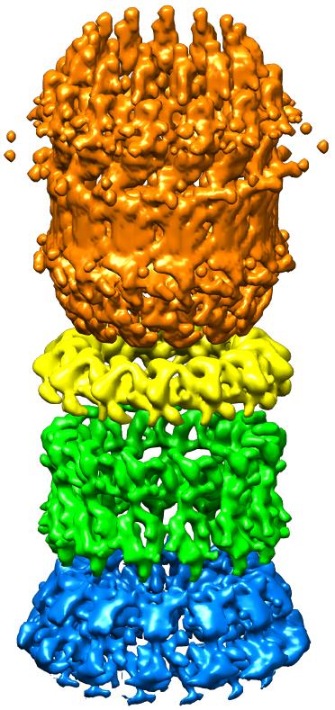
9 Example 2 Figure: Example of low resolution map ( 15 Å or 1.5 nm) 3 of different conformations of the P-glycoprotein. 3 G. Frank et al., Cryo-EM Analysis of the Conformational Landscape of Human P-glycoprotein (ABCB1) During its Catalytic Cycle. 2016
 fitted into the different 3D")
10 Example 2 Figure: Atomic model (obtained with crystallography) fitted into the different 3D maps.

11 Example 2 Figure: Schematic of hp-gp at major steps during the ATPase reaction cycle.
 4. 4 S. Subramaniam et al.")
12 Example 3 - Enzymes Figure: Example of 3D maps of 20 aminoacids and their atomic model. The resolution of each 3D map is 1.8 Å (0.18 nm) 4. 4 S. Subramaniam et al., Resolution advances in cryo-em enable application to drug discovery. 2016
13 Resolution in Structural Biology Figure: Structures to study in biology and how to sample them.
14 Electron Microscope source C1 C2 condenser aperture condenser system sample holder sample objective lens objective aperture projector system imaging Figure: Scheme of a modern Electron Microscope.
.")
15 Electron Microscope: Source High coherence of electron beam. Acceleration voltage: 300 kv. Wavelength: 0.02 Å (2 pm). Figure: Field emission tip.
16 Electron Microscope: Lenses Uses magnetic field to change the trajectory of the electron. Equivalent to light microscope lenses. Low magnification (50x). Figure: Magnetic lens.
17 Electron Microscope: Detector Figure: Direct detector allowed the Resolution Revolution. Gatan K2 Summit detector: 400 fps, 1.06Å pixel size, 8k 8k images.

18 Electron Microscope: Sample holder Figure: Sample holder of Titan KRIOS electron microscope.
19 Sample Preparation! Get a Biochemist. Get sample of protein. Put sample on grid. Plunge freeze it!. Put grid in microscope. Take images.??? Profit.
20 Sample preparation! Figure: Sample too thick.
21 Sample preparation! Figure: Sample too thin.

22 Sample preparation! Figure: Bad luck.

23 Sample preparation! Figure: Sample perfect.

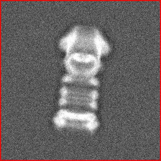
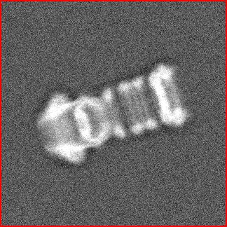
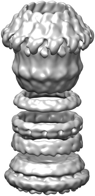
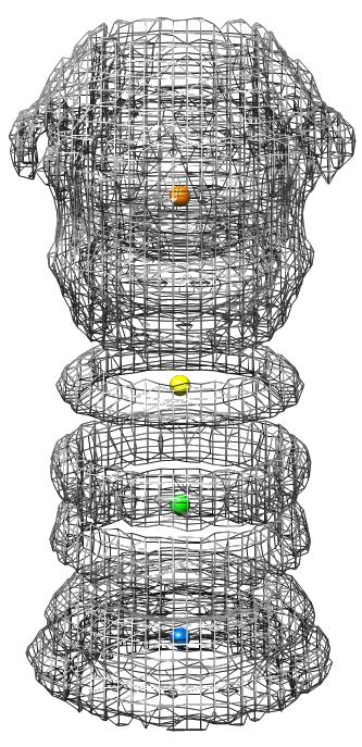
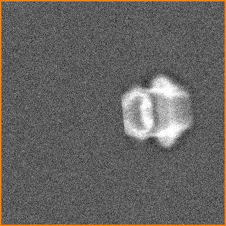
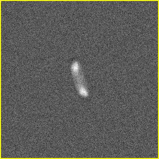
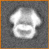
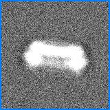
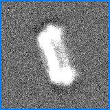
24 Record a micrograph! Figure: Reference molecule (Defocus: 70nm).

25 Record a micrograph! Figure: Recorded micrograph.

26 Record a micrograph! Figure: Filtering?.
27 Image Formation

28 Sample interaction with electron beam. Figure: Radiation destroys the sample. Limits the electron dose (25 e /Å 2 ).

29 Sample interaction with electron beam. Figure: The sample modifies the phase of the electron beam, not its amplitude.
30 Sample interaction with electron beam: Defocus. Figure: Electron beam interaction with sample. Figure: Changing the image plane adds extra phase shift.

31 Sample interaction with electron beam: Defocus. Figure: Defocus and the Contrast Transfer Function (CTF)
32 Image formation model ( ) I(x, y) = I rn + I dc + Poiss Φ e F 1 {F{Ψ exit }CTF (q)mtf (q)} Where I rn is the detector s readout noise, I dc is the dark current, Φ e is the electron flux, and Poiss(A) returns a random number from a Poisson distribution with expected value A 5. 5 M. Vulović, Image Formation modeling in Cryo-Electron Microscopy. 2013

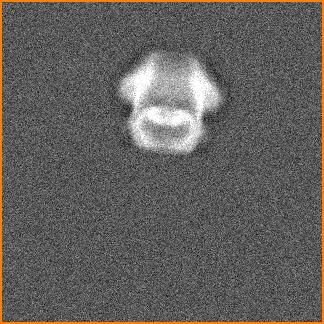
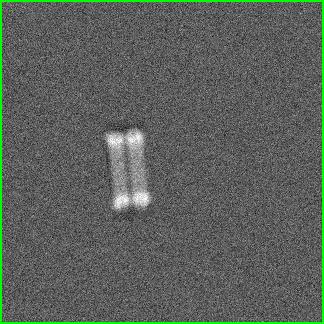
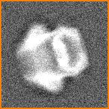
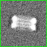
33 Record a micrograph (again)! Figure: Reference molecule with 2000nm of defocus.

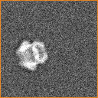
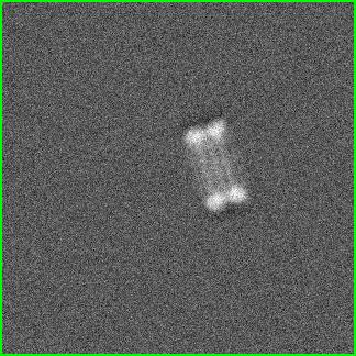
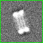
34 Record a micrograph (again)! Figure: Imaged molecule with 2000nm of defocus.

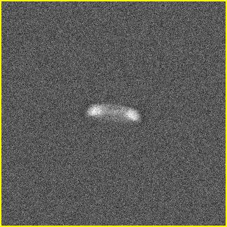
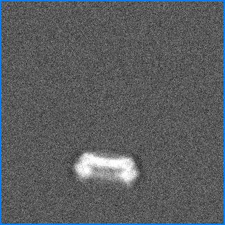
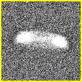
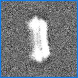
35 Record a micrograph (again)! Figure: Imaged molecule filtered

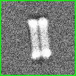
36 Remember: Radiation damage Figure: 3D maps of the same helix for different frames 6. 6 T. Grant, Measuring the optimal exposure for single particle CryoEM using 2.6 Å reconstruction of rotavirus VP
37 CryoEM: Single Particle Analysis
38 Single Particle Analysis (SPA) Goal: Generate High Resolution 3D map from Noisy Micrographs. How: Get orientation of each particle on the micrographs. Assumptions: Rigid protein. Homogeneity. No preferential orientation (Fourier space fully sampled). A lot of data: Millions of small images ( pixels, typically).

39 Fourier Central Slice Theorem Figure: Example of 2D Central Slice Theorem 7. 7 G. Zeng, Medical Image Reconstruction: A Concept Tutorial

40 SPA: Fourier Reconstruction Figure: The fourier transform of each projection is added to the fourier transform of the volume. The reconstruced volume is reconstructed by applying the inverse fourier transform.
41 The SPA problem. Problem: Given the unknown 3D structures V 1,..., V K, and the observed images X 1,..., X N, where X n = proj(v k, φ n ). Find φ n and V n. Solution (Bayesian likelihood framework): arg max V 1,...,V K log p(v 1,..., V K X 1,..., X N ) = arg max V 1,...,V K N log n=1 K k=1 1 K p(x n, φ n V k )dφ n + log p(v 1...K ) Solve by Regularized Expectation Maximization algorithm 8. Issue: Needs an initial guess of the volume. 8 S. Scheres, RELION: Implementation of a bayesian approach to cryoem structure determination. 2012

42 The SPA problem: Overfitting Figure: Image obtained from noise by using the Mona Lisa as initial guess.

43 The SPA problem: Speed of execution Figure: Using Stochastic Gradient Descent algorithm 9. 9 A. Punjabi et al., cryosparc: algorithms for rapid unsupervised cryoem structure determination. 2016

44 The SPA problem: Speed of execution Figure: Branch and Bound: Define a inexpensive lower bound over all poses A. Punjabi et al., cryosparc: algorithms for rapid unsupervised cryoem structure determination. 2016

45 SPA: Procedure Figure: CryoEM micrograph and a particle image F. Sigworth, Principles of cryoem single particle image processing. 2016
46 SPA: Procedure Figure: Proposed SPA workflow S. Scheres, RELION: Implementation of a bayesian approach to cryoem structure determination. 2012
47 SPA: My simplified workflow Correct for particle drifting. Correct CTF. Pick particles. Get an initial model. Get orientations and 3D maps. Classify 3D maps (Heterogeneity). Refine and polish 3D maps. Get resolution. Cure cancer (or publish somewhere, at least).
48 SPA: Procedure Figure: Proposed SPA workflow S. Scheres, RELION: Implementation of a bayesian approach to cryoem structure determination. 2012
49 SPA: Observations/Problems CTF is corrected typically by phase flipping. Particle picking is done by a highly trained machine: a biophysicist. There is no reliable autopicking program. 2D classification is needed. The proper selection of the initial model is crucial. Pose/orientation estimation was slow (?). Sometimes you get scooped.


50 SPA: Local Refinement Global 3D Refinement Global Reconstruction Estimated center of subunits RSS REP (proposed) Figure: Proposed Local refinement scheme. Local 3D Refinements and Local Reconstructions
51 CryoET: Subtomogram Averaging
52 Subtomogram Averaging (StA) Goal: Generate High Resolution 3D map of a particle in situ. How: Use tomograms instead of micrographs. Limitations: Limited views, typically from 60 to +60 with steps of 2 or 3. Tomograms are huge: 8k 8k 2k (480 GB). Alignment is done with volumes, no images. No need of projection matching. Artifacts: Missing Wedge. SLOW.

53 Tomogram creation

54 Tomogram creation

55 Tomogram creation Figure: Frames need to be aligned.
56 Weighted Back Projection 1 Figure: Back Projection G. Zeng, Medical Image Reconstruction. Springer 2016.
57 Weighted Back Projection 2 Figure: Weighted Back Projection G. Zeng, Medical Image Reconstruction. Springer 2016.

58 Algebraic Reconstruction Techniques 1 Figure: Mathematical Model for ART 16 For one projection: p θ = A x 16 G. Zeng, Medical Image Reconstruction. Springer 2016.
59 Algebraic Reconstruction Techniques 2 For multiple projections: p θ1 p θ2 p θ3 p θtotal = A 1 A 2 A 3 x = A total x Underdetermined system: More variables than equations. Cannot be solved by direct algorithms. Iterative algorithms are used: Slow. Mathematical model allows constrains to the solution, like: Solution ˆx must be smooth. Highlight the edges of solution ˆx. Consider pθtotal is noisy.

60 Progressive Stochastic Reconstruction Technique 1 Figure: Graphical description of the PSRT algorithm B. Turonova, Progressive Stochastic Reconstruction Technique for Cryo Electron Tomography. Journal of Structural Biology, 2015
, x 2 is out of range, and x 3 is rejected (does not decrease energy). 18 18 J. Gregson.")
61 Progressive Stochastic Reconstruction Technique 2 Figure: Example of three densities. x 1 is accepted (decrease energy), x 2 is out of range, and x 3 is rejected (does not decrease energy) J. Gregson. Stochastic Tomography and ist Application in 3D imaging of Mixing Fluids
62 StA: My simplified workflow Collect RAW series. Align them using fiducials. Create full tomogram. Pick particles (cannot be avoided, yet?). Crop Subtomograms. Align subtomograms. Publish if not scooped.

63 Tilt series Alignment Figure: Gold beads picked in a tilt series.
64 Subtomogram Averaging Scheme. Figure: Scheme of the StA procedure A. Crowther, Methods in Enzymology - The Resolution Revolution: Recent Advances In CryoEM. 2016
65 StA: Observations/Problems Uses Brute Force to find orientations. Alignment of the tilt series is not accurate. Even lower electron dose than SPA: very low SNR. Low SNR: Difficult to estimate CTF and pick particles.
66 Final Remarks CryoEM is a relatively new technique with a lot of room for improvement. CryoET is even younger!. Most of the problems in CryoEM/CryoET are ill-posed. Most of the tools used in CryoEM/CryoET are based in basic ideas in image processing. Thanks.
Tomography and Subtomogram Averaging
 Tomography and Subtomogram Averaging John Briggs EM course 2017 What do we need to get a 3D structure? Sample preparation methods A transmission electron microscope Different views of our object of interest
Structural Information obtained
 Structural Information obtained from Electron Microscopy Christiane Schaffitzel, 09.05.2013 Including slides from John Briggs, Bettina Boettcher, Nicolas Boisset, Andy Hoenger, Michael Schatz, and more
From cryo-em images of protein complexes to high-resolution 3D structures
 From cryo-em images of protein complexes to high-resolution 3D structures Rouslan Efremov www.cryo-em.be 2018/01/23 Gent What you should do in order for us to make more rapid progress is to make the electron
3DEM > GENERAL METHODS. SINGLE PARTICLE RECONSTRUCTION (SPA): Introduction
: Introduction") 3DEM > GENERAL METHODS SINGLE PARTICLE RECONSTRUCTION (SPA): Introduction SINGLE PARTICLE RECONSTRUCTION > INTRODUCTION General principle of single particle reconstruction o 2D projection images of a 3D
3DEM > GENERAL METHODS SINGLE PARTICLE RECONSTRUCTION (SPA): Introduction SINGLE PARTICLE RECONSTRUCTION > INTRODUCTION General principle of single particle reconstruction o 2D projection images of a 3D
II: Single particle cryoem - an averaging technique
 II: Single particle cryoem - an averaging technique Radiation damage limits the total electron dose that can be used to image biological sample. Thus, images of frozen hydrated macromolecules are very
Cover Page. The handle holds various files of this Leiden University dissertation
 Cover Page The handle http://hdl.handle.net/1887/48877 holds various files of this Leiden University dissertation Author: Li, Y. Title: A new method to reconstruct the structure from crystal images Issue
Cryo-electron microscopy Cryo-EM. Garry Taylor
 Cryo-electron microscopy Cryo-EM Garry Taylor www.st-andrews.ac.uk/~glt2/bl3301 Electron has a wavelength de Broglie relationship: m v = h / λ or λ = h / mv Accelerate e - in a field of potential V, it
Single-particle electron microscopy (cryo-electron microscopy) CS/CME/BioE/Biophys/BMI 279 Nov. 16 and 28, 2017 Ron Dror
 CS/CME/BioE/Biophys/BMI 279 Nov. 16 and 28, 2017 Ron Dror") Single-particle electron microscopy (cryo-electron microscopy) CS/CME/BioE/Biophys/BMI 279 Nov. 16 and 28, 2017 Ron Dror 1 Last month s Nobel Prize in Chemistry Awarded to Jacques Dubochet, Joachim Frank
Single-particle electron microscopy (cryo-electron microscopy) CS/CME/BioE/Biophys/BMI 279 Nov. 16 and 28, 2017 Ron Dror 1 Last month s Nobel Prize in Chemistry Awarded to Jacques Dubochet, Joachim Frank
II: Single particle cryoem - an averaging technique
 II: Single particle cryoem - an averaging technique Radiation damage limits the total electron dose that can be used to image biological sample. Thus, images of frozen hydrated macromolecules are very
III: Single particle cryoem - practical approaches
 III: Single particle cryoem - practical approaches Single particle EM analysis can be performed at both 2D and 3D. Single particle EM (both negative stain and cryo) is to extract structural information
THREE-DIMENSIONA L ELECTRON MICROSCOP Y OF MACROMOLECULAR ASSEMBLIE S. Visualization of Biological Molecules in Their Native Stat e.
 THREE-DIMENSIONA L ELECTRON MICROSCOP Y OF MACROMOLECULAR ASSEMBLIE S Visualization of Biological Molecules in Their Native Stat e Joachim Frank CHAPTER 1 Introduction 1 1 The Electron Microscope and
Single Particle Reconstruction Techniques
 T H E U N I V E R S I T Y of T E X A S S C H O O L O F H E A L T H I N F O R M A T I O N S C I E N C E S A T H O U S T O N Single Particle Reconstruction Techniques For students of HI 6001-125 Computational
Single-particle electron microscopy (cryo-electron microscopy) CS/CME/BioE/Biophys/BMI 279 Nov. 16 and 28, 2017 Ron Dror
 CS/CME/BioE/Biophys/BMI 279 Nov. 16 and 28, 2017 Ron Dror") Single-particle electron microscopy (cryo-electron microscopy) CS/CME/BioE/Biophys/BMI 279 Nov. 16 and 28, 2017 Ron Dror 1 Last month s Nobel Prize in Chemistry Awarded to Jacques Dubochet, Joachim Frank
Single-particle electron microscopy (cryo-electron microscopy) CS/CME/BioE/Biophys/BMI 279 Nov. 16 and 28, 2017 Ron Dror 1 Last month s Nobel Prize in Chemistry Awarded to Jacques Dubochet, Joachim Frank
3. Image formation, Fourier analysis and CTF theory. Paula da Fonseca
 3. Image formation, Fourier analysis and CTF theory Paula da Fonseca EM course 2017 - Agenda - Overview of: Introduction to Fourier analysis o o o o Sine waves Fourier transform (simple examples of 1D
SUPPLEMENTARY INFORMATION
 SUPPLEMENTARY INFORMATION doi:10.1038/nature10934 Supplementary Methods Mathematical implementation of the EST method. The EST method begins with padding each projection with zeros (that is, embedding
Alignment and Other Challenges in Reconstructing Cryotomograms with IMOD
 Alignment and Other Challenges in Reconstructing Cryotomograms with IMOD Challenges in Cryotomography Alignment, alignment, alignment It can be hard to get fiducials onto/in the sample The low SNR makes
Likelihood-based optimization of cryo-em data. Sjors Scheres National Center for Biotechnology CSIC Madrid, Spain
 Likelihood-based optimization of cryo-em data Sjors Scheres National Center for Biotechnology CSIC Madrid, Spain Life based on molecular machines DNA replication Protein synthesis Dynein motion Molecular
EMBO Practical Course on Image Processing for Cryo EM 1-11 September 2015
 EMBO Practical Course on Image Processing for Cryo EM 1-11 September 2015 Practical 4: Optional part for experienced IMOD users - Reconstructing a cryo tomogram and sub-tomogram averaging of GroEL IMOD
BMB/Bi/Ch 173 Winter 2017
 BMB/Bi/Ch 173 Winter 2017 Homework Set 3.2 Assigned 1/26/2017, Due 1/31/17 by 10:30am TA - Sara Weaver sjweaver [a] Caltech.edu Office hours Broad 3rd floor kitchen - Friday 1/27 1:30pm-2:30pm, Monday
Thomas Abraham, PhD
 Thomas Abraham, PhD (tabraham1@hmc.psu.edu) What is Deconvolution? Deconvolution, also termed as Restoration or Deblurring is an image processing technique used in a wide variety of fields from 1D spectroscopy
2D Image Alignment and Classification
 Structural Biology from Cells to Atoms Optical microscopy D Image Alignment and Classification Yao Cong cryotomography cryomicroscopy 8nm 00 nm 50nm 1nm 5nm Crystallography 0.1nm 0.35nm 0.6nm 0.3nm Shanghai
Image Alignment and Application of 2D Fast Rotational Matching in Single Particle cryo-electron Microscopy. Yao Cong. Baylor College of Medicine
 Image Alignment and Application of 2D Fast Rotational Matching in Single Particle cryo-electron Microscopy Yao Cong Baylor College of Medicine cryo-em & single particle analysis Single-particle electron
Vision in the Small: Reconstructing the Structure of Protein Macromolecules from Cryo-Electron Micrographs
 Vision in the Small: Reconstructing the Structure of Protein Macromolecules from Cryo-Electron Micrographs Satya Mallick Sameer Agarwal David Kriegman Serge Belongie Computer Science and Engineering University
3D Reconstruction in EM
 3D Reconstruction in EM C.O.S. Sorzano Biocomputing Unit, CNB-CSIC Instruct Image Processing Center Contents Initial volume Angular assignment 3D Reconstruction 3D Classification Resolution Amplitude correction
Crystallography & Cryo-electron microscopy
 Crystallography & Cryo-electron microscopy Methods in Molecular Biophysics, Spring 2010 Sample preparation Symmetries and diffraction Single-particle reconstruction Image manipulation Basic idea of diffraction:
Lecture 8. Conical tilt reconstruction
 Lecture 8 Conical tilt reconstruction Central section theorem Euler angles Conical tilt series Missing cone artefact Early D studies and negative staining problems Perspectives and new trends Tilt series
Chapter 1 Introduction
 Chapter 1 Introduction 1.1 Structural biology, cryo-em and image processing Structural biology is a branch of life science which focuses on the structures of biological macromolecules, investigating what
Batch Processing of Tomograms with IMOD
 Batch Processing of Tomograms with IMOD Batch Processing Can Be Useful for a Wide Range of Tilt Series Routine plastic section tilt series can be fully reconstructed automatically with 80-95% success rate
Image Processing in Single Particle Analysis
 Image Processing in Single Particle Analysis EM Meeting 04/06/14 Single Particle Analysis P. Thuman-Commike 2001 Single Particle Analysis Key steps Particle Selection Alignment- Centering Classification
STEM electron tomography in the Scanning Electron Microscope
 Journal of Physics: Conference Series PAPER OPEN ACCESS STEM electron tomography in the Scanning Electron Microscope To cite this article: M Ferroni et al 2015 J. Phys.: Conf. Ser. 644 012012 Recent citations
Maximum-likelihood methods
 Maximum-likelihood methods Fred Sigworth, Andrew Barthel, Hemant Tagare Yale University Maximum Likelihood solves all problems. a b Frequency µ 1 µ 2 µ 3 Measured value 3D reconstruction as an optimization
An easy-to-follow tutorial for anyone new to cistem
 Page 1 of 17 An easy-to-follow tutorial for anyone new to cistem Introduction Welcome to cistem! cistem is user-friendly software to process cryo-em images and obtain high-resolution 3D reconstructions
Transmission Electron Microscopy 3D Construction by IPET Method TCBG GPU Workshop
 Transmission Electron Microscopy 3D Construction by IPET Method TCBG GPU Workshop Xing Zhang 8/4/2013 Molecular Foundry Lawrence Berkeley National Laboratory Department of Applied Physics Xi an Jiaotong
Supplementary Materials
 Supplementary Materials Microtubule doublets isolated from sea urchin sperm were studied by cryoelectron tomography, producing a density map in which we can identify a number of nontubulin components,
Initial Model Generation
 Initial Model Generation Workshop on Advanced Topics in EM Structure Determination The Scripps Research Institute La Jolla, November 2007 The issue: Structures of the IP3 receptor as determined by single
arxiv: v3 [q-bio.qm] 1 Oct 2015
![arxiv: v3 [q-bio.qm] 1 Oct 2015](/thumbs/81/84375529.jpg "arxiv: v3 [q-bio.qm] 1 Oct 2015") Alignment of cryo-em movies of individual particles by optimization of image translations John L. Rubinstein 1,2,3,* and Marcus A. Brubaker 4 arxiv:1409.6789v3 q-bio.qm] 1 Oct 2015 1 Molecular Structure
Alignment of cryo-em movies of individual particles by optimization of image translations John L. Rubinstein 1,2,3,* and Marcus A. Brubaker 4 arxiv:1409.6789v3 q-bio.qm] 1 Oct 2015 1 Molecular Structure
Optical Ptychography Imaging
 Optical Ptychography Imaging Summer Project Annafee Azad Supervisors: Dr Fucai Zhang Prof Ian Robinson Summer 2014 23 October 2014 Optical Ptychography Imaging P a g e 2 Abstract This report details a
Processing: Introduction and new approaches. Sjors H.W. Scheres NRAMM cryo-em workshop, NYSBC, 1 November 2017
 Processing 09:00 Sjors Scheres: Intro and new approaches 10:00 Coffee Break 10:30 Niko Grigorieff: New challenges 11:15 Steve Ludtke: Deep learning methods 12:00 Lunch 13:00 Marcus Brubaker: Bayesian methods
Supporting Information
 Supporting Information Schweitzer et al. 10.1073/pnas.1608050113 SI Materials and Methods 26S Proteasome Purification and Characterization. Fresh human blood from a healthy donor was collected under medical
Master of Science Thesis
 Master of Science Thesis Experimental design for cryo-transmission electron microscopy single particle reconstruction: a simulation study on the effect of the defocus and electron dose August 6, 2010 Experimental
Nature Methods doi: /nmeth Supplementary Figure 1
 Supplementary Figure 1 A doming model describes the motion of frozen hydrated samples induced by the high-energy electron beam. (a) Traces of the projected motion measured at three different tilt angles
NCCR TransCure Single Particle Cryo-EM Workshop 2018 at C-CINA
 Page 1 of 21 An easy-to-follow tutorial for anyone new to cistem NCCR TransCure Single Particle Cryo-EM Workshop 2018 at C-CINA Disclaimer This tutorial is based on the original document written by the
Mix-and-Match Tutorial
 Tutorial Series National Center for Biotechnology Biocomputing Unit Scipion Team Intended audience This tutorial presents a general image processing workflow to obtain 3D models of macromolecular complexes
MEDICAL IMAGE ANALYSIS
 SECOND EDITION MEDICAL IMAGE ANALYSIS ATAM P. DHAWAN g, A B IEEE Engineering in Medicine and Biology Society, Sponsor IEEE Press Series in Biomedical Engineering Metin Akay, Series Editor +IEEE IEEE PRESS
Difficult Problems & EMAN2 Introduction
 Difficult Problems & EMAN2 Introduction Steve Ludtke National Center for Macromolecular Imaging Biochemistry & Molecular Biology Baylor College of Medicine sludtke@bcm.edu Initial Model Bias? EMAN2 'monte
Structural Basis for RNA Processing by the Human. RISC-Loading Complex
 Supplementary Information Structural Basis for RNA Processing by the Human RISC-Loading Complex Hong-Wei Wang, Cameron Noland, Bunpote Siridechadilok, David W. Taylor, Enbo Ma, Karin Felderer, Jennifer
X-RAY crystallography [1], [2] and nuclear magnetic. Computational Approaches for Automatic Structural Analysis of Large Bio-molecular Complexes
![X-RAY crystallography [1], [2] and nuclear magnetic. Computational Approaches for Automatic Structural Analysis of Large Bio-molecular Complexes](/thumbs/84/89164539.jpg "X-RAY crystallography [1], [2] and nuclear magnetic. Computational Approaches for Automatic Structural Analysis of Large Bio-molecular Complexes") IEEE/ACM TRANSACTIONS ON COMPUTATIONAL BIOLOGY AND BIOINFORMATICS 1 Computational Approaches for Automatic Structural Analysis of Large Bio-molecular Complexes Zeyun Yu, Student Member, IEEE, Chandrajit
IEEE/ACM TRANSACTIONS ON COMPUTATIONAL BIOLOGY AND BIOINFORMATICS 1 Computational Approaches for Automatic Structural Analysis of Large Bio-molecular Complexes Zeyun Yu, Student Member, IEEE, Chandrajit
The Electron Microscopy Data Bank and OME
 The Electron Microscopy Data Bank and OME Rich data, quality assessment, and cloud computing Christoph Best European Bioinformatics Institute, Cambridge, UK Transmossion Electron Microscope ADVANTAGES
CryoEM: From Biomedical impact to Cloud deployment. Laura del Caño Jesús Cuenca CNB CSIC, Madrid
 CryoEM: From Biomedical impact to Cloud deployment Laura del Caño Jesús Cuenca CNB CSIC, Madrid How can one see a virus? 250 nm Confocal optical microscope 0.1 nm (1 Å) X-ray crystallography 0.5 nm (5
arxiv: v3 [physics.ins-det] 7 Aug 2015
![arxiv: v3 [physics.ins-det] 7 Aug 2015](/thumbs/87/97388151.jpg "arxiv: v3 [physics.ins-det] 7 Aug 2015") Description and comparison of algorithms for correcting anisotropic magnification in cryo-em images Jianhua Zhao 1,, Marcus A. Brubaker 3, Samir Benlekbir 1, and John L. Rubinstein 1,,4,* arxiv:1501.0598v3
Description and comparison of algorithms for correcting anisotropic magnification in cryo-em images Jianhua Zhao 1,, Marcus A. Brubaker 3, Samir Benlekbir 1, and John L. Rubinstein 1,,4,* arxiv:1501.0598v3
Coherent Diffraction Imaging of Biological Materials
 Coherent Diffraction Imaging of Biological Materials Jianwei Miao Dept. of Physics and Astronomy & California NanoSystems Institute UCLA Workshop on Science with Free Electron Lasers SINAP, Shanghai, Aug.
Random Conical tilt 3D reconstruction
 Random Conical tilt 3D reconstruction Central section theorem Euler angles Principle of conical tilt series Missing cone artefact Multivariate statistical analysis Early 3D studies and negative staining
SUPPLEMENTARY INFORMATION
 doi:10.1038/nature12009 Supplementary Figure 1. Experimental tilt series of 104 projections with a tilt range of ±72.6 and equal slope increments, acquired from a Pt nanoparticle using HAADF- STEM (energy:
XRADIA microxct Manual
 XRADIA microxct Manual Multiscale CT Lab Table of Contents 1. Introduction and Basics 1.1 Instrument Parts 1.2 Powering up the system 1.3 Preparing your sample 2. TXM Controller 2.1 Starting up 2.2 Finding
Compressed Sensing for Electron Tomography
 University of Maryland, College Park Department of Mathematics February 10, 2015 1/33 Outline I Introduction 1 Introduction 2 3 4 2/33 1 Introduction 2 3 4 3/33 Tomography Introduction Tomography - Producing
BME I5000: Biomedical Imaging
 1 Lucas Parra, CCNY BME I5000: Biomedical Imaging Lecture 4 Computed Tomography Lucas C. Parra, parra@ccny.cuny.edu some slides inspired by lecture notes of Andreas H. Hilscher at Columbia University.
Segger Tutorial. NCMI Workshop on Single Particle Reconstruction, Validation and Analysis. March 14-17, 2011
 NCMI Workshop on Single Particle Reconstruction, Validation and Analysis March 14-17, 2011 Segger Tutorial Segmentation of Density Maps and Rigid Body Docking of Structures Greg Pintilie Baylor College
ADVANCED RECONSTRUCTION FOR ELECTRON MICROSCOPY
 1 ADVANCED RECONSTRUCTION FOR ELECTRON MICROSCOPY SUHAS SREEHARI S. V. VENKATAKRISHNAN (VENKAT) CHARLES A. BOUMAN PURDUE UNIVERSITY AUGUST 15, 2014 2 OUTLINE 1. Overview of MBIR 2. Previous work 3. Leading
Random Conical tilt 3D reconstruction
 Sali A, Glaeser R, Earnest T, Baumeister W. (00) From Words to literature in structural proteomics. Nature (98): 1-. Random Conical tilt D reconstruction Central section theorem Euler angles Principle
Time-of-flight basics
 Contents 1. Introduction... 2 2. Glossary of Terms... 3 3. Recovering phase from cross-correlation... 4 4. Time-of-flight operating principle: the lock-in amplifier... 6 5. The time-of-flight sensor pixel...
Index. aliasing artifacts and noise in CT images, 200 measurement of projection data, nondiffracting
 Index Algebraic equations solution by Kaczmarz method, 278 Algebraic reconstruction techniques, 283-84 sequential, 289, 293 simultaneous, 285-92 Algebraic techniques reconstruction algorithms, 275-96 Algorithms
AUTOMATIC PLUNGE FREEZER. Leica EM GP
 AUTOMATIC PLUNGE FREEZER Leica EM GP 2 Leica EM GP Automated Plunge Freezing For the preparation of vitrified samples for cryo-tem. The Leica EM GP can be used to plunge freeze biological samples in suspensions
Tomographic Reconstruction
 Tomographic Reconstruction 3D Image Processing Torsten Möller Reading Gonzales + Woods, Chapter 5.11 2 Overview Physics History Reconstruction basic idea Radon transform Fourier-Slice theorem (Parallel-beam)
Machine Learning : supervised versus unsupervised
 Machine Learning : supervised versus unsupervised Neural Networks: supervised learning makes use of a known property of the data: the digit as classified by a human the ground truth needs a training set,
Role of Parallel Imaging in High Field Functional MRI
 Role of Parallel Imaging in High Field Functional MRI Douglas C. Noll & Bradley P. Sutton Department of Biomedical Engineering, University of Michigan Supported by NIH Grant DA15410 & The Whitaker Foundation
Contents. 1 Test dataset: House Centipede (Scutigera coleoptrata) Hemocyanin 3
 Hemocyanin 3") IMAGIC-5 Hands-On Tutorial Contents 1 Test dataset: House Centipede (Scutigera coleoptrata) Hemocyanin 3 2 Hands-On: Contrast Transfer Function (CTF) 4 2.1 Playing around with EM parameters and their influence
IMAGIC-5 Hands-On Tutorial Contents 1 Test dataset: House Centipede (Scutigera coleoptrata) Hemocyanin 3 2 Hands-On: Contrast Transfer Function (CTF) 4 2.1 Playing around with EM parameters and their influence
Advanced materials research using the Real-Time 3D Analytical FIB-SEM 'NX9000'
 SCIENTIFIC INSTRUMENT NEWS 2017 Vol. 9 SEPTEMBER Technical magazine of Electron Microscope and Analytical Instruments. Technical Explanation Advanced materials research using the Real-Time 3D Analytical
Supporting information SI 0: TEM & STEM acquisition parameters, in 2D and 3D
 Electronic Supplementary Material (ESI) for Nanoscale. This journal is The Royal Society of Chemistry 2015 Supporting information SI 0: TEM & STEM acquisition parameters, in 2D and 3D Electron tomography
Marcel Worring Intelligent Sensory Information Systems
 Marcel Worring worring@science.uva.nl Intelligent Sensory Information Systems University of Amsterdam Information and Communication Technology archives of documentaries, film, or training material, video
Introduction to Inverse Problems
 Introduction to Inverse Problems What is an image? Attributes and Representations Forward vs Inverse Optical Imaging as Inverse Problem Incoherent and Coherent limits Dimensional mismatch: continuous vs
Refinement into cryo-em maps. Garib Murshudov MRC-LMB, Cambridge, UK
 Refinement into cryo-em maps Garib Murshudov MRC-LMB, Cambridge, UK Contents About REFMAC Fit into EM maps Effect of oversharpening About REFMAC Refmac is a program for refinement of atomic models into
Super-Resolution from Image Sequences A Review
 Super-Resolution from Image Sequences A Review Sean Borman, Robert L. Stevenson Department of Electrical Engineering University of Notre Dame 1 Introduction Seminal work by Tsai and Huang 1984 More information
Méthodes d imagerie pour les écoulements et le CND
 Méthodes d imagerie pour les écoulements et le CND Journée scientifique FED3G CEA LIST/Lab Imagerie Tomographie et Traitement Samuel Legoupil 15 juin 2012 2D/3D imaging tomography Example Petrochemical
Central Slice Theorem
 Central Slice Theorem Incident X-rays y f(x,y) R x r x Detected p(, x ) The thick line is described by xcos +ysin =R Properties of Fourier Transform F [ f ( x a)] F [ f ( x)] e j 2 a Spatial Domain Spatial
Supplementary Information. Structure of a RSC nucleosome Complex and Insights into Chromatin Remodeling
 Supplementary Information Structure of a RSC nucleosome Complex and Insights into Chromatin Remodeling Yuriy Chaban 1, Chukwudi Ezeokonkwo 1, Wen-Hsiang Chung 1, Fan Zhang 1, Roger D. Kornberg 2, Barbara
Gengsheng Lawrence Zeng. Medical Image Reconstruction. A Conceptual Tutorial
 Gengsheng Lawrence Zeng Medical Image Reconstruction A Conceptual Tutorial Gengsheng Lawrence Zeng Medical Image Reconstruction A Conceptual Tutorial With 163 Figures Author Prof. Dr. Gengsheng Lawrence
FAST MULTIRESOLUTION PHOTON-LIMITED IMAGE RECONSTRUCTION
 FAST MULTIRESOLUTION PHOTON-LIMITED IMAGE RECONSTRUCTION Rebecca Willett and Robert Nowak March 18, 2004 Abstract The techniques described in this paper allow multiscale photon-limited image reconstruction
A Weighted Least Squares PET Image Reconstruction Method Using Iterative Coordinate Descent Algorithms
 A Weighted Least Squares PET Image Reconstruction Method Using Iterative Coordinate Descent Algorithms Hongqing Zhu, Huazhong Shu, Jian Zhou and Limin Luo Department of Biological Science and Medical Engineering,
1. ABOUT INSTALLATION COMPATIBILITY SURESIM WORKFLOWS a. Workflow b. Workflow SURESIM TUTORIAL...
 SuReSim manual 1. ABOUT... 2 2. INSTALLATION... 2 3. COMPATIBILITY... 2 4. SURESIM WORKFLOWS... 2 a. Workflow 1... 3 b. Workflow 2... 4 5. SURESIM TUTORIAL... 5 a. Import Data... 5 b. Parameter Selection...
Methods for heterogeneity analysis
 Methods for heterogeneity analysis (with bias) Sjors H.W. Scheres EMAN workshop, Houston, October 2015 An example protein Jan Experimental setup e- sample detector Electron microscopy imaging e- 3D object
Assessing the homo- or heterogeneity of noisy experimental data. Kay Diederichs Konstanz, 01/06/2017
 Assessing the homo- or heterogeneity of noisy experimental data Kay Diederichs Konstanz, 01/06/2017 What is the problem? Why do an experiment? because we want to find out a property (or several) of an
EMBO Practical Course on Image Processing for Cryo EM 1 11 September Practicals 2 and 3: Single particle refinement with RELION
 EMBO Practical Course on Image Processing for Cryo EM 1 11 September 2015 Practicals 2 and 3: Single particle refinement with RELION This document will take you through the Relion 1.3 tutorial. All instructions
3D Energy Dispersive Spectroscopy Elemental Tomography in the Scanning Transmission Electron Microscope
 3D Energy Dispersive Spectroscopy Elemental Tomography in the Scanning Transmission Electron Microscope Brian Van Devener Topics 1.Introduction to EDS in the STEM 2.Extending EDS into three dimensions
Detector systems for light microscopy
 Detector systems for light microscopy The human eye the perfect detector? Resolution: 0.1-0.3mm @25cm object distance Spectral sensitivity ~400-700nm Has a dynamic range of 10 decades Two detectors: rods
FASART: An iterative reconstruction algorithm with inter-iteration adaptive NAD filter
 Bio-Medical Materials and Engineering 26 (2015) S1409 S1415 DOI 10.3233/BME-151439 IOS Press S1409 FASART: An iterative reconstruction algorithm with inter-iteration adaptive NAD filter Ziying Zhou a,
UC San Francisco UC San Francisco Previously Published Works
 UC San Francisco UC San Francisco Previously Published Works Title MotionCor2: anisotropic correction of beam-induced motion for improved cryo-electron microscopy Permalink https://escholarship.org/uc/item/0rx7g4p1
Phase problem and the Radon transform
 Phase problem and the Radon transform Andrei V. Bronnikov Bronnikov Algorithms The Netherlands The Radon transform and applications Inverse problem of phase-contrast CT Fundamental theorem Image reconstruction
Nature Methods: doi: /nmeth Supplementary Figure 1
 Supplementary Figure 1 Schematic demonstrating the preferred orientation sampling problem in single-particle cryo-em. Specimens sticking to the air-water interface or a substrate support of a cryo-em grid
Monte Carlo Method for Solving Inverse Problems of Radiation Transfer
 INVERSE AND ILL-POSED PROBLEMS SERIES Monte Carlo Method for Solving Inverse Problems of Radiation Transfer V.S.Antyufeev. ///VSP/// UTRECHT BOSTON KÖLN TOKYO 2000 Contents Chapter 1. Monte Carlo modifications
Level Set Method in a Finite Element Setting
 Level Set Method in a Finite Element Setting John Shopple University of California, San Diego November 6, 2007 Outline 1 Level Set Method 2 Solute-Solvent Model 3 Reinitialization 4 Conclusion Types of
Tomography. Introduction to Tomography TEM Tilt-Series Tomography in Life Science STEM Tomography in Materials Science
 Tomography Introduction to Tomography TEM Tilt-Series Tomography in Life Science STEM Tomography in Materials Science Introduction to Tomography Tomography is imaging by sections or sectioning. A device
Supplementary Figure 1
 Supplementary Figure 1 Experimental unmodified 2D and astigmatic 3D PSFs. (a) The averaged experimental unmodified 2D PSF used in this study. PSFs at axial positions from -800 nm to 800 nm are shown. The
Determination of the image orientation
 Lecture E. Orlova Determination of the image orientation Images of molecules in vitreous ice Images: shadow, projection Euler angles Methods of orientation determination: Common lines in Fourier space
Analysis of ARES Data using ML-EM
 Analysis of ARES Data using ML-EM Nicole Eikmeier Hosting Site: Lawrence Berkeley National Laboratory Mentor(s): Brian Quiter, Mark Bandstra Abstract. Imaging analysis of background data collected from
Refractive Index Map Reconstruction in Optical Deflectometry Using Total-Variation Regularization
 Refractive Index Map Reconstruction in Optical Deflectometry Using Total-Variation Regularization L. Jacques 1, A. González 2, E. Foumouo 2 and P. Antoine 2 1 Institute of Information and Communication
EXPLORING THE CONFORMATIONAL FLEXIBILITY OF MACROMOLECULAR NANOMACHINES
 EXPLORING THE CONFORMATIONAL FLEXIBILITY OF MACROMOLECULAR NANOMACHINES Jose-Maria Carazo Sjors Scheres, Paul Eggermont & Gabor Herman National Center for Biotechnology CSIC Madrid, Spain Life based on
Both equations are solved using a finite differences (iterative relaxation) method, which takes some time to converge.
 method, which takes some time to converge.") WEIGHTFIELD 2D Silicon Strip Detector Simulation V0.04 14 November 2011 markus.friedl@oeaw.ac.at Abstract WEIGHTFIELD is a program that allows simulating a silicon strip detector in two dimensions (crosssection).
WEIGHTFIELD 2D Silicon Strip Detector Simulation V0.04 14 November 2011 markus.friedl@oeaw.ac.at Abstract WEIGHTFIELD is a program that allows simulating a silicon strip detector in two dimensions (crosssection).
Acknowledgments and financial disclosure
 AAPM 2012 Annual Meeting Digital breast tomosynthesis: basic understanding of physics principles James T. Dobbins III, Ph.D., FAAPM Director, Medical Physics Graduate Program Ravin Advanced Imaging Laboratories
Formulas of possible interest
 Name: PHYS 3410/6750: Modern Optics Final Exam Thursday 15 December 2011 Prof. Bolton No books, calculators, notes, etc. Formulas of possible interest I = ɛ 0 c E 2 T = 1 2 ɛ 0cE 2 0 E γ = hν γ n = c/v
Digital Volume Correlation for Materials Characterization
 19 th World Conference on Non-Destructive Testing 2016 Digital Volume Correlation for Materials Characterization Enrico QUINTANA, Phillip REU, Edward JIMENEZ, Kyle THOMPSON, Sharlotte KRAMER Sandia National