PH880 Topics in Physics
|
|
|
- Karen Simon
- 6 years ago
- Views:
Transcription
1 PH880 Topics in Physics Modern Optical Imaging (Fall 2010) KAIST PH880 11/10/2010
2 Overview of week 11 Monday: Digital Holographic Tomography Optical Coherence Tomography Wednesday: PhotoacousticTomography KAIST PH880 11/10/2010


3 Rotating the beam angle + high speed (Feld Group, MIT, Nat. Methods, 2006) KAIST PH880 11/10/2010


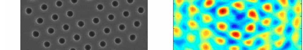
4 Projection (Radon) Diffraction (exact) k y f max k z SEM diffraction Radon KAIST PH880 11/10/2010
5 KAIST PH880 11/10/2010 Time-domain OCT
6 Coherence gating
7 Optical Coherence Microscopy KAIST PH880 11/10/2010 J A Izatt et al, OPTICS LETTERS / Vol. 19, No. 8 / April 15, 1994
8 Optical Coherence Microscopy OCT: Low NA b Δx x Lt Lateral resolution lti 4λ f Δ x = π d Depth of ffocus OCM: High NA b Δx 2 x b= 2z = π Δ R 2λ f=focal length d= lens diameter 8
 pp 355")
9 Fourier-domain OCT no scanning of reference mirror the spectrum of the backscattered sample light amplitude FercherAF, HitzenbergerCK, DrexlerW, Kamp G, Strasser I, LiHC1993b Medical Optical Tomography: Functional Imaging and Monitoring vol IS 11, ed G M uller et al (Bellingham: SPIE Press) pp
10 Parallel OCT set-up with a 2D detector array Bourquin S, Seitz P and Salathe R P 2001 El Lett
11 Overview of week 11 Monday: Digital Holographic Tomography Optical Coherence Tomography Wednesday: Photoacoustic Tomography* KAIST PH880 11/10/2010 * Slides are modified from L Wang s lecture slides
12 High Relative Resolution: Depth-to-Resolution Ratio > 100 Modality Max depth Axial resolution Depth / Resolution Confocal/two-photon microscopy ~ mm ~1-2 microns >100 Optical coherence tomography ~1 mm ~10 microns >100 Magnetic resonance imaging / Ultrasonography ~ mm ~1 mm >100 X-ray CT ~200 mm ~0.1 mm >100
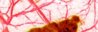

13 Photoacoustic imaging of cancer in vivo Melanoma Melanoma 1 mm Melanoma Histology B-scan image at 764 nm Melanoma 1 mm KAIST PH880 11/10/2010 Nature Biotech. 24, 848 (2006).
14 Photoacoustic Tomography: principle (1) Laser pulse (<ANSI limit: e.g., 20 mj/cm 2 ) (2) Local heating (~ mk) (4) Ultrasonic detection (scattering/100) (3) Ultrasonic emission (~ mbar) Physical Review E 71, (2005). Phys. Rev. Letters 92, (2004). Lihong Wang group, Washington University
15 Photoacoustic Tomography: principle 1. Short laser pulse (~ ns range) is spatially broadened and then used to irradiate biological tissue 2. Produces a temperature rise (~ mk in short time frame) 3. Thermo-elastic expansion causes emission of acoustic wave (discovered by Alexander Graham Bell) 4. Acoustic wave is measured by wideband ultrasonic transducers 5. Acquired signal is combined mathematically to reconstruct the distribution of optical energy absorption KAIST PH880 11/10/2010 V Ntziachristos et al, Nature Biotechnology, 23 3, (2005)
16 Reflection-mode Photoacoustic Microscopy: Illustration Sphere Ph hotoacousti ic signal Surface Sphere Time
17 Reflection mode Dark field Confocal Photoacoustic Microscopy: System Tunable laser Nd:YAG pump laser Motor driver Photodiode Translation stages Amplifier Optical illumination Ultrasonic transducer Conical llens Sample holder Base AD Computer Mirror Heater & temperature controller Dual lfoci Annular illumination i with a dark center Sample Optics Letters 30, 625 (2005) Nature Biotech. 24, 848 (2006).


18 Imaging Depth and Resolution in Photoacoustic microscopy 3 mm B scan of a black double stranded cotton thread embedded in rat Imaging depth: ~3 mm Axial resolution: ~15 microns Depth/resolution: ~200 pixels Lateral resolution: ~45 microns Acquisition time: 2 ms/a scan No signal averaging Optics Letters 30, 625 (2005).


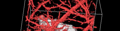
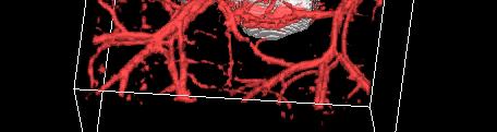
19 Volumetric Imaging of Rat Microvasculature In Vivo Maximum amplitude projection onto the skin 1 mm Volume: 10 mm x 8 mm x 3 mm Optics Express 14, 9317 (2006).
 burn in pig skin in vivo.")


![e [a.u.] PA ampl 0.2 0.1 0 Burn depth ~1.](/docs-images/77/75677709/images/20-4.jpg "7 mm Hyperemic bowl Skin surface 55 5.")
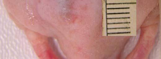
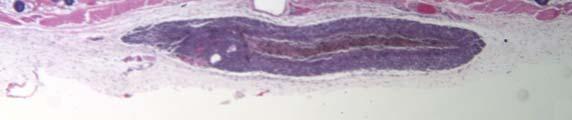
20 Imaging of Skin: Burn in Pigs Acute thermal (175 o C, 20 s) burn in pig skin in vivo. Postmortem imaging at 584 nm optical wavelength. Photograph Healthy Coagulated ated tissue tissue Photoacoustic image B scan image Hyperemic bowl 1 mm 1 mm Hyperemic ring Histology Hyperemic bowl 1 mm itude [a.u.] PA ampl Burn depth ~1.7 mm Hyperemic bowl Skin surface Distance [mm] J Biomed Optics 11, (2006).


21 Imaging of Hemoglobin Oxygen Total hemoglobin concentration Saturation (SO 2 ) In Vivo SO 2 in segmented venules and arterioles Histology mm Arterial microsphere perfusion A 1 4 V Nature Biotech. 24, 848 (2006). 1 mm


22 Hemodynamics In Vivo (578, 584, 590, and 596 nm) Total hemoglobin Oxygen saturation Arteries and veins 1 mm Imaged SO Change in oxygenation Artery Vein Hypoxia Normoxia Hyperoxia Physiological states monitor SO2 change over time. Appl. Phys. Lett. 90, (2007).

 3.")


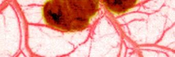
23 In Vivo Genetic Imaging: Gene Expression in Gliosarcoma Tumor in Rat 1. LacZ (gene) 2. Bt Beta galactosidase t (enzyme ) 3. X gal (colorless substrate) 4. Blue product Image of blood vessels at 584 nm wavelength Image of expression of LacZ reporter gene at 635 nm wavelength Composite image 1 mm J Biomed Optics 12(2), (2007).


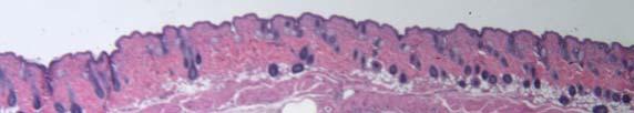
24 Imaging of Human Palm In Vivo Photo Maximum amplitude projection onto the skin Skin surface 0.3 mm 0.13 mm Skin surface B scan image mm Optical absorption Stratum corneum Nature Biotech. 24, 848 (2006).
25 Reading List 1. Ntziachristos i V, Ripoll J, Wang L, & Weissleder R (2005) Looking and listening i to light: the evolution of whole body photonic imaging. Nature biotechnology 23(3): KAIST PH880 11/10/2010
Implementing the probe beam deflection technique for acoustic sensing in photoacoustic and ultrasound imaging
 Implementing the probe beam deflection technique for acoustic sensing in photoacoustic and ultrasound imaging Ronald A. Barnes Jr. The University of Texas at San Antonio This work is a collaboration between
Light and tissue: 2. Two-photon, Optical Coherence Tomography, Photoacoustics
 Light and tissue: 2 Two-photon, Optical Coherence Tomography, Photoacoustics Last lecture: Absorption and Scattering Last lecture: Mean free path Mean free path Transport mean free path Ntziachristos (2010)
University of Lübeck, Medical Laser Center Lübeck GmbH Optical Coherence Tomography
 University of Lübeck, Medical Laser Center Lübeck GmbH Optical Coherence Tomography. Theory Dr. Gereon Hüttmann / 009 What is OCT? ( for the MD ) Lichtquelle Probe Detektor Display OCT is Ultrasound with
Optical Sectioning. Bo Huang. Pharmaceutical Chemistry
 Optical Sectioning Bo Huang Pharmaceutical Chemistry Approaches to 3D imaging Physical cutting Technical difficulty Highest resolution Highest sensitivity Optical sectioning Simple sample prep. No physical
Optical coherence tomography with the "Spectral Radar" - Fast optical analysis in volume scatterers by short coherence interferometry
 Optical coherence tomography with the "Spectral Radar" - Fast optical analysis in volume scatterers by short coherence interferometry M. Bail, G. Häusler, J. M. Herrmann, M. W. Lindner, R. Ringler Physics
High Resolution Multi-modal in vivo Imaging Platform
 High Resolution Multi-modal in vivo Imaging Platform The world s only customizable imaging platform combining ultra high frequency ultrasound and photoacoustics Experience the next generation of in vivo
BME I5000: Biomedical Imaging
 1 Lucas Parra, CCNY BME I5000: Biomedical Imaging Lecture 4 Computed Tomography Lucas C. Parra, parra@ccny.cuny.edu some slides inspired by lecture notes of Andreas H. Hilscher at Columbia University.
HOLOGRAPHIC FEMTOSECOND LASER PROCESSING AND THREE-DIMENSIONAL RECORDING IN BIOLOGICAL TISSUES
 Progress In Electromagnetics Research Letters, Vol. 2, 115 123, 2008 HOLOGRAPHIC FEMTOSECOND LASER PROCESSING AND THREE-DIMENSIONAL RECORDING IN BIOLOGICAL TISSUES Y. Hayasaki Department of Optical Science
Outline. Introduction to photoacoustic computed tomography (PACT) Imaging models and iterative image reconstruction. Success with small animal imaging
 Imaging models and iterative image reconstruction. Success with small animal imaging") Outline Advantages of PACT Photoacoustic Computed Tomography with Applications to Breast Imaging Mark A. Anastasio Department of Biomedical Engineering Washington University in St. Louis St. Louis, MO
Outline Advantages of PACT Photoacoustic Computed Tomography with Applications to Breast Imaging Mark A. Anastasio Department of Biomedical Engineering Washington University in St. Louis St. Louis, MO
NIH Public Access Author Manuscript IEEE Photonics J. Author manuscript; available in PMC 2010 October 14.
 NIH Public Access Author Manuscript Published in final edited form as: IEEE Photonics J. 2010 February 17; 2(1): 57 66. doi:10.1109/jphot.2010.2042801. Fast and robust deconvolution-based image reconstruction
Development and validation of a short-lag spatial coherence theory for photoacoustic imaging
 Development and validation of a short-lag spatial coherence theory for photoacoustic imaging Michelle T. Graham 1 and Muyinatu A. Lediju Bell 1,2 1 Department of Electrical and Computer Engineering, Johns
Advanced Image Reconstruction Methods for Photoacoustic Tomography
 Advanced Image Reconstruction Methods for Photoacoustic Tomography Mark A. Anastasio, Kun Wang, and Robert Schoonover Department of Biomedical Engineering Washington University in St. Louis 1 Outline Photoacoustic/thermoacoustic
HIGH-PERFORMANCE TOMOGRAPHIC IMAGING AND APPLICATIONS
 HIGH-PERFORMANCE TOMOGRAPHIC IMAGING AND APPLICATIONS Hua Lee and Yuan-Fang Wang Department of Electrical and Computer Engineering University of California, Santa Barbara ABSTRACT Tomographic imaging systems
PH880 Topics in Physics
 PH880 Topics in Physics Modern Optical Imaging (Fall 2010) Overview of week 8 Monday Nonlinear Microscopy Wednesday No class (Mid term week) Quantum Optics Electro- magnetic Optics Wave Optics Ray Optics
Coherent Gradient Sensing Microscopy: Microinterferometric Technique. for Quantitative Cell Detection
 Coherent Gradient Sensing Microscopy: Microinterferometric Technique for Quantitative Cell Detection Proceedings of the SEM Annual Conference June 7-10, 010 Indianapolis, Indiana USA 010 Society for Experimental
Efficient Signal Processing Algorithms for Optical Doppler Tomography Literature Survey
 Efficient Signal Processing Algorithms for Optical Doppler Tomography Literature Survey University of Texas at Austin Department of Electrical and Computer Engineering Milos Milosevic Wade Schwartzkopf
Effect of Sensor Directionality on Photoacoustic Imaging: A Study Using the k-wave Toolbox
 Effect of Sensor Directionality on Photoacoustic Imaging: A Study Using the k-wave Toolbox B.T. Cox and B.E. Treeby Department of Medical Physics and Bioengineering, University College London, Gower Street,
Other Laser Surgery Laser Tonsillectomy Use CO 2 with mirror bouncing system Operation takes 15 minutes, no pain Cauterizes blood vessels & Lymphatic
 Other Laser Surgery Laser Tonsillectomy Use CO 2 with mirror bouncing system Operation takes 15 minutes, no pain Cauterizes blood vessels & Lymphatic vessels no blood in throat Patient eat & drink just
Techniques of Noninvasive Optical Tomographic Imaging
 Techniques of Noninvasive Optical Tomographic Imaging Joseph Rosen*, David Abookasis and Mark Gokhler Ben-Gurion University of the Negev Department of Electrical and Computer Engineering P. O. Box 653,
MEDICAL IMAGE ANALYSIS
 SECOND EDITION MEDICAL IMAGE ANALYSIS ATAM P. DHAWAN g, A B IEEE Engineering in Medicine and Biology Society, Sponsor IEEE Press Series in Biomedical Engineering Metin Akay, Series Editor +IEEE IEEE PRESS
Image Acquisition Systems
 Image Acquisition Systems Goals and Terminology Conventional Radiography Axial Tomography Computer Axial Tomography (CAT) Magnetic Resonance Imaging (MRI) PET, SPECT Ultrasound Microscopy Imaging ITCS
Lecture 6: Medical imaging and image-guided interventions
 ME 328: Medical Robotics Winter 2019 Lecture 6: Medical imaging and image-guided interventions Allison Okamura Stanford University Updates Assignment 3 Due this Thursday, Jan. 31 Note that this assignment
Enhanced optical absorptance of metals using interferometric femtosecond ablation
 Enhanced optical absorptance of metals using interferometric femtosecond ablation K. Paivasaari, J. J. J. Kaakkunen, M. Kuittinen and T. Jaaskelainen Department of Physics and Mathematics, University of
Optical Active 3D Scanning. Gianpaolo Palma
 Optical Active 3D Scanning Gianpaolo Palma 3D Scanning Taxonomy SHAPE ACQUISTION CONTACT NO-CONTACT NO DESTRUCTIVE DESTRUCTIVE X-RAY MAGNETIC OPTICAL ACOUSTIC CMM ROBOTIC GANTRY SLICING ACTIVE PASSIVE
Supplementary Figure 1 Guide stars of progressively longer wavelengths can be used for direct wavefront sensing at increasingly large depth in the
 Supplementary Figure 1 Guide stars of progressively longer wavelengths can be used for direct wavefront sensing at increasingly large depth in the cortex of the living mouse. Typical SH images of guide
OPTICAL COHERENCE TOMOGRAPHY:SIGNAL PROCESSING AND ALGORITHM
 OPTICAL COHERENCE TOMOGRAPHY:SIGNAL PROCESSING AND ALGORITHM OCT Medical imaging modality with 1-10 µ m resolutions and 1-2 mm penetration depths High-resolution, sub-surface non-invasive or minimally
QUANTITATIVE PHASE IMAGING OF BIOLOGICAL CELLS USING OFF-AXIS METHOD OF WIDE FIELD DIGITAL INTERFERENCE MICROSCOPY (WFDIM)
") http:// QUANTITATIVE PHASE IMAGING OF BIOLOGICAL CELLS USING OFF-AXIS METHOD OF WIDE FIELD DIGITAL INTERFERENCE MICROSCOPY (WFDIM) Pradeep Kumar Behera 1, Dalip Singh Mehta 2 1,2 Physics,Indian Institute
http:// QUANTITATIVE PHASE IMAGING OF BIOLOGICAL CELLS USING OFF-AXIS METHOD OF WIDE FIELD DIGITAL INTERFERENCE MICROSCOPY (WFDIM) Pradeep Kumar Behera 1, Dalip Singh Mehta 2 1,2 Physics,Indian Institute
Physics 625 Femtosecond laser Project
 Physics 625 Femtosecond laser Project The purpose of this project is for each person to gain experience in designing part of a femtosecond laser system for pump-probe experiments. The system diagram is
PH880 Topics in Physics
 PH880 Topics in Physics Modern Optical Imaging (Fall 2010) Overview of week 4 Monday PSF, OTF Bright field microscopy Resolution/NA Deconvolution Wednesday : holiday Impulse response (PSF) in imaging system
Study of Air Bubble Induced Light Scattering Effect On Image Quality in 193 nm Immersion Lithography
 Study of Air Bubble Induced Light Scattering Effect On Image Quality in 193 nm Immersion Lithography Y. Fan, N. Lafferty, A. Bourov, L. Zavyalova, B. W. Smith Rochester Institute of Technology Microelectronic
Digital Image Processing
 Digital Image Processing SPECIAL TOPICS CT IMAGES Hamid R. Rabiee Fall 2015 What is an image? 2 Are images only about visual concepts? We ve already seen that there are other kinds of image. In this lecture
4D Technology Corporation
 4D Technology Corporation Dynamic Laser Interferometry for Company Profile Disk Shape Characterization DiskCon Asia-Pacific 2006 Chip Ragan chip.ragan@4dtechnology.com www.4dtechnology.com Interferometry
Confocal Raman Imaging with WITec Sensitivity - Resolution - Speed. Always - Provable - Routinely
 Confocal Raman Imaging with WITec Sensitivity - Resolution - Speed Always - Provable - Routinely WITec GmbH, Ulm, Germany, info@witec.de, www.witec.de A modular microscope series An Example: FLIM optical
THE DICOM 2013 INTERNATIONAL CONFERENCE & SEMINAR. DICOM Fields of Use. Klaus Neuner. Brainlab AG. Software Project Manager Feldkirchen, Germany
 THE DICOM 2013 INTERNATIONAL CONFERENCE & SEMINAR March 14-16 Bangalore, India DICOM Fields of Use Klaus Neuner Brainlab AG Software Project Manager Feldkirchen, Germany Introduction This presentation
Coherent Diffraction Imaging with Nano- and Microbeams
 Diffraction Imaging with Nano- and Microbeams Why does lensless need? Mark A Pfeifer Cornell High Energy Synchrotron Source Cornell University Ithaca, NY 14850 map322@cornell.edu XLD 2011 June 28, 2011
The Future of OCT? Swept Source. Todd J. Purkiss, MD, PhD River City Retina Club July 16, 2015
 The Future of OCT? Swept Source Todd J. Purkiss, MD, PhD River City Retina Club July 16, 2015 Background on OCT Optical Coherence Tomography Optical When a light wave meets the interface of two differing
Today s Outline - April 17, C. Segre (IIT) PHYS Spring 2018 April 17, / 22
 PHYS Spring 2018 April 17, / 22") Today s Outline - April 17, 2018 C. Segre (IIT) PHYS 570 - Spring 2018 April 17, 2018 1 / 22 Today s Outline - April 17, 2018 Diffraction enhanced imaging C. Segre (IIT) PHYS 570 - Spring 2018 April 17,
Today s Outline - April 17, 2018 C. Segre (IIT) PHYS 570 - Spring 2018 April 17, 2018 1 / 22 Today s Outline - April 17, 2018 Diffraction enhanced imaging C. Segre (IIT) PHYS 570 - Spring 2018 April 17,
HOLOGRAPHIC SCANNING LASER ACOUSTIC MICROSCOPY (HOLOSLAM): A NEW QNDE TOOL. A. C. Wey, and L. W. Kessler
: A NEW QNDE TOOL. A. C. Wey, and L. W. Kessler") HOLOGRAPHIC SCANNING LASER ACOUSTIC MICROSCOPY (HOLOSLAM): A NEW QNDE TOOL A. C. Wey, and L. W. Kessler Sonoscan, Inc. 530 E. Green Street Bensenville, Illinois INTRODUCTION Acoustic microscopy is the
HOLOGRAPHIC SCANNING LASER ACOUSTIC MICROSCOPY (HOLOSLAM): A NEW QNDE TOOL A. C. Wey, and L. W. Kessler Sonoscan, Inc. 530 E. Green Street Bensenville, Illinois INTRODUCTION Acoustic microscopy is the
SIMULATION AND VISUALIZATION IN THE EDUCATION OF COHERENT OPTICS
 SIMULATION AND VISUALIZATION IN THE EDUCATION OF COHERENT OPTICS J. KORNIS, P. PACHER Department of Physics Technical University of Budapest H-1111 Budafoki út 8., Hungary e-mail: kornis@phy.bme.hu, pacher@phy.bme.hu
Biophysical Techniques (BPHS 4090/PHYS 5800)
") Biophysical Techniques (BPHS 4090/PHYS 5800) Instructors: Prof. Christopher Bergevin (cberge@yorku.ca) Schedule: MWF 1:30-2:30 (CB 122) Website: http://www.yorku.ca/cberge/4090w2017.html York University
Biophysical Techniques (BPHS 4090/PHYS 5800) Instructors: Prof. Christopher Bergevin (cberge@yorku.ca) Schedule: MWF 1:30-2:30 (CB 122) Website: http://www.yorku.ca/cberge/4090w2017.html York University
Orthogonal Fabry-Pérot sensor array system for minimal-artifact 3D photoacoustic tomography
 Orthogonal Fabry-Pérot sensor array system for minimal-artifact 3D photoacoustic tomography Robert Ellwood, Edward Zhang, Paul Beard and Ben Cox Department of Medical Physics and Biomedical Engineering,
LABORATORY SYSTEM FOR X-RAY NANOTOMOGRAPHY
 79 LABORATORY SYSTEM FOR X-RAY NANOTOMOGRAPHY Alexander Sasov, SkyScan, Vluchtenburgstraat 3, Aartselaar B2630, Belgium, www.skyscan.be. ABSTRACT Using advanced X-ray technologies and X-ray scattering
Tutorial Solutions. 10 Holographic Applications Holographic Zone-Plate
 10 Holographic Applications 10.1 Holographic Zone-Plate Tutorial Solutions Show that if the intensity pattern for on on-axis holographic lens is recorded in lithographic film, then a one-plate results.
Coherent Microscopy and Optical Coherence Tomography for Biomedical Applications
 Coherent Microscopy and Optical Coherence Tomography for Biomedical Applications Jeremy M. Coupland Tel.: ++44 (0)1509 227506; Fax: ++44 (0)1509 227502; Email: j.m.coupland@lboro.ac.uk Wolfson School of
Fast and Robust Deconvolution-Based Image Reconstruction for Photoacoustic Tomography in Circular Geometry: Experimental Validation
 Fast and Robust Deconvolution-Based Image Reconstruction for Photoacoustic Tomography in Circular Geometry: Experimental Validation Volume 2, Number 1, February 2010 Invited Paper C. Zhang C. Li L. V.
Range Sensors (time of flight) (1)
 (1)") Range Sensors (time of flight) (1) Large range distance measurement -> called range sensors Range information: key element for localization and environment modeling Ultrasonic sensors, infra-red sensors
Range Sensors (time of flight) (1) Large range distance measurement -> called range sensors Range information: key element for localization and environment modeling Ultrasonic sensors, infra-red sensors
3D TeraHertz Tomography
 3D TeraHertz Tomography B. Recur, 3 A. Younus, 1 S. Salort, 2 P. Mounaix, 1 B. Chassagne, 2 P. Desbarats, 3 1 LOMA, Université de Bordeaux / CNRS 2 ALPhANOV, Centre Technologique Optique et Lasers, Université
Text for the class, Pump-Probe Technique for Picosecond Time-resolved X-ray Diffraction at Cheiron School
 BL19LXU Yoshihito Tanaka, Oct. 2013 Text for the class, Pump-Probe Technique for Picosecond Time-resolved X-ray Diffraction at Cheiron School Abstract The pulsed time structure of synchrotron radiation
Formulas of possible interest
 Name: PHYS 3410/6750: Modern Optics Final Exam Thursday 15 December 2011 Prof. Bolton No books, calculators, notes, etc. Formulas of possible interest I = ɛ 0 c E 2 T = 1 2 ɛ 0cE 2 0 E γ = hν γ n = c/v
Text for the class, Pump and probe technique for picosecond time-resolved x-ray diffraction at the Cheiron School
 Yoshihito Tanaka, Kiminori Ito Oct. 3-4, 2011 Text for the class, Pump and probe technique for picosecond time-resolved x-ray diffraction at the Cheiron School 1. Introduction 1-1. Purpose The pulsed nature
The University of Chicago. Center for EPR Imaging in Vivo Physiology. Image Registration. Boris Epel
 The University of Chicago Center for EPR Imaging in Vivo Physiology Image Registration Boris Epel Imaging Methods are Complimentary CT MRI EPRI High resolution anatomic images Quantitative Poor soft tissue
BME I5000: Biomedical Imaging
 BME I5000: Biomedical Imaging Lecture 1 Introduction Lucas C. Parra, parra@ccny.cuny.edu 1 Content Topics: Physics of medial imaging modalities (blue) Digital Image Processing (black) Schedule: 1. Introduction,
The use of acoustic reflectors to enlarge the effective area of planar sensor arrays
 The use of acoustic reflectors to enlarge the effective area of planar sensor arrays R. Ellwood 1, E.Z. Zhang 1, P.C. Beard 1 & B.T. Cox 1 1 Department of Medical Physics and Bioengineering, University
Silicon Avalanche Photodiodes in Dynamic Light Scattering
 Silicon Avalanche Photodiodes in Dynamic Light Scattering August 2016 Introduction This application note describes the use of the ID100 single photon counting detector for the measurement of light scattered
PHYSICS. Chapter 33 Lecture FOR SCIENTISTS AND ENGINEERS A STRATEGIC APPROACH 4/E RANDALL D. KNIGHT
 PHYSICS FOR SCIENTISTS AND ENGINEERS A STRATEGIC APPROACH 4/E Chapter 33 Lecture RANDALL D. KNIGHT Chapter 33 Wave Optics IN THIS CHAPTER, you will learn about and apply the wave model of light. Slide
Near Field Observation of a Refractive Index Grating and a Topographical Grating by an Optically Trapped Gold Particle
 Near Field Observation of a Refractive Index Grating and a Topographical Grating by an Optically Trapped Gold Particle Hiroo UKITA and Hirotaka UEMI Ritsumeikan University, Kusatsu-shi, Shiga, 2 Japan
Quantitative Phase Imaging in Microscopy
 Invited Paper Quantitative Phase Imaging in Microscopy Colin JR Sheppard, Shan S Kou and Shalin Mehta Division of Bioengineering National University of Singapore Singapore 117574 1 Introduction There are
Phase-Contrast Imaging and Tomography at 60 kev using a Conventional X-ray Tube
 Phase-Contrast Imaging and Tomography at 60 kev using a Conventional X-ray Tube T. Donath* a, F. Pfeiffer a,b, O. Bunk a, W. Groot a, M. Bednarzik a, C. Grünzweig a, E. Hempel c, S. Popescu c, M. Hoheisel
Advanced materials research using the Real-Time 3D Analytical FIB-SEM 'NX9000'
 SCIENTIFIC INSTRUMENT NEWS 2017 Vol. 9 SEPTEMBER Technical magazine of Electron Microscope and Analytical Instruments. Technical Explanation Advanced materials research using the Real-Time 3D Analytical
Mirror Example Consider a concave mirror radius -10 cm then = = Now consider a 1 cm candle s = 15 cm from the vertex Where is the image.
 Mirror Example Consider a concave mirror radius -10 cm then r 10 f = = = 5 cm 2 2 Now consider a 1 cm candle s = 15 cm from the vertex Where is the image 1 s 2 1 = = r s 1 1 2 + = = s s r 1 1 = 0.13333
3D Energy Dispersive Spectroscopy Elemental Tomography in the Scanning Transmission Electron Microscope
 3D Energy Dispersive Spectroscopy Elemental Tomography in the Scanning Transmission Electron Microscope Brian Van Devener Topics 1.Introduction to EDS in the STEM 2.Extending EDS into three dimensions
Lightsheet Z.1. Light Sheet Fluorescence Microscopy by Carl Zeiss. Fabrice Schmitt, Sales Manager Carl ZEISS France
 Lightsheet Z.1 Light Sheet Fluorescence Microscopy by Carl Zeiss Fabrice Schmitt, Sales Manager Carl ZEISS France 12.12.2012 Light Sheet Fluorescence Microscopy (LSFM) Principle The Principle of Light
Blue Skies Blue Eyes Blue Butterflies
 Blue Skies Blue Eyes Blue Butterflies Friday, April 19 Homework #9 due in class Lecture: Blue Skies, Blue Eyes & Blue Butterflies: Interaction of electromagnetic waves with matter. Week of April 22 Lab:
Exact Frequency-Domain Reconstruction for Thermoacoustic Tomography I: Planar Geometry
 IEEE TRANSACTIONS ON MEDICAL IMAGING, VOL. 21, NO. 7, JULY 2002 823 Exact Frequency-Domain Reconstruction for Thermoacoustic Tomography I: Planar Geometry Yuan Xu, Dazi Feng, and Lihong V. Wang* Abstract
Applications of adaptive optics in femtosecond laser material processing
 Applications of adaptive optics in femtosecond laser material processing STFC / Photonics KTN - Laser Applications of Adaptive Optics Professor Derryck T. Reid Ultrafast Optics Group School of Engineering
Quantitative Analysis of Cardiomyocyte Dynamics with Optical Coherence Phase Microscopy
 Quantitative Analysis of Cardiomyocyte Dynamics with Optical Coherence Phase Microscopy Rehman Ansari* a,c, Redouane Aherrahrou b, Zouhair Aherrahrou b, Jeanette Erdmann b, Gereon Hüttmann a, and Achim
Digitalna Holografija i Primjene
 Digitalna Holografija i Primjene Hrvoje Skenderović Institut za fiziku 5. PIF Radionica, IRB, 16.12.2014. Holography Dennis Gabor invented holography in 1948 as a method for recording and reconstructing
High count rate multichannel TCSPC for optical tomography
 High count rate multichannel TCSPC for optical tomography Wolfgang Becker *a, Axel Bergmann a, Heidrun Wabnitz **b, Dirk Grosenick b, Adam Liebert ***b a Becker & Hickl GmbH; b Physikalisch-Technische
Absorption distribution of an optical beam focused into a turbid medium
 Absorption distribution of an optical beam focused into a turbid medium Lihong V. Wang and Gan Liang The focusing of light into a turbid medium was studied with Monte Carlo simulations. Focusing was found
Time-Resolved Optical Tomography in Preclinical Studies: Propagation of Excitation and Fluorescence Photons.
 Excerpt from the Proceedings of the COMSOL Conference 2008 Hannover Time-Resolved Optical Tomography in Preclinical Studies: Propagation of Excitation and Fluorescence Photons. F. Nouizi 1, R. Chabrier
Diffraction and Interference of Plane Light Waves
 PHY 92 Diffraction and Interference of Plane Light Waves Diffraction and Interference of Plane Light Waves Introduction In this experiment you will become familiar with diffraction patterns created when
45 µm polystyrene bead embedded in scattering tissue phantom. (a,b) raw images under oblique
 raw images under oblique") Phase gradient microscopy in thick tissue with oblique back-illumination Tim N Ford, Kengyeh K Chu & Jerome Mertz Supplementary Figure 1: Comparison of added versus subtracted raw OBM images 45 µm polystyrene
Phase gradient microscopy in thick tissue with oblique back-illumination Tim N Ford, Kengyeh K Chu & Jerome Mertz Supplementary Figure 1: Comparison of added versus subtracted raw OBM images 45 µm polystyrene
Miniature Mirau interferometry for swept-source OCT imaging with applications in dermatology
 Miniature Mirau interferometry for swept-source OCT imaging with applications in dermatology C. Gorecki 1, S. Bargiel 1, J. Lullin 1,, J. Albero 1, N. Passilly 1, S. Perrin 1, L. Froehly 1, W.- S. Wang
Index. aliasing artifacts and noise in CT images, 200 measurement of projection data, nondiffracting
 Index Algebraic equations solution by Kaczmarz method, 278 Algebraic reconstruction techniques, 283-84 sequential, 289, 293 simultaneous, 285-92 Algebraic techniques reconstruction algorithms, 275-96 Algorithms
Computed tomography - outline
 Computed tomography - outline Computed Tomography Systems Jørgen Arendt Jensen and Mikael Jensen (DTU Nutech) October 6, 216 Center for Fast Ultrasound Imaging, Build 349 Department of Electrical Engineering
Lecture 05. First Example: A Real Lidar
 Lecture 05. First Example: A Real Lidar Brief review of lidar basics K Doppler lidar system architecture K lidar signal estimate from lidar equation Comparison of estimate to reality Summary Review of
Classification of Hyperspectral Breast Images for Cancer Detection. Sander Parawira December 4, 2009
 1 Introduction Classification of Hyperspectral Breast Images for Cancer Detection Sander Parawira December 4, 2009 parawira@stanford.edu In 2009 approximately one out of eight women has breast cancer.
The role of light source in coherence scanning interferometry and optical coherence tomography
 The role of light source in coherence scanning interferometry and optical coherence tomography Dr Rong Su Research Fellow Advanced Manufacturing Research Group Manufacturing Metrology Team Precision manufacturing
Holography & Coherence For Holography need coherent beams Two waves coherent if fixed phase relationship between them for some period of time
 Holography & Coherence For Holography need coherent beams Two waves coherent if fixed phase relationship between them for some period of time Coherence Coherence appear in two ways Spatial Coherence Waves
What is Frequency Domain Analysis?
 R&D Technical Bulletin P. de Groot 9/3/93 What is Frequency Domain Analysis? Abstract: The Zygo NewView is a scanning white-light interferometer that uses frequency domain analysis (FDA) to generate quantitative
Simulation of confocal microscopy through scattering media with and without time gating
 Magnor et al. Vol. 18, No. 11/November 2001/J. Opt. Soc. Am. B 1695 Simulation of confocal microscopy through scattering media with and without time gating Marcus Magnor Computer Graphics Laboratory, Stanford
Material for Chapter 6: Basic Principles of Tomography M I A Integral Equations in Visual Computing Material
 Material for Chapter : Integral Equations in Visual Computing Material Basic Principles of Tomography c 00 Bernhard Burgeth 0 Source: Images Figure : Radon Transform: ttenuation http://en.wikimedia.org/wiki/image:radon_transform.png
Ultrasound-mediated biophotonic imaging: A review of acousto-optical tomography and photo-acoustic tomography
 Disease Markers 19 (2003,2004) 123 138 123 IOS Press Ultrasound-mediated biophotonic imaging: A review of acousto-optical tomography and photo-acoustic tomography Lihong V. Wang Optical Imaging Laboratory,
UNIVERSITY OF SOUTHAMPTON
 UNIVERSITY OF SOUTHAMPTON PHYS2007W1 SEMESTER 2 EXAMINATION 2014-2015 MEDICAL PHYSICS Duration: 120 MINS (2 hours) This paper contains 10 questions. Answer all questions in Section A and only two questions
EPFL SV PTBIOP BIOP COURSE 2015 OPTICAL SLICING METHODS
 BIOP COURSE 2015 OPTICAL SLICING METHODS OPTICAL SLICING METHODS Scanning Methods Wide field Methods Point Scanning Deconvolution Line Scanning Multiple Beam Scanning Single Photon Multiple Photon Total
Simulation of Diffuse Optical Tomography using COMSOL Multiphysics
 Simulation of Diffuse Optical Tomography using COMSOL Multiphysics SAM Kirmani *1 L Velmanickam 1 D Nawarathna 1 SS Sherif 2 and IT Lima Jr 1 1 Department of Electrical and Computer Engineering North Dakota
Physical & Electromagnetic Optics: Diffraction Gratings
 31/05/2018 Physical & Electromagnetic Optics: Diffraction Gratings Optical Engineering Prof. Elias N. Glytsis School of Electrical & Computer Engineering National Technical University of Athens Multiple
MEASUREMENT OF WIGNER DISTRIBUTION FUNCTION FOR BEAM CHARACTERIZATION OF FELs*
 MEASUREMENT OF WIGNER DISTRIBUTION FUNCTION FOR BEAM CHARACTERIZATION OF FELs* T. Mey #, B. Schäfer and K. Mann, Laser-Laboratorium e.v., Göttingen, Germany B. Keitel, S. Kreis, M. Kuhlmann, E. Plönjes
Time-Resolved measurements by FEL spontaneous emission: A proposal for sub-picosecond pumps & probe structural and spectrometric investigations
 Time-Resolved measurements by FEL spontaneous emission: A proposal for sub-picosecond pumps & probe structural and spectrometric investigations V. Rossi Albertini, B. Paci & P. Perfetti Istituto di Struttura
Computed tomography of simple objects. Related topics. Principle. Equipment TEP Beam hardening, artefacts, and algorithms
 Related topics Beam hardening, artefacts, and algorithms Principle The CT principle is demonstrated with the aid of simple objects. In the case of very simple targets, only a few images need to be taken
Signal post processing in frequency domain OCT and OCM using a filter bank approach
 Signal post processing in frequency domain OCT and OCM using a filter bank approach Bernd Hofer 1,2, Boris Považay 1, Boris Hermann 1, Angelika Unterhuber 1, Gerald Matz 2, Franz Hlawatsch 2, Wolfgang
SAFT DATA PROCESSING APPLIED TO LASER-ULTRASONIC INSPECTION
 SAFT DATA PROCESSING APPLIED TO LASER-ULTRASONIC INSPECTION A. Blouin, D. Levesque, C. Neron, F. Enguehard, D. Drolet, and I-P. Monchalin National Research Council of Canada, Industrial Materials Institute,
LSM510 Confocal Microscope Standard Operation Protocol Basic Operation
 LSM510 Confocal Microscope Standard Operation Protocol Basic Operation Please make sure that the COMPRESSED AIR has been TURNED ON prior to the use of the equipment. Kindly inform the administrator if
Cherenkov Radiation. Doctoral Thesis. Rok Dolenec. Supervisor: Prof. Dr. Samo Korpar
 Doctoral Thesis Time-of-Flight Time-of-Flight Positron Positron Emission Emission Tomography Tomography Using Using Cherenkov Cherenkov Radiation Radiation Rok Dolenec Supervisor: Prof. Dr. Samo Korpar
Use of a laser beam with an oblique angle of incidence to measure the reduced scattering coefficient of a turbid medium
 Use of a laser beam with an oblique angle of incidence to measure the reduced scattering coefficient of a turbid medium Lihong Wang and Steven L. Jacques A simple and quick approach is used to measure
PICOSECOND TRANSIENT THERMAL IMAGING USING A CCD BASED THERMOREFLECTANCE SYSTEM
 Proceedings of the 14th International Heat Transfer Conference IHTC14 August 8-13, 21, Washington, DC, USA IHTC14- PICOSECOND TRANSIENT THERMAL IMAGING USING A CCD BASED THERMOREFLECTANCE SYSTEM James
Real Time Process Control with Optical Coherence Tomography
 Real Time Process Control with Optical Coherence Tomography 16th of June 2016, Ch. Meier Bern HUCE, University OPTOLAB of Applied Science HuCE-optoLabt Overview Short introduction to OCT Systems Resolution
3. Image formation, Fourier analysis and CTF theory. Paula da Fonseca
 3. Image formation, Fourier analysis and CTF theory Paula da Fonseca EM course 2017 - Agenda - Overview of: Introduction to Fourier analysis o o o o Sine waves Fourier transform (simple examples of 1D
10/5/09 1. d = 2. Range Sensors (time of flight) (2) Ultrasonic Sensor (time of flight, sound) (1) Ultrasonic Sensor (time of flight, sound) (2) 4.1.
 (2) Ultrasonic Sensor (time of flight, sound) (1) Ultrasonic Sensor (time of flight, sound) (2) 4.1.") Range Sensors (time of flight) (1) Range Sensors (time of flight) (2) arge range distance measurement -> called range sensors Range information: key element for localization and environment modeling Ultrasonic
Range Sensors (time of flight) (1) Range Sensors (time of flight) (2) arge range distance measurement -> called range sensors Range information: key element for localization and environment modeling Ultrasonic
Quantitative evaluation of systematic imaging error due to uncertainty in tissue optical properties in high-density diffuse optical tomography
 Quantitative evaluation of systematic imaging error due to uncertainty in tissue optical properties in high-density diffuse optical tomography Yuxuan Zhan* a, Adam Eggebrecht b, Hamid Dehghani a, Joseph