Confocal Raman Imaging with WITec Sensitivity - Resolution - Speed. Always - Provable - Routinely
|
|
|
- Karin Gray
- 5 years ago
- Views:
Transcription


1 Confocal Raman Imaging with WITec Sensitivity - Resolution - Speed Always - Provable - Routinely WITec GmbH, Ulm, Germany, info@witec.de,


2 A modular microscope series An Example: FLIM optical profiling Raman-SEM


3 Diffraction Limit and Confocal Microscopy Detection Pinhole Detection Lens Point Light Source Dichroic beam splitter Conjugate planes Objective Focal plane λ=532nm, NA=0.9 ΔX l = 360nm, ΔX a = 920nm
4 Confocal Raman Microscopy: Confocal Microscope + Raman Spectrometer => Imaging with chemical sensitivity
5 Challenges of the Confocal Raman Imaging Y= 20µm X= 20µm The ideal pixel size for an XY scan λ=532nm, NA=0.9 ΔX l = 360nm, ΔX a = 920nm Scan size Number of pixels Integration time Imaging time 20µm x 20µm 150 x 150 = ms 23 s ΔX l 3 = 120nm 50 ms 18 min 1 s > 6 h


6 Challenges of the Confocal Raman Imaging Number of spectra 20 x 20 = 400 Number of spectra 100 x 100 = x 40 [µm 2 ]
7 Confocal Raman Imaging with WITec Sensitivity - Resolution - Speed Always - Provable - Routinely WITec GmbH, Ulm, Germany, info@witec.de,

8 multi-mode fiber UHTS 300 CCD laser single-mode fiber Raman edge filter dichroic beam-splitter Z-stage piezo scanner


9 WITec Raman Spectroscopy System» Lens-based excitation optimized Raman Spectrometer Transmission >60% Superior peak shape conservation» Back-illuminated CCD or EM-CCD detectors highest Quantum efficiency 3 rd order 4 th order UHTS 300 Ultrahigh-Throughput Raman Spectrometer Detecting the 4 th order of Silicon

10 Resolution in Optical Microscopy - XY 1 50 nm Confocal Raman Image of a Carbon Nanotube Scan Range: 1.85 x 0.83 μm 65 x 27 pixels 34 ms integration/pixel 532 nm frequency-doubled Nd:YAG laser Objective: 100 x NA 0.9 Pinhole size: 50 μm Copyright WITec GmbH

11 Resolution in Optical Microscopy - XZ Depth Profiling on a suspended Graphene multilayer Point distance 50 nm 2 s integration/pixel 532 nm frequency-doubled Nd:YAG laser Objective: 100 x NA 0.9 Pinhole size: 10 μm Copyright WITec GmbH

12 Sensitivity & Speed - EM CCD Sample: Toothpaste 760 µs / spectrum, 40,000 spectra 1 µm Raman. AFM. SNOM True Surface => 42 s / image Image acquisition real-time demo Copyright WITec GmbH


13 Data acquisition and analysis

14 True Surface Microscopy The solution for confocal Raman imaging of large and very rough samples
15 True Surface Microscopy The solution for confocal Raman imaging of large and very rough samples

16 True Surface Microscopy The solution for confocal Raman imaging of large and very rough samples

17 True Surface Microscopy The solution for confocal Raman imaging of large and very rough samples

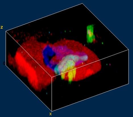
18 True Surface Microscopy 3D Confocal Raman Imaging of Transparent and Opaques Samples Tablet True Surface True Surface Confocal Raman image Raman Spectra 300µm 0µm High resolution Raman imaging without loss of signal due to the sample roughness
19 RISE Microscopy: Correlative Raman Imaging and Scanning Electron Microscopy
20 typical setup for Raman/SEM e-beam off-axis parabolic mirror advantages: can be retro-fitted to several SEM s disadvantages: detection excitation only single spectra => no imaging!! resolution (claim: 2µm; reality: 4-5µm) optical beam not visible in SEM => adjustment necessary offset between e-beam and Raman spot unknown (thermal drift, charging) restricted e-beam image size white light image has bad quality (max. 25µm Ø) not confocal!


21 what about EDX (energy dispersive X-ray spectroscopy)? - very powerful technique - X-rays are generated by the e-beam - X-ray spectrum characteristic for atoms - resolution typ. 1-2 µm Problem: only atoms can be distinguished - no chemistry (e.g. polymers) PMMA: PS: 1-2 µm characteristic X-radiation
22 RISE: Correlative Raman Imaging and Scanning Electron Microscopy


23 RISE: Correlative Raman Imaging and Scanning Electron Microscopy
 gray white brain")
24 RISE: Correlative Raman Imaging and Scanning Electron Microscopy hamster brain (white and gray brain matter) gray white brain matter

")
25 RISE: Correlative Raman Imaging and Scanning Electron Microscopy carbon plagioclase (feldspar) apatite plagioclase (feldspar) Diorite

26 4 G FS V 20 1 RISE: Correlative Raman Imaging and Scanning Electron Microscopy graphene on Si

27 4 G FS V 20 1 RISE: Correlative Raman Imaging and Scanning Electron Microscopy graphene on Si
28 RISE: Correlative Raman Imaging and Scanning Electron Microscopy D blue, dark green: light green, red: yellow: G 1 layer >1 layer Si D

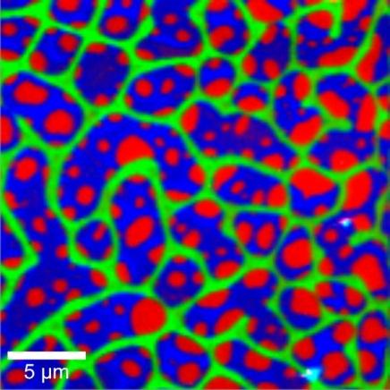
29 RISE: Correlative Raman Imaging and Scanning Electron Microscopy PS PMMA


30 Al, K Fe, Ca, K FS V O Na, K G Si SEM
31 A modular microscope series An Example: FLIM optical profiling Raman-SEM


32 High Resolution - High Sensitivity Fast Imaging 1 µm Raman. AFM. SNOM True Surface True Surface-Raman 3D-Raman Fast Imaging acquisition real-time AFM-Raman SEM-Raman Copyright WITec GmbH
33 MERCI
WITec Raman Spectroscopy Solutions
 Confocal Raman Imaging WITec Raman Spectroscopy Solutions -40-20 0 20 CCD cts 500 1000 1500 2000 2500 3000 3000 relative wavenumbers (cm -1 ) www.witec.de WITec UHTS Ultra-High Throughput Spectrometers
Chemical Characterization of Diverse Pharmaceutical Samples by Confocal Raman Microscopy
 Whitepaper Chemical Characterization of Diverse Pharmaceutical Samples by Confocal Raman Microscopy WITec GmbH, Lise-Meitner-Str. 6, 89081 Ulm, Germany, www.witec.de Introduction The development and production
Chemical Characterization of Pharmaceutical Samples by Confocal Raman Microscopy and Correlative Techniques
 APPLICATION NOTE Chemical Characterization of Pharmaceutical Samples by Confocal Raman Microscopy and Correlative Techniques WITec GmbH, Lise-Meitner-Str. 6, 89081 Ulm, Germany fon +49 (0) 731 140 700,
TrueSurface Microscopy. Confocal Microscopy Along with Large Area Optical Profiling.
 TrueSurface Microscopy Confocal Microscopy Along with Large Area Optical Profiling www.witec.de alpha500 with sensor for profilometry Features & Benefits Extension for the WITec alpha500 series that combines
WITec Suite FIVE. Project FIVE Control FIVE Project FIVE+
 Project FIVE Control FIVE Project FIVE+ For further information about WITec Suite please contact us: by phone: +49 (0) 731 140700 by email: info@witec.de WITec Suite FIVE Data Acquisition, Evaluation and
FLEX 2 NEW. Key points. 3D Confocal Raman, 2 lasers, fiber based, AFM combined
 FLEX 2 3D Confocal Raman, 2 lasers, fiber based, AFM combined NEW Key points Compact size 2 lasers, easily switchable 2 confocal operation modes, easily switchable : High Spatial Resolution 35 nm High
SPECTRUM. The world s first fully automated Raman AFM. AFM - confocal Raman - SNOM - TERS AFM KPFM. Raman. AFM-Raman characterization of PS-PVAC
 Raman KPFM AFM AFM-Raman characterization of PS-PVAC polymer blend film SPECTRUM The world s first fully automated Raman AFM AFM - confocal Raman - SNOM - TERS The first fully integrated & automated AFM
MonoVista CRS+ Raman Microscopes
 MonoVista CRS+ Benefits Deep UV to NIR wavelength range Up to 4 integrated multi-line lasers plus port for large external lasers Dual beam path for UV and VIS/NIR Motorized Laser selection Auto Alignment
Confocal Raman Microscope RAMOS
 Confocal Raman Microscope RAMOS 1 future`s Confocal Raman Microscope RAMOS 2 Confocal Raman Microscope RAMOS Ostec Corporate Group produces and offers hi-tech innovative scientific and analytical equipment.
Optical Sectioning. Bo Huang. Pharmaceutical Chemistry
 Optical Sectioning Bo Huang Pharmaceutical Chemistry Approaches to 3D imaging Physical cutting Technical difficulty Highest resolution Highest sensitivity Optical sectioning Simple sample prep. No physical
Depth analysis for laminated film by confocal Raman mapping
 Newest modular 3D Imaging Raman Microspectroscopy System Simple operation and low cost with all the basic features of our top of the line system. Structural images of transparent samples (plastic, film,
MultiView 2000 TM. The First Tip and Sample Scanning Probe Microscope. The Next Evolution in SPM. The Next Evolution in SPM
 MultiView 2000 TM The First Tip and Sample Scanning Probe Microscope MultiView 2000 TM Using Two Award Winning Nanonics 3D FlatScan Stages MultiView 2000TM Top-View (Top) and open position (Bottom). The
LSM 5 MP, LSM 510 and LSM 510 META Laser Scanning Microscopes
 LSM 5 MP, LSM 510 and LSM 510 META Laser Scanning Microscopes Brief Operating Manual Release 4.2 January 2007 Contents Page Starting the System...3 Setting the microscope...6 Configuring the beam path
Improvement of the correlative AFM and ToF-SIMS approach using an empirical sputter model for 3D chemical characterization
 Improvement of the correlative AFM and ToF-SIMS approach using an empirical sputter model for 3D chemical characterization T. Terlier 1, J. Lee 1, K. Lee 2, and Y. Lee 1 * 1 Advanced Analysis Center, Korea
NDD FLIM Systems for Leica SP2 MP and SP5 MP Multiphoton Microscopes
 NDD FLIM Systems for Leica SP2 MP and SP5 MP Multiphoton Microscopes bh FLIM systems for the confocal and the multiphoton versions of the Leica SP2 and SP5 microscopes are available since 2002 [4]. These
Confocal Microscopy Imaging of Single Emitter Fluorescence and Hanbury Brown, and Twiss Setup for Photon Antibunching. Abstract
 James Maslek 10/26/12 Confocal Microscopy Imaging of Single Emitter Fluorescence and Hanbury Brown, and Twiss Setup for Photon Antibunching Abstract The purpose of this experiment was to observe fluorescence
Introduction to. 3D Scanning Confocal Microscope with Spectrometer
 Introduction to Nanofinder-S 3D Scanning Confocal Microscope with Spectrometer Alexei Kuzmin E-mail: a.kuzmin@cfi.lu.lv Principle of Confocal Microscopy Laser X-Y Excitation Pinhole Excitation Filter Objective
QUANTAX EDS SYSTEM SOP
 QUANTAX EDS SYSTEM SOP December 2017 Energy-Dispersive X-Ray Spectroscopy (EDS, EDX, EDXS or XEDS), is an analytical technique used for the elemental analysis or chemical characterization of a sample.
LIFA KEY FEATURES APPLICATIONS. Fluorescence Lifetime Attachment LIFA 15001A02 16/03/2015
 LIFA Fluorescence Lifetime Attachment LIFA 151A2 16/3/215 The LIFA is a dedicated system for Fluorescence Lifetime Imaging Microscopy (FLIM). It allows the generation of lifetime images on any widefield
Confocal Raman Systems SPECTROSCOPY GROUP
 Confocal Raman Systems SPECTROSCOPY GROUP MonoVista CRS Configuration Option PIXIS TE-Cooled CCD Camera Acton Spectrograph Micro-Raman Interface Witness Camera Optional Laser Micro/Macro Transfer Optics
Introductory Guide to Light Microscopy - Biomedical Confocal Microscopy
 Introductory Guide to Light Microscopy - Biomedical Confocal Microscopy 7 May 2007 Michael Hooker Microscopy Facility Michael Chua microscopy@unc.edu 843-3268 6007 Thurston Bowles Wendy Salmon wendy_salmon@med.unc.edu
Renishaw invia Raman Microscope (April 2006)
") Renishaw invia Raman Microscope (April 2006) I. Starting the System 1. The main system unit is ON all the time. 2. Switch on the Leica microscope and light source for reflective bright field (BF) imaging.
Renishaw invia Raman Microscope (April 2006) I. Starting the System 1. The main system unit is ON all the time. 2. Switch on the Leica microscope and light source for reflective bright field (BF) imaging.
Versatile laser Raman Spectrometer. RMP-500 series
 Versatile laser Raman Spectrometer RMP-500 series About RMP-500 RMP-500 series is a compact and versatile laser Raman spectrometer consisting of a micro Raman probe connected through fiber optics to the
Operating Procedure for Horiba Raman Microscope
 Operating Procedure for Horiba Raman Microscope SAFETY Be aware of Laser radiation at all times! Do not remove the covers of the instrument. Components are supplied with 110V electric source. Do not touch
Exam Microscopic Measurement Techniques 4T th of April, 2008
 Exam Microscopic Measurement Techniques 4T300 29 th of April, 2008 Name / Initials: Ident. #: Education: This exam consists of 5 questions. Questions and sub questions will be rewarded with the amount
12/7/2012. Biomolecular structure. Diffraction, X-ray crystallography, light- and electron microscopy. CD spectroscopy, mass spectrometry
 phase difference at a given distance constructive/destructive interference Biomolecular structure. Diffraction, X-ray crystallography, light- and electron microscopy. CD spectroscopy, mass spectrometry
MultiView Tomorrow s Systems. for Today s Challenges. Benefits of the Multiprobe System
 MultiView 4000 Tomorrow s Systems for Today s Challenges The integration of multiple probes in scanning probe microscopy (SPM) has been a dream since its earliest days of development. Nano-structure research
Confocal Microscope Imaging of Single-Emitter Fluorescence and Hanbury Brown & Twiss Setup for Photon Antibunching. Edward Pei
 Confocal Microscope Imaging of Single-Emitter Fluorescence and Hanbury Brown & Twiss Setup for Photon Antibunching Edward Pei Abstract The purpose of these labs was to study single photon sources and measure
TissueFAXS SL Confocal high throughput configuration (actual appearance of the product may differ)
") TISSUEFAXS CONFOCAL TissueFAXS SL Confocal high throughput configuration (actual appearance of the product may differ) TissueFAXS Confocal provides a unique combination of digital slide scanning and laser
TISSUEFAXS CONFOCAL TissueFAXS SL Confocal high throughput configuration (actual appearance of the product may differ) TissueFAXS Confocal provides a unique combination of digital slide scanning and laser
Indiana Center for Biological Microscopy. BioRad MRC 1024 MP Confocal & Multi-Photon Microscope
 Indiana Center for Biological Microscopy BioRad MRC 1024 MP Confocal & Multi-Photon Microscope Microscope and the Attached Accessories A: B: C: D: E: F: G: H: Mercury Lamp Transmission Light Kr/Ar Laser
Single Photon Counting Module COUNT blue -Series
 Single Photon Counting Module COUNT blue -Series Description Laser Components COUNT blue series of Single Photon Counting Modules has been developed to offer a unique combination of high quantum efficiency,
Thermo Scientific DXR2 Raman Family
 MOLECULAR SPECTROSCOPY Thermo Scientific DXR2 Raman Family Focus on answers, not the technique Product Specifications Easily adapt to any sample challenge using the Thermo Scientific DXR 2 family of Raman
NIRvana: 640. Applications: Nanotube fluorescence, emission, absorption, non-destructive testing and singlet oxygen detection
 NIRvana: 64 The NIRvana: 64 from Princeton Instruments is the world s first scientific grade, deep-cooled, large format InGaAs camera for low-light scientific SWIR imaging and spectroscopy applications.
Easy integration into complex experimental setup
 NIRvana: 64ST The NIRvana: 64ST from Princeton Instruments is the world s first scientific grade, deep-cooled, large format InGaAs camera for low-light scientific SWIR imaging and spectroscopy applications.
picoemerald Tunable Two-Color ps Light Source Microscopy & Spectroscopy CARS SRS
 picoemerald Tunable Two-Color ps Light Source Microscopy & Spectroscopy CARS SRS 1 picoemerald Two Colors in One Box Microscopy and Spectroscopy with a Tunable Two-Color Source CARS and SRS microscopy
Imaging Spectrometers
 JOBIN YVON Imaging Spectrometers ihr Series Uniquely shaped for uniquely superior performance. ihr Series Imaging Spectrometers A Unique Shape for a Unique Spectrometer The difference between ihr spectrometers
XRF MAPPING: NEW TOOLS FOR DISTRIBUTION ANALYSIS
 19 19 XRF MAPNG: NEW TOOLS FOR DISTRIBUTION ANALYSIS B. Scruggd, M. Haschke2, L. Herczeg, J. Nicolosi Edax Inc; Mahwah, NJ 2Riintgenanalytik MeJtechnik GmbH; Taunusstein, Germany INTRODUCTION With the
Introduction to Microeletromechanical Systems (MEMS) Lecture 8 Topics. MEMS Overview
 Lecture 8 Topics. MEMS Overview") Introduction to Microeletromechanical Systems (MEMS) Lecture 8 Topics MicroOptoElectroMechanical Systems (MOEMS) Scanning D Micromirrors TI Digital Light Projection Device Basic Optics: Refraction and
Introduction to Microeletromechanical Systems (MEMS) Lecture 8 Topics MicroOptoElectroMechanical Systems (MOEMS) Scanning D Micromirrors TI Digital Light Projection Device Basic Optics: Refraction and
NIRvana: 640ST. Applications: Nanotube fluorescence, emission, absorption, non-destructive testing and singlet oxygen detection
 Powered by LightField The NIRvana: 64ST from Princeton Instruments is the world s first scientific grade, deep-cooled, large format InGaAs camera for low-light scientific SWIR imaging and spectroscopy
Vision Based Metal Spectral Analysis using
 1/27 Vision Based Metal Spectral Analysis using Eranga Ukwatta Department of Electrical and Computer Engineering The University of Western Ontario May 25, 2009 2/27 Outline 1 Overview of Element Spectroscopy
Modular Raman Spectrometers
 Modular Raman Flexible Raman from the Raman Experts horiba.com/scientific Flexible and Affordable Raman The new range of modular Raman spectrometers from HORIBA Scientific allows the user to have a flexible
LECTURE 37: Ray model of light and Snell's law
 Lectures Page 1 Select LEARNING OBJECTIVES: LECTURE 37: Ray model of light and Snell's law Understand when the ray model of light is applicable. Be able to apply Snell's Law of Refraction to any system.
LIFA. SPECIFICATIONs. Fluorescence Lifetime Attachment LIFA14001A02 25/02/2014
 LIFA Fluorescence Lifetime Attachment The LIFA is a dedicated system for Fluorescence Lifetime Imaging Microscopy (FLIM). It allows the generation of lifetime images on any widefield fluorescence microscope
1. Motivation 2. Nanopositioning and Nanomeasuring Machine 3. Multi-Sensor Approach 4. Conclusion and Outlook
 Prospects of multi-sensor technology for large-area applications in micro- and nanometrology 08/21/2011-08/25/2011, National Harbor E. Manske 1, G. Jäger 1, T. Hausotte 2 1 Ilmenau University of Technology,
Mag.x system 125 A new high end modular microscope. Dr. Ralf Großkloß QIOPTIQ
 Mag.x system 125 A new high end modular microscope Dr. Ralf Großkloß QIOPTIQ Mag.x system 125 A new high end modular microscope Dr. Ralf Großkloß QIOPTIQ Resolution Speed Sensitivity Qioptiq 2011 3 Optical
AUTOFOCUS SENSORS & MICROSCOPY AUTOMATION IR LASER SCANNING CONFOCAL MICROSCOPE IRLC DEEP SEE. Now See Deeper than ever before
 AUTOFOCUS SENSORS & MICROSCOPY AUTOMATION IR LASER SCANNING CONFOCAL MICROSCOPE IRLC DEEP SEE Now See Deeper than ever before Review and inspection of non visible subsurface defects Non visible and subsurface
Modular Approach and Customized solutions (near field, UV-VIS, etc.)
") Compact & Flexible System Configuration High Resolution: < 0.3cm Measurement down to 10cm Confocal Optics for Microscope and Remote Probe Fully Automated 2D, 3D & 4D Raman Imaging Attachable to AFM, XRD,
Compact & Flexible System Configuration High Resolution: < 0.3cm Measurement down to 10cm Confocal Optics for Microscope and Remote Probe Fully Automated 2D, 3D & 4D Raman Imaging Attachable to AFM, XRD,
Last class. This class. Single molecule imaging Deconvolution. FLIM Confocal
 FLIM, Confocal Last class Single molecule imaging Deconvolution This class FLIM Confocal FLIM Fluorescence Lifetime IMaging FLIM Fluorescence lifetime imaging Easier and more accurate quantitation Much
Leica TCS SPE. Spectacular Imaging! Technical Documentation
 Leica TCS SPE Spectacular Imaging! Technical Documentation Leica TCS SPE Spectacular Imaging Easy to Achieve A Reliable System Affordable Excellence The high resolution spectral confocal Leica TCS SPE
Non-Descanned FLIM Systems for Olympus FV-1000 and FV-300 Multiphoton Microscopes
 Non-Descanned FLIM Systems for Olympus FV-1000 and FV-300 Multiphoton Microscopes Abstract. Recently multiphoton versions of the Olympus FV 1000 and FV 300 laser scanning microscopes have become available.
The Anfatec Level AFM a short description. Atomic Force Microscopy - approved devices for affordable prices
 The Anfatec Level AFM a short description Atomic Force Microscopy - approved devices for affordable prices Our system is complete for almost all typical applications. It provides all basic modes as: contact
Series Spectrometers PARTICLE CHARACTERIZATION ELEMENTAL ANALYSIS FLUORESCENCE GRATINGS & OEM SPECTROMETERS OPTICAL COMPONENTS RAMAN
 Series Spectrometers ELEMENTAL ANALYSIS FLUORESCENCE GRATINGS & OEM SPECTROMETERS OPTICAL COMPONENTS PARTICLE CHARACTERIZATION RAMAN SPECTROSCOPIC ELLIPSOMETRY SPR IMAGING ihr Series Imaging Spectrometers
Video frame rates and higher to efficiently synchronize with high repetition rate lasers
 PI-MAX3:1024 x 256 The PI-MAX3:1024 x 256 from Princeton Instruments is the next generation, fully-integrated scientific intensified CCD camera (ICCD) system featuring a 1024 x 256 spectroscopy CCD fiber-coupled
Single Photon Counting Module
 Description Laser Components COUNT series of s has been developed to offer a unique combination of high quantum efficiency, wide dynamic range and ease of use for photon counting applications. Combining
Optics Vac Work MT 2008
 Optics Vac Work MT 2008 1. Explain what is meant by the Fraunhofer condition for diffraction. [4] An aperture lies in the plane z = 0 and has amplitude transmission function T(y) independent of x. It is
Principles of Light Microscopy
 Monday 8 August 2011 Principles of Light Microscopy 09:00 09:30 Introduction 09:30 10:15 The story of the microscope / 10:15 Coffee 10:30 12:45 Limitations of the eye. Resolution, contrast, magnification.
Chapter 2: Wave Optics
 Chapter : Wave Optics P-1. We can write a plane wave with the z axis taken in the direction of the wave vector k as u(,) r t Acos tkzarg( A) As c /, T 1/ and k / we can rewrite the plane wave as t z u(,)
Apex High Performance Spectrometer
 Apex High Performance Spectrometer 1 Elite High Performance Spectrometers Challenge Integrated, high end instruments are required to detect low light levels for challenging Fluorescence and Raman applications
Leica TCS STED. The Fast Track to Superresolution Technical Documentation
 Leica TCS STED The Fast Track to Superresolution Technical Documentation 8 9 6 7 Inverted research microscope Leica DMI6000 CS Scan head Laser and power supply Computer table Air damped optical table 6
Optical properties and characterization
 Optical properties and characterization Name Picture Description Site Responsible 1 Laser Nd:YAG MAPLE (Matrix Assisted Pulsed Laser Evaporation) system for biomaterials and polymeric thin film deposition
Geometric Field Tracing through an Off- Axis Parabolic Mirror
 UseCase.0077 (1.0) Geometric Field Tracing through an Off- Axis Parabolic Mirror Keywords: focus, geometric field tracing, diffractive field tracing Description This use case explains the usage of the
P recise Eye. High resolution, diffraction-limited f/4.5 optical quality for high precision measurement and inspection.
 High resolution, diffraction-limited f/4.5 optical quality for high precision measurement and inspection. Long working distance makes lighting and handling easier. Compact size. Coaxial lighting available
ZEISS Smartproof 5 Your Integrated Widefield Confocal Microscope for Surface Analysis in Quality Assurance and Quality Control
 Product Information Version 1.0 ZEISS Smartproof 5 Your Integrated Widefield Confocal Microscope for Surface Analysis in Quality Assurance and Quality Control Dedicated Design. Guided Workflow. Trusted
Compatible with Windows 8/7/XP, and Linux; Universal programming interfaces for easy custom programming.
 PI-MAX 4: 1024f The PI-MAX4:1024f from Princeton Instruments is the next generation, fully-integrated scientific intensified CCD camera (ICCD) system featuring a 1k x 1k full-frame CCD fiberoptically coupled
Independent Resolution Test of
 Independent Resolution Test of as conducted and published by Dr. Adam Puche, PhD University of Maryland June 2005 as presented by (formerly Thales Optem Inc.) www.qioptiqimaging.com Independent Resolution
3D Energy Dispersive Spectroscopy Elemental Tomography in the Scanning Transmission Electron Microscope
 3D Energy Dispersive Spectroscopy Elemental Tomography in the Scanning Transmission Electron Microscope Brian Van Devener Topics 1.Introduction to EDS in the STEM 2.Extending EDS into three dimensions
Attachable to other Advanced analytical tools (e.g AFM, XRD, SEM etc.) Detect and measure deposits in liquid as it is (Particle ID Detection)
 Detect and measure deposits in liquid as it is (Particle ID Detection)") Compact & Flexible System Configuration High Resolution: < 0.3cm Measurement down to 10cm Confocal Optics for Microscope and Remote Probe Fully Automated 2D, 3D & 4D Raman Imaging Attachable to other Advanced
Compact & Flexible System Configuration High Resolution: < 0.3cm Measurement down to 10cm Confocal Optics for Microscope and Remote Probe Fully Automated 2D, 3D & 4D Raman Imaging Attachable to other Advanced
NEW OPTICAL MEASUREMENT TECHNIQUE FOR SI WAFER SURFACE DEFECTS USING ANNULAR ILLUMINATION WITH CROSSED NICOLS
 NEW OPTICAL MEASUREMENT TECHNIQUE FOR SI WAFER SURFACE DEFECTS USING ANNULAR ILLUMINATION WITH CROSSED NICOLS Satoru Takahashi 1, Takashi Miyoshi 1, Yasuhiro Takaya 1, and Takahiro Abe 2 1 Department of
H.-J. Jordan (NanoFocus Messtechnik GmbH), R. Brodmann (Brodmann Marketing & Vertrieb)
, R. Brodmann (Brodmann Marketing & Vertrieb)") Highly accurate surface measurement by means of white light confocal microscopy Hochgenaue Oberflächenmessung mit Hilfe von konfokalen Weißlichttechniken H.-J. Jordan (NanoFocus Messtechnik GmbH), R. Brodmann
Highly accurate surface measurement by means of white light confocal microscopy Hochgenaue Oberflächenmessung mit Hilfe von konfokalen Weißlichttechniken H.-J. Jordan (NanoFocus Messtechnik GmbH), R. Brodmann
Delayline Detectors. Imaging Detection of Electrons, Ions & Photons with Picosecond Time Resolution. e - I +
 Delayline Detectors Imaging Detection of Electrons, Ions & Photons with Picosecond Time Resolution e - I + Delayline Readout of MCPs - The Technical Approach - Microchannel-Plate (MCP) detectors provide
Lightsheet Z.1. Light Sheet Fluorescence Microscopy by Carl Zeiss. Fabrice Schmitt, Sales Manager Carl ZEISS France
 Lightsheet Z.1 Light Sheet Fluorescence Microscopy by Carl Zeiss Fabrice Schmitt, Sales Manager Carl ZEISS France 12.12.2012 Light Sheet Fluorescence Microscopy (LSFM) Principle The Principle of Light
Advances in Disk Metrology
 Advances in Disk Metrology Robert Kertayasa Zeta Instruments March 2011 www.zeta-inst.com 1909 Concourse Drive San Jose CA 95131 PHONE (408) 577-1888 FAX (408) 577-0588 Agenda Introduction Technology Sample
Optical Topography Measurement of Patterned Wafers
 Optical Topography Measurement of Patterned Wafers Xavier Colonna de Lega and Peter de Groot Zygo Corporation, Laurel Brook Road, Middlefield CT 6455, USA xcolonna@zygo.com Abstract. We model the measurement
X-Ray fluorescence and Raman spectroscopy
 X-Ray fluorescence and Raman spectroscopy Advanced physics laboratory (nd part) 4CFU Catalini Letizia, De Angelis Giulia Vittoria, Piselli Verdiana Abstract In this paper we report about two different
HOLOGRAPHIC FEMTOSECOND LASER PROCESSING AND THREE-DIMENSIONAL RECORDING IN BIOLOGICAL TISSUES
 Progress In Electromagnetics Research Letters, Vol. 2, 115 123, 2008 HOLOGRAPHIC FEMTOSECOND LASER PROCESSING AND THREE-DIMENSIONAL RECORDING IN BIOLOGICAL TISSUES Y. Hayasaki Department of Optical Science
FEATURES BENEFITS 1024 x 1024 Imaging Array High resolution imaging and spectroscopy
 PI-MAX3:1024i The PI-MAX3:1024i from Princeton Instruments is the next generation, fully-integrated scientific intensified CCD camera (ICCD) system featuring a 1k x 1k interline CCD fiberoptically coupled
PI-MAX 4: 1024i-RF. Compatible with Windows 8/7/XP, and Linux; Universal programming interfaces for easy custom programming.
 The PI-MAX4: 1024i-RF from Princeton Instruments is the ultimate scientific, intensified CCD camera (ICCD) system, featuring a 1k x 1k interline CCD fiberoptically coupled to Gen III filmless intensifiers.
CFIM MICROSCOPY COURSE TIMETABLE PRINCIPLES OF MICROSCOPY MONDAY 6 TH OF JANUARY 2014 FRIDAY 10 TH OF JANUARY 2014
 MICROSCOPY COURSE TIMETABLE PRINCIPLES OF MICROSCOPY MONDAY 6 TH OF JANUARY 2014 FRIDAY 10 TH OF JANUARY 2014 CONFOCAL AND FLUORESCENCE MICROSCOPY MONDAY 20 TH OF JANUARY 2014 FRIDAY 24 TH OF JANUARY 2014
3. Scanning BRILLOUIN microscopy
 3. Scanning BRILLOUIN microscopy 3.1. Principles of scanning BRILLOUIN microscopy versus ultrasonic pulse-echo techniques Acoustic microscopy spatially resolves the variation of acoustic properties of
Hyperspectral Microscope based on Imaging Fourier transform Spectrometry. Dr. Li Jianping, Claude. Copyright: Claude.
 The Claude Research Group Hyperspectral Microscope based on Imaging Fourier transform Spectrometry Dr. Li Jianping, Claude jp.li@siat.ac.cn June 2017 Outline Spectral Imaging Fourier transform Imaging
Characterization of stratified media using high-resolution thin film measurement techniques
 Characterization of stratified media using high-resolution thin film measurement techniques Alberto Aguerri Sensofar-Tech, S.L. Crt. N150 Km14.5 IPCT Mòdul TR-20, 08227 Terrassa (Barcelona), Spain E-mail:
Fourier Transform Imaging Spectrometer at Visible Wavelengths
 Fourier Transform Imaging Spectrometer at Visible Wavelengths Noah R. Block Advisor: Dr. Roger Easton Chester F. Carlson Center for Imaging Science Rochester Institute of Technology May 20, 2002 1 Abstract
specular diffuse reflection.
 Lesson 8 Light and Optics The Nature of Light Properties of Light: Reflection Refraction Interference Diffraction Polarization Dispersion and Prisms Total Internal Reflection Huygens s Principle The Nature
Certus Light. NanoScanTechnology. Basic Datasheet. reasoned innovations. Entry Level Scanning Probe Microscope. Scanning Probe Microscope
 NanoScanTechnology reasoned innovations Nano Scan Technology Ltd. Russia, 141700, Dolgoprudny, Zavodskaya St, 7 Phone: +7 (495) 642-40-68 +7 (495) 642-40-67 Skype: NanoScanTech E-mail: info@nanoscantech.ru
INFINITY-CORRECTED TUBE LENSES
 INFINITY-CORRECTED TUBE LENSES For use with Infinity-Corrected Objectives Available in Focal Lengths Used by Thorlabs, Nikon, Leica, Olympus, and Zeiss Designs for Widefield and Laser Scanning Applications
Ray Optics. Lecture 23. Chapter 23. Physics II. Course website:
 Lecture 23 Chapter 23 Physics II Ray Optics Course website: http://faculty.uml.edu/andriy_danylov/teaching/physicsii Let s finish talking about a diffraction grating Diffraction Grating Let s improve (more
mag.x system 125 High Resolution Wide Field Micro-Inspection System
 mag.x system 125 High Resolution Wide Field Micro-Inspection System High Resolution Micro-Inspection System Modular System 02 High resolution inspection is being used in many applications. Each application
Development of automated ultraviolet laser beam profiling system using fluorometric technique
 Development of automated ultraviolet laser beam profiling system using fluorometric technique BB Shrivastava*, NS Benerji, P Bhatnagar, HS Vora a and U Nundy Chemical and Excimer Laser Section a Laser
PyLoN: Applications: Astronomy, Chemiluminescence, Bioluminescence, Phosphor Imaging, Ultra-low light Imaging and Spectroscopy.
 Now Powered by LightField PyLoN: 2048 2048 x 2048 The PyLoN: 2048 is a controllerless, cryogenically-cooled CCD camera designed for quantitative scientific imaging applications demanding the highest possible
Nonlinear optics and two photon microscopy. Table of contents. Sam Whiteley and Seth Parker PHYS 173/BGGN 266 July 13, 2014
 Nonlinear optics and two photon microscopy Sam Whiteley and Seth Parker PHYS 173/BGGN 266 July 13, 2014 Table of contents 1. Introduction 2. Optical setup 3. Initial images and troubleshooting 4. Determining
Optical Active 3D Scanning. Gianpaolo Palma
 Optical Active 3D Scanning Gianpaolo Palma 3D Scanning Taxonomy SHAPE ACQUISTION CONTACT NO-CONTACT NO DESTRUCTIVE DESTRUCTIVE X-RAY MAGNETIC OPTICAL ACOUSTIC CMM ROBOTIC GANTRY SLICING ACTIVE PASSIVE
Sensor based adaptive laser micromachining using ultrashort pulse lasers for zero-failure manufacturing
 Sensor based adaptive laser micromachining using ultrashort pulse lasers for zero-failure manufacturing Fraunhofer Institute for Production Technology, Aachen M. Sc. Guilherme Mallmann Prof. Dr.-Ing. Robert
Characterization of MEMS Devices
 MEMS: Characterization Characterization of MEMS Devices Prasanna S. Gandhi Assistant Professor, Department of Mechanical Engineering, Indian Institute of Technology, Bombay, Recap Fabrication of MEMS Conventional
4D IMAGING AT YOUR FINGERTIPS Real-time, Portable, High-Resolution Solutions for your Quality Control Needs
 STDO Dynamic 3D 4D Imaging Microscopy Instrument Systems 4D IMAGING AT YOUR FINGERTIPS Real-time, Portable, High-Resolution Solutions for your Quality Control Needs STDO-HOLO Overview: STDO-HOLO enables
Minimizes reflection losses from UV - IR; Optional AR coatings & wedge windows are available.
 Now Powered by LightField PyLoN:100 1340 x 100 The PyLoN :100 is a controllerless, cryogenically-cooled CCD camera designed for quantitative scientific spectroscopy applications demanding the highest possible
Instytut Fizyki Doświadczalnej Wydział Matematyki, Fizyki i Informatyki UNIWERSYTET GDAŃSKI
 Instytut Fizyki Doświadczalnej Wydział Matematyki, Fizyki i Informatyki UNIWERSYTET GDAŃSKI I. Background theory. 1. Characteristics of the apparatus: prismatic, grating, interferometers. 2. Operating
PLASTIC FILM TEXTURE MEASUREMENT USING 3D PROFILOMETRY
 PLASTIC FILM TEXTURE MEASUREMENT USING 3D PROFILOMETRY Prepared by Jorge Ramirez 6 Morgan, Ste156, Irvine CA 92618 P: 949.461.9292 F: 949.461.9232 nanovea.com Today's standard for tomorrow's materials.
Detectors for Future Light Sources. Gerhard Grübel Deutsches Elektronen Synchrotron (DESY) Notke-Str. 85, Hamburg
 Notke-Str. 85, Hamburg") Detectors for Future Light Sources Gerhard Grübel Deutsches Elektronen Synchrotron (DESY) Notke-Str. 85, 22607 Hamburg Overview Radiation from X-Ray Free Electron lasers (XFEL, LCLS) Ultrafast detectors
Detectors for Future Light Sources Gerhard Grübel Deutsches Elektronen Synchrotron (DESY) Notke-Str. 85, 22607 Hamburg Overview Radiation from X-Ray Free Electron lasers (XFEL, LCLS) Ultrafast detectors
ihr Series horiba.com/osd Research Grade Spectrometers Simply the best imaging spectrometers with no compromise
 ihr Series Research Grade Spectrometers Simply the best imaging spectrometers with no compromise horiba.com/osd Unmatched Flexibility in Applications HORIBA Scientific s Optical Spectroscopy Division
MATERIALS CHARACTERIZATION FACILITY
 MATERIALS CHARACTERIZATION FACILITY LOOK DEEPER AT THE GEORGIA TECH MATERIALS CHARACTERIZATION FACILITY Shared Imaging Facilities & Analytical Services Available to academic, industry, and government users,