Hierarchical, Learning-based Automatic Liver Segmentation
|
|
|
- Gordon Norton
- 5 years ago
- Views:
Transcription


1 Hierarchical, Learning-based Automatic Liver Segmentation Haibin Ling 1, S. Kevin Zhou 1, Yefeng Zheng 1, Bogdan Georgescu 1 Michael Suehling 2, and Dorin Comaniciu 1 1 Integrated Data Systems Department 2 Siemens Medical Solutions Siemens Corporate Research, USA Germany {haibin.ling, shaohua.zhou, yefeng.zheng, bogdan.georgescu,michael.suehling dorin.comaniciu}@siemens.com Abstract In this paper we present a hierarchical, learning-based approach for automatic and accurate liver segmentation from 3D CT volumes. We target CT volumes that come from largely diverse sources (e.g., diseased in six different organs) and are generated by different scanning protocols (e.g., contrast and non-contrast, various resolution and position). Three key ingredients are combined to solve the segmentation problem. First, a hierarchical framework is used to efficiently and effectively monitor the accuracy propagation in a coarse-to-fine fashion. Second, two new learning techniques, marginal space learning and steerable features, are applied for robust boundary inference. This enables handling of highly heterogeneous texture pattern. Third, a novel shape space initialization is proposed to improve traditional methods that are limited to similarity transformation. The proposed approach is tested on a challenging dataset containing 174 volumes. Our approach not only produces excellent segmentation accuracy, but also runs about fifty times faster than state-of-the-art solutions [7, 9]. 1. Introduction Liver analysis plays an important role in the therapeutic strategies for hepatic diseases. Segmentation of a liver from a three dimensional computed tomography (CT) volume often serves as the first step in image-based hepatic studies and continuously attracts research attention [19, 6, 16, 11, 1, 13, 7, 5, 18]. Despite a large body of literature, fully automatic liver segmentation from a 3D volume remains a challenge, due to the large variations in liver shapes and in the intensity pattern inside and along liver boundaries. Unlike in previous work, the CT volumes in our study come from patients with diseases in different organs, and are scanned under largely diverse protocols. Specifically, there are several diversities in our dataset: 1) The volumes (a) mm. (b) mm. (c) mm. (d) mm. Figure 1. Examples of CT volumes in our dataset and their resolutions. Note the variance in the intensity, position of livers, volume dimensions. Hounsfield unit (HU) window [-15, 155] is used for visualization purpose (same for other figures). have diseases in six different organs (the liver counts only one third); 2) Some volumes are enhanced by contrast agent while some not; 3) The volume dimension and liver position in the volume substantially vary; 4) The inter-slice resolution changes from 1.0 mm to 5.0 mm. Some examples are shown in Figures 1 and 6. More details can be found in Sec These diversities increase the variability of livers in both shape and texture patterns, and therefore render the problem more challenging. Furthermore, the automatic detection (or initialization) is made more difficult especially because of 3). To the best of our knowledge, there is no previous work that handles all the above issues in an automatic and robust fashion (cf. Sec. 2 and Table 2). We propose an automatic segmentation approach that ad /08/$ IEEE
2 dresses all the challenges mentioned above. There are three major contributions in our system. First, the system uses a hierarchical shape representation, which efficiently and effectively handles the shape inference and accuracy propagation. Second, a learning-based boundary localization technique is utilized. By using this technique, the system not only achieves accurate boundary responses, but also becomes reliable to the heterogeneous intensity patterns. In addition, a liver surface is decomposed into patches according to surrounding anatomic structures, and patch dependent classifiers are used for further improvement. Third, a subspace shape initialization is introduced to improve traditional pose initialization. The proposed approach is tested on a database containing 174 3D CT volumes. Our method demonstrates excellent performance, achieving an accuracy of 1.76±0.92 mm (or 1.59±0.49 mm after excluding outliers). In addition, it is more than fifty times faster than state-of-the-art approaches (e.g., [7] and [9]). The rest of the paper is organized as follow. Sec. 2 summarizes related work. Sec. 3 introduces the hierarchical representation for liver shapes. Sec. 4 describes the detection and robust initialization in our approach. Then, Sec. 5 describes the hierarchical and learning-based boundary refinement. Sec. 6 gives the experiments on the challenging liver database. Finally, Sec. 7 concludes the paper. 2. Related Work Early work on liver segmentation mainly focused on two dimensional images and often required manual or semimanual interaction. For example, Gao et al. [6] presented a semiautomatic liver segmentation system for CT data that combines several image processing techniques. Some recent works on 3D liver segmentation systems were built from the processing of 2D slices. Soler et al. [16] proposed a framework for a complete anatomical, pathological and functional segmentation of the liver from CT scans. Liu et al. [13] proposed a semiautomatic method for liver segmentation from contrast-enhanced CT images using a gradient vector flow field [20]. Florin et al. [5] used the levelset techniques for 2D key slice segmentation and then a 3D shape was interpolated from 2D contours. Some recent work used probabilistic atlas for the task [24, 14]. While convenient and fast, these methods deal with volumes with the same dimension and are roughly aligned (usually acquired by the same scanning protocol). For example, volumes used in both [24] and [14] areof fixed size and resolution. Consequently, these approaches have difficulty in generalizing to datasets such as the one in our experiment. Thework[11, 9, 7, 14] usingstatistical shape model (SSM) [4] are the most related to ours. Lamecker et al. [11] modeled liver surface with SSM and applied it for segmentation tasks. It was later extended [9] with some detailed processing on boundary intensity. Another work using SSM is proposed by Heimann et al. [7], where an evolution algorithm is applied for better shape initialization. The SSM used in [14] is in a multi-level fashion, where a liver surface is recursively decomposed into subsurfaces (patches) and each subsurface is attached with a local SSM. More recently, several approaches have been tested in the MICCAI workshop on segmentation challenge [18]. The SSM based approach in [9] achieved the best accuracy, and a region growing approach in [15] ranked the second. Our work is different from previous approaches in several aspects. First, a hierarchical shape representation is presented. Note that our hierarchy lies in the shape (and volume) representation while the multilevel in [14] means multilevel patch decomposition. Second, learning based techniques are used for boundary deformation as well shape detection. Third, we use the marginal space learning [22] for the pose and subspace initialization. In addition, as mentioned in the introduction, none of previous works has been tested on a heterogeneous dataset as ours. A summary of some related works is given in Table Hierarchical Representation for Livers 3.1. Mesh Representation We represent the shape of a liver by a closed triangle mesh M(P, T), where P = {p i R 3 } n i=1 is the set of n mesh points, and T = {t i Z 3 +} m i=1 is the set of m triangle indices. A canonical mapping from the liver surface to a unit sphere is built. Specifically, a continuous liver surface S can be parameterized as S(θ, ϕ) :[0, 2π) [ π/2,π/2] R 3. With this representation, a dense mesh is generated by uniformly sampling the space of spherical coordinates (θ, ϕ). In other words, the point set P is generated by first cutting S into half-circle slices and then uniformly sampling along each half-circle slice. Accordingly, the triangle set T is built by connecting points in neighboring slices sequentially. In practice, we use a dense mesh for a liver shape with 120 half-slices and 42 points per slice. This mesh serves as a reference mesh that provides point correspondence and spherical coordinates. For this reason, it is called a base mesh and denoted as M b =(P b,t b ).An example of base mesh is shown in Fig. 2(a). The base mesh is inappropriate for direct use. First, the dimension of its shape space is too high for effective shape modeling. Second, it is computationally expensive for boundary deformation. Third, it is not efficient because it is not uniformly sampled along surfaces. For example, the sampling around the lung region is much denser than that around the cusp area. To avoid these problems, a new mesh M 0 =(P 0,T 0 ) is sampled from M b (approximately) uniformly along mesh surfaces, i.e., in the sense of geodesic distance. This is
![(c) M 1 (detected) (a) M b (annotated groundtruth) (b) M 0 (detected result) Figure 2. Mesh pyramid. (d) M 2 (detected) done through a standard mesh simplification routine [8].](/docs-images/83/87657349/images/3-0.jpg "We denote this downsampling process with operator, i.e., M 0 = (M b ). One example is shown in Fig. 2 (b). Note that P 0 is actually a subset of P b.")


3 (c) M 1 (detected) (a) M b (annotated groundtruth) (b) M 0 (detected result) Figure 2. Mesh pyramid. (d) M 2 (detected) done through a standard mesh simplification routine [8]. We denote this downsampling process with operator, i.e., M 0 = (M b ). One example is shown in Fig. 2 (b). Note that P 0 is actually a subset of P b. Therefore, it can be written as P 0 = P b (I 0 ) =. {p b i P b : i I 0 }, where I 0 {1,..., P b } is called index set for M 0. The index set implicitly maintains correspondence from a sparse mesh to the unit sphere. This is important because it is used to compute the spherical coordinates that are needed for boundary inference (Sec. 5) The Hierarchical Shape Model Starting from the mesh M 0, a mesh pyramid is built by recursively applying the downsampling process mentioned above. As a result, our hierarchical mesh representation includes a dense mesh M b and a mesh pyramid {M l } L l=0, where L +1is the number of pyramid layers (L =2in our implementation). Specifically, we have M 0 = (M b ) M l = (M l 1 ) l =1...L P l = P b (I l ) l =0...L. In practice, about half of mesh points are kept during downsampling. That is, n l n l 1 /2, where n l = P l denoting the number of points for layer l. A volume pyramid is built such that meshes at different layers correspond to volumes at different resolutions. An example of mesh pyramid is illustrated in Fig. 2. Using the hierarchical shape representation, we build statistical shape models [4] for each layer. Specifically, the model for layer l contains a pair (µ l,v l =[ν l 1,...,ν l k l ]), where µ l R 3n l is the mean shape, and V l R 3n l k l contains k l modes that capture sufficiently large shape variations at layer l. In summary, our hierarchical model is denoted by (µ l,v l,p l,t l ),l=0,..., L. (1) 4. Detection and Initialization In this section we describe our approach to automatic liver detection and initialization. Both steps are performed on the coarsest layer. This not only helps improving the efficiency, but also implicitly captures more global information that is important for the initialization purpose Liver Detection Via Marginal Space Learning The task of detection is to find the best pose of a liver in a given volume vol. The pose of a liver is determined by nine parameters, p = (p 1,p 2,p 3 ),θ = (θ 1,θ 2,θ 3 ), s =(s 1,s 2,s 3 ), corresponding to location, orientation, and (anisotropic) scale respectively. Using a probabilistic framework, our task can be formulated as (ˆp, ˆθ, ŝ) = arg max Pr(p,θ,s vol). (2) p,θ,s Solving equation (2) involves a search in a 9D space, which is too expensive in practice. Instead, an efficient inference scheme, marginal space learning (MSL) [22], is applied. Intuitively, MSL reduces the size of the searching space by marginal space inference and sequentially propagates to the whole space. In our task, the 9D parameter space is decomposed to three marginal spaces as follows: Pr(p,θ,s vol) =Pr(p vol)pr(θ p,vol)pr(s θ, p,vol), (3) which is consistent with the decomposition used in [22]. To learn the marginal probabilities (i.e., Pr(p vol), Pr(θ p,vol), and Pr(s θ, p,vol)),the probabilistic boosting tree (PBT) [17] is used. Another choice is the probabilistic boosting network [21]. Moreover, 3D Haar features are used for location detection and steerable features are used for orientation and scale inferences. The detected shape can be described as: x = fŝ fˆθ fˆp (µ L ), (4)
![process. For this purpose, we use intensity-based features as in [22], but the features are sampled along the surface of mesh M L c. Fig. 3 shows an example before and after subspace initialization.](/docs-images/83/87657349/images/4-0.jpg "The improvement is obvious.")
4 process. For this purpose, we use intensity-based features as in [22], but the features are sampled along the surface of mesh M L c. Fig. 3 shows an example before and after subspace initialization. The improvement is obvious. Furthermore, compared to the evolution algorithm used in [7] that requires about six minutes, our solution runs as fast as about four seconds using similar machine configurations (Sec. 6). Figure 3. Initialization. Left: after pose detection. Right: after shape space initialization. where fŝ,fˆθ, and fˆp denote the scaling, rotation, and translation using the detected parameters respectively. In other words, we simply put the mean shape in the detected box. An example detection is shown in Fig Subspace Initialization SSM-based methods usually start boundary refinement right after the pose detection (or manual initialization). However, as noted in [7], for highly deformable shapes such as livers, the pose detection can be improved by further initialization [23]. We propose using learning techniques for the inference of shape initialization. The basic idea is to learn the coefficients corresponding to the first three shape components 1. In other words, the initialization now estimates another three parameters c =(c 1,c 2,c 3 ) after pose detection: ) 3 x = f s f θ f p (µ L + c i νi L. (5) i=1 In the MSL framework, this can be viewed as an additional inference of a conditional probability Pr(c s,θ,p,vol). The PBT is used again for learning. The problems left are how to obtain training samples and what features to use. The training samples are extracted from the 3D parameter spaces C =[c. 1,0,c 1,1 ] [c 2,0,c 2,1 ] [c 3,0,c 3,1 ], where [c i,0,c i,1 ] is the range for coefficient c i determined by the corresponding eigenvalue. Positive samples in C are extracted from annotated meshes by standard projection in the subspace. Let C + be positive sample set. The negative sample set C is built by first uniformly sampling the space C, and then eliminating any samples that are within a certain distance to positive samples in C +. Given the shape model (µ L,V L ) and warping parameters (p,θ,s), each c Ccorresponds to a mesh, denoted as M L c. The feature set for c should be robust to the warping 1 More coefficients can be included, but we did not observe significant improvement after three. 5. Boundary Refinement After initialization, boundary refinement is used for accurate boundary localization. There are three key components in our refinement method: hierarchical scheme, probabilistic boundary response, and patch-based boundary classifiers Hierarchical Boundary Refinement Starting from the coarsest layer M L, the mesh at current layer is first refined and then upsampled to a finer layer. The procedure continues till the finest layer M 0 is reached. Algorithm 1 summarizes the algorithm, where Π l denotes the subspace projection at the l-th layer. There are several advantages by this hierarchical scheme. First, the coarse-to-fine fashion helps to achieve reliability as well as accuracy. In our application, the refinement at a coarser level gathers more information from a larger surrounding (note that volumes are organized hierarchically too). As a result, the boundary accuracy improves gradually and in a steady fashion. This can be seen in our experiments summarized in Table 3. Second, efficiency is improved as a standard benefit from the hierarchical scheme. This is verified by the fact that our solution is more than fifty times faster than state-of-the-art solutions. Third, the framework introduces the flexibility of treating layers differently. For example, as shown in the following subsection, at the finest layer, we use an array of boundary classifiers for different regions of a liver (see 5.3). For the upsampling between layers, the thin plate spline (TPS) warping [2] is used. Given two point sets with correspondence between them, TPS finds a nonlinear warping by minimizing a second order bending energy. In our task, mesh points at a coarse level correspond to a subset of mesh points at the finer level. Therefore, we can use the mean shapes at different levels for the warping. For a given point x R 3, its TPS warping has the following formula 3 n a T (x; a, b, c, Q)= a i x + b i + c i,j x q j, j=1 i=1 where subscript i denote the i-th coordinate; Q = {q j R 3 } na j=1 is the anchoring point set, and a = (a 1,a 2,a 3 ), b = (b 1,b 2,b 3 ), c = {c i,j } are warping parameters.

. 13: end if 14: end for Specifically, for the mesh M l =(P l,t l ), the parameters (a, b, c) are estimated by warping from µ l to P l.")
5 Algorithm 1 Hierarchical Mesh Refinement 1: M L (P L,T L ) initialization. 2: for l = L down to 0 do 3: for i =1to i max do {/* iteration for i max times*/} 4: for p P l do {/* local boundary refinement */} 5: Q p candidate points close to p. 6: p arg max q Qp Pr(bdry q,vol l ). 7: end for 8: P l Π l (P l ). {/* subspace projection */} 9: end for 10: if l>0then {/* upsampling with TPS */} 11: Estimate TPS parameters (a, b, c). 12: P l 1 T(µ l 1 ; a, b, c,µ l ). 13: end if 14: end for Specifically, for the mesh M l =(P l,t l ), the parameters (a, b, c) are estimated by warping from µ l to P l. Then P l is upsampled to get P l 1 by T (µ l 1 ; a, b, c,µ l ) Learning Based Boundary Detection A key issue in boundary refinement is boundary localization, which usually involves locally searching around current shape boundaries. Specifically, for a current boundary point p P l, a candidate point set Q p is formed by including points along the normal direction at p and within some distance. Then the point in Q p with the maximum boundary probability 2 (response) is used to replace p, i.e., p arg max q Q p Pr(bdry q,vol l ). (6) Previous works usually approximate the boundary response by simple checking gradients or intensity distribution along surface normals. However, the information gathered this way is not enough for our task because the texture pattern of livers has a large variability. When dealing with data from different scanning protocols, the variability is even larger. To attack this problem, we decide to learn Pr(bdry q,vol l ) using PBT and steerable features, similarly to [22]. In addition, the spherical coordinates of mesh points are included as features. These coordinates provide important distinctive information because the intensity patterns around boundary are closely tied to their positions on the liver surface (see for example Fig. 4). By checking the learned PBT classifiers, we found that these coordinates have been frequently selected, which validates our idea. Computation of spherical coordinates is through the mapping from the base mesh M b to a unit sphere. In particular, let p i P l be the i-th point in the l-th layer. Its spherical coordinates is determined by sph(ii l ), where 2 Strictly speaking, it is also conditioned on the normal direction at p. Figure 4. Patch clustering. Different colors indicate points from different patches. I l i is the i-th index in index set Il and sph(.) is a function that converts an index to spherical coordinates via the base mesh (cf. Sec. 3.1) Patch Based Boundary Refinement The heterogeneity of texture pattern along liver boundaries suggests the use of patch dependent boundary classifiers. To this end, we decompose a liver surface to five patches: liver-lung, liver-heart, liver-kidney, liver-tissue, and liver-misc. We annotate twenty groundtruth (base) meshes. Two slices of such annotation are shown in Fig. 4. Forthei-th point in the base mesh, its prior probability belonging to the k-th patch is estimated as the patch frequency w i (k) = n i,k n all, where n i,k is the number of base meshes that its i-th point is annotated as belonging to the k-th patch; n all =20; and k =1,..., 5 corresponds to the five different patches. There are two schemes to use the prior information w i when computing boundary responses (6): soft-patch and hard-patch. The soft-patch method computes a weighted probability as Pr(bdry q,vol)= 5 w q (k)pr k (bdry q,vol), (7) k=1 where Pr k is the learned conditional probability for the k-th patch, w q is the prior probability at point q. In contrast, the hard-patch scheme takes only the response from the patch with maximum prior probability, i.e. Pr(bdry q,vol)=prˆk(q) (bdry q,vol), (8) where ˆk(q). = arg max 1 k 5 w q (k) is precomputable. While the soft-patch scheme sounds more natural than the hard-patch one, there is no significant difference observed in our experiments. There are two reasons for this. First, the patch decomposition of liver surfaces is relatively stable. Second, when training classifiers for each patch, we also include sample points from its neighborhood. Considering its apparent speed benefit, the hard-patch scheme is chosen in our final system. Note that, the patch-dependent boundary classifiers are used only for the finest layer due to the sparseness of meshes at coarse layers and the lack of training samples.
6 Table 1. Disease distribution of the dataset. diseased organ liver colon lymphnode kidney pancreas peritoneum percentage 33% 39% 19% 4% 4% 1% number of volumes volume height (millimeter) 1 Z 0 0 X 1 Figure 5. Left: the distribution of volume sizes of our dataset. Right: the distribution of liver locations. Each circle indicates the normalized x and z coordinates of a liver centroid (i.e., a volume dimension is normalized to 1 1 1). 6. Experiments 6.1. Dataset Our database contains 174 3D CT volumes, each with an annotated groundtruth dense mesh. As mentioned in the introduction, the dataset is very challenging in that the volumes come from largely diverse sources. In particular, the patients have diseases in six different organs (cf. Table 1) and therefore are often scanned under different protocols. This heterogeneity causes a large variation in both shape deformation and texture patterns of livers. Moreover, diagnosis of different diseases often request different contrast agent to be injected into the patient, or no contrast at all. For example, in our database, most volumes with liver diseases are enhanced with contrast agent, while those with colon diseases are usually not. Due to the different scanning protocols, the volumes have various dimensionality: the inter-slice resolution varies from 1.0 mm to 5.0 mm; the number of slices varies from 105 to 524; the actual volume height varies from 183 mm to 766 mm. Moreover, the position of livers changes a lot, which presents significant challenges to atlas based approaches such as [24, 14]. The distributions of volume heights and liver center positions are shown in Fig. 5. Some example volumes are shown in Figures 1 and Evaluation Two experiments are conducted for evaluation. The first experiment uses the whole dataset for both training and testing. The second one uses five-fold cross validation. Errors are measured using the average symmetric surface distance. Experiment I: testing with all volumes. The purpose of this experiment is two-fold. First, it aims to study the limit of our approach, which provides a reasonable expectation when there are enough training samples. Second, it illustrates how the hierarchical scheme helps in the procedure. Table 3. Performance using all 174 volumes. The fourth and fifth columns show the average symmetric surface errors in millimeters. The last column shows the average running time of the corresponding stage (initialization is included in layer 2). Layer # points Voxel After init. After mesh Run time size or upsamping refinement (seconds) mm 5.29± ± mm 3.11± ± varies 2.01± ± The average segmentation performance at different stages are shown in Table 3. The table shows clearly how the segmentation accuracy increases in the hierarchical framework. At the coarser layers, our system efficiently reduces the error to nearly a halfvoxel precision. The results are then propagated to finer layers for further refinement. At the final layer, the accuracy reaches 1.26 millimeter, which is smaller than the average inter-slice resolution of the database. The average running time for one volume, including all steps, is around 12 seconds (on an Intel 3.2 GHz processor). This is much faster than many state-of-the-art solutions (cf. Table 2). Experiment II: cross validation. In most previous studies, liver segmentation approaches were evaluated by dividing the dataset into training and testing sets. This can be easily biased given the limited number of samples (cf. Table 2) and the large shape and texture variation of livers. To give a thorough evaluation, we conduct a five-fold cross validation on 75 volumes selected from the dataset 3. Specifically, the dataset is divided to five sets, each containing 15 volumes. Each time one set is chosen as the testing set and the rest as the training set (for both the shape model and boundary classifiers). This is done for five times and the average performance is reported. The mean error measured in the average symmetric surface distance is 1.76±0.99 mm, and the median is 1.45 mm. For comparison reason, after removing five outliers (similar to [7]), the mean error becomes 1.59±0.50 mm, and the median becomes 1.38 mm. Some typical segmentation results are shown in Fig. 6. Comparison to previous works. It is difficult to directly compare different liver segmentation approaches due to the use of different datasets as well as different annotations. That said, it is worth summarizing the previous experiments to comprehend the status of the study. Table 2 summarizes recent works of fully automatic liver segmentation with reported average symmetric surface distances 4 along with datasets used. The table shows clearly that our precision is among the the best reported. Furthermore, compared to the two methods [9, 7] with similar reported precisions, our approach have two apparent strengths. First, the dataset used in our experiments has more diversities than previous tested datasets. Second, our method runs more than fifty 3 These are all the volumes we had when we conduct cross-validation. 4 We exclude [24] because it uses different error measures.
7 Table 2. Comparison to recent automatic liver segmentation experiments, sorted by the reporting time. Note: Experiments from the MICCAI liver segmentation challenge [18]. The online testing scores are shown ( The top two scores are included. The shape model is built from additional 43 volumes as in [11]. Reported scores are after excluding outliers. Method Mean Error (mm) Run time # volume involved # volume tested contrast Interslice reso. # slice Soler et al. [16] 2 15min Yes 2-3 n/a Lamecker et al. [11] 2.3±0.3 n/a Yes 5 n/a Heimann et al. [7] 1.6± min Yes Okada et al. [14] 2.15 n/a 28 8 Yes Ruskó et al. [15] sec Yes 1-3 n/a Kainmueller et al. [9] min 40(+43) 10 Yes 1-3 n/a Our approach 1.59±0.50 /1.76± sec Mixed times faster. 7. Conclusion In this paper we propose a hierarchical, learning based approach for automatic liver segmentation from 3D CT volume. We target on general data from patients with difference diseased organs and scanned under different protocols. Despite the challenges, our approach demonstrates excellent performance in accuracy and runs more than fifty times faster than state-of-the-art solutions. There are two important future directions along our study. First, more data should be included to achieve further improvement. Second, post-processing steps can potentially achieve better accuracy. In particular, graph theory could be used for boundary adjustment [7, 12, 3]. References [1] J. M. Blackall, G. P. Penney, A. P. King, and D. J. Hawkes. Alignment of sparse freehand 3-D ultrasound with preoperative images of the liver using models of respiratory motion and deformation. IEEE Trans. Med. Imaging, 24(11): , [2] F. Bookstein. Principal Warps: Thin-Plate-Splines and Decomposition of Deformations, IEEE Trans. PAMI, 11(6): , [3] Y. Boykov and G. Funka-Lea. Graph Cuts and Efficient N-D Image Segmentation. IJCV, 70(2): , [4] T. F. Cootes, C. J. Taylor, D. H. Cooper, and J. Graham, Active shape models - their training and application, Comput. Vis. Image Underst., 61(1):38-59, , 3 [5] C. Florin, N. Paragios, G. Funka-Lea, and J. Williams. Liver Segmentation Using Sparse 3D Prior Models with Optimal Data Support. IPMI, , 2 [6] L. Gao, D. G. Heath, B. S. Kuszyk, and E. K. Fishman. Automatic liver segmentation technique for three-dimensional visualization of CT data, Radiology. 201(2):359-64, , 2 [7] T. Heimann, S. Münzing, H.-P. Meinzer, I. Wolf. A Shape-Guided Deformable Model with Evolutionary Algorithm Initialization for 3D Soft Tissue Segmentation, IPMI, , 2, 4, 6, 7 [8] H. Hoppe. Progressive Meshes. SIGGRAPH, , [9] D. Kainmueller, T. Lange, and H. Lamecker. Shape Constrained Automatic Segmentation of the Liver based on a Heuristic Intensity Model, MICCAI Wshp. 3D Segmentation in the Clinic: A Grand Challenge, , 2, 6, 7 [10] H. Lamecker, T. Lange, and M. Seebass. A Statistical Shape Model for the Liver. MICCAI, 2: , [11] H. Lamecker, T. Lange, and M. Seebaee. Segmentation of the Liver using a 3D Statistical Shape Model, ZIB Tech. Report, , 2, 7 [12] K. Li, X. Wu, D. Z. Chen, and M. Sonka. Optimal Surface Segmentation in Volumetric Images A Graph-Theoretic Approach. PAMI, 28(1) , [13] F. Liu, B. Zhao, P. K. Kijewski, L. Wang, and L. H. Schwartz. Liver segmentation for CT images using GVF snake, Medical Physics, 32(12): , , 2 [14] T. Okada, R. Shimada, Y. Sato, M. Hori, K. Yokota, M. Nakamoto, Y. Chen, H. Nakamura, and S. Tamura. Automated segmentation of the liver from 3D CT images using probabilistic atlas and multilevel statistical shape model, MICCAI, , 6, 7 [15] L. Ruskó, G. Bekes, G. Németh, and M. Fidrich. Fully automatic liver segmentation for contrast-enhanced CT images, MICCAI Wshp. 3D Segmentation in the Clinic: A Grand Challenge, , 7 [16] L. Soler, H. Delingette, G. Malandain, J. Montagnat, N. Ayache, C. Koehl, O. Dourthe, B. Malassagne, M. Smith, D. Mutter, and J. Marescaux, Fully automatic anatomical, pathological, and functional segmentation from CT scans for hepatic surgery, Computer Aided Surgery, 6(3): , , 2, 7 [17] Z. Tu, Probabilistic Boosting-Tree: Learning Discriminative Models for Classification, Recognition, and Clustering, ICCV, II: , [18] B. van Ginneken, T. Heimann, and M. Styner. 3D Segmentation in the Clinic: A Grand Challenge, MICCAI Wshp. 3D Segmentation in the Clinic: A Grand Challenge, , 2, 7 [19] C-M Wu, Y-C Chen, and K-S Hsieh. Texture Features for Classification of Ultrasonic Liver Images. IEEE Trans. Med. Imaging, 11(2): , [20] C. Xu and J. L. Prince. Snakes, shapes, and gradient vector flow. IEEE Transactions on Image Processing, 7(3): , [21] J. Zhang, S. Zhou, L. McMillan, and D. Comaniciu. Joint real-time object detection and pose estimation using probabilistic boosting network, CVPR, [22] Y. Zheng, A. Barbu, B. Georgescu, M. Scheuering, and D. Comaniciu. Fast Automatic Heart Chamber Segmentation from 3D CT Data Using Marginal Space Learning and Steerable Features, ICCV, , 3, 4, 5 [23] S. Zhou, D. Comaniciu. Shape regression machine, IPMI, [24] X. Zhou, T. Kitagawa, T. Hara, H. Fujita, X. Zhang, R. Yokoyama, H. Kondo, M. Kanematsu, H. Hoshi. Constructing a Probabilistic Model for Automated Liver Region Segmentation Using Noncontrast X-Ray Torso CT images, MICCAI, , 6


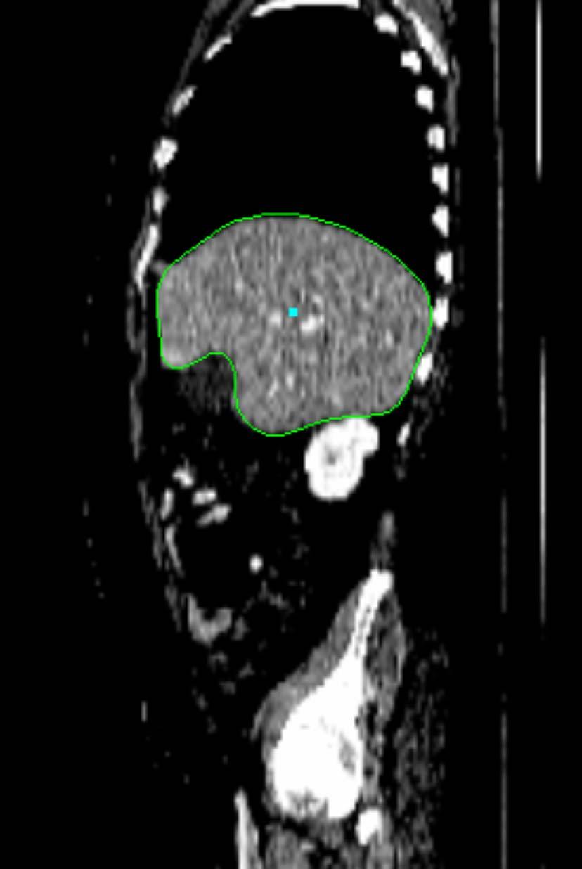


8 Figure 6. Typical results. From left to right: sagittal, coronal, and transversal slices. The errors (inter-slice resolutions, diseased organs) for from top to bottom: 1.09 (1.5, liver) mm, 1.30 (1.0, colon) mm, 1.63 (1.0, liver) mm, and 2.38 (5.0, lymphnode) mm.
Marginal Space Learning for Efficient Detection of 2D/3D Anatomical Structures in Medical Images
 Marginal Space Learning for Efficient Detection of 2D/3D Anatomical Structures in Medical Images Yefeng Zheng, Bogdan Georgescu, and Dorin Comaniciu Integrated Data Systems Department, Siemens Corporate
Universities of Leeds, Sheffield and York
 promoting access to White Rose research papers Universities of Leeds, Sheffield and York http://eprints.whiterose.ac.uk/ This is an author produced version of a paper published in Lecture Notes in Computer
Global-to-Local Shape Matching for Liver Segmentation in CT Imaging
 Global-to-Local Shape Matching for Liver Segmentation in CT Imaging Kinda Anna Saddi 1,2, Mikaël Rousson 1, Christophe Chefd hotel 1, and Farida Cheriet 2 1 Department of Imaging and Visualization, Siemens
A Generic Probabilistic Active Shape Model for Organ Segmentation
 A Generic Probabilistic Active Shape Model for Organ Segmentation Andreas Wimmer 1,2, Grzegorz Soza 2, and Joachim Hornegger 1 1 Chair of Pattern Recognition, Department of Computer Science, Friedrich-Alexander
Left Ventricle Endocardium Segmentation for Cardiac CT Volumes Using an Optimal Smooth Surface
 Left Ventricle Endocardium Segmentation for Cardiac CT Volumes Using an Optimal Smooth Surface Yefeng Zheng a, Bogdan Georgescu a, Fernando Vega-Higuera b, and Dorin Comaniciu a a Integrated Data Systems
3D Statistical Shape Model Building using Consistent Parameterization
 3D Statistical Shape Model Building using Consistent Parameterization Matthias Kirschner, Stefan Wesarg Graphisch Interaktive Systeme, TU Darmstadt matthias.kirschner@gris.tu-darmstadt.de Abstract. We
INTEGRATED DETECTION NETWORK (IDN) FOR POSE AND BOUNDARY ESTIMATION IN MEDICAL IMAGES
 FOR POSE AND BOUNDARY ESTIMATION IN MEDICAL IMAGES") INTEGRATED DETECTION NETWORK (IDN) FOR POSE AND BOUNDARY ESTIMATION IN MEDICAL IMAGES Michal Sofka Kristóf Ralovich Neil Birkbeck Jingdan Zhang S.Kevin Zhou Siemens Corporate Research, 775 College Road
INTEGRATED DETECTION NETWORK (IDN) FOR POSE AND BOUNDARY ESTIMATION IN MEDICAL IMAGES Michal Sofka Kristóf Ralovich Neil Birkbeck Jingdan Zhang S.Kevin Zhou Siemens Corporate Research, 775 College Road
Fully Automatic Multi-organ Segmentation based on Multi-boost Learning and Statistical Shape Model Search
 Fully Automatic Multi-organ Segmentation based on Multi-boost Learning and Statistical Shape Model Search Baochun He, Cheng Huang, Fucang Jia Shenzhen Institutes of Advanced Technology, Chinese Academy
Nonrigid Surface Modelling. and Fast Recovery. Department of Computer Science and Engineering. Committee: Prof. Leo J. Jia and Prof. K. H.
 Nonrigid Surface Modelling and Fast Recovery Zhu Jianke Supervisor: Prof. Michael R. Lyu Committee: Prof. Leo J. Jia and Prof. K. H. Wong Department of Computer Science and Engineering May 11, 2007 1 2
Automated segmentation methods for liver analysis in oncology applications
 University of Szeged Department of Image Processing and Computer Graphics Automated segmentation methods for liver analysis in oncology applications Ph. D. Thesis László Ruskó Thesis Advisor Dr. Antal
Methodological progress in image registration for ventilation estimation, segmentation propagation and multi-modal fusion
 Methodological progress in image registration for ventilation estimation, segmentation propagation and multi-modal fusion Mattias P. Heinrich Julia A. Schnabel, Mark Jenkinson, Sir Michael Brady 2 Clinical
Fully Automatic Model Creation for Object Localization utilizing the Generalized Hough Transform
 Fully Automatic Model Creation for Object Localization utilizing the Generalized Hough Transform Heike Ruppertshofen 1,2,3, Cristian Lorenz 2, Peter Beyerlein 4, Zein Salah 3, Georg Rose 3, Hauke Schramm
Semantic Context Forests for Learning- Based Knee Cartilage Segmentation in 3D MR Images
 Semantic Context Forests for Learning- Based Knee Cartilage Segmentation in 3D MR Images MICCAI 2013: Workshop on Medical Computer Vision Authors: Quan Wang, Dijia Wu, Le Lu, Meizhu Liu, Kim L. Boyer,
Model-Based Organ Segmentation in CT Scans
 Model-Based Organ Segmentation in CT Scans Jiun-Hung Chen General Exam/ Thesis Proposal University of Washington 2009 Program Authorized to Offer Degree: UW Computer Science and Engineering TABLE OF CONTENTS
Multi-stage Learning for Robust Lung Segmentation in Challenging CT Volumes
 Multi-stage Learning for Robust Lung Segmentation in Challenging CT Volumes Michal Sofka 1, Jens Wetzl 1,NeilBirkbeck 1, Jingdan Zhang 1, Timo Kohlberger 1,JensKaftan 2,Jérôme Declerck 2, and S. Kevin
Robust and Accurate Coronary Artery Centerline Extraction in CTA by Combining Model-Driven and Data-Driven Approaches
 Robust and Accurate Coronary Artery Centerline Extraction in CTA by Combining Model-Driven and Data-Driven Approaches Yefeng Zheng, Huseyin Tek, and Gareth Funka-Lea Imaging and Computer Vision, Siemens
Prostate Detection Using Principal Component Analysis
 Prostate Detection Using Principal Component Analysis Aamir Virani (avirani@stanford.edu) CS 229 Machine Learning Stanford University 16 December 2005 Introduction During the past two decades, computed
Automatic Rapid Segmentation of Human Lung from 2D Chest X-Ray Images
 Automatic Rapid Segmentation of Human Lung from 2D Chest X-Ray Images Abstract. In this paper, we propose a complete framework that segments lungs from 2D Chest X-Ray (CXR) images automatically and rapidly.
Robust 3D Organ Localization with Dual Learning Architectures and Fusion
 Robust 3D Organ Localization with Dual Learning Architectures and Fusion Xiaoguang Lu (B), Daguang Xu, and David Liu Medical Imaging Technologies, Siemens Medical Solutions, Inc., Princeton, NJ, USA xiaoguang.lu@siemens.com
Automatic Mitral Valve Inflow Measurements from Doppler Echocardiography
 Automatic Mitral Valve Inflow Measurements from Doppler Echocardiography JinHyeong Park 1, S. Kevin Zhou 1, John Jackson 2, and Dorin Comaniciu 1 1 Integrated Data Systems,Siemens Corporate Research, Inc.,
Auto-contouring the Prostate for Online Adaptive Radiotherapy
 Auto-contouring the Prostate for Online Adaptive Radiotherapy Yan Zhou 1 and Xiao Han 1 Elekta Inc., Maryland Heights, MO, USA yan.zhou@elekta.com, xiao.han@elekta.com, Abstract. Among all the organs under
Landmark-based 3D Elastic Registration of Pre- and Postoperative Liver CT Data
 Landmark-based 3D Elastic Registration of Pre- and Postoperative Liver CT Data An Experimental Comparison Thomas Lange 1, Stefan Wörz 2, Karl Rohr 2, Peter M. Schlag 3 1 Experimental and Clinical Research
Automatic Detection and Segmentation of Axillary Lymph Nodes
 Automatic Detection and Segmentation of Axillary Lymph Nodes Adrian Barbu 1, Michael Suehling 2,XunXu 2,DavidLiu 2, S. Kevin Zhou 2, and Dorin Comaniciu 2 1 Statistics Department, Florida State Univ.,
Manifold Learning-based Data Sampling for Model Training
 Manifold Learning-based Data Sampling for Model Training Shuqing Chen 1, Sabrina Dorn 2, Michael Lell 3, Marc Kachelrieß 2,Andreas Maier 1 1 Pattern Recognition Lab, FAU Erlangen-Nürnberg 2 German Cancer
Image Segmentation and Registration
 Image Segmentation and Registration Dr. Christine Tanner (tanner@vision.ee.ethz.ch) Computer Vision Laboratory, ETH Zürich Dr. Verena Kaynig, Machine Learning Laboratory, ETH Zürich Outline Segmentation
Translation Symmetry Detection: A Repetitive Pattern Analysis Approach
 2013 IEEE Conference on Computer Vision and Pattern Recognition Workshops Translation Symmetry Detection: A Repetitive Pattern Analysis Approach Yunliang Cai and George Baciu GAMA Lab, Department of Computing
4D Cardiac Reconstruction Using High Resolution CT Images
 4D Cardiac Reconstruction Using High Resolution CT Images Mingchen Gao 1, Junzhou Huang 1, Shaoting Zhang 1, Zhen Qian 2, Szilard Voros 2, Dimitris Metaxas 1, and Leon Axel 3 1 CBIM Center, Rutgers University,
Pathology Hinting as the Combination of Automatic Segmentation with a Statistical Shape Model
 Pathology Hinting as the Combination of Automatic Segmentation with a Statistical Shape Model Pascal A. Dufour 12,HannanAbdillahi 3, Lala Ceklic 3,Ute Wolf-Schnurrbusch 23,JensKowal 12 1 ARTORG Center
A Multiple-Layer Flexible Mesh Template Matching Method for Nonrigid Registration between a Pelvis Model and CT Images
 A Multiple-Layer Flexible Mesh Template Matching Method for Nonrigid Registration between a Pelvis Model and CT Images Jianhua Yao 1, Russell Taylor 2 1. Diagnostic Radiology Department, Clinical Center,
Automatic Multi-organ Segmentation Using Learning-Based Segmentation and Level Set Optimization
 Automatic Multi-organ Segmentation Using Learning-Based Segmentation and Level Set Optimization Timo Kohlberger 1,MichalSofka 1, Jingdan Zhang 1,NeilBirkbeck 1, Jens Wetzl 1,JensKaftan 2,Jérôme Declerck
A Systematic Analysis System for CT Liver Image Classification and Image Segmentation by Local Entropy Method
 A Systematic Analysis System for CT Liver Image Classification and Image Segmentation by Local Entropy Method A.Anuja Merlyn 1, A.Anuba Merlyn 2 1 PG Scholar, Department of Computer Science and Engineering,
Robust Discriminative Wire Structure Modeling with Application to Stent Enhancement in Fluoroscopy
 Robust Discriminative Wire Structure Modeling with Application to Stent Enhancement in Fluoroscopy Xiaoguang Lu, Terrence Chen, Dorin Comaniciu Image Analytics and Informatics, Siemens Corporate Research
Liver Segmentation in CT Data: A Segmentation Refinement Approach
 Liver Segmentation in CT Data: A Segmentation Refinement Approach Reinhard Beichel 12, Christian Bauer 3, Alexander Bornik 3, Erich Sorantin 4, and Horst Bischof 3 1 Dept. of Electrical and Computer Engineering,
Semantic Annotation of Medical Images
 Semantic Annotation of Medical Images Sascha Seifert a, Michael Kelm a, Manuel Moeller b, Saikat Mukherjee c, Alexander Cavallaro d, Martin Huber a, and Dorin Comaniciu c a Integrated Data Systems, Siemens
Eye Detection by Haar wavelets and cascaded Support Vector Machine
 Eye Detection by Haar wavelets and cascaded Support Vector Machine Vishal Agrawal B.Tech 4th Year Guide: Simant Dubey / Amitabha Mukherjee Dept of Computer Science and Engineering IIT Kanpur - 208 016
Hierarchical Segmentation and Identification of Thoracic Vertebra Using Learning-Based Edge Detection and Coarse-to-Fine Deformable Model
 Hierarchical Segmentation and Identification of Thoracic Vertebra Using Learning-Based Edge Detection and Coarse-to-Fine Deformable Model Jun Ma, Le Lu, Yiqiang Zhan, Xiang Zhou, Marcos Salganicoff, and
Hierarchical Shape Statistical Model for Segmentation of Lung Fields in Chest Radiographs
 Hierarchical Shape Statistical Model for Segmentation of Lung Fields in Chest Radiographs Yonghong Shi 1 and Dinggang Shen 2,*1 1 Digital Medical Research Center, Fudan University, Shanghai, 232, China
Pathology Hinting as the Combination of Automatic Segmentation with a Statistical Shape Model
 Pathology Hinting as the Combination of Automatic Segmentation with a Statistical Shape Model Pascal A. Dufour 1,2, Hannan Abdillahi 3, Lala Ceklic 3, Ute Wolf-Schnurrbusch 2,3, and Jens Kowal 1,2 1 ARTORG
Tracking the Left Ventricle through Collaborative Trackers and Sparse Shape Model
 Tracking the Left Ventricle through Collaborative Trackers and Sparse Shape Model Yan Zhou IMPAC Medical Systems, Elekta Inc., Maryland Heights, MO, USA Abstract. Tracking the left ventricle plays an important
Classification of Subject Motion for Improved Reconstruction of Dynamic Magnetic Resonance Imaging
 1 CS 9 Final Project Classification of Subject Motion for Improved Reconstruction of Dynamic Magnetic Resonance Imaging Feiyu Chen Department of Electrical Engineering ABSTRACT Subject motion is a significant
TOWARDS REDUCTION OF THE TRAINING AND SEARCH RUNNING TIME COMPLEXITIES FOR NON-RIGID OBJECT SEGMENTATION
 TOWDS REDUTION OF THE TRAINING AND SEH RUNNING TIME OMPLEXITIES FOR NON-RIGID OBJET SEGMENTATION Jacinto. Nascimento (a) Gustavo arneiro (b) Instituto de Sistemas e Robótica, Instituto Superior Técnico,
A Non-Linear Image Registration Scheme for Real-Time Liver Ultrasound Tracking using Normalized Gradient Fields
 A Non-Linear Image Registration Scheme for Real-Time Liver Ultrasound Tracking using Normalized Gradient Fields Lars König, Till Kipshagen and Jan Rühaak Fraunhofer MEVIS Project Group Image Registration,
AN essential part of any computer-aided surgery is planning
 1 A Model Based Validation Scheme for Organ Segmentation in CT Scan Volumes Hossein Badakhshannoory, Student Member, IEEE, and Parvaneh Saeedi, Member, IEEE Abstract In this work, we propose a novel approach
Computational Radiology Lab, Children s Hospital, Harvard Medical School, Boston, MA.
 Shape prior integration in discrete optimization segmentation algorithms M. Freiman Computational Radiology Lab, Children s Hospital, Harvard Medical School, Boston, MA. Email: moti.freiman@childrens.harvard.edu
Automatic Delineation of Left and Right Ventricles in Cardiac MRI Sequences Using a Joint Ventricular Model
 Automatic Delineation of Left and Right Ventricles in Cardiac MRI Sequences Using a Joint Ventricular Model Xiaoguang Lu 1,, Yang Wang 1, Bogdan Georgescu 1, Arne Littman 2, and Dorin Comaniciu 1 1 Siemens
Generation of Triangle Meshes from Time-of-Flight Data for Surface Registration
 Generation of Triangle Meshes from Time-of-Flight Data for Surface Registration Thomas Kilgus, Thiago R. dos Santos, Alexander Seitel, Kwong Yung, Alfred M. Franz, Anja Groch, Ivo Wolf, Hans-Peter Meinzer,
Segmentation of Bony Structures with Ligament Attachment Sites
 Segmentation of Bony Structures with Ligament Attachment Sites Heiko Seim 1, Hans Lamecker 1, Markus Heller 2, Stefan Zachow 1 1 Visualisierung und Datenanalyse, Zuse-Institut Berlin (ZIB), 14195 Berlin
Automatic Extraction of 3D Dynamic Left Ventricle Model from 2D Rotational Angiocardiogram
 Automatic Extraction of 3D Dynamic Left Ventricle Model from 2D Rotational Angiocardiogram Mingqing Chen 1, Yefeng Zheng 1, Kerstin Mueller 2,3, Christopher Rohkohl 2, Guenter Lauritsch 2,JanBoese 2, Gareth
Available Online through
 Available Online through www.ijptonline.com ISSN: 0975-766X CODEN: IJPTFI Research Article ANALYSIS OF CT LIVER IMAGES FOR TUMOUR DIAGNOSIS BASED ON CLUSTERING TECHNIQUE AND TEXTURE FEATURES M.Krithika
The Anatomical Equivalence Class Formulation and its Application to Shape-based Computational Neuroanatomy
 The Anatomical Equivalence Class Formulation and its Application to Shape-based Computational Neuroanatomy Sokratis K. Makrogiannis, PhD From post-doctoral research at SBIA lab, Department of Radiology,
Non-rigid Registration using Discrete MRFs: Application to Thoracic CT Images
 Non-rigid Registration using Discrete MRFs: Application to Thoracic CT Images Ben Glocker 1, Nikos Komodakis 2, Nikos Paragios 3,4, and Nassir Navab 1 1 Computer Aided Medical Procedures (CAMP), TU Mï
Simultaneous Segmentation and Correspondence Establishment for Statistical Shape Models
 Simultaneous Segmentation and Correspondence Establishment for Statistical Shape Models Marius Erdt 1, Matthias Kirschner 2, and Stefan Wesarg 2 1 Fraunhofer Institute for Computer Graphics, Darmstadt,
Discrete Optimization of Ray Potentials for Semantic 3D Reconstruction
 Discrete Optimization of Ray Potentials for Semantic 3D Reconstruction Marc Pollefeys Joined work with Nikolay Savinov, Christian Haene, Lubor Ladicky 2 Comparison to Volumetric Fusion Higher-order ray
Open-Curve Shape Correspondence Without Endpoint Correspondence
 Open-Curve Shape Correspondence Without Endpoint Correspondence Theodor Richardson and Song Wang Department of Computer Science and Engineering, University of South Carolina, Columbia, SC 29208, USA richa268@cse.sc.edu,
Mobile Human Detection Systems based on Sliding Windows Approach-A Review
 Mobile Human Detection Systems based on Sliding Windows Approach-A Review Seminar: Mobile Human detection systems Njieutcheu Tassi cedrique Rovile Department of Computer Engineering University of Heidelberg
Generic Face Alignment Using an Improved Active Shape Model
 Generic Face Alignment Using an Improved Active Shape Model Liting Wang, Xiaoqing Ding, Chi Fang Electronic Engineering Department, Tsinghua University, Beijing, China {wanglt, dxq, fangchi} @ocrserv.ee.tsinghua.edu.cn
An Adaptive Eigenshape Model
 An Adaptive Eigenshape Model Adam Baumberg and David Hogg School of Computer Studies University of Leeds, Leeds LS2 9JT, U.K. amb@scs.leeds.ac.uk Abstract There has been a great deal of recent interest
NIH Public Access Author Manuscript Proc Soc Photo Opt Instrum Eng. Author manuscript; available in PMC 2014 October 07.
 NIH Public Access Author Manuscript Published in final edited form as: Proc Soc Photo Opt Instrum Eng. 2014 March 21; 9034: 903442. doi:10.1117/12.2042915. MRI Brain Tumor Segmentation and Necrosis Detection
A Methodology for Constructing Geometric Priors and Likelihoods for Deformable Shape Models
 A Methodology for Constructing Geometric Priors and Likelihoods for Deformable Shape Models Derek Merck, Gregg Tracton, Stephen Pizer, and Sarang Joshi Medical Image Display & Analysis Group, University
Learning and Inferring Depth from Monocular Images. Jiyan Pan April 1, 2009
 Learning and Inferring Depth from Monocular Images Jiyan Pan April 1, 2009 Traditional ways of inferring depth Binocular disparity Structure from motion Defocus Given a single monocular image, how to infer
Hybrid Spline-based Multimodal Registration using a Local Measure for Mutual Information
 Hybrid Spline-based Multimodal Registration using a Local Measure for Mutual Information Andreas Biesdorf 1, Stefan Wörz 1, Hans-Jürgen Kaiser 2, Karl Rohr 1 1 University of Heidelberg, BIOQUANT, IPMB,
Biomedical Image Analysis Using Markov Random Fields & Efficient Linear Programing
 Biomedical Image Analysis Using Markov Random Fields & Efficient Linear Programing Nikos Komodakis Ahmed Besbes Ben Glocker Nikos Paragios Abstract Computer-aided diagnosis through biomedical image analysis
Organ Surface Reconstruction using B-Splines and Hu Moments
 Organ Surface Reconstruction using B-Splines and Hu Moments Andrzej Wytyczak-Partyka Institute of Computer Engineering Control and Robotics, Wroclaw University of Technology, 27 Wybrzeze Wyspianskiego
arxiv: v1 [cs.cv] 11 Apr 2018
![arxiv: v1 [cs.cv] 11 Apr 2018](/thumbs/83/88598484.jpg "arxiv: v1 [cs.cv] 11 Apr 2018") Unsupervised Segmentation of 3D Medical Images Based on Clustering and Deep Representation Learning Takayasu Moriya a, Holger R. Roth a, Shota Nakamura b, Hirohisa Oda c, Kai Nagara c, Masahiro Oda a,
Unsupervised Segmentation of 3D Medical Images Based on Clustering and Deep Representation Learning Takayasu Moriya a, Holger R. Roth a, Shota Nakamura b, Hirohisa Oda c, Kai Nagara c, Masahiro Oda a,
Non-rigid Segmentation using Sparse Low Dimensional Manifolds and Deep Belief Networks
 Non-rigid Segmentation using Sparse Low Dimensional Manifolds and Deep Belief Networks Jacinto C. Nascimento Instituto de Sistemas e Robótica Instituto Superior Técnico, Portugal Gustavo Carneiro Australian
Medical Image Analysis Active Shape Models
 Medical Image Analysis Active Shape Models Mauricio Reyes, Ph.D. mauricio.reyes@istb.unibe.ch ISTB - Institute for Surgical Technology and Biomechanics University of Bern Lecture Overview! Statistical
CHAPTER 6 PROPOSED HYBRID MEDICAL IMAGE RETRIEVAL SYSTEM USING SEMANTIC AND VISUAL FEATURES
 188 CHAPTER 6 PROPOSED HYBRID MEDICAL IMAGE RETRIEVAL SYSTEM USING SEMANTIC AND VISUAL FEATURES 6.1 INTRODUCTION Image representation schemes designed for image retrieval systems are categorized into two
3D Volume Mesh Generation of Human Organs Using Surface Geometries Created from the Visible Human Data Set
 3D Volume Mesh Generation of Human Organs Using Surface Geometries Created from the Visible Human Data Set John M. Sullivan, Jr., Ziji Wu, and Anand Kulkarni Worcester Polytechnic Institute Worcester,
Cognition Network Technology for a Fully Automated 3D Segmentation of Liver
 Cognition Network Technology for a Fully Automated 3D Segmentation of Liver Günter Schmidt, Maria Athelogou, Ralf Schönmeyer, Rene Korn, and Gerd Binnig Definiens AG, Research, Trappentreustr. 1, 80339
Human Heart Coronary Arteries Segmentation
 Human Heart Coronary Arteries Segmentation Qian Huang Wright State University, Computer Science Department Abstract The volume information extracted from computed tomography angiogram (CTA) datasets makes
Improving Image Segmentation Quality Via Graph Theory
 International Symposium on Computers & Informatics (ISCI 05) Improving Image Segmentation Quality Via Graph Theory Xiangxiang Li, Songhao Zhu School of Automatic, Nanjing University of Post and Telecommunications,
Using Probability Maps for Multi organ Automatic Segmentation
 Using Probability Maps for Multi organ Automatic Segmentation Ranveer Joyseeree 1,2, Óscar Jiménez del Toro1, and Henning Müller 1,3 1 University of Applied Sciences Western Switzerland (HES SO), Sierre,
Deformable Segmentation via Sparse Shape Representation
 Deformable Segmentation via Sparse Shape Representation Shaoting Zhang 2, Yiqiang Zhan 1, Maneesh Dewan 1, Junzhou Huang 2, Dimitris N. Metaxas 2, and Xiang Sean Zhou 1 1 Siemens Medical Solutions, Malvern,
TUBULAR SURFACES EXTRACTION WITH MINIMAL ACTION SURFACES
 TUBULAR SURFACES EXTRACTION WITH MINIMAL ACTION SURFACES XIANGJUN GAO Department of Computer and Information Technology, Shangqiu Normal University, Shangqiu 476000, Henan, China ABSTRACT This paper presents
Learning-based Neuroimage Registration
 Learning-based Neuroimage Registration Leonid Teverovskiy and Yanxi Liu 1 October 2004 CMU-CALD-04-108, CMU-RI-TR-04-59 School of Computer Science Carnegie Mellon University Pittsburgh, PA 15213 Abstract
Keywords: Delaunay, Haar, LymphNodes, Nonmaximal suppression INTRODUCTION
 Automatic Detection and Segmentation of Lymph Nodes Using NonMaximal Suppression Algorithm From CT Data Brahmya Joseph PG Scholar, P.Madhavan Assistant Professor Abstract Image segmentation is a difficult
Segmentation of Images
 Segmentation of Images SEGMENTATION If an image has been preprocessed appropriately to remove noise and artifacts, segmentation is often the key step in interpreting the image. Image segmentation is a
Face Alignment Under Various Poses and Expressions
 Face Alignment Under Various Poses and Expressions Shengjun Xin and Haizhou Ai Computer Science and Technology Department, Tsinghua University, Beijing 100084, China ahz@mail.tsinghua.edu.cn Abstract.
Using temporal seeding to constrain the disparity search range in stereo matching
 Using temporal seeding to constrain the disparity search range in stereo matching Thulani Ndhlovu Mobile Intelligent Autonomous Systems CSIR South Africa Email: tndhlovu@csir.co.za Fred Nicolls Department
Automated Model-Based Rib Cage Segmentation and Labeling in CT Images
 Automated Model-Based Rib Cage Segmentation and Labeling in CT Images Tobias Klinder 1,2,CristianLorenz 2,JensvonBerg 2, Sebastian P.M. Dries 2, Thomas Bülow 2,andJörn Ostermann 1 1 Institut für Informationsverarbeitung,
Texture-Based Detection of Myositis in Ultrasonographies
 Texture-Based Detection of Myositis in Ultrasonographies Tim König 1, Marko Rak 1, Johannes Steffen 1, Grit Neumann 2, Ludwig von Rohden 2, Klaus D. Tönnies 1 1 Institut für Simulation & Graphik, Otto-von-Guericke-Universität
An efficient face recognition algorithm based on multi-kernel regularization learning
 Acta Technica 61, No. 4A/2016, 75 84 c 2017 Institute of Thermomechanics CAS, v.v.i. An efficient face recognition algorithm based on multi-kernel regularization learning Bi Rongrong 1 Abstract. A novel
Rule-Based Ventral Cavity Multi-organ Automatic Segmentation in CT Scans
 Rule-Based Ventral Cavity Multi-organ Automatic Segmentation in CT Scans Assaf B. Spanier (B) and Leo Joskowicz The Rachel and Selim Benin School of Computer Science and Engineering, The Hebrew University
Erlangen-Nuremberg, Germany
 Automatic 3D Motion Estimation of Left Ventricle from C-arm Rotational Angiocardiography Using a Prior Motion Model and Learning Based Boundary Detector Mingqing Chen 1, Yefeng Zheng 1, Yang Wang 1, Kerstin
Nonrigid Registration using Free-Form Deformations
 Nonrigid Registration using Free-Form Deformations Hongchang Peng April 20th Paper Presented: Rueckert et al., TMI 1999: Nonrigid registration using freeform deformations: Application to breast MR images
Deformable Segmentation using Sparse Shape Representation. Shaoting Zhang
 Deformable Segmentation using Sparse Shape Representation Shaoting Zhang Introduction Outline Our methods Segmentation framework Sparse shape representation Applications 2D lung localization in X-ray 3D
Cost-alleviative Learning for Deep Convolutional Neural Network-based Facial Part Labeling
 [DOI: 10.2197/ipsjtcva.7.99] Express Paper Cost-alleviative Learning for Deep Convolutional Neural Network-based Facial Part Labeling Takayoshi Yamashita 1,a) Takaya Nakamura 1 Hiroshi Fukui 1,b) Yuji
2 Michael E. Leventon and Sarah F. F. Gibson a b c d Fig. 1. (a, b) Two MR scans of a person's knee. Both images have high resolution in-plane, but ha
 Two MR scans of a person's knee. Both images have high resolution in-plane, but ha") Model Generation from Multiple Volumes using Constrained Elastic SurfaceNets Michael E. Leventon and Sarah F. F. Gibson 1 MIT Artificial Intelligence Laboratory, Cambridge, MA 02139, USA leventon@ai.mit.edu
Model Generation from Multiple Volumes using Constrained Elastic SurfaceNets Michael E. Leventon and Sarah F. F. Gibson 1 MIT Artificial Intelligence Laboratory, Cambridge, MA 02139, USA leventon@ai.mit.edu
8/3/2017. Contour Assessment for Quality Assurance and Data Mining. Objective. Outline. Tom Purdie, PhD, MCCPM
 Contour Assessment for Quality Assurance and Data Mining Tom Purdie, PhD, MCCPM Objective Understand the state-of-the-art in contour assessment for quality assurance including data mining-based techniques
HYBRID MULTISCALE LANDMARK AND DEFORMABLE IMAGE REGISTRATION. Dana Paquin. Doron Levy. Lei Xing. (Communicated by Yang Kuang)
") MATHEMATICAL BIOSCIENCES http://www.mbejournal.org/ AND ENGINEERING Volume 4, Number 4, October 2007 pp. 711 737 HYBRID MULTISCALE LANDMARK AND DEFORMABLE IMAGE REGISTRATION Dana Paquin Department of Mathematics,
MATHEMATICAL BIOSCIENCES http://www.mbejournal.org/ AND ENGINEERING Volume 4, Number 4, October 2007 pp. 711 737 HYBRID MULTISCALE LANDMARK AND DEFORMABLE IMAGE REGISTRATION Dana Paquin Department of Mathematics,
Fully Automated Knowledge-Based Segmentation of the Caudate Nuclei in 3-D MRI
 Fully Automated Knowledge-Based Segmentation of the Caudate Nuclei in 3-D MRI Michael Wels 1, Martin Huber 2, and Joachim Hornegger 1 1 Institute of Pattern Recognition, University Erlangen-Nuremberg,
PATTERN SYNTHESIS FOR PLANAR ARRAY BASED ON ELEMENTS ROTATION
 Progress In Electromagnetics Research Letters, Vol. 11, 55 64, 2009 PATTERN SYNTHESIS FOR PLANAR ARRAY BASED ON ELEMENTS ROTATION F. Zhang, F.-S. Zhang, C. Lin, G. Zhao, and Y.-C. Jiao National Key Laboratory
MEDICAL IMAGE NOISE REDUCTION AND REGION CONTRAST ENHANCEMENT USING PARTIAL DIFFERENTIAL EQUATIONS
 MEDICAL IMAGE NOISE REDUCTION AND REGION CONTRAST ENHANCEMENT USING PARTIAL DIFFERENTIAL EQUATIONS Miguel Alemán-Flores, Luis Álvarez-León Departamento de Informática y Sistemas, Universidad de Las Palmas
Automatized & Interactive. Muscle tissues characterization using. Na MRI
 Automatized & Interactive Human Skeletal Muscle Segmentation Muscle tissues characterization using 23 Na MRI Noura Azzabou 30 April 2013 What is muscle segmentation? Axial slice of the thigh of a healthy
A New Shape Matching Measure for Nonlinear Distorted Object Recognition
 A New Shape Matching Measure for Nonlinear Distorted Object Recognition S. Srisuky, M. Tamsriy, R. Fooprateepsiri?, P. Sookavatanay and K. Sunaty Department of Computer Engineeringy, Department of Information
MR IMAGE SEGMENTATION
 MR IMAGE SEGMENTATION Prepared by : Monil Shah What is Segmentation? Partitioning a region or regions of interest in images such that each region corresponds to one or more anatomic structures Classification
Detecting Bone Lesions in Multiple Myeloma Patients using Transfer Learning
 Detecting Bone Lesions in Multiple Myeloma Patients using Transfer Learning Matthias Perkonigg 1, Johannes Hofmanninger 1, Björn Menze 2, Marc-André Weber 3, and Georg Langs 1 1 Computational Imaging Research
Prototype of Silver Corpus Merging Framework
 www.visceral.eu Prototype of Silver Corpus Merging Framework Deliverable number D3.3 Dissemination level Public Delivery data 30.4.2014 Status Authors Final Markus Krenn, Allan Hanbury, Georg Langs This
Journal of Asian Scientific Research FEATURES COMPOSITION FOR PROFICIENT AND REAL TIME RETRIEVAL IN CBIR SYSTEM. Tohid Sedghi
 Journal of Asian Scientific Research, 013, 3(1):68-74 Journal of Asian Scientific Research journal homepage: http://aessweb.com/journal-detail.php?id=5003 FEATURES COMPOSTON FOR PROFCENT AND REAL TME RETREVAL
Colored Point Cloud Registration Revisited Supplementary Material
 Colored Point Cloud Registration Revisited Supplementary Material Jaesik Park Qian-Yi Zhou Vladlen Koltun Intel Labs A. RGB-D Image Alignment Section introduced a joint photometric and geometric objective