Tutorial on access to, and use of OAI Images
|
|
|
- Ferdinand Parks
- 5 years ago
- Views:
Transcription
1 Tutorial on access to, and use of OAI Images John Lynch, PhD OAI Coordinating Center UCSF, San Francisco
2 Overview Image Releases: process and nomenclature What has been released & What is still to be released Types of Images Available: - Imaging schedule - Exam types (radiographs/ MRI sequences) Images on Hard Drives: - Barcodes and meta-data to find images - Operations Manuals & DICOM Image Release Notes - Contents of DICOM headers & images on disk Matching Clinical/Demographics to Images: - Imaging meta-data, and Barcodes - Clinical/Demographic datasets, Search & Browse, My Cart - Case Study selecting on baseline characteristics Exploring existing Biomarkers: - Selecting MRIs for examination based on x-ray readings
3 Process and organization of image releases Images received at the Imaging QC Center (Synarc) - undergo QC and cleaning - sent to the CC in batches as processing completed At CC, images undergo further QC checks - additional blinding and removal of private DICOM tags - packaged for public release Images in each release correspond to a designated subset of participants (ppts) ppts (progression/incidence) ppts (progression) - first half of the cohort (all ppts) - second half of the cohort (all ppts) ppts (progression, with 18-month interim visit)
4 OAI Image Release Nomenclature Image Releases identified by 3 pieces of information: Visit at which images were obtained: - Baseline (0), 12-month (1), 18-month (2), 24-month (3), 30-month (4), 36-month (5), 48-month (6) Group of participants: progression/incidence (Group A) progression (Group B) - First Half of Cohort (Group C) - Participants with 18-month interim visit (Group D) - Second Half of Cohort (Group E) Version number of release: - Sequentially numbered beginning with 1
5 OAI Current Image Releases #1: 0.A.2 - Baseline images from Group A participants - (N=200) 2 nd version of the release #2: 0.B B.2 - Baseline & 12-month images from Group B participants - (N=160) 2 nd version of the release #3: 0.C C.1 - Baseline & 12-month images from Group C participants - (N=2686) 1 st version of the release #4: 2.D.1-18-month interim visit images from Group D (N=288) 1 st version of release - Baseline and 12m can be provided (subset of #3)
6 Current Images Available (0.C.1 & 1.C.1) FU images acquired BL-FU Images available for release 1 st half ppts n = 2,691 MRI: 2,340 (87%) Knee x-ray: 2,394 (89%) Both: 2,309 (86%) MRI: 2,328 (86%) Knee x-ray: 2,206 (84%) Both: 2,111 (80%)
7 OAI Future Image Releases #5: 3.C.1 (Group C 1 st half of cohort) - 24-month images - release early followed later by: 36-month (5.C.1), 48-month (6.C.1) - Groups A and B are subsets of Group C: later images will be in the C releases #6: 0.E.1 (Group E 2 nd half of cohort) - Baseline images remainder of cohort (N=2110) mid May be paired with 12-month images (1.E.1) - Followed later by 3.E.1, 5.E.1, 6.E.1 Releases for special groups of participants: - Group F (participants with 30-month interim visits) - Thigh MRIs
8 OAI Annual Knee Imaging All participants, baseline, 12mo, 24mo, 36mo, 48mo Knee Radiographs: - Bilateral Fixed Flexion: baseline and then annually Knee MRI (3T Siemens Trio): - baseline and then annually - R knee: long protocol, L knee: shorter protocol Pulse Sequence Right Knee (mins) Left Knee (mins) Localizer (3 plane) SAG 3D DESS WE (with Cor & Axial MPRs) COR IW 2D TSE SAG IW 2D TSE (with fat suppression) COR T1W 3D FLASH WE SAG 2D muti-echo (T2 MAP) TOTAL
9 OAI Additional Imaging Additional Knee Radiographs: - Fluoro-guided L and R knee: Enrollment and annually* - Lateral L & R knee: enrollment and 36-month (controls) Additional Knee MRI visits: - Interim 18-month visit in 287 (unilateral MRI) - Interim 30-month visit in ~500 ppts (unilateral MRI) - Also with clinical outcomes and biospecimen collection Pelvis and Hand Radiographs: - Enrollment visit and 48-month visit Bilateral Full-Limb Radiograph: - 12-month, 24-month or 36-month visit Thigh MRI: - Enrollment, 24-month and 48-month * Fluoro in subset of progression subcohort only
10 OAI Imaging Details Imaging Schedule: - visits and examination schedules: Operations Manuals: - detailed information on how the images were acquired DICOM Image Release Notes: - image releases have a set of notes on the hard drive - includes types of images, pulse sequence parameters - they are also downloadable at: Bilateral Fixed Flexion Knee Radiographs: - Synaflexer Frame allows correction for magnification differences between visits allows for assessment of beam angle

11 OAI - Synaflexer Frame

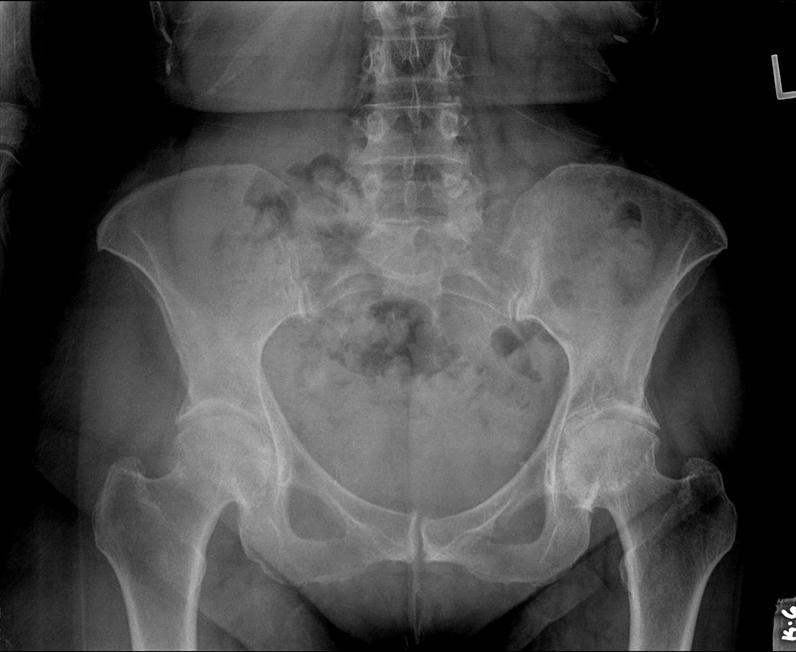
12 OAI Hand and Pelvis Radiographs

13 OAI Full Limb and Lateral Knee Radiographs

14 OAI MRI IW TSE Sequences

15 OAI MRI 3D DESS Sequence 3D - reconstructed as 0.7mm sagittal slices MPR Reformats


16 OAI Special Sequences Coronal FLASH Multi-echo sequence for T2 Maps
17 OAI Images on Hard Drives Provided by Group (A,B,C, etc) - separate folder for each annual visit (0,1,3,5,6) - interim visits (2 = 18month, 4=30month) Participant ID / Exam Date/ Barcode folders - participant ID is UID (7 digits starting with 9 ) - barcode is UID for a particular radiograph or MR sequence Each barcode has downloadable imaging meta-data - can be used to find out location of images on the hard drives - imaging meta-data also has QC ratings for images - if we decide current arrangement is stable, we could consider storing folder name in meta-data - for 0.A.2, 0.B.2, 1.B.2 you also need the spreadsheets that go with each release to find images on disk Each release has DICOM Image Release Notes
18 DICOM Image Headers Patient ID & Name: - OAI ID (9nnnnnn), prefixed with OAI for Patient Name Study Descriptions: - Visit + Exam Type + Anatomy/Side eg: Enrollment Visit Left Knee MRI eg: 12-month Visit Knee x-ray Series Descriptions: - MRI: describes pulse sequence used eg: COR_IW_TSE - X-Ray: eg: Bilateral Fixed Flexion Knee ONE x-ray image per series and only one series per study DICOM Series identified by Barcode: - Barcode is in Accession Number since often easy to search on
19 Image Acquisition Details DICOM Image Release Notes (DIRN) - give some information on image acquisition - some background on what particular type of images can be used for Operations Manuals (OM) - set of instructions on how to perform the acquisition - detailed specifics such as: special positioning for knee radiographs internal rotation for pelvis radiographs selection of imaging plane orientation for specific MRI sequences DIRN and OM both downloadable from OAI Online: one OM for radiograhy - one OM for MRI - separate DIRN for each image release
20 X-Ray OM Example

21 MRI OM Example
22 OAI Imaging Meta-Data Images of 11,090 Radiographs released Images from 61,950 MRI acquisitions released Imaging meta-data is provided online: - information on who has which types of images at a particular visit - if no image, gives you the reason the image isn t available - gives you a QC rating (if selected for visual QC) BL Meta-Data Variable (XR / MR) ID V00XRCOMP or V00MRCOMP V00XNDREAS or V00MNDREAS V00EXAMTP or V00MEXAMTP V00XRDATE or V00MRDATE V00XRBARCD or V00MRBARCD V00ACCEPT or V00QCRESLT Description Participant's ID Was exam completed & available? If Not Available, Reason Type of x-ray / MRI sequence Date of exam Unique identifier (barcode) QC rating

23 OAI Data, Variables and Documentation can use Variable Guides to explore data offline
 for My")
24 Search/Browse Frequent Knee BL Select FKP (by person) for My Cart

25 OAI Shopping Cart

26 OAI My Codebook


27 OAI Data Explorer Baseline Demographics: Knee Pain Status (KSX) vs Radiographic OA (XRKAO) Only in elderly people who aren t overweight
28 Data Explorer Results (Simplified) Table of P01KSX by P01XRKOA P01XRKOA P01KSX 0: Neither 1: Right knee only 2: Left knee only 3: Both knees Total.M: Missing : No pain either knee : Infreq pain 1 knee, no pain other knee : Infreq pain both knees : Freq pain 1 knee, no pain other knee : Freq pain 1 knee, infreq pain other knee : Freq pain both knees TOTAL
29 OAI Matching Clinical Data to Images Use Search and Browse to identify variables - Variable Guide is an off-line alternative Add variables to your Cart Download relevant datasets - always make sure you have current ENROLLEES dataset Use datasets to find a subset of participants/knees that fit your inclusion/exclusion criteria - can use Data Explorer to determine approximate #s who match - cannot analyze whether particular ppt has particular images Download imaging meta-data: - merge with clinical/demographic data - find who has particular types of images - find the barcodes (UIDs) and hence folders on disks - find QC ratings (or reasons not done)
30 OAI Images Case Study Want to select based on baseline criteria: - Gender Enrollees dataset - Body Mass Index PhysExam00 dataset - Age at Enrollment SubjectChar00 dataset - Radiographic Knee OA Biomarkers00 dataset - Frequent knee pain JointSx00 dataset Need to merge based on ID: - Remember imaging meta-data is multiple records per participant - Enrollees dataset is master of who is in study - Can merge easily in SAS - Import ASCII into database and use left joins on ID To find relevant variable names/values: - Use Search/Browse online - or Variable Guides offline
31 OAI Images Case Study Choose based on baseline criteria: - Female Gender (P02SEX = 2) Body Mass Index (P01BMI = 22.5) - No Radiographic Knee OA (P01XRKOA = 0) - Frequent pain in at least one knee (P01KSX > 2) - Older than 60 at Enrollment (V00AGE > 60) Gives the following 4 participants: ID P02SEX P01BMI V00AGE P01KSX P01XRKOA

32 OAI Baseline Imaging Meta-Data Example ID=
33 OAI Baseline Imaging Meta-Data XRAY00 shows that this person has (V00XRCOMP=1) - Fixed flexion knee radiograph - Right Hand radiograph - AP Pelvis radiograph - Each radiograph has a date and a unique barcode (V00XRBARCD) MRI00 shows Left Knee MRI sequences not available: - V00MRCOMP=0 for all Left knee sequences - 2 sequences were not expected (V00MNDREAS=8) MRI Sequence Not Expected (L knee has short protocol) - 5 sequences were not done (V00MNDREAS=2) MRI Stopped at Participant request MRI00 shows that this person DID have R knee MRI: - V00MRCOMP=1 for all Right Knee sequences - There is a date for each sequences - There is a unique barcode (V00MRBARCD) for each sequence
34 OAI Images Case Study Choose based on baseline criteria: - Female Gender (P02SEX = 2) Body Mass Index (P01BMI = 22.5) - No Radiographic Knee OA (P01XRKOA = 0) - Frequent pain in at least one knee (P01KSX > 2) - Older than 60 at Enrollment (V00AGE > 60) Include requirement that there is: - Baseline fixed flexion radiograph - Baseline R knee Sagittal DESS Find 2 participants who have images that match: ID V00EXAMTP V00XRBARCD V00ACCEPT V00MEXAMTP V00MRBARCD V00QCRESLT Bilateral PA Fixed Flexion Knee Bilateral PA Fixed Flexion Knee YD R SAG 3D DESS WE YD R SAG 3D DESS WE Y Y
35 OAI Images Case Study SAS Code data temp; merge current.enrollees current.physexam00 current.subjectchar00 current.biomarkers00 current.jointsx00; by id; run; data who; set temp; where P02SEX=2 and P01BMI=22.5 and V00AGE>60 and P01XRKOA=0 and P01KSX>2; run; data images; merge who (in=in keep=id) current.xray00 (where=(v00examtp='bilateral PA Fixed Flexion Knee') in=inx) current.mri00 (where=(v00mexamtp='r SAG 3D DESS WE') in=inm); by id; if in and inx and inm; run; proc print; var ID V00EXAMTP V00XRBARCD V00ACCEPT V00MEXAMTP V00MRBARCD V00QCRESLT; run;
36 OAI Biomarkers Datasets Clinic Baseline X-ray readings (for whole cohort) Central paired knee x-ray readings (Group B, 160ppts): - OARSI gradings and K&L Grades at baseline and 12-months - identified by V00IMAGESB=1 / V01IMAGESB=1 - images are in 0.B.2 and 1.B.2 (are also a subset of 0.C.1 and 1.C.1) Biomarkers dataset will grow: - Joint Space Width measurements (Group B) - MRI Readings and/or Cartilage Volumes (Group B) - Further analyses of larger groups of participants - Any results/readings given back to OAI (eg: Hip-Knee-Ankle angle) Matching Paired Readings to Clinical/Demographics: - Images analyzed identified by barcode - V00XRBCODE / V01XRBCODE in knee x-ray readings - Matches to barcode variables in XRAY00 / XRAY01 meta-data
37 Summary Use clinical, demographic, biomarker datasets to select participants or knees Search & Browse or Variable Guides to explore My Cart, My Codebook to customize Download data available as SAS or ASCII Imaging meta-data to find who has required images - you may only require specific image types Request images from Coordinating Center - small subsets on UCSF provided hard drives - larger releases requestor provides hard drive Future Releases - additional visits, 2 nd half of cohort - images for specific lists of participants
38 In Future Further Releases: - additional visits for 1st half of cohort (Group C) - images for 2 nd half of cohort (Group E) - thigh MRIs Food for thought: - Images for specific lists of participants selection based on downloadable datasets would be subsets of existing releases about ½ GB per participant per visit requestor keeps track of images already provided - Online access to downloadable images about 1TB of data per visit for 1 st half of cohort (Group C) can compress, but only to about 75% of original size
Maximizing Statistical Interactions Part II: Database Issues Provided by: The Biostatistics Collaboration Center (BCC) at Northwestern University
 at Northwestern University") Maximizing Statistical Interactions Part II: Database Issues Provided by: The Biostatistics Collaboration Center (BCC) at Northwestern University While your data tables or spreadsheets may look good to
Maximizing Statistical Interactions Part II: Database Issues Provided by: The Biostatistics Collaboration Center (BCC) at Northwestern University While your data tables or spreadsheets may look good to
CT Protocol Clinical Graphics Move Forward 3D motion analysis service
 CT Protocol Clinical Graphics Move Forward 3D motion analysis service Version 1.4 Description of contents This document describes the CT protocol of scans for Clinical Graphics Move Forward 3D Motion Simulation
Supplementary methods
 Supplementary methods This section provides additional technical details on the sample, the applied imaging and analysis steps and methods. Structural imaging Trained radiographers placed all participants
The objective of this tutorial is to present Model Based Calculations. These are calculations that only make sense relative to rigid segments.
 C-Motion Online Documentation Visual3D : Model Based Computations Objectives (# 1388) The objective of this tutorial is to present Model Based Calculations. These are calculations that only make sense
Technical Publications
 GE Medical Systems Technical Publications Direction 2188003-100 Revision 0 Tissue Volume Analysis DICOM for DICOM V3.0 Copyright 1997 By General Electric Co. Do not duplicate REVISION HISTORY REV DATE
Orthopedic MRI Protocols. Philips Panorama HFO
 Orthopedic MRI Protocols Philips Panorama HFO 1 2 Prepared in collaboration with Dr. John F. Feller, Medical Director of Desert Medical Imaging, Palm Springs, CA. Desert Medical Imaging will provide the
SYNAPSE 3D Conformance Statement FUJIFILM SYNAPSE 3D V3.0. Conformance Statement. Revision Z45N
 FUJIFILM SYNAPSE 3D V3.0 Conformance Statement Revision 1.0.0 1 Revision History SYNAPSE 3D Conformance Statement Revision Date Description 1.0 2011/07/01 Initial revision. 2 Table of Contents 1 Introduction...4
syngo MR E11 Operator Manual Ortho Answers for life.
 www.siemens.com/healthcare syngo MR E11 Operator Manual Ortho Answers for life. syngo MR E11 Operator Manual Ortho Legend Indicates a hint Is used to provide information on how to avoid operating errors
The protocols used to scan the musculoskeletal system are tailored to each patient and
 Chapter 22. Musculoskeletal Protocols The protocols used to scan the musculoskeletal system are tailored to each patient and region being examined. The clinical indication for the examination will also
New Enhanced Multi-frame DICOM CT and MR Objects to Enhance Performance and Image Processing on PACS and Workstations
 New Enhanced Multi-frame DICOM CT and MR Objects to Enhance Performance and Image Processing on PACS and Workstations SCAR 2004 Hot Topics - 22 May 2004 David Clunie, RadPharm Charles Parisot,, GE Healthcare
DICOM Correction Item
 DICOM Correction Item Correction Number CP-668 Log Summary: Type of Modification Addition Name of Standard PS 3.3, 3.17 2006 Rationale for Correction The term axial is common in practice, but is incorrectly
Technical Publications
 g GE Medical Systems Technical Publications Direction 2275362-100 Revision 0 DICOM for DICOM V3.0 Copyright 2000 By General Electric Co. Do not duplicate REVISION HISTORY REV DATE REASON FOR CHANGE 0 May
GE Healthcare CLINICAL GALLERY. Discovery * MR750w 3.0T. This brochure is intended for European healthcare professionals.
 GE Healthcare CLINICAL GALLERY Discovery * MR750w 3.0T This brochure is intended for European healthcare professionals. NEURO PROPELLER delivers high resolution, motion insensitive imaging in all planes.
Athena Radiology Medical Workstation
 High productivity and integration Athena DICOM Viewer is being designed according with the suggestions and the necessities of medical radiologists. From the design of its interface to the advanced image
Andrew H. Karp Sierra Information Services, Inc. San Francisco, California USA
 Indexing and Compressing SAS Data Sets: How, Why, and Why Not Andrew H. Karp Sierra Information Services, Inc. San Francisco, California USA Many users of SAS System software, especially those working
icatvision Quick Reference
 icatvision Quick Reference Navigating the i-cat Interface This guide shows how to: View reconstructed images Use main features and tools to optimize an image. REMINDER Images are displayed as if you are
Qualitative Comparison of Conventional and Oblique MRI for Detection of Herniated Spinal Discs
 Qualitative Comparison of Conventional and Oblique MRI for Detection of Herniated Spinal Discs Doug Dean Final Project Presentation ENGN 2500: Medical Image Analysis May 16, 2011 Outline Review of the
DICOM CONFORMANCE STATEMENT MEDIA STORAGE FOR TOSHIBA DIGITAL FLUOROGRAPHY SYSTEM MODEL DFP-2000A. with XIDF-037A and XIDF-038A or SRS-1000A
 DICOM CONFORMANCE STATEMENT MEDIA STORAGE FOR TOSHIBA DIGITAL FLUOROGRAPHY SYSTEM MODEL DFP-2000A with XIDF-037A and XIDF-038A or SRS-1000A (MIIXR0002EAC) 2001 IMPORTANT! (1) No part of this manual may
ThE ultimate, INTuITIVE Mr INTErFAcE
 ThE ultimate, INTuITIVE Mr INTErFAcE Empowering you to do more The revolutionary Toshiba M-power user interface takes Mr performance and flexibility to levels higher than ever before. M-power is able to
These rules are specific in their settings and numbers and form designers should adhere to them in all cases.
 Flowed Form Strategy Form Services uses Adobe LiveCycle in conjunction with Adobe form fragments to create forms. However, to successfully maximize the capabilities of these technologies, forms must be
Technical Publications
 g GE Medical Systems Technical Publications 2365523-100 Revision 1 DICOM for DICOM V3.0 Copyright 2003 By General Electric Co. Do not duplicate REVISION HISTORY REV DATE REASON FOR CHANGE 0 Nov. 15, 2002
Emergency Patient in Image Pilot. Image Pilot & Drangonfly PACS
 Emergency Patient in Image Pilot Image Pilot & Drangonfly PACS Step 1: To add a new patient as Emergency you click New Exam to start. Step 2: Enter anything into the Patient ID and Patient Name field.
Lucy Phantom MR Grid Evaluation
 Lucy Phantom MR Grid Evaluation Anil Sethi, PhD Loyola University Medical Center, Maywood, IL 60153 November 2015 I. Introduction: The MR distortion grid, used as an insert with Lucy 3D QA phantom, is
DICOM Conformance Statement
 DICOM Conformance Statement SCENARIA Tokyo, Japan E1E-HC0002-01 Hitachi, Ltd. 2016. All rights reserved. History Revision Description Date Rev. 1.0 Initial. 2016/04/01 ii E1E-HC0002 Contents History...
Basal Ganglia Matching Tools 2- Tutorial
 Basal Ganglia Matching Tools 2- Tutorial Minimum system hardware requirements: - Processor: Pentium 4 or Athlon with speed of 1 GHz - RAM: 256 MB - Hard disk space: 40 MB. System requirements 1. Insert
Whole Body X-ray CT System Supria. DICOM Conformance Statement
 Whole Body X-ray CT System Supria DICOM Conformance Statement Tokyo, Japan (SN-C633E) Copyright Hitachi Medical Corporation. 2013, 2014. All rights reserved. History Revision Description Date Rev. 1.0
DICOM Correction Item
 DICOM Correction Item Correction Number CP-567 Log Summary: Type of Modification Clarification Name of Standard PS 3.3 2004 Rationale for Correction The order of signficance of dimension indices (i.e.,which
Carestream s 2 nd Generation Metal Artifact Reduction Software (CMAR 2)
") Carestream s 2 nd Generation Metal Artifact Reduction Software (CMAR 2) Author: Levon Vogelsang Introduction Cone beam computed tomography (CBCT), or cone beam CT technology, offers considerable promise
Carestream s 2 nd Generation Metal Artifact Reduction Software (CMAR 2) Author: Levon Vogelsang Introduction Cone beam computed tomography (CBCT), or cone beam CT technology, offers considerable promise
Lab Location: MRI, B2, Cardinal Carter Wing, St. Michael s Hospital, 30 Bond Street
 Lab Location: MRI, B2, Cardinal Carter Wing, St. Michael s Hospital, 30 Bond Street MRI is located in the sub basement of CC wing. From Queen or Victoria, follow the baby blue arrows and ride the CC south
DICOM Conformance Statement
 For SonixTouch Q+, SonixMDP/SP/OP Q+ and SonixOne Diagnostic Ultrasound Systems (and Systems with Software Version 6.1.0 and Newer) Analogic Corporation 8 Centennial Drive Peabody, MA 01960 USA bkultrasound.com
X-Porte DICOM Conformance Statement
 21919 30th Dr. SE, Bothell, WA 98021-3904 USA Telephone 1.425.951.1200 Facsimile 1.425.951.1201 www.sonosite.com Document Number: Revision: Title: D10711 D X-Porte DICOM Conformance Statement CHANGE HISTORY:
E-MRI a standard in imaging
 The Compact E-MRI E-MRI a standard in imaging Thanks to an excellent cost/benefit ratio, dedicated MRI systems have become a reality in the world of diagnostic imaging. The aging of the population together
Biomechanics Laboratory School of Human Kinetics University of Ottawa
 Biomechanics Laboratory School of Human Kinetics University of Ottawa Visual3D Quick Reference Guide D. Gordon E. Robertson, PhD, FCSB Last revised: 1 November 2006 Table of Contents 1: Static Trial....
COBRE Scan Information
 COBRE Scan Information Below is more information on the directory structure for the COBRE imaging data. Also below are the imaging parameters for each series. Directory structure: var/www/html/dropbox/1139_anonymized/human:
Submission Guidelines
 Submission Guidelines Clinical Trial Results invites the submission of phase I, II, and III clinical trials for publication in a brief print format, with full trials results online. We encourage the submission
E. Mark Haacke, PhD. The MRI Institute for Biomedical Research Detroit, Michigan Wayne State University Detroit, Michigan 48201
 E. Mark Haacke, PhD The MRI Institute for Biomedical Research Detroit, Michigan 48202 Wayne State University Detroit, Michigan 48201 Acknowledgements The testing and establishment of these protocols has
QIBA PET Amyloid BC March 11, Agenda
 QIBA PET Amyloid BC March 11, 2016 - Agenda 1. QIBA Round 6 Funding a. Deadlines b. What projects can be funded, what cannot c. Discussion of projects Mechanical phantom and DRO Paul & John? Any Profile
DICOM Conformance Statement
 1 of 6 DICOM Conformance Statement DICOM Conformance Statement Review Station software Review Station (ERS) software is an image reviewing software application that reads and displays DICOM- compliant
Enhanced DICOM MR for spectroscopy, structural and functional imaging
 The Medicine Behind the Image Enhanced DICOM MR for spectroscopy, structural and functional imaging Dr. David A. Clunie, MB.,BS., FRACR Chief Technology Officer RadPharm, Inc. Acknowledgments Mark Day,
SAS Programs SAS Lecture 4 Procedures. Aidan McDermott, April 18, Outline. Internal SAS formats. SAS Formats
 SAS Programs SAS Lecture 4 Procedures Aidan McDermott, April 18, 2006 A SAS program is in an imperative language consisting of statements. Each statement ends in a semi-colon. Programs consist of (at least)
X-Rays, Videos and Document Filing
 X-Rays, Videos and Document Filing X-rays and Other Images... 2 The Main Image File... 2 Location of Images... 2 Thumb Nails views of images... 3 Importing and storing MRI s... 3 Importing X-rays and Other
DICOM Conformance Statement
 DICOM Conformance Statement ECLOS Tokyo, Japan (SN-C357E) Copyright Hitachi Medical Corporation. 2007, 2008. All rights reserved. History Revision Description Date Rev. 1.0 Initial. 2007/02/01 Rev 2.0
Digital Image Processing
 Digital Image Processing SPECIAL TOPICS CT IMAGES Hamid R. Rabiee Fall 2015 What is an image? 2 Are images only about visual concepts? We ve already seen that there are other kinds of image. In this lecture
Technical Publications
 g GE Healthcare Technical Publications 2391078-2-800 Revision 1 Seno Advantage2 WorkStation for DICOM V3.0 do not duplicate Copyright 2006 By General Electric Co. THIS PAGE LEFT INTENTIONALLY BLANK REVISION
Technical Publications
 g GE Medical Systems Technical Publications Direction 2264272-100 Revision 1 DICOM for DICOM V3.0 Copyright ª 2000 By General Electric Co. Do not duplicate THIS PAGE LEFT INTENTIONALLY BLANK TABLE OF
CT Protocol Review: Practical Tips for the Imaging Physicist Physicist
 CT Protocol Review: Practical Tips for the Imaging Physicist Physicist Dianna Cody, Ph.D., DABR, FAAPM U.T.M.D. Anderson Cancer Center August 8, 2013 AAPM Annual Meeting Goals Understand purpose and importance
Slide 1. Technical Aspects of Quality Control in Magnetic Resonance Imaging. Slide 2. Annual Compliance Testing. of MRI Systems.
 Slide 1 Technical Aspects of Quality Control in Magnetic Resonance Imaging Slide 2 Compliance Testing of MRI Systems, Ph.D. Department of Radiology Henry Ford Hospital, Detroit, MI Slide 3 Compliance Testing
A Study of Medical Image Analysis System
 Indian Journal of Science and Technology, Vol 8(25), DOI: 10.17485/ijst/2015/v8i25/80492, October 2015 ISSN (Print) : 0974-6846 ISSN (Online) : 0974-5645 A Study of Medical Image Analysis System Kim Tae-Eun
CFB: A Programming Pattern for Creating Change from Baseline Datasets Lei Zhang, Celgene Corporation, Summit, NJ
 Paper TT13 CFB: A Programming Pattern for Creating Change from Baseline Datasets Lei Zhang, Celgene Corporation, Summit, NJ ABSTRACT In many clinical studies, Change from Baseline analysis is frequently
eschoolplus+ Medical Training Guide
 eschoolplus+ Medical Training Guide Version 3.1 August 2016 Arkansas Public School Computer Network This page intentionally left blank Page 2 of 101 Table of Contents Student Medical Records Overview...
DICOM Conformance Statement
 DICOM Conformance Statement -- Offline media storage support -- PRO NM, VISUCAM VISUCAM and VISUCAM NM FA Opt AF SW - Version 4.2 NM FA Carl Zeiss Meditec AG Göschwitzer Str. 51-52 07745 Jena Germany +49
Local Coordinate Systems From Motion Capture Data
 Local Coordinate Systems From Motion Capture Data Anatomical Coordinate Systems (thigh) How to calculate the anatomical coordinate systems 1) Find the Hip center 2) Find the Knee center 3) Find the Inferior/Superior
Equipment Specification
 MULTISLICE CT SCANNER ( 16 Slices ) Merk : Hitachi Japan Model : SUPRIA 5 MHU Price : Rp 6.512.360.215,27 No Equipment Specification 1 2 3 Scanner Gantry Object for scanning : Whole body including head
DICOM 3.0 Conformance Statement
 DICOM 3.0 Conformance Statement DICOMLink v1.2 for ICON Siemens Medical Systems, Inc. Nuclear Medicine Group DICOM Conformance Statement DICOM 3.0 Conformance Statement Siemens ICON DICOMlink v1.2 for
SSID User Guide and Policy
 OSPI SSID User Guide and Policy Using the Comprehensive Education Data and Research System to obtain State Student Identifiers Customer Support September 2017 Table of Contents Introduction... 3 Using
OneView. User s Guide
 OneView User s Guide Welcome to OneView. This user guide will show you everything you need to know to access and utilize the wealth of information available from OneView. The OneView program is an Internet-based
DICOM Enhanced XA/XRF Object New Dimensions for X-Ray Projection Imaging. (DICOM Supplement 83)
") DICOM Enhanced XA/XRF Object New Dimensions for X-Ray Projection Imaging (DICOM Supplement 83) Authors: Heinz Blendinger Bas Revet Francisco Sureda Rainer Thieme Speaker: Heinz Blendinger Siemens Medical
DICOM Enhanced XA/XRF Object New Dimensions for X-Ray Projection Imaging (DICOM Supplement 83) Authors: Heinz Blendinger Bas Revet Francisco Sureda Rainer Thieme Speaker: Heinz Blendinger Siemens Medical
EXCELLING WITH ANALYSIS AND VISUALIZATION
 EXCELLING WITH ANALYSIS AND VISUALIZATION A PRACTICAL GUIDE FOR DEALING WITH DATA Prepared by Ann K. Emery July 2016 Ann K. Emery 1 Welcome Hello there! In July 2016, I led two workshops Excel Basics for
To access the images that accompany a Radiology result from Memorial Health System select the document in Chart Viewer and click on View.
 ImageLink To access the images that accompany a Radiology result from Memorial Health System select the document in Chart Viewer and click on View. On the upper right side of the Order Viewer select the
Archiving Full Resolution Images
 Archiving Full Resolution Images Archival or full resolution files are very large and are either uncompressed or minimally compressed. This tutorial explains how to use CONTENTdm and the Project Client
Site Imaging Manual DECAMP
 Site Imaging Manual DECAMP 1 4703 Detection of Early lung Cancer Among Military Personnel Study 1 (DECAMP 1): Diagnosis and Surveillance of Indeterminate Pulmonary Nodules Version: Final Version 2.0 Date:
Unit-of-Analysis Programming Vanessa Hayden, Fidelity Investments, Boston, MA
 Unit-of-Analysis Programming Vanessa Hayden, Fidelity Investments, Boston, MA ABSTRACT There are many possible ways to organize your data, but some ways are more amenable to end-reporting and analysis
TBS CALCULATOR USER GUIDE
 Med-Imaps SA PTIB Hôpital Xavier Arnozan Avenue du Haut-Lévêque F 33600 PESSAC Tel. +33 (0)5 57 10 28 56 www.med-imaps.com TBS CALCULATOR USER GUIDE User Guide version 2.0 This User Guide is based on TBS
Using PROC SQL to Calculate FIRSTOBS David C. Tabano, Kaiser Permanente, Denver, CO
 Using PROC SQL to Calculate FIRSTOBS David C. Tabano, Kaiser Permanente, Denver, CO ABSTRACT The power of SAS programming can at times be greatly improved using PROC SQL statements for formatting and manipulating
RAD. Experiences Using the Wireless FPD-Equipped MobileDaRt Evolution and Its Usefulness. 1. Introduction
 RAD Experiences Using the Wireless FPD-Equipped MobileDaRt Evolution and Its Usefulness Department of Radiological Technology, Kitasato University Kitasato Institute Medical Center Hospital Satoshi Yanagita
Raffaello Conformance Statement for DICOM V3.0
 Raffaello Conformance Statement for DICOM V3.0 Copyright 2003-2006 by I.M.S. s.r.l. DOCUMENT VERSIONS Version Date Author Changes 1.00 04-Oct-03 IMS s.r.l. First Version 1.01 27-Feb-04 IMS s.r.l. Updated
Medical Image Processing: Image Reconstruction and 3D Renderings
 Medical Image Processing: Image Reconstruction and 3D Renderings 김보형 서울대학교컴퓨터공학부 Computer Graphics and Image Processing Lab. 2011. 3. 23 1 Computer Graphics & Image Processing Computer Graphics : Create,
Optimisation of Toshiba Aquilion ONE Volume Imaging
 Optimisation of Toshiba Aquilion ONE Volume Imaging Jane Edwards, RPRSG Royal Free London NHS Foundation Trust Dr Mufudzi Maviki, Plymouth Hospitals NHS Trust Background In 2011/12 Radiology at RFH was
DICOM Conformance Statement for Scanner Interface. Release 4.7.1
 DICOM Conformance Statement for Scanner Interface Release 4.7.1 Document: Pd088CONFSTAT edition 3 11 August 2003 2003 Elekta. All rights reserved. Issued by: P. O. BOX 7593 SE-103 93 STOCKHOLM SWEDEN Tel:
Technical Publications
 g GE Medical Systems Technical Publications 2391078-800 Revision 1 Seno Advantage WorkStation for DICOM V3.0 do not duplicate Copyright 2003 By General Electric Co. THIS PAGE LEFT INTENTIONALLY BLANK REVISION
icatvision Software Manual
 University of Minnesota School of Dentistry icatvision Software Manual April 19, 2013 Mansur Ahmad, BDS, PhD Associate Professor, University of Minnesota School of Dentistry Director, American Board of
TITAN DICOM Conformance Statement
 TITAN DICOM Conformance Statement This document contains confidential information that is proprietary to SonoSite. Neither the document nor the information contained therein should be disclosed or reproduced
The DICOM Standard. Miloš Šrámek Austrian Academy of Sciences
 The DICOM Standard Miloš Šrámek Austrian Academy of Sciences Medical Image Formats Typical information present in a file: Image data (unmodified or compressed) Patient identification and demographics Technical
Technical Publication. DICOM Conformance Statement. Patient Browser 2.1. Document Revision 2. April 13, Copyright BrainLAB AG
 Technical Publication DICOM Conformance Statement 2.1 Document Revision 2 April 13, 2011 2011 Copyright BrainLAB AG 1 Conformance Statement Overview This is a conformance statement for the BrainLAB software.
TARDIS Working Practice Document No: 035
 TARDIS Working Practice Document No: 035 Uploading CT or MR Images All stroke patients should have a baseline scan taken before randomisation. Any other CT scans or MRIs, taken up until completion of the
Tutorial BOLD Module
 m a k i n g f u n c t i o n a l M R I e a s y n o r d i c B r a i n E x Tutorial BOLD Module Please note that this tutorial is for the latest released nordicbrainex. If you are using an older version please
Create a SAS Program to create the following files from the PREC2 sas data set created in LAB2.
 Topics: Data step Subsetting Concatenation and Merging Reference: Little SAS Book - Chapter 5, Section 3.6 and 2.2 Online documentation Exercise I LAB EXERCISE The following is a lab exercise to give you
Shadow casting. What is the problem? Cone Beam Computed Tomography THE OBJECTIVES OF DIAGNOSTIC IMAGING IDEAL DIAGNOSTIC IMAGING STUDY LIMITATIONS
 Cone Beam Computed Tomography THE OBJECTIVES OF DIAGNOSTIC IMAGING Reveal pathology Reveal the anatomic truth Steven R. Singer, DDS srs2@columbia.edu IDEAL DIAGNOSTIC IMAGING STUDY Provides desired diagnostic
Whole Body X-ray CT System Supria. DICOM Conformance Statement
 Whole Body X-ray CT System Supria DICOM Conformance Statement Tokyo, Japan E1E-HC0001-02 Hitachi, Ltd. 2016. All rights reserved. ii E1E-HC0001 History Revision Description Date Rev. 1.0 Initial. 2016/4/1
EPILOG PREOP. Viewer Interpretation Guideline
 EPILOG PREOP Viewer Interpretation Guideline Starting the viewer Launch the viewer for a patient by clicking this icon. A new browser tab or window will open. Data loading To ensure smooth switching between
Breast MRI Accreditation Program Clinical Image Quality Guide
 Breast MRI Accreditation Program Clinical Image Quality Guide Introduction This document provides guidance on breast MRI clinical image quality and describes the criteria used by the ACR Breast MRI Accreditation
186 Statistics, Data Analysis and Modeling. Proceedings of MWSUG '95
 A Statistical Analysis Macro Library in SAS Carl R. Haske, Ph.D., STATPROBE, nc., Ann Arbor, M Vivienne Ward, M.S., STATPROBE, nc., Ann Arbor, M ABSTRACT Statistical analysis plays a major role in pharmaceutical
Computed tomography of simple objects. Related topics. Principle. Equipment TEP Beam hardening, artefacts, and algorithms
 Related topics Beam hardening, artefacts, and algorithms Principle The CT principle is demonstrated with the aid of simple objects. In the case of very simple targets, only a few images need to be taken
n o r d i c B r a i n E x Tutorial DSC Module
 m a k i n g f u n c t i o n a l M R I e a s y n o r d i c B r a i n E x Tutorial DSC Module Please note that this tutorial is for the latest released nordicbrainex. If you are using an older version please
STAT 7000: Experimental Statistics I
 STAT 7000: Experimental Statistics I 2. A Short SAS Tutorial Peng Zeng Department of Mathematics and Statistics Auburn University Fall 2009 Peng Zeng (Auburn University) STAT 7000 Lecture Notes Fall 2009
McKesson Radiology Display Protocol Manual
 McKesson Radiology Display Protocol Manual Table of Contents Preparing a Site for Display Protocols... 5 Best Practices for Creating Display Protocols... 6 Creating a New Display Protocol... 7 Creating
Background. Outline. Radiographic Tomosynthesis: Image Quality and Artifacts Reduction 1 / GE /
 Radiographic Tomosynthesis: Image Quality and Artifacts Reduction Baojun Li, Ph.D Department of Radiology Boston University Medical Center 2012 AAPM Annual Meeting Background Linear Trajectory Tomosynthesis
MRI. When to use What sequences. Outline 2012/09/19. Sequence: Definition. Basic Principles: Step 2. Basic Principles: Step 1. Govind Chavhan, MD
 MRI When to use What sequences Govind Chavhan, MD Assistant Professor and Staff Radiologist The Hospital For Sick Children, Toronto Planning Acquisition Post processing Interpretation Patient history and
New Technology Allows Multiple Image Contrasts in a Single Scan
 These images were acquired with an investigational device. PD T2 T2 FLAIR T1 MAP T1 FLAIR PSIR T1 New Technology Allows Multiple Image Contrasts in a Single Scan MR exams can be time consuming. A typical
MEDICAL IMAGING 2nd Part Computed Tomography
 MEDICAL IMAGING 2nd Part Computed Tomography Introduction 2 In the last 30 years X-ray Computed Tomography development produced a great change in the role of diagnostic imaging in medicine. In convetional
Working with Composite Endpoints: Constructing Analysis Data Pushpa Saranadasa, Merck & Co., Inc., Upper Gwynedd, PA
 PharmaSug2016- Paper HA03 Working with Composite Endpoints: Constructing Analysis Data Pushpa Saranadasa, Merck & Co., Inc., Upper Gwynedd, PA ABSTRACT A composite endpoint in a Randomized Clinical Trial
iportal 2.0 Introduction and Frequently Asked Questions
 iportal 2.0 Introduction and Frequently Asked Questions Table of Contents Introduction... 2 Frequently Asked Questions... 4 Account Management... 4 Q: Can I change my own password?... 4 Q: Am I able to
CT Basics Principles of Spiral CT Dose. Always Thinking Ahead.
 1 CT Basics Principles of Spiral CT Dose 2 Who invented CT? 1963 - Alan Cormack developed a mathematical method of reconstructing images from x-ray projections Sir Godfrey Hounsfield worked for the Central
Project Requirements
 MVS 330 The University of Michigan Division of Kinesiology Department of Movement Science Project Requirements Professor Melissa Gross Edited by Tom Hoogendyk Revised 9/99 1996 Melissa Gross, Ph.D. Table
DICOM Conformance Statement for GALILEI. Software Version V6.0
 DICOM Conformance Statement for GALILEI Software Version V6.0 Version 1.0, December 6 th, 2011 Contents Revision History... 2 Purpose... 2 References... 2 Terms and Definitions... 2 Abbreviations... 3
AAPM Standard of Practice: CT Protocol Review Physicist
 AAPM Standard of Practice: CT Protocol Review Physicist Dianna Cody, Ph.D., DABR, FAAPM U.T.M.D. Anderson Cancer Center September 11, 2014 2014 Texas Radiation Regulatory Conference Goals Understand purpose
XRADIA microxct Manual
 XRADIA microxct Manual Multiscale CT Lab Table of Contents 1. Introduction and Basics 1.1 Instrument Parts 1.2 Powering up the system 1.3 Preparing your sample 2. TXM Controller 2.1 Starting up 2.2 Finding
ACRIN 4703 Detection of Early lung Cancer Among Military Personnel Study 1 (DECAMP 1): Diagnosis and Surveillance of Indeterminate Pulmonary Nodules
: Diagnosis and Surveillance of Indeterminate Pulmonary Nodules") ACRIN 4703 Detection of Early lung Cancer Among Military Personnel Study 1 (DECAMP 1): Diagnosis and Surveillance of Indeterminate Pulmonary Nodules Data Management Training Lindsey Dymond, Senior Research
ACRIN 4703 Detection of Early lung Cancer Among Military Personnel Study 1 (DECAMP 1): Diagnosis and Surveillance of Indeterminate Pulmonary Nodules Data Management Training Lindsey Dymond, Senior Research
CHILI /Workstation. Reporting multi modal digital images. Product Specification
 CHILI /Workstation Reporting multi modal digital images Product Specification CHILI /Workstation CHILI/Workstation is a diagnostic PACS workstation with additional functions for teleradiology. CHILI/Workstation