Closeout Action: v/w Date n=f-«submitted
|
|
|
- Alexis Palmer
- 5 years ago
- Views:
Transcription

1 CA8120 Georgia Institute of Technology Office of Contract Administration PROJECT CLOSEOUT - NOTICE ' Page: 1 24-FEB :32 Closeout Notice Date 24-FEB-1998 Project Number E-18-T33 Doch Id Center Number 10/24-6-R0881-0A0 Project Director BERTA, YOLANDE Project Unit MSE Sponsor COLUMBIAN CHEMICALS COMPANY/SWARTZ, LO Division Id Contract Number AGR DTD 29 DEC 1997 Contract Entity GTRC Prime Contract Number Title CARBON NANOPARTICLE ANALYSIS Effective Completion Date 20-FEB-1998 (Performance) 20-FEB-1998 (Reports) Closeout Action: v/w Date n=f-«submitted Final Invoice or Copy of Final Invoice Final Report of Inventions and/or Subcontracts Government Property Inventory and Related Certificate Classified Material Certificate Release and Assignment Other Comments Distribution Required: Project Director/Principal Investigator Research Administrative Network Accounting Research Security Department Reports Coordinator Research Property Team Supply Services Department/Procurement Georgia Tech Research Corporation Project File
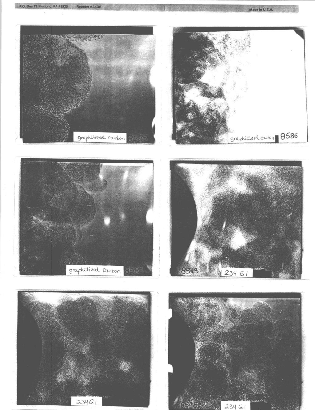
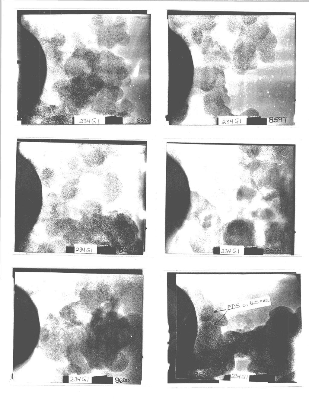
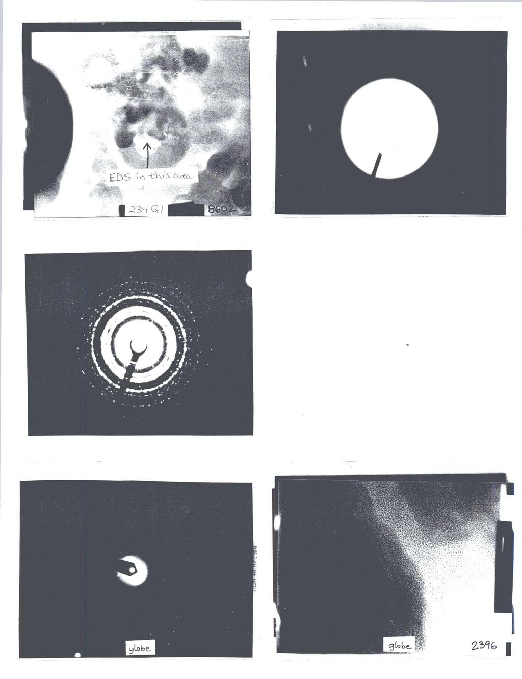
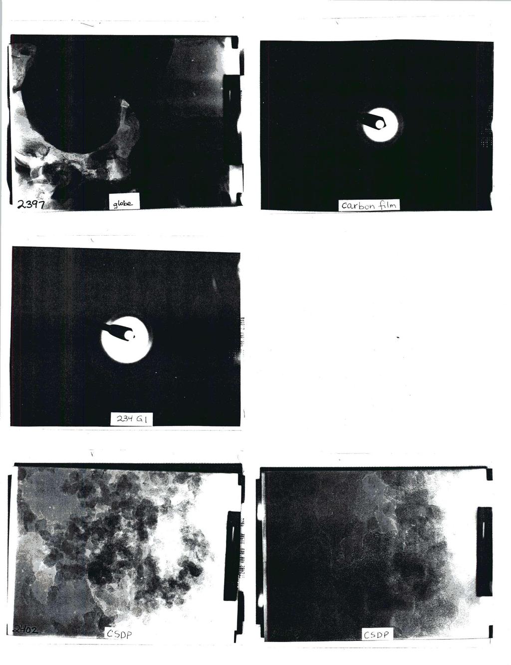
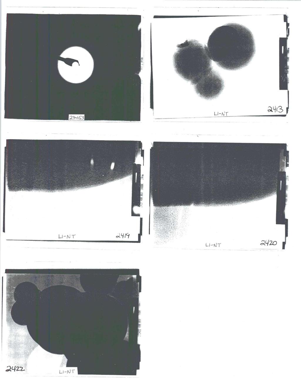
2 'GeorgiaDL^ {S(M^ / J t?tech[ji](o)d(o)@]^7 =3 School of Materials Science and Engineering February 13, 1998 Dr. Charles Herd Columbian Chemicals Company P.O. Box 96 Swartz, LA Dear Charles and Paul, Enclosed are 9 printouts from EDS analyses, and 37 negatives, with corresponding contact prints, for images obtained of your samples with the field emission and high voltage microscopes, January 20 - February 6. The bulk of the work was completed January 20-22, while you were both here. The follow-up imaging was done in the last week of January and the first week of February, after maintenance on our instruments. The 5 samples successfully imaged are 1) 234 G1, 2) globes, 3) CSDP, 4) 234 G3, and 5) LI-NT. The test material, graphitized carbon black, also produced 3 great images. The sample without usable negatives is "234 standard"; EDS analysis indicated trace amounts of oxygen and sulfur present in this sample (see analysis sheets 2 and 3, and compare to analysis sheet 1, pure carbon film). Following is a list of negative numbers with corresponding sample descriptions and experimental notes. HF2000 Field emission microscope Neq# mag description M graphitized carbon black K graphitized carbon black K graphitized carbon black 8593* 500K 234 G1 * black mark due to light exposure before developing 8594* 500K 234 G1 8595* 500K 234 G1 8596* 500K 234 G1 8597* 500K 234 G1 image slightly distorted by astigmatism 8598* 500K 234 G1 8599* 500K 234 G1 image slightly distorted by astigmatism 8600* 500K 234 G1 image slightly distorted by astigmatism 8601* 500K 234 G1 EDS spectrum taken in region indicated on contact print; - spectrum (sheet 7a) and analysis (sheet 7b) show that O, Si and S were detected School of Materials Science and Engineering, Atlanta, Georgia U.S.A., fax A Unit of the University System of Georgia An Equal Education and Employment Opportunity Institution
3 8602* 200K 234 G1 EDS spectrum taken in region indicated on contact print; spectrum (sheet 8a) and analysis (sheet 8b) show O, Si and S were detected; Si: O ratio is 1 : 2, suggesting that the compound Si0 2 is present; these findings were confirmed by analysis sheets 4 and 6b analysis sheet 5 only confirms Si; sheets 7b and 9b confirm all three elements, but not the Si: O ratio m diff pattern of EDS area (too bright, but perhaps still useful) m Au standard for camera length calculation for #8603 JEM 4000EX hiqh voltaqe microscope n*m globe at edge, 2.0 is the camera constant, LX, in units of nm*cm K globe from which diff pattern taken K globe overview n*m carbon film n*m 234 G1 particles K CSDP K CSDP n*m CSDP K 234 G3 density of negative is light; may be difficult to print K 234 G3 density of negative is light; may be difficult to print K 234 G3 density of negative is light; may be difficult to print K 234 G3 density of negative is light; may be difficult to print n*m 234 G K LI-NT particle overview, high mag images didn't turn out K LI-NT; high mag of particle in # K LI-NT; high mag of particle in # K LI-NT particle overview K 234 G K 234 G K 234 G n*m particles from previous image, using smallest aperture K 234 G3
4 If you have any questions about the data, analyses, or negatives, please contact at (404) It was a pleasure working with you both. Thank you for the graphitized carbon black sample; perhaps it will help in finding the reason for the sample contamination in our field emission instrument. Yolande Berta Research Scientist II Georgia Institute oftechnology (404) Enclosures
5 Refit _0 -K' _0 -K" _S -K' S -K" Refit _0 -K _S -K Wed Jan 21 16:28: Chi-sqd = 4.91 Livetime = 68.0 Sec, Standardless Analysis Element Net Error Counts (1-Sigma) C -K / K 0 +/- 0 Cu-K 262 +/- 22 Cu-L 45 +/- 12 S -K 0 +/- 0 Adjustment Factors K L M Z-Balance: Shell: Metallurgical and Biological Thin Section Correction Acceleration Voltage =200 kv Take-off Angle = deg Element Counts K-Rel K-Std Wt % Atom % x C x C C -K K S -K Total
6 ** Warning ** Undefined detector subtype. Using default value subtype= 2 Refit _0 -K' _0 -K" Refit _0 -K Wed Jan 21 16:20: Chi-sqd =4.70 Livetime =59.0 Sec Standardless Analysis Element Net Error Counts (1-Sigma) C -K /- 52 O -K 0 +/- 0 Cu-K 209 +/- 25 Cu-L 90 +/- 14 S -K 42 +/- re- Adjustment Factors K L M Z-Balance: Shell: Metallurgical and Biological Thin Section Correction Acceleration Voltage =200 kv Take-off Angle = deg Element Counts K-Rel K-Std Wt % Atom % X C X C C -K K S -K Total
7 Refit 0 -K' 0 -K Wed Jan 21 16:24: Chi-sqd =3.72 Livetime = Sec. Standardless Analysis Element Net Error Counts (1-Sigma) C -K /- 50 O -K 2 +/- 23 Cu-K 252 +/- 26 Cu-L 68 +/- 14 S -K 41 +/- 16 Adjustment Factors K L M Z-Balance: Shell: Metallurgical and Biological Thin Section Correction Acceleration Voltage =200 kv Take-off Angle = deg Element Counts K-Rel K-Std Wt % Atom % x C x C C -K K S -K Total
8 Refit Refit _0 -K" _S -K" _Si-K' _Si-K" Thu Jan 22 10:16: Chi-sqd = 2.82 Livetime = 71.0 Sec, Standardless Analysis Element Net Error Counts (1-Sigma) C -K 983 +/- 39 O -K 62 +/- 11 Cu-K 488 +/- 33 Cu-L 9 +/- 11 S -K 56 +/- 8, Si-K 201 +/- 14 Adjustment Factors XM L M Z-Balance: Shell: Metallurgical and Biological Thin Section Correction Acceleration Voltage =200 kv Take-off Angle = deg Element Counts K-Rel K-Std Wt % Atom % x,c x,c C -K K S -K Si-K Total
9 Refit _0 -K' _0 -K" _S -K' _S -K" _Si-K / Si-K" Refit _0 -K _S -K Wed Jan 21 17:25: Chi-sqd =5.30 Livetime = 72.0 Sec, Standardless Analysis Element Net Error Counts (1-Sigma) C -K / K 0 +/- 0 Cu-K 414 +/- 30 Cu-L 41 +/- 14 S -K 0 +/- _ J ^. Si-K 10 +/- 9 Adjustment Factors Z-Balance: Shell: T L M Metallurgical and Biological Thin Section Correction Acceleration Voltage =200 kv Take-off Angle = deg Element Counts K-Rel K-Std C -K 2348 x,c x,c 0 -K S -K Si-K Total Wt % Atom %
10 75 70A ] 55 c o u n t s ^ (M 5 OA Energy (kev) 2.5 Co limn Take-off angle Acquisition type Creation tine Livetime Deadt ime Channels Channel width Detector type Window type Window thickness Contamination material Contamination thickness File name : HITACHI,GEORGIA TECH 33 Accelerating voltage 200 eds Magnification 1 98/01/21 17:06 Charge Beam current Beam spot size Beam location 0,0 10 Working distance 13.4 Stage X 0 Stage Y Stage Z 0 none Stage tilt 0 0 Stage rotation 0 Notes: < > 3 > ^ / < S - Wed Jan 21 17:08: Livetime : 79.0 Sec. Technique: Peak Location Elements Present: C ( 6), Si(14), Cu(29) Possible Additional Elements: 0s(76) Energy Intensity Element unidentified C Ka Si Ka Cu Kal Cu Kbl or 0s Lai
11 ** Warning ** Undefined detector subtype. Using default value subtype= 2. Refit _0 -K' _0 -K" _S -K' _S -K" Refit _Si-K' _Si-K" Wed Jan 21 17:10: Chi-sqd = 4.47 Livetime = 79.0 Sec. Standardless Analysis Element Net Error Counts (1-Sigma) C -K /- 56 O -K 24 +/- 14 Cu-K 518 +/- 33 Cu-L 72 +/- 16 S -K 31 +/- 11 Si-K 89 +/- 12" Adjustment Factors K L M Z-Balance: Shell: Metallurgical and Biological Thin Section Correction Acceleration Voltage =200 kv Take-off Angle = deg Element Counts K-Rel K-Std Wt % Atom % X C X C C -K K S -K Si-K Total
12 C 140 o u n 120 t s Energy (kev) Column Take-off angle Acquisition type Creation time Livetime Deadtime Channels Channel width Detector type Window type Window thickness Contamination material Contamination thickness File name : HITACHI,GEORGIA TECH 33 eds 98/01/22 10: none 0 Accelerating voltage 200 Magni fication 1 Charge 69 Beam current 1 Beam spot size 0 Beam location 0,0 Working distance 13.4 Stage X 0 Stage Y 0 Stage Z 0 Stage tilt 0 Stage rotation 0 Notes: ^3>W O f ) C{ IA Qjc^L P6o ) Refit _0 -K' _0 -K" Refit _Si-K" Thu Jan 22 10:04: Chi-sqd Livetime Sec. Standardless Analysis Element Net Error Counts (1-Sigma) C -K / K 2 +/- 23 Cu-K 549 +/- 37 Cu-L 59 +/- 14 S -K 70 +/- 18 Si-K 45 +/- 11
13 Refit _0 -K' 0 -K" Refit _Si-K" Thu Jan 22 10: 04: Chi-sqd = 4.21 Livetime = 69.0 Sec, Standardless Analysis Element Net Error Counts (1-Sigma) C -K / K 2 +/- 23 Cu-K 549 +/- 37 Cu-L 59 +/- 14 S -K 70 +/- 18 Si-K 45 +/- 11 Adjustment Factors K L M Z-Balance: Shell: Metallurgical and Biological Thin Section Correction Acceleration Voltage =200 kv Take-off Angle = deg Element Counts K-Rel K-Std Wt % Atom % x,c x,c C -K K S -K Si-K Total
14 Energy (kev) Column Take-off angle Acquisition type Creation time Livetime Deadtime Channels Channel width Detector type Window type Window thickness Contamination material Contamination thickness File name : Notes: 3 ^ G> / ^ ^ : HITACHI,GEORGIA TECH : 33 Accelerating voltage 200 : eds Magni f ication 1 : 98/01/22 10:16 Charge 71 : 71 Beam current 1 : Beam spot size 0 : 2048 Beam location 0,0 10 Working distance 13.4 Stage X 0 Stage Y Stage Z 0 Refit / Refit _0 -K" _S -K" _Si-K' _Si-K" Thu Jan 22 10:16: none Stage tilt 0 0 Stage rotation 0 Chi-sqd Livetime Sec. Standardless Analysis Element Net Error Counts (1-Sigma) C -K 983 +/ K 62 +/- 11 Cu-K 488 +/- 33 Cu-L 9 +/- 11 S -K 56 +/- 8 Si-K 201 +/- 14
15 ** Warning ** Undefined detector subtype. Using default value subtype= 2. Ref it Refit _S -K" _Si-K' _Si-K" Thu Jan 22 10:20: Chi-sqd = =3.23 " J Livetime = 71.0 Sec, Standardless Analysis Element Net Error Counts (1-Sigma) C -K 992 +/- 38 Cu-K 488 +/- 33 Cu-L 10 +/- 11 S -K 56 +/- 8 Si-K 201 +/- 14 Adjustment Factors Z-Balance: Shell: K L M Metallurgical and Biological Thin Section Correction Acceleration Voltage =200 kv Take-off Angle = deg Element C -K S -K Si-K 0 -K Total Counts K-Rel X,C K-Std x,c Wt % Atom % Formula Compound% Stoichiometry C S Si Stoichiometry results are based upon 24 Oxygen atoms Table Symbols: S wt.% calculated by Stoichiometry
16 c Column Take-off angle Acquisition type Creation time Livetime Deadtime Channels Channel width Detector type Window type Window thickness Contamination material Contamination thickness File name : HITACHI,GEORGIA TECH 33 Accelerating voltage 200 eds Magnification 1 98/01/22 10:09 Charge Beam current Beam spot size Beam location 0,0 10 Working distance 13.4 Stage X 0 Stage Y Stage Z 0 none Stage tilt 0 0 Stage rotation 0 Notes: A^^F Q, \ IAX) Refit.0 -K' «0 0 -K" Refit.Si-K" Thu Jan 22 10:09: Chi-sqd Livetime Sec. Standardless Analysis Element Net Error Counts (1-Sigma) C -K / K 16 +/- 11 Cu-K 452 +/- 32 Cu-L 30 +/- 11 S -K 59 +/- 17 Si-K 136 +/- 12
17 Refit _0 -K' _0 -K" Refit _Si-K" Thu Jan 22 10:09: Chi-sqd = 2.69 Livetime = 66.0 Sec Standardless Analysis Element Net Error Counts (1-Sigma) C -K / K 16 +/- 11 Cu-K 452 +/- 32 Cu-L 30 +/- 11 S -K 59 +/- -IX.- Si-K 136 +/- 12 Adjustment Factors K L M Z-Balance: Shell: Metallurgical and Biological Thin Section Correction Acceleration Voltage =200 kv Take-off Angle = deg Element Counts K-Rel K-Std Wt % Atom % x,c x,c C -K K S -K Si-K Total
18
19
20
21

22
23

24
Evex NanoAnalysis EDS Acquisition
 NanoAnalysis EDS Acquisition 1. Proper Kv for the sample 2. Correct working distance for EDS 3. Good Image correct magnification & in focus 4. Make note if sample is tilted 5. Adjust beam current for proper
QUANTAX EDS SYSTEM SOP
 QUANTAX EDS SYSTEM SOP December 2017 Energy-Dispersive X-Ray Spectroscopy (EDS, EDX, EDXS or XEDS), is an analytical technique used for the elemental analysis or chemical characterization of a sample.
CHEM-E5225 :Electron Microscopy Imaging I
 CHEM-E5225 :Electron Microscopy Imaging I 2018.11 Yanling Ge Outline Amplitude Contrast Phase Contrast Images Thickness and Bending Effects Amplitude Contrast Amplitude phase TEM STEM Incoherent elastic
Maintenance Package Measurement
 Maintenance Package Measurement Contents Contents 1. Package measurement flow...1 2. Measurement procedures...3 2.1 Startup... 3 2.2 Hardware setup... 4 2.3 Setting Package measurement conditions... 7
Introduction to Biomedical Imaging
 Alejandro Frangi, PhD Computational Imaging Lab Department of Information & Communication Technology Pompeu Fabra University www.cilab.upf.edu X-ray Projection Imaging Computed Tomography Digital X-ray
Cover Page. The handle holds various files of this Leiden University dissertation
 Cover Page The handle http://hdl.handle.net/1887/48877 holds various files of this Leiden University dissertation Author: Li, Y. Title: A new method to reconstruct the structure from crystal images Issue
Enhanced material contrast by dual-energy microct imaging
 Enhanced material contrast by dual-energy microct imaging Method note Page 1 of 12 2 Method note: Dual-energy microct analysis 1. Introduction 1.1. The basis for dual energy imaging Micro-computed tomography
Spherical Crystal X-ray Imaging for MTW, OMEGA, and OMEGA EP
 Spherical Crystal X-ray Imaging for MTW, OMEGA, and OMEGA EP C.STOECKL, G. FISKEL, R. K. JUNGQUIST, P. M. NILSON, AND W. THEOBALD University of Rochester, Laboratory for Laser Energetics Spherical Crystal
Supporting information SI 0: TEM & STEM acquisition parameters, in 2D and 3D
 Electronic Supplementary Material (ESI) for Nanoscale. This journal is The Royal Society of Chemistry 2015 Supporting information SI 0: TEM & STEM acquisition parameters, in 2D and 3D Electron tomography
Probe for EPMA: Software for Electron Probe MicroAnalysis
 Probe Software www.probesoftware.com Probe for EPMA: Software for Electron Probe MicroAnalysis Navigate your sample graphically using the StageMap and PictureSnap click and go features! User definable
XRAY SHARK xpb X-ray Scanner for transformer paperboards
 XRAY SHARK xpb X-ray Scanner for transformer paperboards Introduction Generator Collimator Beam Detector XRAY SHARK - The Better Solution The XRAY SHARK is an x-ray device which is to be integrated in
Spectroscopic Ellipsometer --- J. A. Woollam alpha-se
 Spectroscopic Ellipsometer --- J. A. Woollam alpha-se Introduction Figure 1: J. A. Woollam alpha-se spectroscopic ellipsometer An ellipsometer measures the change in polarization as light reflects or transmits
diffraction patterns obtained with convergent electron beams yield more information than patterns obtained with parallel electron beams:
 CBED-Patterns Principle of CBED diffraction patterns obtained with convergent electron beams yield more information than patterns obtained with parallel electron beams: specimen thickness more precise
Probe for EPMA Purchase Justification
 Probe Software Software for MicroAnalysis Probe Software, Inc. 885 Crest Drive Eugene, OR 97405 USA (541) 343-3400 sales@probesoftware.com www.probesoftware.com Probe for EPMA Purchase Justification See
DAMAGE INSPECTION AND EVALUATION IN THE WHOLE VIEW FIELD USING LASER
 DAMAGE INSPECTION AND EVALUATION IN THE WHOLE VIEW FIELD USING LASER A. Kato and T. A. Moe Department of Mechanical Engineering Chubu University Kasugai, Aichi 487-8501, Japan ABSTRACT In this study, we
SUPPLEMENTARY INFORMATION
 SUPPLEMENTARY INFORMATION doi:10.1038/nature10934 Supplementary Methods Mathematical implementation of the EST method. The EST method begins with padding each projection with zeros (that is, embedding
Scanned by CamScanner
 Scanned by CamScanner Scanned by CamScanner Annexure I Name of the equipment: Field Emission Scanning Electron Microscope (FE-SEM) along with Energy Dispersive Spectroscope (EDS) and accessories. Technical
CT Reconstruction with Good-Orientation and Layer Separation for Multilayer Objects
 17th World Conference on Nondestructive Testing, 25-28 Oct 2008, Shanghai, China CT Reconstruction with Good-Orientation and Layer Separation for Multilayer Objects Tong LIU 1, Brian Stephan WONG 2, Tai
Application of MCNP Code in Shielding Design for Radioactive Sources
 Application of MCNP Code in Shielding Design for Radioactive Sources Ibrahim A. Alrammah Abstract This paper presents three tasks: Task 1 explores: the detected number of as a function of polythene moderator
G4beamline Simulations for H8
 G4beamline Simulations for H8 Author: Freja Thoresen EN-MEF-LE, Univ. of Copenhagen & CERN Supervisor: Nikolaos Charitonidis CERN (Dated: December 15, 2015) Electronic address: frejathoresen@gmail.com
ION BEAM MILLING SYSTEM FOR TEM, SEM AND LM PREPARATION. Leica EM RES102
 ION BEAM MILLING SYSTEM FOR TEM, SEM AND LM PREPARATION Leica EM RES102 ION BEAM MILLING In recent years, ion milling has been developed into the most applicable method of sample preparation for the analysis
Advanced materials research using the Real-Time 3D Analytical FIB-SEM 'NX9000'
 SCIENTIFIC INSTRUMENT NEWS 2017 Vol. 9 SEPTEMBER Technical magazine of Electron Microscope and Analytical Instruments. Technical Explanation Advanced materials research using the Real-Time 3D Analytical
NEW OPTICAL MEASUREMENT TECHNIQUE FOR SI WAFER SURFACE DEFECTS USING ANNULAR ILLUMINATION WITH CROSSED NICOLS
 NEW OPTICAL MEASUREMENT TECHNIQUE FOR SI WAFER SURFACE DEFECTS USING ANNULAR ILLUMINATION WITH CROSSED NICOLS Satoru Takahashi 1, Takashi Miyoshi 1, Yasuhiro Takaya 1, and Takahiro Abe 2 1 Department of
3D Energy Dispersive Spectroscopy Elemental Tomography in the Scanning Transmission Electron Microscope
 3D Energy Dispersive Spectroscopy Elemental Tomography in the Scanning Transmission Electron Microscope Brian Van Devener Topics 1.Introduction to EDS in the STEM 2.Extending EDS into three dimensions
CCD Image Acquisition Tutorial
 CCD Image Acquisition Tutorial Gatan, Inc. 5933 Coronado Lane, Pleasanton, CA 94588 Tel: (925) 463-0200 Fax: (925) 463-0204 May 2001 1. Introduction This document will guide users of Gatan CCD camera through
Shielding factors for traditional safety glasses
 Shielding factors for traditional safety glasses Malcolm McEwen, Hong Shen and Ernesto Mainegra-Hing Ionizing Radiation Standards, National Research Council Canada Alan DuSautoy, Radiation and Health Sciences
The Electrochemical Innovation Lab X-ray Suite: from macro- to nano-ct
 The Electrochemical Innovation Lab X-ray Suite: from macro- to nano-ct Dr. Francesco Iacoviello, Toby Neville & the Electrochemical Innovation Lab f.iacoviello@ucl.ac.uk MANIFEST - 2017 November 10 th,
Structural Information obtained
 Structural Information obtained from Electron Microscopy Christiane Schaffitzel, 09.05.2013 Including slides from John Briggs, Bettina Boettcher, Nicolas Boisset, Andy Hoenger, Michael Schatz, and more
Physics 123 Optics Review
 Physics 123 Optics Review I. Definitions & Facts concave converging convex diverging real image virtual image real object virtual object upright inverted dispersion nearsighted, farsighted near point,
Probe for EPMA Quick Start Instructions
 Probe for EPMA Quick Start Instructions Setting up a New Run for Quantitative Acquisition This guide is intended as a quick start and just covers the basic steps for creating a new Probe for EPMA run.
XRADIA microxct Manual
 XRADIA microxct Manual Multiscale CT Lab Table of Contents 1. Introduction and Basics 1.1 Instrument Parts 1.2 Powering up the system 1.3 Preparing your sample 2. TXM Controller 2.1 Starting up 2.2 Finding
Raith e_line Electron Beam Lithography
 Raith e_line Electron Beam Lithography Standard Operating Procedure 1 (For an un-patterned sample) Revision: 7.0 Last Updated: Feb.18/2015, Revised by Mohamad Rezaei Overview This document will provide
INCAGSR. Gun Shot Residue
 INCAGSR GSR Gun Shot Residue INCAGSR G A dedicated solution for automated detection and analysis of Gun Shot Residue using the scanning electron microscope This product has been designed in conjunction
JEOL CarryScope SEM Revision /07/17 Page 1 of 7. JEOL CarryScope SEM
 Page 1 of 7 JEOL CarryScope SEM The JEOL CarryScope is a compact and portable SEM that utilizes a standard tungsten filament. It is to be used for inspecting and measuring samples processed in the NRF
PARTICLE IMAGE VELOCIMETRY (PIV) AND VOLUMETRIC VELOCIMETRY (V3V) SYSTEMS
 AND VOLUMETRIC VELOCIMETRY (V3V) SYSTEMS") PARTICLE IMAGE VELOCIMETRY (PIV) AND VOLUMETRIC VELOCIMETRY (V3V) SYSTEMS VERSATILE, UPGRADEABLE FLUID MECHANICS MEASUREMENT SOLUTIONS UNDERSTANDING, ACCELERATED FULL SPECTRUM OF GLOBAL VELOCITY SYSTEMS
PARTICLE IMAGE VELOCIMETRY (PIV) AND VOLUMETRIC VELOCIMETRY (V3V) SYSTEMS VERSATILE, UPGRADEABLE FLUID MECHANICS MEASUREMENT SOLUTIONS UNDERSTANDING, ACCELERATED FULL SPECTRUM OF GLOBAL VELOCITY SYSTEMS
The same X-ray transparent in situ pouch cell design and sample holder plates were used for both 2D
 Electronic Supplementary Material (ESI) for Energy & Environmental Science. This journal is The Royal Society of Chemistry 2014 Supplementary Information Experimental Materials and Electrochemistry The
Crystal Quality Analysis Group
 Crystal Quality Analysis Group Contents Contents 1. Overview...1 2. Measurement principles...3 2.1 Considerations related to orientation and diffraction conditions... 3 2.2 Rocking curve measurement...
Optics Final Exam Name
 Instructions: Place your name on all of the pages. Do all of your work in this booklet. Do not tear off any sheets. Show all of your steps in the problems for full credit. Be clear and neat in your work.
SUPPLEMENTARY INFORMATION
 doi:10.1038/nature12009 Supplementary Figure 1. Experimental tilt series of 104 projections with a tilt range of ±72.6 and equal slope increments, acquired from a Pt nanoparticle using HAADF- STEM (energy:
NSS Spectral Analysis System Handbook Version 3.0
 NSS Spectral Analysis System Handbook Version 3.0 The information in this publication is provided for reference only. All information contained in this publication is believed to be correct and complete.
räíê~_êáöüí Microfocus X-Ray Source Technical Data Sheet X-Ray Tube Unit Specifications
 Technical Data Sheet räíê~_êáöüí 4 mm Anode to Object distance True round spot Grounded target = High power, 80W Integrated design No HV cable The choice for use with an x-ray optic due to close coupling
LABORATORY SYSTEM FOR X-RAY NANOTOMOGRAPHY
 79 LABORATORY SYSTEM FOR X-RAY NANOTOMOGRAPHY Alexander Sasov, SkyScan, Vluchtenburgstraat 3, Aartselaar B2630, Belgium, www.skyscan.be. ABSTRACT Using advanced X-ray technologies and X-ray scattering
ANOMALOUS SCATTERING FROM SINGLE CRYSTAL SUBSTRATE
 177 ANOMALOUS SCATTERING FROM SINGLE CRYSTAL SUBSTRATE L. K. Bekessy, N. A. Raftery, and S. Russell Faculty of Science, Queensland University of Technology, GPO Box 2434, Brisbane, Queensland, Australia
Product Description. Leica RC30 Aerial Camera System. Overview
 Leica RC30 Aerial Camera System Overview The Leica RC30 is an aerial film camera system of top quality and performance, capable of acquiring the very best aerial photographs with black and white, color
Manual Infrared viewers ABRIS M series M1300 M1700 M2000
 Manual Infrared viewers ABRIS M series M1300 M1700 M2000 Content Content... 3 Safety requirements... 4 About... 4 Applications... 5 How does it work?... 5 Operation... 6 IR viewer in comparison with a
GEORGIA INSTITUTE OF TECHNOLOGY PROJECT ADMINISTRATION DATA SHEET. To 8'? This all gl
 GEORGIA INSTITUTE OF TECHNOLOGY PROJECT ADMINISTRATION DATA SHEET OFFICE OF CONTRACT ADMINISTRATION ORIGINAL REVISION NO. Project No. E-21-A1 (R659-A2) GTRC/I/DER DATE 1 /24 /85 Project Director: Gail
Scattering/Wave Terminology A few terms show up throughout the discussion of electron microscopy:
 1. Scattering and Diffraction Scattering/Wave Terology A few terms show up throughout the discussion of electron microscopy: First, what do we mean by the terms elastic and inelastic? These are both related
Rietveld refinements collection strategies!
 Rietveld refinements collection strategies! Luca Lutterotti! Department of Materials Engineering and Industrial Technologies! University of Trento - Italy! Quality of the experiment! A good refinement,
All forms of EM waves travel at the speed of light in a vacuum = 3.00 x 10 8 m/s This speed is constant in air as well
 Pre AP Physics Light & Optics Chapters 14-16 Light is an electromagnetic wave Electromagnetic waves: Oscillating electric and magnetic fields that are perpendicular to the direction the wave moves Difference
Development of Ultrafast CXRS system in Heliotron J. Graduate School of Energy Science Kyoto University LU XIANGXUN 03/15/2016
 1 Development of Ultrafast CXRS system in Heliotron J Graduate School of Energy Science Kyoto University LU XIANGXUN 03/15/2016 2 Outline 1. Introduction 2. Charge exchange Recombination Spectroscopy (CXRS)
Diffraction I - Geometry. Chapter 3
 Diffraction I - Geometry Chapter 3 Outline ❽ Diffraction basics ❽ Braggs law ❽ Laue equations ❽ Reciprocal space and diffraction ❽ Units for x-ray wavelengths ❽ Diffraction methods Laue photographs Rotation
NIRSpec Technical Note NTN / ESA-JWST-TN Author(s): G. Giardino Date of Issue: November 11, 2013 Version: 1.0
: G. Giardino Date of Issue: November 11, 2013 Version: 1.0") NIRSpec Technical Note NTN-013-011/ ESA-JWST-TN-093 Author(s): G. Giardino Date of Issue: November 11, 013 Version: 1.0 estec European Space Research and Technology Centre Keplerlaan 1 01 AZ Noordwijk
NIRSpec Technical Note NTN-013-011/ ESA-JWST-TN-093 Author(s): G. Giardino Date of Issue: November 11, 013 Version: 1.0 estec European Space Research and Technology Centre Keplerlaan 1 01 AZ Noordwijk
Solved with COMSOL Multiphysics 4.3a
 Magnetic Lens Introduction Scanning electron microscopes image samples by scanning with a high-energy beam of electrons. The subsequent electron interactions produce signals such as secondary and back-scattered
OPSE FINAL EXAM Fall CLOSED BOOK. Two pages (front/back of both pages) of equations are allowed.
 of equations are allowed.") CLOSED BOOK. Two pages (front/back of both pages) of equations are allowed. YOU MUST SHOW YOUR WORK. ANSWERS THAT ARE NOT JUSTIFIED WILL BE GIVEN ZERO CREDIT. ALL NUMERICAL ANSERS MUST HAVE UNITS INDICATED.
CLOSED BOOK. Two pages (front/back of both pages) of equations are allowed. YOU MUST SHOW YOUR WORK. ANSWERS THAT ARE NOT JUSTIFIED WILL BE GIVEN ZERO CREDIT. ALL NUMERICAL ANSERS MUST HAVE UNITS INDICATED.
The User may not distribute, share, and otherwise convey the copyrighted documents to any other persons, corporations
 LICENSE AGREEMENT Static Control Components, Inc. (Static Control) grants this limited license to the person, firm or corporation (hereinafter "User) downloading electronically or by printing this file
Operating Procedure for Horiba Raman Microscope
 Operating Procedure for Horiba Raman Microscope SAFETY Be aware of Laser radiation at all times! Do not remove the covers of the instrument. Components are supplied with 110V electric source. Do not touch
SEM topography. Live quantitative surface topography in SEM
 SEM topography Live quantitative surface topography in SEM 2 Measure surface topography with SEM. n Use conventional segmented BSE signals. n Get immediate feedback with automated topographic reconstruction.
EM Sample Preparation Coating Technology. April 2013
 EM Sample Preparation Coating Technology April 2013 Mosquito antenna Courtesy of: Dr. Daniela Gruber, Vienna, Austria EM SAMPLE PREPARATION COATING TECHNOLOGY 4 Coating Technology Coating of samples is
JSM-7900F. Scientific / Metrology Instruments. Schottky Field Emission Scanning Electron Microscope. Ultimate Analytical tool
 Scientific / Metrology Instruments Schottky Field Emission Scanning Electron Microscope Ultimate Analytical tool JSM-7900F High-Performance FE-SEM successfully combining ultrahigh resolution and unprecedented
Study of Mode Characteristics of Glass Optical Fibers LABORATORY MANUAL: TESTER MCS-04
 Study of Mode Characteristics of Glass Optical Fibers LABORATORY MANUAL: TESTER MCS-04 1. Definition and Computation of Numerical Aperture 2. Definition and Computation of V Number & Computation 3. Specifications
Introduction of Hitachi SU1510
 Introduction of Hitachi SU1510 1 1. Main Features of the SU1510 SU1510 Same high-performance optics as S-3400N Variable Pressure mode as standard Large samples up to 153mm in diameter (observation range
PRE: See Yun Jaan 15 Jul 08 Rev C. APP: Leong See Fan DCR No. D _438039
 Application 114-1114 Specification PRE: See Yun Jaan 15 Jul 08 Rev C APP: Leong See Fan DCR No. D20080715001838_438039 SO(Small Outline) DIMM (Dual-In Memory Module) Sockets, 144 Positions with 0.8mm Centerline
HKL Flamenco EBSD Data Acquisition Flow
 HKL Flamenco EBSD Data Acquisition Flow Basic steps for automatic data acquisition (e.g. orientation mapping) This is basic routine for the new operator. Feel free to experiment with different settings
LEXT 3D Measuring LASER Microscope
 LEXT 3D Measuring LASER Microscope Warning: This instrument may only be operated by those who have been trained by AAF staff and have read and signed the AAF laboratory policies. A) STARTUP 1. Computer
H.-J. Jordan (NanoFocus Messtechnik GmbH), R. Brodmann (Brodmann Marketing & Vertrieb)
, R. Brodmann (Brodmann Marketing & Vertrieb)") Highly accurate surface measurement by means of white light confocal microscopy Hochgenaue Oberflächenmessung mit Hilfe von konfokalen Weißlichttechniken H.-J. Jordan (NanoFocus Messtechnik GmbH), R. Brodmann
Highly accurate surface measurement by means of white light confocal microscopy Hochgenaue Oberflächenmessung mit Hilfe von konfokalen Weißlichttechniken H.-J. Jordan (NanoFocus Messtechnik GmbH), R. Brodmann
SPECTRAL IMAGING VIEWER SOFTWARE MANUAL
 103 Quality Circle, Suite 215 Huntsville, AL 35806 Phone: (256) 704-3332 Fax: (256) 971-2073 E-Mail: info@axometrics.com Website: http://www.axometrics.com SPECTRAL IMAGING VIEWER SOFTWARE MANUAL 2012
HIGH-SPEED THEE-DIMENSIONAL TOMOGRAPHIC IMAGING OF FRAGMENTS AND PRECISE STATISTICS FROM AN AUTOMATED ANALYSIS
 23 RD INTERNATIONAL SYMPOSIUM ON BALLISTICS TARRAGONA, SPAIN 16-20 APRIL 2007 HIGH-SPEED THEE-DIMENSIONAL TOMOGRAPHIC IMAGING OF FRAGMENTS AND PRECISE STATISTICS FROM AN AUTOMATED ANALYSIS P. Helberg 1,
Chapter 7: Geometrical Optics
 Chapter 7: Geometrical Optics 7. Reflection at a Spherical Surface L.O 7.. State laws of reflection Laws of reflection state: L.O The incident ray, the reflected ray and the normal all lie in the same
TEAMS National Competition High School Version Photometry 25 Questions
 TEAMS National Competition High School Version Photometry 25 Questions Page 1 of 14 Telescopes and their Lenses Although telescopes provide us with the extraordinary power to see objects miles away, the
1
 In the following tutorial we will determine by fitting the standard instrumental broadening supposing that the LaB 6 NIST powder sample broadening is negligible. This can be achieved in the MAUD program
Beam Analysis Camera Based Beam Propagation Analyzer: M Automatic M 2 - at Production Speeds. Manual M 2
 3.7.1 Camera Based Beam Propagation Analyzer: M 2 ISO compliant Automatically measure your beam quality in under 1 minutes Tune your laser for best operation Specifically developed for continuous usage
Hidenobu Tachibana The Cancer Institute Hospital of JFCR, Radiology Dept. The Cancer Institute of JFCR, Physics Dept.
 2-D D Dose-CT Mapping in Geant4 Hidenobu Tachibana The Cancer Institute Hospital of JFCR, Radiology Dept. The Cancer Institute of JFCR, Physics Dept. Table of Contents Background & Purpose Materials Methods
Single-particle electron microscopy (cryo-electron microscopy) CS/CME/BioE/Biophys/BMI 279 Nov. 16 and 28, 2017 Ron Dror
 CS/CME/BioE/Biophys/BMI 279 Nov. 16 and 28, 2017 Ron Dror") Single-particle electron microscopy (cryo-electron microscopy) CS/CME/BioE/Biophys/BMI 279 Nov. 16 and 28, 2017 Ron Dror 1 Last month s Nobel Prize in Chemistry Awarded to Jacques Dubochet, Joachim Frank
Single-particle electron microscopy (cryo-electron microscopy) CS/CME/BioE/Biophys/BMI 279 Nov. 16 and 28, 2017 Ron Dror 1 Last month s Nobel Prize in Chemistry Awarded to Jacques Dubochet, Joachim Frank
Intermediate Physics PHYS102
 Intermediate Physics PHYS102 Dr Richard H. Cyburt Assistant Professor of Physics My office: 402c in the Science Building My phone: (304) 384-6006 My email: rcyburt@concord.edu My webpage: www.concord.edu/rcyburt
Confocal Raman Systems SPECTROSCOPY GROUP
 Confocal Raman Systems SPECTROSCOPY GROUP MonoVista CRS Configuration Option PIXIS TE-Cooled CCD Camera Acton Spectrograph Micro-Raman Interface Witness Camera Optional Laser Micro/Macro Transfer Optics
JSM-IT100 Series. InTouchScope. Scientific/Metrology Instruments. Scanning Electron Microscope
 Scientific/Metrology Instruments Scanning Electron Microscope InTouchScope JSM-IT100 Series The JSM-IT100 represents 50 years of SEM expertise and innovation at JEOL. This scaleable, compact, easy-to-use
NORAN System 7. Spectral Imaging X-ray Microanalysis System. Spectral Analysis. Point & Shoot. Spectral Imaging
 m i c r o a n a l y s i s NORAN System 7 Spectral Imaging X-ray Microanalysis System Spectral Analysis Point & Shoot Spectral Imaging COMPASS: Component Analysis of Spectral Images Part of Thermo Fisher
MODEL: MD7TC. Lightning Surge Protectors for Electronics Equipment M-RESTER
 MODEL: MDTC Lightning Surge Protectors for Electronics Equipment M-RESTER LIGHTNING SURGE PROTECTOR FOR THERMOCOUPLE USE (ultra-slim) Functions & Features High discharge current capacity 0 ka ( / 0 μs),
Guide to the Tantalus Synchrotron Radiation Source Collection
 Guide to the Tantalus Synchrotron Radiation Source Collection Alison Oswald. Archives Center, National Museum of American History P.O. Box 37012 Suite 1100, MRC 601 Washington, D.C. 20013-7012 archivescenter@si.edu
High spatial resolution measurement of volume holographic gratings
 High spatial resolution measurement of volume holographic gratings Gregory J. Steckman, Frank Havermeyer Ondax, Inc., 8 E. Duarte Rd., Monrovia, CA, USA 9116 ABSTRACT The conventional approach for measuring
Checklist for your microscope - your requirements
 Checklist for your microscope - your requirements 1) Which kind of microscope do you need? Compound microscope: (primarily used for transparent/translucent preparation) (Page: 1-3) Stereo microscope (surface
MICHELSON S INTERFEROMETER
 MICHELSON S INTERFEROMETER Objectives: 1. Alignment of Michelson s Interferometer using He-Ne laser to observe concentric circular fringes 2. Measurement of the wavelength of He-Ne Laser and Na lamp using
Chapter 24 - The Wave Nature of Light
 Chapter 24 - The Wave Nature of Light Summary Four Consequences of the Wave nature of Light: Diffraction Dispersion Interference Polarization Huygens principle: every point on a wavefront is a source of
General Physics Experiment 11
 Physics Labs General Physics Experiment 11 Interference and Diffraction of Light Objectives: To measure the wavelength of light emitted by a Helium-Neon laser. To observe the character of single slit diffraction.
UNIVERSITY OF PLYMOUTH
 UNIVERSITY OF PLYMOUTH Olympus LEXT offers a new view for engineering students Olympus LEXT: The Educator Traditionally, undergraduate courses give students an excellent grounding in the basics of a broad
Revision History. Applicable Documents
 Revision History Version Date Revision History Remarks 1.0 2011.11-1.1 2013.1 Update of the processing algorithm of CAI Level 3 NDVI, which yields the NDVI product Ver. 01.00. The major updates of this
Fischerscope X Ray CIRCULARS 07/01
 Fischerscope X Ray CIRCULARS 07/01 New instrument Fischerscope X-Ray XDVM-P As we informed in December, both the instruments Fischerscope X-RAY XDVM-T7-W 602-636 (measuring head 602-557) and Fischerscope
Primary Use. Operating Principle
 Primary Use The Leica DVM6 is an optical microscope that has the ability observe samples at a high magnification at a high resolution. The microscope allows users to view their sample with up to a 2350x
3DEM > GENERAL METHODS. SINGLE PARTICLE RECONSTRUCTION (SPA): Introduction
: Introduction") 3DEM > GENERAL METHODS SINGLE PARTICLE RECONSTRUCTION (SPA): Introduction SINGLE PARTICLE RECONSTRUCTION > INTRODUCTION General principle of single particle reconstruction o 2D projection images of a 3D
3DEM > GENERAL METHODS SINGLE PARTICLE RECONSTRUCTION (SPA): Introduction SINGLE PARTICLE RECONSTRUCTION > INTRODUCTION General principle of single particle reconstruction o 2D projection images of a 3D
3-D. Here red spheres show the location of gold nanoparticles inside/around a cell nucleus.
 3-D The CytoViva 3-D System allows the user can locate objects of interest in a 3-D space. It does this by acquiring multiple Z planes and performing our custom software routines to locate and observe
Name: Jonathan Smartt Title: Thin Lenses Investigation Date of Lesson: Week 2, Day 2 Technology Lesson: Yes Length: 75 minutes Course: Physics Grade
 Name: Jonathan Smartt Title: Thin Lenses Investigation Date of Lesson: Week 2, Day 2 Technology Lesson: Yes Length: 75 minutes Course: Physics Grade Level: 11 th or 12 th Source: Some information taken
Experiences from CRYRING diagnostics
 Workshop on Low Current, Low Energy Beam Diagnostics November 23-25 th 2009, Groβsachsen, Germany Experiences from CRYRING diagnostics A. Källberg Manne Siegbahn Laboratory Stockholm University Frescativägen
FEI Helios NanoLab 600 TEM specimen prep recipe Nicholas G. Rudawski (352) (office) (805) (cell) Last updated: 07/16/18
 (office) (805) (cell) Last updated: 07/16/18") FEI Helios NanoLab 600 TEM specimen prep recipe Nicholas G. Rudawski ngr@ufl.edu (352) 392 3077 (office) (805) 252-4916 (cell) Last updated: 07/16/18 This recipe is essentially a composite of several established
FEI Helios NanoLab 600 TEM specimen prep recipe Nicholas G. Rudawski ngr@ufl.edu (352) 392 3077 (office) (805) 252-4916 (cell) Last updated: 07/16/18 This recipe is essentially a composite of several established
Supporting Information. High-Throughput, Algorithmic Determination of Nanoparticle Structure From Electron Microscopy Images
 Supporting Information High-Throughput, Algorithmic Determination of Nanoparticle Structure From Electron Microscopy Images Christine R. Laramy, 1, Keith A. Brown, 2, Matthew N. O Brien, 2 and Chad. A.
PHYS 219 General Physics: Electricity, Light and Modern Physics
 PHYS 219 General Physics: Electricity, Light and Modern Physics Exam 2 is scheduled on Tuesday, March 26 @ 8 10 PM In Physics 114 It will cover four Chapters 21, 22, 23, and 24. Start reviewing lecture
Cornell Spectrum Imager (CSI) Open Source Spectrum Analysis with ImageJ Tutorial
 Open Source Spectrum Analysis with ImageJ Tutorial") Cornell Spectrum Imager (CSI) Open Source Spectrum Analysis with ImageJ Tutorial Electron Microscopy Summer School 2017 Why CSI Current Software Black box Expensive Steep learning curve Cornell Spectrum
Cornell Spectrum Imager (CSI) Open Source Spectrum Analysis with ImageJ Tutorial Electron Microscopy Summer School 2017 Why CSI Current Software Black box Expensive Steep learning curve Cornell Spectrum
PROVIDE A UNIFORM DOSE IN THE SMALL ANIMAL.
 Considerations in the Use of the RS 2000 X ray Irradiator for Biological Research (Primarily Small Animal, tissue, and cells) and the fallacy of the High KV spectrum. The performance goal for a small animal
STEM electron tomography in the Scanning Electron Microscope
 Journal of Physics: Conference Series PAPER OPEN ACCESS STEM electron tomography in the Scanning Electron Microscope To cite this article: M Ferroni et al 2015 J. Phys.: Conf. Ser. 644 012012 Recent citations
Chapter 24. Wave Optics
 Chapter 24 Wave Optics Wave Optics The wave nature of light is needed to explain various phenomena Interference Diffraction Polarization The particle nature of light was the basis for ray (geometric) optics
Lecture 4 Recap of PHYS110-1 lecture Physical Optics - 4 lectures EM spectrum and colour Light sources Interference and diffraction Polarization
 Lecture 4 Recap of PHYS110-1 lecture Physical Optics - 4 lectures EM spectrum and colour Light sources Interference and diffraction Polarization Lens Aberrations - 3 lectures Spherical aberrations Coma,