Automatized & Interactive. Muscle tissues characterization using. Na MRI
|
|
|
- Emery Franklin
- 6 years ago
- Views:
Transcription


1 Automatized & Interactive Human Skeletal Muscle Segmentation Muscle tissues characterization using 23 Na MRI Noura Azzabou 30 April 2013
2 What is muscle segmentation? Axial slice of the thigh of a healthy volunteer (Dixon image) A non segmented view A manually segmented view
3 Purposes of muscle segmentation: To study each muscle selectively, for a better characterization of neuromuscular disorder. To access volumetric muscle changes.

4 From manual to automated segmentation Today: segmentation of all muscles of a humanthighis: Manual Extremely long (4 hours) Tedious WhyisautomaticSegmentation challenging? Multi-object, partial contours, no distinctive texture Large inter-subject variations Variable (Inter-operator volume variability: 3 %) Example of missing contours
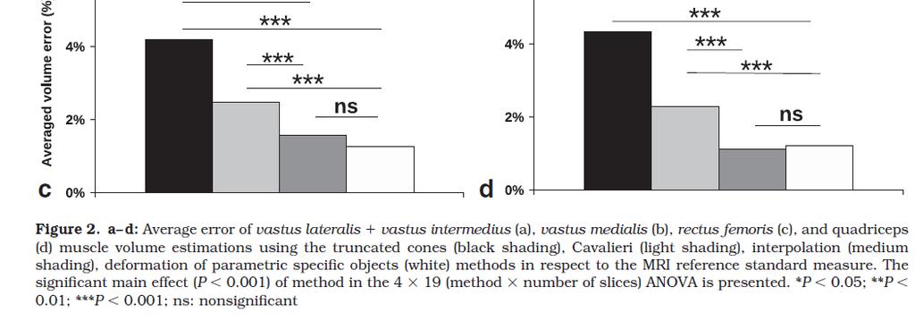
5 For comparison and volume assessement: The vastus lateralis (VL), vastus intermedius (VI), vastus medialis (VM), and rectus femoris (RF) muscles of the 10 subjects were manually outlined for all of the slices by the same investigator Because substantial fusion may be found between VL and VI on some slices (3), these two muscles were outlined Together.

6
7
8
9 The techniques described in this work are dedicated to muscle volume assessement and does not offer an accurate segmentation technique. Accurate volume estimation requires the manual segmentation of an important number of slices Need of accurate segmentation with little intreaction
10 Automatic muscle group segmentation through the registration of a model on the image and using as internal forces an average of rigid local transforms.
11 Gilles et al (MICCAI 2008) Model construction 1- manuallysegment a given reference image. Model building Reference

12 Gilles et al (MICCAI 2008) Model construction Intensity pro ofile computation 2- For eachvertex of the mesh, Learn intensityprofile in the normal direction
13 Gilles et al (MICCAI 2008) Shape registration : 1-First rough registration basedon bone position Initialis sation Step Source:
14 Gilles et al (MICCAI 2008) Shape registration : 1-First rough registration basedon bone position Initialis sation Step Source:
15 Gilles et al (MICCAI 2008) Shape registration : 2-For each vertex of the mesh search for : The voxelthat have the closest intensity profile to the reference one. The voxelsthat have a maximum gradient The target point is the mean of these two point.

16 Gilles et al (MICCAI 2008) Shape registration : Define external forces as the forces that will deform the mesh toward the target points defined previously!!! Important deformation may lead to a non smooth contours and to a solution that not correspond to the expected form of a muscle
17 Gilles et al (MICCAI 2008) Shape registration : internal forces Aims to constrain the deformations of the shape due to external forces and to keep it similar to the reference one. Performs a mapping of the model vertex to the target pixels based on local rigid transforms. To insure smoothness of deformation, the neighboring vertexes in the reference model are clustered and each cluster is deformed using a rigid transform to much the target points.

")
18 Gilles et al (MICCAI 2010)

19 Gilles et al (MICCAI 2008)
20 Gilles et al (MICCAI 2008) Segmentation result ++ amongthe first solutions for muscle segmentation tation Result ++ manymuscles are well delineated Segment -- inspiteof a good contrast some segmentation errorare observedand theyare due to the model Source:
21 1- Shape Model is discrete and based on landmarks. 2-Segmentation is equivalent to a registration of set of landmark on the image to segment. 3- Only one muscle is segmented.
22 Model learning : shape Wang et al (MICCAI 2010) 1-Register all the training image together 2- Find a consistent set of landmarks, between all the training image
23 Model learning : shape Wang et al (MICCAI 2010) 1-Register all the training image together 2- Find a consistent set of landmarks, between all the training image How to describe the model : 1- Consider the length of couple of landmarks (pb de scale invariance)
 2- Consider the normalized length of all triplets of landmark.")
24 Model learning : shape Wang et al (MICCAI 2010) 1-Register all the training image together 2- Find a consistent set of landmarks, between all the training image How to describe the model : 1- Consider the length of couple of landmarks (pb de scale invariance) 2- Consider the normalized length of all triplets of landmark. Over the training set we can probability distribution function of these distances.
25 Model learning : appearance Wang et al (MICCAI 2010) 1-In addition to geometrical properties, local appearance properties of each landmark are learnt.

26 Wang et al (MICCAI 2010) Segmentation a correspondence problem that requires detection for the model points in the image or finding a set of correspondences for each landmarks. 1-For a new image to segment and for each landmark, find the corresponding one using block matching technique. If the best much is selected, we can find many false positive and the shape model is not respected. consider many candidates that have similar appearance and then select the ones that they are close to the shape model......
27 L1 L3 L3 that minimize Use discrete optimization technique applied to high order Markov field [1]. [1] N. Komodakis, G. Tziritas, and N. Paragios. Performance vs computational efficiency for optimizing single and dynamic mrfs: Setting the state of the art with primal dual strategies. Comput. Vis. Image Und. (CVIU), 112(1):14 29, 2008
28 Wang et al (MICCAI 2010) Conclusions : No segmentation results are provided, the evaluation was expressed in terms of how resulting landmarks are close to the manually placed landmark. Only one muscle is segmented but this technqiue could be easily extended to deal with several muscle segmentations. It is hard to imagine that a software can find in a consistent manner landmarks in different volumes, because muscle surface is very homogeneous and smooth.
29 1-Construct a Model using PCA 2-Align the model to the image using two information 2.1 image contours obtained using an edge detector 2.2 intensity model to detect, background, subcuteneous fat and muscles.

30 Model definition : A probabilistic map that assigns for each pixel the probability of belonging to one specific muscles.

31 Andrews et al (MICCAI 2011) Model construction : 1- realign the different training segmentation using the anatomical images. 2- Apply the ILR transform which is a projection of the probability vector in a multi-dimensional with real value and lower dimension.
32 Andrews et al (MICCAI 2011) Model construction : 1- realign the different training segmentation using the anatomical images. 2- Apply the ILR transform which is a projection of the probability vector in a multi-dimensional with real value and lower dimension. 3-performing a PCA on the training sample, the shape is parameterized as : 4-Shape energy term enforces a Mahalanobis type penalty to allow components corresponding to eigenmodes of greater variance to vary more


33 Registration of the model to the image 1-extract image contours Andrews et al (MICCAI 2011)

34 Andrews et al (MICCAI 2011) Registration of the model to the image
 Registration of")

35 Andrews et al (MICCAI 2011) Registration of the model to the image Results & Validation
36 Results & Validation Andrews et al (MICCAI 2011)
37 Segmentation with random walks (Grady, 2006 ) Let s consider a walker that is moving randomly on the ground, to progress from one position, he had many possibilities. The probability of moving to a specefic position is inversily proportionnel to the effort spent to make the step. The walker will prefer easy path (flat) then more difficult ones (bumpy ground)
38 Segmentation with random walks (Grady, 2006 ) Assume that there are some landmarks on the ground. Q: What is the probability that the random walks arrives first to the green/red/ purple landmark?


39 Segmentation with random walks (Grady, 2006 ) A:Therandomwalkalgorithms
40 Segmentation with random walks (Grady, 2006 ) Non segmented view with drawn seeds automatic segmentation


41 Segmentation with random walks (Grady, 2006 ) Graph based formulation
 Transition probabilities")
42 Segmentation with random walks (Grady, 2006 ) Transition probabilities definition
")
43 Labelling Segmentation with random walks (Grady, 2006 )
44 Segmentation with random walks (Grady, 2006 ) Objective function :
45 Segmentation with random walks (Grady, 2006 ) Objective function
46 Segmentation with random walks (Grady, 2006 ) Objective function


47 Segmentation with random walks (Grady, 2006 ) Results : it naturally respects weaks boundary
48 Segmentation with random walks (Grady, 2006 ) Application to muscle segmentation non segmented view with drawn seeds automatic segmentation
49 Segmentation with random walks (Grady, 2006 ) Application to muscle segmentation a non segmented view manual segmentation
50 Segmentation with random walks (Grady, 2006 ) Application to muscle segmentation User interaction: drawing seeds 20 min instead of 4 hours Random Walk computation (< 5 min) Accuracy of the Random Walk segmentation: 90%


51 RW & Prior Shape Model (Baudin et al MICCAI 2012) Objective function : Mean shape The random walker is guided by the muscle shape model instead of seeds.
 Objective function")
52 RW & Prior Shape Model (Baudin et al MICCAI 2012) Objective function :

53 RW & Prior Shape Model (Baudin et al MICCAI 2012) Weight s of the prior knowledge : constant weight
 Weight s of the prior knowledge :")
54 RW & Prior Shape Model (Baudin et al MICCAI 2012) Weight s of the prior knowledge : Entropy
 Weight s of the prior knowledge : Gaussian")
55 RW & Prior Shape Model (Baudin et al, MICCAI 2012 ) Weight s of the prior knowledge : Gaussian weights
 Weight s of the prior knowledge : Confidence")
56 RW & Prior Shape Model (Baudin et al, MICCAI 2012 ) Weight s of the prior knowledge : Confidence map
57 RW & Prior Shape Model (Baudin et al, MICCAI 2012 ) Experimental results Comparing performances for various weighting schemes. the boxplots are generated from Dice coefficients for all labels (4 muscle database).
58 RW & Prior Shape Model (Baudin et al, MICCAI 2012 ) Experimental results Comparison of our method (bottom box) with the registration method (top box) and the method from (Gilles and Pai, 2008) (middle box). Dataset : subset of the 4 muscle dataset with only 15 test volumes.
 Experimental")
59 RW & Prior Shape Model (Baudin et al, MICCAI 2012 ) Experimental results


60 RW & Prior Shape Model (Baudin et al, MICCAI 2012 ) Experimental results
 Mean shape and principal")

61 RW & Prior Shape Model (Baudin et al, BMVC 2012 ) Mean shape and principal variation mode The random walker is guided by the muscle shape model instead of seeds.
 Model construction using")
62 RW & Prior Shape Model (Baudin et al, BMVC 2012 ) Model construction using PCA
63 RW & Prior Shape Model (Baudin et al, BMVC 2012 ) Model construction using PCA
 Objective")
64 RW & Prior Shape Model (Baudin et al, BMVC 2012 ) Objective function
65 RW & Prior Shape Model (Baudin et al, BMVC 2012 ) Experimental results


66 RW & Prior Shape Model (Baudin et al, BMVC 2012 )
Image Segmentation and Registration
 Image Segmentation and Registration Dr. Christine Tanner (tanner@vision.ee.ethz.ch) Computer Vision Laboratory, ETH Zürich Dr. Verena Kaynig, Machine Learning Laboratory, ETH Zürich Outline Segmentation
Modern Medical Image Analysis 8DC00 Exam
 Parts of answers are inside square brackets [... ]. These parts are optional. Answers can be written in Dutch or in English, as you prefer. You can use drawings and diagrams to support your textual answers.
COSC160: Detection and Classification. Jeremy Bolton, PhD Assistant Teaching Professor
 COSC160: Detection and Classification Jeremy Bolton, PhD Assistant Teaching Professor Outline I. Problem I. Strategies II. Features for training III. Using spatial information? IV. Reducing dimensionality
Inter and Intra-Modal Deformable Registration:
 Inter and Intra-Modal Deformable Registration: Continuous Deformations Meet Efficient Optimal Linear Programming Ben Glocker 1,2, Nikos Komodakis 1,3, Nikos Paragios 1, Georgios Tziritas 3, Nassir Navab
Interactive Differential Segmentation of the Prostate using Graph-Cuts with a Feature Detector-based Boundary Term
 MOSCHIDIS, GRAHAM: GRAPH-CUTS WITH FEATURE DETECTORS 1 Interactive Differential Segmentation of the Prostate using Graph-Cuts with a Feature Detector-based Boundary Term Emmanouil Moschidis emmanouil.moschidis@postgrad.manchester.ac.uk
Semantic Context Forests for Learning- Based Knee Cartilage Segmentation in 3D MR Images
 Semantic Context Forests for Learning- Based Knee Cartilage Segmentation in 3D MR Images MICCAI 2013: Workshop on Medical Computer Vision Authors: Quan Wang, Dijia Wu, Le Lu, Meizhu Liu, Kim L. Boyer,
8/3/2017. Contour Assessment for Quality Assurance and Data Mining. Objective. Outline. Tom Purdie, PhD, MCCPM
 Contour Assessment for Quality Assurance and Data Mining Tom Purdie, PhD, MCCPM Objective Understand the state-of-the-art in contour assessment for quality assurance including data mining-based techniques
Biomedical Image Analysis Using Markov Random Fields & Efficient Linear Programing
 Biomedical Image Analysis Using Markov Random Fields & Efficient Linear Programing Nikos Komodakis Ahmed Besbes Ben Glocker Nikos Paragios Abstract Computer-aided diagnosis through biomedical image analysis
Scene-Based Segmentation of Multiple Muscles from MRI in MITK
 Scene-Based Segmentation of Multiple Muscles from MRI in MITK Yan Geng 1, Sebastian Ullrich 2, Oliver Grottke 3, Rolf Rossaint 3, Torsten Kuhlen 2, Thomas M. Deserno 1 1 Department of Medical Informatics,
Joint Tumor Segmentation and Dense Deformable Registration of Brain MR Images
 Joint Tumor Segmentation and Dense Deformable Registration of Brain MR Images Sarah Parisot 1,2,3, Hugues Duffau 4, Stéphane Chemouny 3, Nikos Paragios 1,2 1. Center for Visual Computing, Ecole Centrale
MR IMAGE SEGMENTATION
 MR IMAGE SEGMENTATION Prepared by : Monil Shah What is Segmentation? Partitioning a region or regions of interest in images such that each region corresponds to one or more anatomic structures Classification
Active Learning for Interactive 3D Image Segmentation
 Active Learning for Interactive 3D Image Segmentation Andrew Top 1, Ghassan Hamarneh 1 and Rafeef Abugharbieh 2 1 Medical Image Analysis Lab, Simon Fraser University 2 Biomedical Signal and Image Computing
A Multiple-Layer Flexible Mesh Template Matching Method for Nonrigid Registration between a Pelvis Model and CT Images
 A Multiple-Layer Flexible Mesh Template Matching Method for Nonrigid Registration between a Pelvis Model and CT Images Jianhua Yao 1, Russell Taylor 2 1. Diagnostic Radiology Department, Clinical Center,
Generic Face Alignment Using an Improved Active Shape Model
 Generic Face Alignment Using an Improved Active Shape Model Liting Wang, Xiaoqing Ding, Chi Fang Electronic Engineering Department, Tsinghua University, Beijing, China {wanglt, dxq, fangchi} @ocrserv.ee.tsinghua.edu.cn
The Generalized Log-Ratio Transformation: Learning Shape and Adjacency Priors for Simultaneous Thigh Muscle Segmentation
 IEEE TRANSACTIONS ON MEDICAL IMAGING, 2015 1 The Generalized Log-Ratio Transformation: Learning Shape and Adjacency Priors for Simultaneous Thigh Muscle Segmentation Shawn Andrews and Ghassan Hamarneh
Methodological progress in image registration for ventilation estimation, segmentation propagation and multi-modal fusion
 Methodological progress in image registration for ventilation estimation, segmentation propagation and multi-modal fusion Mattias P. Heinrich Julia A. Schnabel, Mark Jenkinson, Sir Michael Brady 2 Clinical
Region-based Segmentation
 Region-based Segmentation Image Segmentation Group similar components (such as, pixels in an image, image frames in a video) to obtain a compact representation. Applications: Finding tumors, veins, etc.
Automatic Segmentation of Parotids from CT Scans Using Multiple Atlases
 Automatic Segmentation of Parotids from CT Scans Using Multiple Atlases Jinzhong Yang, Yongbin Zhang, Lifei Zhang, and Lei Dong Department of Radiation Physics, University of Texas MD Anderson Cancer Center
Computational Radiology Lab, Children s Hospital, Harvard Medical School, Boston, MA.
 Shape prior integration in discrete optimization segmentation algorithms M. Freiman Computational Radiology Lab, Children s Hospital, Harvard Medical School, Boston, MA. Email: moti.freiman@childrens.harvard.edu
Nonrigid Surface Modelling. and Fast Recovery. Department of Computer Science and Engineering. Committee: Prof. Leo J. Jia and Prof. K. H.
 Nonrigid Surface Modelling and Fast Recovery Zhu Jianke Supervisor: Prof. Michael R. Lyu Committee: Prof. Leo J. Jia and Prof. K. H. Wong Department of Computer Science and Engineering May 11, 2007 1 2
Learning and Inferring Depth from Monocular Images. Jiyan Pan April 1, 2009
 Learning and Inferring Depth from Monocular Images Jiyan Pan April 1, 2009 Traditional ways of inferring depth Binocular disparity Structure from motion Defocus Given a single monocular image, how to infer
STIC AmSud Project. Graph cut based segmentation of cardiac ventricles in MRI: a shape-prior based approach
 STIC AmSud Project Graph cut based segmentation of cardiac ventricles in MRI: a shape-prior based approach Caroline Petitjean A joint work with Damien Grosgeorge, Pr Su Ruan, Pr JN Dacher, MD October 22,
K-Means Clustering Using Localized Histogram Analysis
 K-Means Clustering Using Localized Histogram Analysis Michael Bryson University of South Carolina, Department of Computer Science Columbia, SC brysonm@cse.sc.edu Abstract. The first step required for many
Estimating Human Pose in Images. Navraj Singh December 11, 2009
 Estimating Human Pose in Images Navraj Singh December 11, 2009 Introduction This project attempts to improve the performance of an existing method of estimating the pose of humans in still images. Tasks
Computer vision: models, learning and inference. Chapter 13 Image preprocessing and feature extraction
 Computer vision: models, learning and inference Chapter 13 Image preprocessing and feature extraction Preprocessing The goal of pre-processing is to try to reduce unwanted variation in image due to lighting,
Development of 3D Model-based Morphometric Method for Assessment of Human Weight-bearing Joint. Taeho Kim
 Development of 3D Model-based Morphometric Method for Assessment of Human Weight-bearing Joint Taeho Kim Introduction Clinical measurement in the foot pathology requires accurate and robust measurement
The Anatomical Equivalence Class Formulation and its Application to Shape-based Computational Neuroanatomy
 The Anatomical Equivalence Class Formulation and its Application to Shape-based Computational Neuroanatomy Sokratis K. Makrogiannis, PhD From post-doctoral research at SBIA lab, Department of Radiology,
Medical Image Analysis Active Shape Models
 Medical Image Analysis Active Shape Models Mauricio Reyes, Ph.D. mauricio.reyes@istb.unibe.ch ISTB - Institute for Surgical Technology and Biomechanics University of Bern Lecture Overview! Statistical
Vertebrae Segmentation in 3D CT Images based on a Variational Framework
 Vertebrae Segmentation in 3D CT Images based on a Variational Framework Kerstin Hammernik, Thomas Ebner, Darko Stern, Martin Urschler, and Thomas Pock Abstract Automatic segmentation of 3D vertebrae is
Registration Techniques
 EMBO Practical Course on Light Sheet Microscopy Junior-Prof. Dr. Olaf Ronneberger Computer Science Department and BIOSS Centre for Biological Signalling Studies University of Freiburg Germany O. Ronneberger,
Learning-based Neuroimage Registration
 Learning-based Neuroimage Registration Leonid Teverovskiy and Yanxi Liu 1 October 2004 CMU-CALD-04-108, CMU-RI-TR-04-59 School of Computer Science Carnegie Mellon University Pittsburgh, PA 15213 Abstract
Anatomical landmark and region mapping based on a template surface deformation for foot bone morphology
 Anatomical landmark and region mapping based on a template surface deformation for foot bone morphology Jaeil Kim 1, Sang Gyo Seo 2, Dong Yeon Lee 2, Jinah Park 1 1 Department of Computer Science, KAIST,
Where are we now? Structural MRI processing and analysis
 Where are we now? Structural MRI processing and analysis Pierre-Louis Bazin bazin@cbs.mpg.de Leipzig, Germany Structural MRI processing: why bother? Just use the standards? SPM FreeSurfer FSL However:
Norbert Schuff VA Medical Center and UCSF
 Norbert Schuff Medical Center and UCSF Norbert.schuff@ucsf.edu Medical Imaging Informatics N.Schuff Course # 170.03 Slide 1/67 Objective Learn the principle segmentation techniques Understand the role
The organization of the human cerebral cortex estimated by intrinsic functional connectivity
 1 The organization of the human cerebral cortex estimated by intrinsic functional connectivity Journal: Journal of Neurophysiology Author: B. T. Thomas Yeo, et al Link: https://www.ncbi.nlm.nih.gov/pubmed/21653723
Prostate Detection Using Principal Component Analysis
 Prostate Detection Using Principal Component Analysis Aamir Virani (avirani@stanford.edu) CS 229 Machine Learning Stanford University 16 December 2005 Introduction During the past two decades, computed
Segmenting the Left Ventricle in 3D Using a Coupled ASM and a Learned Non-Rigid Spatial Model
 Segmenting the Left Ventricle in 3D Using a Coupled ASM and a Learned Non-Rigid Spatial Model Stephen O Brien, Ovidiu Ghita, and Paul F. Whelan Centre for Image Processing and Analysis, Dublin City University,
Fully Automatic Multi-organ Segmentation based on Multi-boost Learning and Statistical Shape Model Search
 Fully Automatic Multi-organ Segmentation based on Multi-boost Learning and Statistical Shape Model Search Baochun He, Cheng Huang, Fucang Jia Shenzhen Institutes of Advanced Technology, Chinese Academy
Computational Design. Stelian Coros
 Computational Design Stelian Coros Schedule for presentations February 3 5 10 12 17 19 24 26 March 3 5 10 12 17 19 24 26 30 April 2 7 9 14 16 21 23 28 30 Send me: ASAP: 3 choices for dates + approximate
Pathology Hinting as the Combination of Automatic Segmentation with a Statistical Shape Model
 Pathology Hinting as the Combination of Automatic Segmentation with a Statistical Shape Model Pascal A. Dufour 12,HannanAbdillahi 3, Lala Ceklic 3,Ute Wolf-Schnurrbusch 23,JensKowal 12 1 ARTORG Center
Nonrigid Registration using Free-Form Deformations
 Nonrigid Registration using Free-Form Deformations Hongchang Peng April 20th Paper Presented: Rueckert et al., TMI 1999: Nonrigid registration using freeform deformations: Application to breast MR images
CHAPTER 2. Morphometry on rodent brains. A.E.H. Scheenstra J. Dijkstra L. van der Weerd
 CHAPTER 2 Morphometry on rodent brains A.E.H. Scheenstra J. Dijkstra L. van der Weerd This chapter was adapted from: Volumetry and other quantitative measurements to assess the rodent brain, In vivo NMR
Su et al. Shape Descriptors - III
 Su et al. Shape Descriptors - III Siddhartha Chaudhuri http://www.cse.iitb.ac.in/~cs749 Funkhouser; Feng, Liu, Gong Recap Global A shape descriptor is a set of numbers that describes a shape in a way that
Segmentation. Separate image into coherent regions
 Segmentation II Segmentation Separate image into coherent regions Berkeley segmentation database: http://www.eecs.berkeley.edu/research/projects/cs/vision/grouping/segbench/ Slide by L. Lazebnik Interactive
3D Volume Mesh Generation of Human Organs Using Surface Geometries Created from the Visible Human Data Set
 3D Volume Mesh Generation of Human Organs Using Surface Geometries Created from the Visible Human Data Set John M. Sullivan, Jr., Ziji Wu, and Anand Kulkarni Worcester Polytechnic Institute Worcester,
Automatic Generation of Shape Models Using Nonrigid Registration with a Single Segmented Template Mesh
 Automatic Generation of Shape Models Using Nonrigid Registration with a Single Segmented Template Mesh Geremy Heitz, Torsten Rohlfing, and Calvin R. Maurer, Jr. Image Guidance Laboratories Department of
STRUCTURAL EDGE LEARNING FOR 3-D RECONSTRUCTION FROM A SINGLE STILL IMAGE. Nan Hu. Stanford University Electrical Engineering
 STRUCTURAL EDGE LEARNING FOR 3-D RECONSTRUCTION FROM A SINGLE STILL IMAGE Nan Hu Stanford University Electrical Engineering nanhu@stanford.edu ABSTRACT Learning 3-D scene structure from a single still
Atlas-Based Segmentation of Abdominal Organs in 3D Ultrasound, and its Application in Automated Kidney Segmentation
 University of Toronto Atlas-Based Segmentation of Abdominal Organs in 3D Ultrasound, and its Application in Automated Kidney Segmentation Authors: M. Marsousi, K. N. Plataniotis, S. Stergiopoulos Presenter:
ANALYSIS OF PULMONARY FIBROSIS IN MRI, USING AN ELASTIC REGISTRATION TECHNIQUE IN A MODEL OF FIBROSIS: Scleroderma
 ANALYSIS OF PULMONARY FIBROSIS IN MRI, USING AN ELASTIC REGISTRATION TECHNIQUE IN A MODEL OF FIBROSIS: Scleroderma ORAL DEFENSE 8 th of September 2017 Charlotte MARTIN Supervisor: Pr. MP REVEL M2 Bio Medical
Norbert Schuff Professor of Radiology VA Medical Center and UCSF
 Norbert Schuff Professor of Radiology Medical Center and UCSF Norbert.schuff@ucsf.edu 2010, N.Schuff Slide 1/67 Overview Definitions Role of Segmentation Segmentation methods Intensity based Shape based
Segmentation of Images
 Segmentation of Images SEGMENTATION If an image has been preprocessed appropriately to remove noise and artifacts, segmentation is often the key step in interpreting the image. Image segmentation is a
Image Analysis Lecture Segmentation. Idar Dyrdal
 Image Analysis Lecture 9.1 - Segmentation Idar Dyrdal Segmentation Image segmentation is the process of partitioning a digital image into multiple parts The goal is to divide the image into meaningful
MACHINE LEARNED BOUNDARY DEFINITIONS... The True Story of A Ten-Year Trail Across the Ph.D. Plains
 MACHINE LEARNED BOUNDARY DEFINITIONS... The True Story of A Ten-Year Trail Across the Ph.D. Plains Stewart Crawford-Hines BMAC 27 October 23 Outline Results () Comparisons (7, ) Quantification (6) Engineering
The Insight Toolkit. Image Registration Algorithms & Frameworks
 The Insight Toolkit Image Registration Algorithms & Frameworks Registration in ITK Image Registration Framework Multi Resolution Registration Framework Components PDE Based Registration FEM Based Registration
Classification. Vladimir Curic. Centre for Image Analysis Swedish University of Agricultural Sciences Uppsala University
 Classification Vladimir Curic Centre for Image Analysis Swedish University of Agricultural Sciences Uppsala University Outline An overview on classification Basics of classification How to choose appropriate
Overview of Proposed TG-132 Recommendations
 Overview of Proposed TG-132 Recommendations Kristy K Brock, Ph.D., DABR Associate Professor Department of Radiation Oncology, University of Michigan Chair, AAPM TG 132: Image Registration and Fusion Conflict
Hierarchical Shape Statistical Model for Segmentation of Lung Fields in Chest Radiographs
 Hierarchical Shape Statistical Model for Segmentation of Lung Fields in Chest Radiographs Yonghong Shi 1 and Dinggang Shen 2,*1 1 Digital Medical Research Center, Fudan University, Shanghai, 232, China
Semi-automatic Segmentation of Vertebral Bodies in Volumetric MR Images Using a Statistical Shape+Pose Model
 Semi-automatic Segmentation of Vertebral Bodies in Volumetric MR Images Using a Statistical Shape+Pose Model A. Suzani, A. Rasoulian S. Fels, R. N. Rohling P. Abolmaesumi Robotics and Control Laboratory,
Statistical Shape Analysis of Anatomical Structures. Polina Golland
 Statistical Shape Analysis of Anatomical Structures by Polina Golland B.A., Technion, Israel (1993) M.Sc., Technion, Israel (1995) Submitted to the Department of Electrical Engineering and Computer Science
Acknowledgements. Atlas-based automatic measurements of the morphology of the tibiofemoral joint
 Atlas-based automatic measurements of the morphology of the tibiofemoral joint M Brehler 1, G Thawait 2, W Shyr 1, J Ramsay 3, JH Siewerdsen 1,2, W Zbijewski 1 1 Dept. of Biomedical Engineering, Johns
Combining Top-down and Bottom-up Segmentation
 Combining Top-down and Bottom-up Segmentation Authors: Eran Borenstein, Eitan Sharon, Shimon Ullman Presenter: Collin McCarthy Introduction Goal Separate object from background Problems Inaccuracies Top-down
COMP 102: Computers and Computing
 COMP 102: Computers and Computing Lecture 23: Computer Vision Instructor: Kaleem Siddiqi (siddiqi@cim.mcgill.ca) Class web page: www.cim.mcgill.ca/~siddiqi/102.html What is computer vision? Broadly speaking,
Deformable Segmentation using Sparse Shape Representation. Shaoting Zhang
 Deformable Segmentation using Sparse Shape Representation Shaoting Zhang Introduction Outline Our methods Segmentation framework Sparse shape representation Applications 2D lung localization in X-ray 3D
Pathology Hinting as the Combination of Automatic Segmentation with a Statistical Shape Model
 Pathology Hinting as the Combination of Automatic Segmentation with a Statistical Shape Model Pascal A. Dufour 1,2, Hannan Abdillahi 3, Lala Ceklic 3, Ute Wolf-Schnurrbusch 2,3, and Jens Kowal 1,2 1 ARTORG
Det De e t cting abnormal event n s Jaechul Kim
 Detecting abnormal events Jaechul Kim Purpose Introduce general methodologies used in abnormality detection Deal with technical details of selected papers Abnormal events Easy to verify, but hard to describe
Problem Solving Assignment 1
 CS6240 Problem Solving Assignment 1 p. 1/20 Problem Solving Assignment 1 CS6240 Multimedia Analysis Daniel Dahlmeier National University of Singapore CS6240 Problem Solving Assignment 1 p. 2/20 Introduction
Image Segmentation. Ross Whitaker SCI Institute, School of Computing University of Utah
 Image Segmentation Ross Whitaker SCI Institute, School of Computing University of Utah What is Segmentation? Partitioning images/volumes into meaningful pieces Partitioning problem Labels Isolating a specific
Supervised texture detection in images
 Supervised texture detection in images Branislav Mičušík and Allan Hanbury Pattern Recognition and Image Processing Group, Institute of Computer Aided Automation, Vienna University of Technology Favoritenstraße
Elastic registration of medical images using finite element meshes
 Elastic registration of medical images using finite element meshes Hartwig Grabowski Institute of Real-Time Computer Systems & Robotics, University of Karlsruhe, D-76128 Karlsruhe, Germany. Email: grabow@ira.uka.de
Comparison Study of Clinical 3D MRI Brain Segmentation Evaluation
 Comparison Study of Clinical 3D MRI Brain Segmentation Evaluation Ting Song 1, Elsa D. Angelini 2, Brett D. Mensh 3, Andrew Laine 1 1 Heffner Biomedical Imaging Laboratory Department of Biomedical Engineering,
CHAPTER VIII SEGMENTATION USING REGION GROWING AND THRESHOLDING ALGORITHM
 CHAPTER VIII SEGMENTATION USING REGION GROWING AND THRESHOLDING ALGORITHM 8.1 Algorithm Requirement The analysis of medical images often requires segmentation prior to visualization or quantification.
Image Comparison on the Base of a Combinatorial Matching Algorithm
 Image Comparison on the Base of a Combinatorial Matching Algorithm Benjamin Drayer Department of Computer Science, University of Freiburg Abstract. In this paper we compare images based on the constellation
Norbert Schuff Professor of Radiology VA Medical Center and UCSF
 Norbert Schuff Professor of Radiology Medical Center and UCSF Norbert.schuff@ucsf.edu Slide 1/67 Overview Definitions Role of Segmentation Segmentation methods Intensity based Shape based Texture based
Marginal Space Learning for Efficient Detection of 2D/3D Anatomical Structures in Medical Images
 Marginal Space Learning for Efficient Detection of 2D/3D Anatomical Structures in Medical Images Yefeng Zheng, Bogdan Georgescu, and Dorin Comaniciu Integrated Data Systems Department, Siemens Corporate
A Generation Methodology for Numerical Phantoms with Statistically Relevant Variability of Geometric and Physical Properties
 A Generation Methodology for Numerical Phantoms with Statistically Relevant Variability of Geometric and Physical Properties Steven Dolly 1, Eric Ehler 1, Yang Lou 2, Mark Anastasio 2, Hua Li 2 (1) University
Auto-Segmentation Using Deformable Image Registration. Disclosure. Objectives 8/4/2011
 Auto-Segmentation Using Deformable Image Registration Lei Dong, Ph.D. Dept. of Radiation Physics University of Texas MD Anderson Cancer Center, Houston, Texas AAPM Therapy Educational Course Aug. 4th 2011
Region-based Segmentation and Object Detection
 Region-based Segmentation and Object Detection Stephen Gould Tianshi Gao Daphne Koller Presented at NIPS 2009 Discussion and Slides by Eric Wang April 23, 2010 Outline Introduction Model Overview Model
Automatically Building Appearance Models from Image Sequences using Salient Features.
 Automatically Building Appearance Models from Image Sequences using Salient Features. K.N.Walker, T.F.Cootes and C.J.Taylor Dept. Medical Biophysics, Manchester University, UK Tel: +44 (0)161 275 5130
Correcting User Guided Image Segmentation
 Correcting User Guided Image Segmentation Garrett Bernstein (gsb29) Karen Ho (ksh33) Advanced Machine Learning: CS 6780 Abstract We tackle the problem of segmenting an image into planes given user input.
Semi-Automatic Detection of Cervical Vertebrae in X-ray Images Using Generalized Hough Transform
 Semi-Automatic Detection of Cervical Vertebrae in X-ray Images Using Generalized Hough Transform Mohamed Amine LARHMAM, Saïd MAHMOUDI and Mohammed BENJELLOUN Faculty of Engineering, University of Mons,
Occluded Facial Expression Tracking
 Occluded Facial Expression Tracking Hugo Mercier 1, Julien Peyras 2, and Patrice Dalle 1 1 Institut de Recherche en Informatique de Toulouse 118, route de Narbonne, F-31062 Toulouse Cedex 9 2 Dipartimento
Model-Based Organ Segmentation in CT Scans
 Model-Based Organ Segmentation in CT Scans Jiun-Hung Chen General Exam/ Thesis Proposal University of Washington 2009 Program Authorized to Offer Degree: UW Computer Science and Engineering TABLE OF CONTENTS
Auto-contouring the Prostate for Online Adaptive Radiotherapy
 Auto-contouring the Prostate for Online Adaptive Radiotherapy Yan Zhou 1 and Xiao Han 1 Elekta Inc., Maryland Heights, MO, USA yan.zhou@elekta.com, xiao.han@elekta.com, Abstract. Among all the organs under
Last week. Multi-Frame Structure from Motion: Multi-View Stereo. Unknown camera viewpoints
 Last week Multi-Frame Structure from Motion: Multi-View Stereo Unknown camera viewpoints Last week PCA Today Recognition Today Recognition Recognition problems What is it? Object detection Who is it? Recognizing
Announcements. Recognition I. Gradient Space (p,q) What is the reflectance map?
 What is the reflectance map?") Announcements I HW 3 due 12 noon, tomorrow. HW 4 to be posted soon recognition Lecture plan recognition for next two lectures, then video and motion. Introduction to Computer Vision CSE 152 Lecture 17
Announcements I HW 3 due 12 noon, tomorrow. HW 4 to be posted soon recognition Lecture plan recognition for next two lectures, then video and motion. Introduction to Computer Vision CSE 152 Lecture 17
Non-rigid Image Registration using Electric Current Flow
 Non-rigid Image Registration using Electric Current Flow Shu Liao, Max W. K. Law and Albert C. S. Chung Lo Kwee-Seong Medical Image Analysis Laboratory, Department of Computer Science and Engineering,
TG 132: Use of Image Registration and Fusion in RT
 TG 132: Use of Image Registration and Fusion in RT Kristy K Brock, PhD, DABR, FAAPM Associate Professor Department of Radiation Oncology, University of Michigan Chair, AAPM TG 132: Image Registration and
Simultaneous Model-based Segmentation of Multiple Objects
 Simultaneous Model-based Segmentation of Multiple Objects Astrid Franz 1, Robin Wolz 1, Tobias Klinder 1,2, Cristian Lorenz 1, Hans Barschdorf 1, Thomas Blaffert 1, Sebastian P. M. Dries 1, Steffen Renisch
AN essential part of any computer-aided surgery is planning
 1 A Model Based Validation Scheme for Organ Segmentation in CT Scan Volumes Hossein Badakhshannoory, Student Member, IEEE, and Parvaneh Saeedi, Member, IEEE Abstract In this work, we propose a novel approach
Digital Volume Correlation for Materials Characterization
 19 th World Conference on Non-Destructive Testing 2016 Digital Volume Correlation for Materials Characterization Enrico QUINTANA, Phillip REU, Edward JIMENEZ, Kyle THOMPSON, Sharlotte KRAMER Sandia National
Fast Interactive Region of Interest Selection for Volume Visualization
 Fast Interactive Region of Interest Selection for Volume Visualization Dominik Sibbing and Leif Kobbelt Lehrstuhl für Informatik 8, RWTH Aachen, 20 Aachen Email: {sibbing,kobbelt}@informatik.rwth-aachen.de
ABSTRACT 1. INTRODUCTION 2. METHODS
 Finding Seeds for Segmentation Using Statistical Fusion Fangxu Xing *a, Andrew J. Asman b, Jerry L. Prince a,c, Bennett A. Landman b,c,d a Department of Electrical and Computer Engineering, Johns Hopkins
Shape Descriptor using Polar Plot for Shape Recognition.
 Shape Descriptor using Polar Plot for Shape Recognition. Brijesh Pillai ECE Graduate Student, Clemson University bpillai@clemson.edu Abstract : This paper presents my work on computing shape models that
Whole Body MRI Intensity Standardization
 Whole Body MRI Intensity Standardization Florian Jäger 1, László Nyúl 1, Bernd Frericks 2, Frank Wacker 2 and Joachim Hornegger 1 1 Institute of Pattern Recognition, University of Erlangen, {jaeger,nyul,hornegger}@informatik.uni-erlangen.de
Regional Manifold Learning for Deformable Registration of Brain MR Images
 Regional Manifold Learning for Deformable Registration of Brain MR Images Dong Hye Ye, Jihun Hamm, Dongjin Kwon, Christos Davatzikos, and Kilian M. Pohl Department of Radiology, University of Pennsylvania,
Wavelet Applications. Texture analysis&synthesis. Gloria Menegaz 1
 Wavelet Applications Texture analysis&synthesis Gloria Menegaz 1 Wavelet based IP Compression and Coding The good approximation properties of wavelets allow to represent reasonably smooth signals with
Comparison of Local Feature Descriptors
 Department of EECS, University of California, Berkeley. December 13, 26 1 Local Features 2 Mikolajczyk s Dataset Caltech 11 Dataset 3 Evaluation of Feature Detectors Evaluation of Feature Deriptors 4 Applications
NIH Public Access Author Manuscript Proc Soc Photo Opt Instrum Eng. Author manuscript; available in PMC 2014 October 07.
 NIH Public Access Author Manuscript Published in final edited form as: Proc Soc Photo Opt Instrum Eng. 2014 March 21; 9034: 903442. doi:10.1117/12.2042915. MRI Brain Tumor Segmentation and Necrosis Detection
Fundamentals of Digital Image Processing
 \L\.6 Gw.i Fundamentals of Digital Image Processing A Practical Approach with Examples in Matlab Chris Solomon School of Physical Sciences, University of Kent, Canterbury, UK Toby Breckon School of Engineering,
Introduction to Medical Image Processing
 Introduction to Medical Image Processing Δ Essential environments of a medical imaging system Subject Image Analysis Energy Imaging System Images Image Processing Feature Images Image processing may be