Acknowledgements. Atlas-based automatic measurements of the morphology of the tibiofemoral joint
|
|
|
- Jesse James
- 6 years ago
- Views:
Transcription

1 Atlas-based automatic measurements of the morphology of the tibiofemoral joint M Brehler 1, G Thawait 2, W Shyr 1, J Ramsay 3, JH Siewerdsen 1,2, W Zbijewski 1 1 Dept. of Biomedical Engineering, Johns Hopkins University, Baltimore, MD USA 2 Russel H. Morgan Dept. of Radiology, Johns Hopkins University, Baltimore, MD USA 3 Natick Soldier Research, Development and Engineering Center (NSRDEC), Natick, MA USA Acknowledgements The I-STAR Laboratory Imaging for Surgery, Therapy, and Radiology Collaborators S Demehri (JHU Radiology) J Keplan (NSRDEC) M Coyne (NSRDEC) T Brown (Boise State University) Funding Support US Army NSRDEC W911QY-14-C-0014 Carestream Health NIH 1R01-EB

![in the orientation of the extremity Projection plane / principal viewing planes [1] [1] Measurements and](/docs-images/78/77678475/images/2-1.jpg "Classifications in Musculoskeletal Radiology (2014), Thieme Motivation Morphology measurements Used in diagnosis,")


2 Motivation AP radiograph projection Coronal CBCT slice Morphology measurements Used in diagnosis, treatment and implant planning Anatomical landmarks are manually set Difficult to identify landmarks Planar to volumetric imaging Variability in the orientation of the extremity Projection plane / principal viewing planes [1] [1] Measurements and Classifications in Musculoskeletal Radiology (2014), Thieme Motivation Morphology measurements Used in diagnosis, treatment and implant planning Anatomical landmarks are manually set Difficult to identify landmarks Planar to volumetric imaging Variability in the orientation of the extremity Projection plane / principal viewing planes Dedicated Extremities CBCT Weight-bearing imaging Sitting and standing configuration Compact, low dose High spatial resolution (~200 m) Soft-tissue contrast resolution Source: 90 kv, 10 mgy / scan Scan time: ~30s Detector: mm pixel FDK: ~20 cm 3 with 0.3 mm voxels 2
 Medial Tibal Slope (MTS) c) Lateral")
 [1] [2] [2] [1]")
,")
![Thieme [2] Atlas of the Patellofemoral](/docs-images/78/77678475/images/3-3.jpg "Joint (2013), Springer Slope (CTS)")


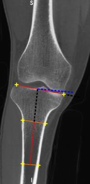
3 Metrics (2D) a) Static Alignment (SA) b) Medial Tibal Slope (MTS) c) Lateral Tibial Slope (LTS) d) Coronal Tibial Slope (CTS) [1] [2] [2] [1] Measurements and Classifications in Musculoskeletal Radiology (2014), Thieme [2] Atlas of the Patellofemoral Joint (2013), Springer Metrics (2D) a) Static Alignment (SA) b) Medial Tibal Slope (MTS) c) Lateral Tibial Slope (LTS) d) Coronal Tibial Slope (CTS) femoral axis α (a) (b) β (c) γ (d) δ tibial axis projected tibial axis projected tibial axis tibial axis Coronal Sagittal Sagittal Coronal 3

 Description Anatomical metrics: Medial, Lateral Tibial Slopes")
 Notch Width Index (NWI) Tibial Tubercle-Trochlear Groove (TT-TG) Bisect Offset (BO) Patellar Tilt (PT) Insall-Salvatti Ratio (ISR) Build and")
4 Metrics (3D) a) Static Alignment (SA) b) Medial Tibal Slope (MTS) c) Lateral Tibial Slope (LTS) d) Coronal Tibial Slope (CTS) Based on 10 anatomical landmarks Lateral plateau Femur Patella Medial plateau Tibia JMAT: Joint Morphology Analysis Tool DICOM-compatible multi-planar viewer Implemented in C++ (Qt, ITK and VTK) Semi-automated analysis of anatomical metrics Guides the user through selection of landmarks Computes metrics from the landmarks Reuses landmarks across metrics Load DICOM volume (CT or MR) Description Anatomical metrics: Medial, Lateral Tibial Slopes (MTS, LTS) Coronal Tibial Slope (CTS) Medial, Lateral Tibial Depth (MTD, LTD) ICD:TPW Ratio (ICD-TPW) Static Alignment (SA) Coronal Femoral Slope (CFS) Notch Width Index (NWI) Tibial Tubercle-Trochlear Groove (TT-TG) Bisect Offset (BO) Patellar Tilt (PT) Insall-Salvatti Ratio (ISR) Build and Save report 4
: ρ r = 0.80 ρ = 0.71 r = 0.9 ρ = 0.9 r = 0.67 ρ = 0.")
5 Intra- and inter-reader variability Intra-reader variability: SA ~0.6 o, MTS 0.5 o, LTS 0.8 o, CTS 0.6 o (6 repeats x 4 subjects) Intra-reader variability: ~0.8 o reported in MRI [1] Inter-reader variability correlation: 0.67 and more SA MTS LTS CTS Pearson correlation coefficient: r ICC(AGREEMENT): ρ r = 0.80 ρ = 0.71 r = 0.9 ρ = 0.9 r = 0.67 ρ = 0.64 r = 0.94 ρ = 0.94 [1] Hashemi J et al, J Bone Joint Surg Am, , 2008 Automatic measurements Disagreement between two observers Difficult to calculate landmarks in 3D Basic idea Use annotated images as atlas Transform landmarks to new images No segmentation of new image needed No large data base needed 5

6 Workflow Scan Atlas Atlas Atlas Atlas Rigid USE THIS AREA TO SHOW THE CURRENT STEP whole image Tibia Femur Initial transform Workflow Scan Atlas Atlas Atlas Atlas whole image Rigid Fixed image Initial USE THIS AREA Single TO bone SHOW THE CURRENT s STEP Tibia Femur Initial transform Single bone Single Single bone Masked bone single Similarity Similarity Similarity bone Similarity pick best atlas 6


7 Workflow Scan Atlas Atlas Atlas Atlas whole image Rigid Fixed image USE THIS AREA TO SHOW THE CURRENT STEP Tibia Femur Initial transform Single bone Single Single bone Masked bone single Similarity Similarity Similarity bone Similarity pick best atlas Surface deformation Transform landmarks Workflow Scan Atlas Atlas Atlas Atlas whole image Rigid Fixed image USE THIS AREA TO SHOW THE CURRENT STEP Tibia Femur Initial transform Single bone Single Single bone Masked bone single Similarity Similarity Similarity bone Similarity pick best atlas Surface deformation Transform landmarks Calculate metrics 7
8 Surface deformation Atlas registered contour Find the best fitting atlas GC I 0, I 1 = 1 3 NCC d dx I d dx I 1 + NCC d dy I d dy I 1 + NCC d dz I d dz I 1 Surface deformation Atlas Find the best fitting atlas registered contour I GC I 0, I 1 = 1 3 NCC d dx I d dx I 1 + NCC d dy I d dy I 1 + NCC d dz I d dz I 1 8

9 Gradient maginitude Gradient maginitude Surface deformation Atlas Find the best fitting atlas 0.15 v i registered contour I v i : Surface normals GC I 0, I 1 = 1 3 NCC d dx I d dx I 1 + NCC d dy I d dy I 1 + NCC d dz I d dz I 1 Surface deformation Atlas 0.15 v i registered contour deformed surface Find the best fitting atlas I Surface deformation v i : Surface normals GC I 0, I 1 = 1 3 NCC d dx I d dx I 1 + NCC d dy I d dy I 1 + NCC d dz I d dz I 1 9


10 Gradient maginitude Gradient maginitude Surface deformation Atlas 0.15 v i registered contour deformed surface Find the best fitting atlas I Surface deformation v i : Surface normals Transform landmarks GC I 0, I 1 = 1 3 NCC d dx I d dx I 1 + NCC d dy I d dy I 1 + NCC d dz I d dz I 1 Surface deformation Atlas 0.15 v i registered contour deformed surface Find the best fitting atlas I Surface deformation v i : Surface normals Transform landmarks GC I 0, I 1 = 1 3 NCC d dx I d dx I 1 + NCC d dy I d dy I 1 + NCC d dz I d dz I 1 10
11 Evaluation 24 healthy subjects (natural standing stance) Manual measurements using JMAT Evaluation methodology Leave-one-out cross-validation as a function of atlas size A: set of all images B: subset A\{p1} from which all possible k-subsets of Atlas images are drawn k: atlas size A p2 B p3 p4 p1 Evaluation 24 healthy subjects (natural standing stance) Manual measurements using JMAT Evaluation methodology Leave-one-out cross-validation as a function of atlas size A: set of all images B: subset A\{p1} from which all possible k-subsets of Atlas images are drawn k: atlas size A p2 B p3 p4 p1 k = 2 Atlas images Measure p1 using: (p2,p3) (p2,p4) (p3,p4) 11
 (p2,p4) (p3,p4) Measure p2 using: (p1,p3) (p1,p4)")
12 MTS Evaluation 24 healthy subjects (natural standing stance) Manual measurements using JMAT Evaluation methodology Leave-one-out cross-validation as a function of atlas size A: set of all images B: subset A\{p1} from which all possible k-subsets of Atlas images are drawn k: atlas size A p2 B p3 p4 p1 k = 2 Atlas images Measure p1 using: (p2,p3) (p2,p4) (p3,p4) Measure p2 using: (p1,p3) (p1,p4) (p3,p4) Atlas size 1 volume / atlas 15 volumes / atlas 20 volumes / atlas image number image number image number 1 Atlas image 15 Atlas images 20 Atlas images 23 data points per box data points per box 1771 data points per box 12
13 Manual (Expert 1) [ ] Manual (Expert 2) [ ] Atlas size vs interquartile range and RMSE Reproducibility of observer measurements Leave-one-out (atlas set of 23) Expert 1 Set atlas landmarks Perfect prediction for a new sample r = 0.99 ρ = 0.98 r = 1.0 ρ = 1.0 r = 0.99 ρ = 0.99 r = 0.99 ρ = 0.99 r = 0.82 ρ = 0.76 r = 0.9 ρ = 0.9 r = 0.65 ρ = 0.63 r = 0.94 ρ = 0.94 Expert 2 different expertise different institution Automatic method within expert-toexpert variability r=0.8, ρ=0.71 r=0.9, ρ=0.9 r=0.67, ρ=0.64 r=0.94, ρ=


, Q. Cao et al.")


 Foot and ankle Correlate with clinical outcomes")
14 Joint Space Morphology Tibial plateau joint space maps Joint space width Load-bearing vs non-load-bearing Articular surfaces modeled as conductor Field lines connect corresponding points [1] Characterization of 3D joint space morphology using an electrostatic model (with application to osteoarthritis), Q. Cao et al. PMB 2015 Conclusions Automated anatomical measurements Transfer of 2D metrics to 3D Analysis of reproducibility with respect to atlas size Correlation with manual readers Reflects agreement between readers Excellent (MTS, CTS ρ >0.9) Ongoing work Include Patellar metrics (Patellar Tilt, TTTG) Foot and ankle Correlate with clinical outcomes Application development Joint Space Maps Load-bearing vs non-load-bearing 14
The protocols used to scan the musculoskeletal system are tailored to each patient and
 Chapter 22. Musculoskeletal Protocols The protocols used to scan the musculoskeletal system are tailored to each patient and region being examined. The clinical indication for the examination will also
Semantic Context Forests for Learning- Based Knee Cartilage Segmentation in 3D MR Images
 Semantic Context Forests for Learning- Based Knee Cartilage Segmentation in 3D MR Images MICCAI 2013: Workshop on Medical Computer Vision Authors: Quan Wang, Dijia Wu, Le Lu, Meizhu Liu, Kim L. Boyer,
Assessing 3D tunnel position in ACL reconstruction using a novel single image 3D-2D registration
 Title Assessing 3D tunnel position in ACL reconstruction using a novel single image 3D-2D registration Author(s) Kang, X; Yau, WP; Otake, Y; Cheung, PYS; Hu, Y; Taylor, RH Citation SPIE Medical Imaging
Acknowledgments. High Performance Cone-Beam CT of Acute Traumatic Brain Injury
 A. Sisniega et al. (presented at RSNA 214) High Performance Cone-Beam CT of Acute Traumatic Brain Injury A. Sisniega 1 W. Zbijewski 1, H. Dang 1, J. Xu 1 J. W. Stayman 1, J. Yorkston 2, N. Aygun 3 V. Koliatsos
Auto-Segmentation Using Deformable Image Registration. Disclosure. Objectives 8/4/2011
 Auto-Segmentation Using Deformable Image Registration Lei Dong, Ph.D. Dept. of Radiation Physics University of Texas MD Anderson Cancer Center, Houston, Texas AAPM Therapy Educational Course Aug. 4th 2011
Acknowledgments. Nesterov s Method for Accelerated Penalized-Likelihood Statistical Reconstruction for C-arm Cone-Beam CT.
 June 5, Nesterov s Method for Accelerated Penalized-Likelihood Statistical Reconstruction for C-arm Cone-Beam CT Adam S. Wang, J. Webster Stayman, Yoshito Otake, Gerhard Kleinszig, Sebastian Vogt, Jeffrey
Overview of Proposed TG-132 Recommendations
 Overview of Proposed TG-132 Recommendations Kristy K Brock, Ph.D., DABR Associate Professor Department of Radiation Oncology, University of Michigan Chair, AAPM TG 132: Image Registration and Fusion Conflict
Hot Topics in Imaging Physics A Key Interdisciplinary Science for 21 st Century Medicine
 Hot Topics in Imaging Physics A Key Interdisciplinary Science for 21 st Century Medicine Jeff Siewerdsen, PhD FAAPM Department of Biomedical Engineering Department of Computer Science Russell H. Morgan
Development of 3D Model-based Morphometric Method for Assessment of Human Weight-bearing Joint. Taeho Kim
 Development of 3D Model-based Morphometric Method for Assessment of Human Weight-bearing Joint Taeho Kim Introduction Clinical measurement in the foot pathology requires accurate and robust measurement
Good Morning! Thank you for joining us
 Good Morning! Thank you for joining us Deformable Registration, Contour Propagation and Dose Mapping: 101 and 201 Marc Kessler, PhD, FAAPM The University of Michigan Conflict of Interest I receive direct
CT Protocol Clinical Graphics Move Forward 3D motion analysis service
 CT Protocol Clinical Graphics Move Forward 3D motion analysis service Version 1.4 Description of contents This document describes the CT protocol of scans for Clinical Graphics Move Forward 3D Motion Simulation
Anatomical landmark and region mapping based on a template surface deformation for foot bone morphology
 Anatomical landmark and region mapping based on a template surface deformation for foot bone morphology Jaeil Kim 1, Sang Gyo Seo 2, Dong Yeon Lee 2, Jinah Park 1 1 Department of Computer Science, KAIST,
Leksell SurgiPlan Overview. Powerful planning for surgical success
 Leksell SurgiPlan Overview Powerful planning for surgical success Making a Difference in Surgical Planning Leksell SurgiPlan Leksell SurgiPlan is an advanced image-based neuro surgical planning software,
Model Based Segmentation of Clinical Knee MRI
 Model Based Segmentation of Clinical Knee MRI Tina Kapur 12, Paul A. Beardsley 2, Sarah F. Gibson 2, W. Eric L. Grimson 1, William M. Wells 13 corresponding author: tkapur@ai.mit.edu Abstract A method
Implementation of Advanced Image Guided Radiation Therapy
 Image Acquisition Course Outline Principles, characteristics& applications of the available modalities Image Processing in the T x room Image guided treatment delivery What can / can t we do in the room
8/3/2017. Contour Assessment for Quality Assurance and Data Mining. Objective. Outline. Tom Purdie, PhD, MCCPM
 Contour Assessment for Quality Assurance and Data Mining Tom Purdie, PhD, MCCPM Objective Understand the state-of-the-art in contour assessment for quality assurance including data mining-based techniques
Carestream s 2 nd Generation Metal Artifact Reduction Software (CMAR 2)
") Carestream s 2 nd Generation Metal Artifact Reduction Software (CMAR 2) Author: Levon Vogelsang Introduction Cone beam computed tomography (CBCT), or cone beam CT technology, offers considerable promise
Carestream s 2 nd Generation Metal Artifact Reduction Software (CMAR 2) Author: Levon Vogelsang Introduction Cone beam computed tomography (CBCT), or cone beam CT technology, offers considerable promise
Assessing Accuracy Factors in Deformable 2D/3D Medical Image Registration Using a Statistical Pelvis Model
 Assessing Accuracy Factors in Deformable 2D/3D Medical Image Registration Using a Statistical Pelvis Model Jianhua Yao National Institute of Health Bethesda, MD USA jyao@cc.nih.gov Russell Taylor The Johns
TG 132: Use of Image Registration and Fusion in RT
 TG 132: Use of Image Registration and Fusion in RT Kristy K Brock, PhD, DABR, FAAPM Associate Professor Department of Radiation Oncology, University of Michigan Chair, AAPM TG 132: Image Registration and
Design and performance characteristics of a Cone Beam CT system for Leksell Gamma Knife Icon
 Design and performance characteristics of a Cone Beam CT system for Leksell Gamma Knife Icon WHITE PAPER Introduction Introducing an image guidance system based on Cone Beam CT (CBCT) and a mask immobilization
3/27/2012 WHY SPECT / CT? SPECT / CT Basic Principles. Advantages of SPECT. Advantages of CT. Dr John C. Dickson, Principal Physicist UCLH
 3/27/212 Advantages of SPECT SPECT / CT Basic Principles Dr John C. Dickson, Principal Physicist UCLH Institute of Nuclear Medicine, University College London Hospitals and University College London john.dickson@uclh.nhs.uk
David Wagner, Kaan Divringi, Can Ozcan Ozen Engineering
 Internal Forces of the Femur: An Automated Procedure for Applying Boundary Conditions Obtained From Inverse Dynamic Analysis to Finite Element Simulations David Wagner, Kaan Divringi, Can Ozcan Ozen Engineering
Image Guidance and Beam Level Imaging in Digital Linacs
 Image Guidance and Beam Level Imaging in Digital Linacs Ruijiang Li, Ph.D. Department of Radiation Oncology Stanford University School of Medicine 2014 AAPM Therapy Educational Course Disclosure Research
CT NOISE POWER SPECTRUM FOR FILTERED BACKPROJECTION AND ITERATIVE RECONSTRUCTION
 CT NOISE POWER SPECTRUM FOR FILTERED BACKPROJECTION AND ITERATIVE RECONSTRUCTION Frank Dong, PhD, DABR Diagnostic Physicist, Imaging Institute Cleveland Clinic Foundation and Associate Professor of Radiology
Self-Calibration of Cone-Beam CT Using 3D-2D Image Registration
 Self-Calibration of Cone-Beam CT Using 3D-2D Image Registration Sarah Ouadah 1 J. Webster Stayman 1 Grace Jianan Gang 1 Ali Uneri 2 Tina Ehtiati 3 Jeffrey H. Siewerdsen 1,2 1. Department of Biomedical
Spiral CT. Protocol Optimization & Quality Assurance. Ge Wang, Ph.D. Department of Radiology University of Iowa Iowa City, Iowa 52242, USA
 Spiral CT Protocol Optimization & Quality Assurance Ge Wang, Ph.D. Department of Radiology University of Iowa Iowa City, Iowa 52242, USA Spiral CT Protocol Optimization & Quality Assurance Protocol optimization
Leksell SurgiPlan. Powerful planning for success
 Leksell SurgiPlan Powerful planning for success Making a difference in surgical planning Leksell SurgiPlan Leksell SurgiPlan is an advanced image-based neurosurgical planning software, specifically designed
Computer-Tomography I: Principles, History, Technology
 Computer-Tomography I: Principles, History, Technology Prof. Dr. U. Oelfke DKFZ Heidelberg Department of Medical Physics (E040) Im Neuenheimer Feld 280 69120 Heidelberg, Germany u.oelfke@dkfz.de History
Incorporation of Prior Knowledge for Region of Change Imaging from Sparse Scan Data in Image-Guided Surgery
 Incorporation of Prior Knowledge for Region of Change Imaging from Sparse Scan Data in Image-Guided Surgery J. Lee a, J. W. Stayman b, Y. Otake c, S. Schafer b, W. Zbijewski b, A. J. Khanna b,d, J. L.
Semi-Automatic Segmentation of the Patellar Cartilage in MRI
 Semi-Automatic Segmentation of the Patellar Cartilage in MRI Lorenz König 1, Martin Groher 1, Andreas Keil 1, Christian Glaser 2, Maximilian Reiser 2, Nassir Navab 1 1 Chair for Computer Aided Medical
Medicale Image Analysis
 Medicale Image Analysis Registration Validation Prof. Dr. Philippe Cattin MIAC, University of Basel Prof. Dr. Philippe Cattin: Registration Validation Contents 1 Validation 1.1 Validation of Registration
Comparison of Different Metrics for Appearance-model-based 2D/3D-registration with X-ray Images
 Comparison of Different Metrics for Appearance-model-based 2D/3D-registration with X-ray Images Philipp Steininger 1, Karl D. Fritscher 1, Gregor Kofler 1, Benedikt Schuler 1, Markus Hänni 2, Karsten Schwieger
CT Basics Principles of Spiral CT Dose. Always Thinking Ahead.
 1 CT Basics Principles of Spiral CT Dose 2 Who invented CT? 1963 - Alan Cormack developed a mathematical method of reconstructing images from x-ray projections Sir Godfrey Hounsfield worked for the Central
Estimating 3D Respiratory Motion from Orbiting Views
 Estimating 3D Respiratory Motion from Orbiting Views Rongping Zeng, Jeffrey A. Fessler, James M. Balter The University of Michigan Oct. 2005 Funding provided by NIH Grant P01 CA59827 Motivation Free-breathing
Methodological progress in image registration for ventilation estimation, segmentation propagation and multi-modal fusion
 Methodological progress in image registration for ventilation estimation, segmentation propagation and multi-modal fusion Mattias P. Heinrich Julia A. Schnabel, Mark Jenkinson, Sir Michael Brady 2 Clinical
3D VISUALIZATION OF SEGMENTED CRUCIATE LIGAMENTS 1. INTRODUCTION
 JOURNAL OF MEDICAL INFORMATICS & TECHNOLOGIES Vol. 10/006, ISSN 164-6037 Paweł BADURA * cruciate ligament, segmentation, fuzzy connectedness,3d visualization 3D VISUALIZATION OF SEGMENTED CRUCIATE LIGAMENTS
Image Registration. Prof. Dr. Lucas Ferrari de Oliveira UFPR Informatics Department
 Image Registration Prof. Dr. Lucas Ferrari de Oliveira UFPR Informatics Department Introduction Visualize objects inside the human body Advances in CS methods to diagnosis, treatment planning and medical
Automated segmentation methods for liver analysis in oncology applications
 University of Szeged Department of Image Processing and Computer Graphics Automated segmentation methods for liver analysis in oncology applications Ph. D. Thesis László Ruskó Thesis Advisor Dr. Antal
1. Learn to incorporate QA for surface imaging
 Hania Al-Hallaq, Ph.D. Assistant Professor Radiation Oncology The University of Chicago ***No disclosures*** 1. Learn to incorporate QA for surface imaging into current QA procedures for IGRT. 2. Understand
Volumetric Analysis of the Heart from Tagged-MRI. Introduction & Background
 Volumetric Analysis of the Heart from Tagged-MRI Dimitris Metaxas Center for Computational Biomedicine, Imaging and Modeling (CBIM) Rutgers University, New Brunswick, NJ Collaboration with Dr. Leon Axel,
Advances in Forensic Anthropology
 Advances in Forensic Anthropology Technology Transition Workshop Improving Forensic Facial Reproduction Using Empirical Modeling During this session, attendees will learn of an approach for forensic facial
Lucy Phantom MR Grid Evaluation
 Lucy Phantom MR Grid Evaluation Anil Sethi, PhD Loyola University Medical Center, Maywood, IL 60153 November 2015 I. Introduction: The MR distortion grid, used as an insert with Lucy 3D QA phantom, is
Automatized & Interactive. Muscle tissues characterization using. Na MRI
 Automatized & Interactive Human Skeletal Muscle Segmentation Muscle tissues characterization using 23 Na MRI Noura Azzabou 30 April 2013 What is muscle segmentation? Axial slice of the thigh of a healthy
A Multiple-Layer Flexible Mesh Template Matching Method for Nonrigid Registration between a Pelvis Model and CT Images
 A Multiple-Layer Flexible Mesh Template Matching Method for Nonrigid Registration between a Pelvis Model and CT Images Jianhua Yao 1, Russell Taylor 2 1. Diagnostic Radiology Department, Clinical Center,
STIC AmSud Project. Graph cut based segmentation of cardiac ventricles in MRI: a shape-prior based approach
 STIC AmSud Project Graph cut based segmentation of cardiac ventricles in MRI: a shape-prior based approach Caroline Petitjean A joint work with Damien Grosgeorge, Pr Su Ruan, Pr JN Dacher, MD October 22,
Hierarchical Multi structure Segmentation Guided by Anatomical Correlations
 Hierarchical Multi structure Segmentation Guided by Anatomical Correlations Oscar Alfonso Jiménez del Toro oscar.jimenez@hevs.ch Henning Müller henningmueller@hevs.ch University of Applied Sciences Western
icatvision Quick Reference
 icatvision Quick Reference Navigating the i-cat Interface This guide shows how to: View reconstructed images Use main features and tools to optimize an image. REMINDER Images are displayed as if you are
Slide 1. Technical Aspects of Quality Control in Magnetic Resonance Imaging. Slide 2. Annual Compliance Testing. of MRI Systems.
 Slide 1 Technical Aspects of Quality Control in Magnetic Resonance Imaging Slide 2 Compliance Testing of MRI Systems, Ph.D. Department of Radiology Henry Ford Hospital, Detroit, MI Slide 3 Compliance Testing
Auto-contouring the Prostate for Online Adaptive Radiotherapy
 Auto-contouring the Prostate for Online Adaptive Radiotherapy Yan Zhou 1 and Xiao Han 1 Elekta Inc., Maryland Heights, MO, USA yan.zhou@elekta.com, xiao.han@elekta.com, Abstract. Among all the organs under
CT Reconstruction Using Spectral and Morphological Prior Knowledge: Application to Imaging the Prosthetic Knee
 CT Reconstruction Using Spectral and Morphological Prior Knowledge: Application to Imaging the Prosthetic Knee Wojciech Zbijewski, J. Webster Stayman, Abdullah Muhit, John Yorkston, John A. Carrino and
Biomet PMI. Patient-Matched Implants. CT Protocols
 CT Protocols One Surgeon. One Patient. Over 1 million times per year, Biomet helps one surgeon provide personalized care to one patient. The science and art of medical care is to provide the right solution
IMSURE QA SOFTWARE FAST, PRECISE QA SOFTWARE
 QA SOFTWARE FAST, PRECISE Software for accurate and independent verification of monitor units, dose, and overall validity of standard, IMRT, VMAT, SRS and brachytherapy plans no film, no phantoms, no linac
Object Identification in Ultrasound Scans
 Object Identification in Ultrasound Scans Wits University Dec 05, 2012 Roadmap Introduction to the problem Motivation Related Work Our approach Expected Results Introduction Nowadays, imaging devices like
Medical Image Registration by Maximization of Mutual Information
 Medical Image Registration by Maximization of Mutual Information EE 591 Introduction to Information Theory Instructor Dr. Donald Adjeroh Submitted by Senthil.P.Ramamurthy Damodaraswamy, Umamaheswari Introduction
MODELLING OF PROSTHETIC HIP JOINT GENERATED FROM CT SCAN DATA Mahender Koduri 1, G Krishna Teja 2, O Rajender 3 1,2,3
 MODELLING OF PROSTHETIC HIP JOINT GENERATED FROM CT SCAN DATA Mahender Koduri 1, G Krishna Teja 2, O Rajender 3 1,2,3 Asst. Professor, Dept. of Mech. Engg. AGI ABSTRACT Total hip arthroplasty is a surgical
Segmentation of 3-D medical image data sets with a combination of region based initial segmentation and active surfaces
 Header for SPIE use Segmentation of 3-D medical image data sets with a combination of region based initial segmentation and active surfaces Regina Pohle, Thomas Behlau, Klaus D. Toennies Otto-von-Guericke
Shadow casting. What is the problem? Cone Beam Computed Tomography THE OBJECTIVES OF DIAGNOSTIC IMAGING IDEAL DIAGNOSTIC IMAGING STUDY LIMITATIONS
 Cone Beam Computed Tomography THE OBJECTIVES OF DIAGNOSTIC IMAGING Reveal pathology Reveal the anatomic truth Steven R. Singer, DDS srs2@columbia.edu IDEAL DIAGNOSTIC IMAGING STUDY Provides desired diagnostic
6th International DAAAM Baltic Conference INDUSTRIAL ENGINEERING April 2008, Tallinn, Estonia. Radu, C. & Roşca, I.C.
 6th International DAAAM Baltic Conference INDUSTRIAL ENGINEERING 24-26 April 2008, Tallinn, Estonia ON THE DESIGN OF A MEDICAL IMPLANT USED FOR OSTEOSYNTHESIS OF THE TRANSSINDESMOTIC FIBULAR FRACTURE PART
Experience Boundless Performance
 Experience Boundless Performance About Samsung Samsung Electronics Co., Ltd. inspires the world and shapes the future with transformative ideas and technologies, redefining the worlds of TVs, smartphones,
CT IMAGE PROCESSING IN HIP ARTHROPLASTY
 U.P.B. Sci. Bull., Series C, Vol. 75, Iss. 3, 2013 ISSN 2286 3540 CT IMAGE PROCESSING IN HIP ARTHROPLASTY Anca MORAR 1, Florica MOLDOVEANU 2, Alin MOLDOVEANU 3, Victor ASAVEI 4, Alexandru EGNER 5 The use
3D Numerical Analysis of an ACL Reconstructed Knee
 3D Numerical Analysis of an ACL Reconstructed Knee M. Chizari, B. Wang School of Engineering, University of Aberdeen, Aberdeen AB24 7QW, UK Abstract: Numerical methods applicable to the tibia bone and
MORPHOLOGY ANALYSIS OF HUMAN KNEE USING MR IMAGERY
 MORPHOLOGY ANALYSIS OF HUMAN KNEE USING MR IMAGERY D. Chetverikov 1,2, G. Renner 1 1 Computer and Automation Research Institute, Budapest, Hungary; 2 Eötvös Loránd University, Budapest, Hungary We present
Measuring Motion and Shape in Biomechanics
 Measuring Motion and Shape in Biomechanics G. Renner*, K. Andronyi** * Computer and Automation Research Institute of HAS, Budapest, Hungary (e-mail: renner@sztaki.hu). **Semmelweis University, Orthopaedic
Weakly Supervised Fully Convolutional Network for PET Lesion Segmentation
 Weakly Supervised Fully Convolutional Network for PET Lesion Segmentation S. Afshari a, A. BenTaieb a, Z. Mirikharaji a, and G. Hamarneh a a Medical Image Analysis Lab, School of Computing Science, Simon
Feasibility of 3D Printed Patient specific Phantoms for IMRT QA and Other Dosimetric Special Procedures
 Feasibility of 3D Printed Patient specific Phantoms for IMRT QA and Other Dosimetric Special Procedures ehler 046@umn.edu Eric Ehler, PhD Assistant Professor Department of Radiation Oncology What is 3D
Limitations of Projection Radiography. Stereoscopic Breast Imaging. Limitations of Projection Radiography. 3-D Breast Imaging Methods
 Stereoscopic Breast Imaging Andrew D. A. Maidment, Ph.D. Chief, Physics Section Department of Radiology University of Pennsylvania Limitations of Projection Radiography Mammography is a projection imaging
NON OB ULTRASOUND MORPHOMETRICS AS BIOMARKERS
 NON OB ULTRASOUND MORPHOMETRICS AS BIOMARKERS Brian S Garra Washington DC VA Medical Center and Division of Imaging & Applied Mathematics, OSEL, CDRH, FDA GOALS Review Major Types of Clinical Ultrasonic
radiotherapy Andrew Godley, Ergun Ahunbay, Cheng Peng, and X. Allen Li NCAAPM Spring Meeting 2010 Madison, WI
 GPU-Accelerated autosegmentation for adaptive radiotherapy Andrew Godley, Ergun Ahunbay, Cheng Peng, and X. Allen Li agodley@mcw.edu NCAAPM Spring Meeting 2010 Madison, WI Overview Motivation Adaptive
shape modeling of the paranasal
 Automatic segmentation and statistical shape modeling of the paranasal sinuses to estimate natural variations Ayushi Sinha a, Simon Leonard a, Austin Reiter a, Masaru Ishii b, Russell H. Taylor a and Gregory
3D registration of micro-ct images for the identification of trabecular fracture region.
 3D registration of micro-ct images for the identification of trabecular fracture region. S. Tassani 1,2, P.A. Asvestas 3, G.K. Matsopoulos 1, and F. Baruffaldi 2 1 Institute of Communication and Computer
Saturn User Manual. Rubén Cárdenes. 29th January 2010 Image Processing Laboratory, University of Valladolid. Abstract
 Saturn User Manual Rubén Cárdenes 29th January 2010 Image Processing Laboratory, University of Valladolid Abstract Saturn is a software package for DTI processing and visualization, provided with a graphic
Semi-automated Basal Ganglia Segmentation Using Large Deformation Diffeomorphic Metric Mapping
 Semi-automated Basal Ganglia Segmentation Using Large Deformation Diffeomorphic Metric Mapping Ali Khan 1, Elizabeth Aylward 2, Patrick Barta 3, Michael Miller 4,andM.FaisalBeg 1 1 School of Engineering
IEEE TRANSACTIONS ON IMAGE PROCESSING, VOL. 26, NO. 10, OCTOBER
 IEEE TRANSACTIONS ON IMAGE PROCESSING, VOL. 26, NO. 10, OCTOBER 2017 4753 Detecting Anatomical Landmarks From Limited Medical Imaging Data Using Two-Stage Task-Oriented Deep Neural Networks Jun Zhang,
MUSCULOSKELETAL SIMULATION :
 TUTORIAL MUSCULOSKELETAL SIMULATION : FROM MOTION CAPTURE TO MUSCULAR ACTIVITY IN LOWER LIMB MODELS Nicolas Pronost and Anders Sandholm Musculoskeletal simulation? What is it? 2 Musculoskeletal simulation?
From DQE to TRE: Image Science, Image Guidance, and Cone-Beam CT
 W task = F task C task Magnification Focal Spot Size Scatter-to-Primary Ratio kvp, Dose Scintillator, Detector Pixel Aperture, 2D Sampling Reconstruction Filter Backprojection Voxel Size 3D Sampling PW,
The 3D Accuitomo 80 offers a minute voxel. size of just 80 μm (micrometer). This super-fine. voxel combined with the unit s 13 bit grayscale
. This super-fine. voxel combined with the unit s 13 bit grayscale") 2 Thinking ahead. Focused on life. REALIZED: Groundbreaking Resolution of 80 µm Voxel 05 The Crystallization of cutting-edge mechatronics and High Definition display algorithm The 3D Accuitomo 80 offers
2 Thinking ahead. Focused on life. REALIZED: Groundbreaking Resolution of 80 µm Voxel 05 The Crystallization of cutting-edge mechatronics and High Definition display algorithm The 3D Accuitomo 80 offers
Image Quality Assessment and Quality Assurance of Advanced Imaging Systems for IGRT. AAPM Penn-Ohio Chapter Sep 25, 2015 Soyoung Lee, PhD
 Image Quality Assessment and Quality Assurance of Advanced Imaging Systems for IGRT AAPM Penn-Ohio Chapter Sep 25, 2015 Soyoung Lee, PhD 1 Outline q Introduction q Imaging performances in 4D-CBCT Image
Financial disclosure. Onboard imaging modality for IGRT
 Tetrahedron Beam Computed Tomography Based On Multi-Pixel X- Ray Source and Its Application in Image Guided Radiotherapy Tiezhi Zhang, Ph.D. Advanced X-ray imaging Lab Financial disclosure Patent royalty
Ch. 4 Physical Principles of CT
 Ch. 4 Physical Principles of CT CLRS 408: Intro to CT Department of Radiation Sciences Review: Why CT? Solution for radiography/tomography limitations Superimposition of structures Distinguishing between
Comparison Study of Clinical 3D MRI Brain Segmentation Evaluation
 Comparison Study of Clinical 3D MRI Brain Segmentation Evaluation Ting Song 1, Elsa D. Angelini 2, Brett D. Mensh 3, Andrew Laine 1 1 Heffner Biomedical Imaging Laboratory Department of Biomedical Engineering,
Detecting Anatomical Landmarks from Limited Medical Imaging Data using Two-Stage Task-Oriented Deep Neural Networks
 IEEE TRANSACTIONS ON IMAGE PROCESSING Detecting Anatomical Landmarks from Limited Medical Imaging Data using Two-Stage Task-Oriented Deep Neural Networks Jun Zhang, Member, IEEE, Mingxia Liu, Member, IEEE,
Automatic Segmentation of Menisci in MR Images Using Pattern Recognition and Graph Cuts. Fredrik Nedmark,
 Automatic Segmentation of Menisci in MR Images Using Pattern Recognition and Graph Cuts Fredrik Nedmark, fredrik_n84@hotmail.com April 14, 2011 Abstract In this Master s thesis a system for automatic segmentation
An Improved Tracking Technique for Assessment of High Resolution Dynamic Radiography Kinematics
 Copyright c 2008 ICCES ICCES, vol.8, no.2, pp.41-46 An Improved Tracking Technique for Assessment of High Resolution Dynamic Radiography Kinematics G. Papaioannou 1, C. Mitrogiannis 1 and G. Nianios 1
REAL-TIME ADAPTIVITY IN HEAD-AND-NECK AND LUNG CANCER RADIOTHERAPY IN A GPU ENVIRONMENT
 REAL-TIME ADAPTIVITY IN HEAD-AND-NECK AND LUNG CANCER RADIOTHERAPY IN A GPU ENVIRONMENT Anand P Santhanam Assistant Professor, Department of Radiation Oncology OUTLINE Adaptive radiotherapy for head and
Knowledge-Based Segmentation of Brain MRI Scans Using the Insight Toolkit
 Knowledge-Based Segmentation of Brain MRI Scans Using the Insight Toolkit John Melonakos 1, Ramsey Al-Hakim 1, James Fallon 2 and Allen Tannenbaum 1 1 Georgia Institute of Technology, Atlanta GA 30332,
CREATION AND VISUALIZATION OF ANATOMICAL MODELS WITH AMIRA CREATION ET VISUALISATION DES MODELES ANATOMIQUES AVEC AMIRA
 CREATION AND VISUALIZATION OF ANATOMICAL MODELS WITH AMIRA CREATION ET VISUALISATION DES MODELES ANATOMIQUES AVEC AMIRA Summary 3D imaging methods are widely used in medicine and biology, mainly for image-guided
Knowledge-Based Segmentation of Brain MRI Scans Using the Insight Toolkit
 Knowledge-Based Segmentation of Brain MRI Scans Using the Insight Toolkit John Melonakos 1, Ramsey Al-Hakim 1, James Fallon 2 and Allen Tannenbaum 1 1 Georgia Institute of Technology, Atlanta GA 30332,
S. Guru Prasad, Ph.D., DABR
 PURPOSE S. Guru Prasad, Ph.D., DABR Director of Medical Physics IAEA Consultant NorthShore University Health System and University of Chicago, Pritzker School of Medicine Current TPS utilize more information
Semi-automatic Segmentation of Vertebral Bodies in Volumetric MR Images Using a Statistical Shape+Pose Model
 Semi-automatic Segmentation of Vertebral Bodies in Volumetric MR Images Using a Statistical Shape+Pose Model A. Suzani, A. Rasoulian S. Fels, R. N. Rohling P. Abolmaesumi Robotics and Control Laboratory,
Theoretical Accuracy of Model-Based Shape Matching for Measuring Natural Knee Kinematics with Single-Plane Fluoroscopy
 Benjamin J. Fregly 1 Phone: (352) 392-8157 Fax: (352) 392-7303 e-mail: fregly@ufl.edu Department of Mechanical and Aerospace Engineering, Department of Biomedical Engineering, Department of Othopaedics
Automatic Segmentation of Parotids from CT Scans Using Multiple Atlases
 Automatic Segmentation of Parotids from CT Scans Using Multiple Atlases Jinzhong Yang, Yongbin Zhang, Lifei Zhang, and Lei Dong Department of Radiation Physics, University of Texas MD Anderson Cancer Center
Supplementary methods
 Supplementary methods This section provides additional technical details on the sample, the applied imaging and analysis steps and methods. Structural imaging Trained radiographers placed all participants
DEEP LEARNING WITH ORTHOGONAL VOLUMETRIC HED SEGMENTATION AND 3D SURFACE RECONSTRUCTION MODEL OF PROSTATE MRI
 DEEP LEARNING WITH ORTHOGONAL VOLUMETRIC HED SEGMENTATION AND 3D SURFACE RECONSTRUCTION MODEL OF PROSTATE MRI Ruida Cheng a, Nathan Lay b, Francesca Mertan c, Baris Turkbey c, Holger R. Roth b, Le Lu b,
The Insight Toolkit. Image Registration Algorithms & Frameworks
 The Insight Toolkit Image Registration Algorithms & Frameworks Registration in ITK Image Registration Framework Multi Resolution Registration Framework Components PDE Based Registration FEM Based Registration
Optimization of CT Simulation Imaging. Ingrid Reiser Dept. of Radiology The University of Chicago
 Optimization of CT Simulation Imaging Ingrid Reiser Dept. of Radiology The University of Chicago Optimization of CT imaging Goal: Achieve image quality that allows to perform the task at hand (diagnostic
ISO Implants for surgery Total knee-joint prostheses Determination of endurance properties of knee tibial trays
 INTERNATIONAL STANDARD ISO 14879-1 First edition 2000-06-01 Implants for surgery Total knee-joint prostheses Part 1: Determination of endurance properties of knee tibial trays Implants chirurgicaux Prothèses
The Use of Unwrapped Phase in MR Image Segmentation: A Preliminary Study
 The Use of Unwrapped Phase in MR Image Segmentation: A Preliminary Study Pierrick Bourgeat 1, Jurgen Fripp 1,3, Andrew Janke 2, Graham Galloway 2, Stuart Crozier 3,andSébastien Ourselin 1 1 Autonomous
Modeling of MR-guided HIFU for Breast and Brain Therapy
 Modeling of MR-guided HIFU for Breast and Brain Therapy Douglas A. Christensen, Allison Payne, Nick Todd, Scott Almquist, Alexis Farrer and Dennis L. Parker University of Utah Salt Lake City, Utah Overview
Computer Aided Design for Osseo-Integration Applications
 Computer Aided Design for Osseo-Integration Applications Anoop Parthasarathy 1, Dr Ramkumar P S 2, Yogesh Jain 3 1 Engineer, Applied Cognition Systems Pvt. Ltd. Bangalore, India, 2 Adjunct Professor,Manipal
Introduction to Medical Image Processing
 Introduction to Medical Image Processing Δ Essential environments of a medical imaging system Subject Image Analysis Energy Imaging System Images Image Processing Feature Images Image processing may be