4D MRI phase-contrast image to determinate blood flow patterns in aorta
|
|
|
- Sibyl Lamb
- 6 years ago
- Views:
Transcription
1 4D MRI phase-contrast image to determinate blood flow patterns in aorta E. Soudah 1, M.R.Cancio 2, H.Yervilla 2, F.Carreras 3, J.S.Ronda 1, E.Oñate 1 1 International Centre for Numerical Methods in Engineering(CIMNE) Campus Norte UPC, Barcelona, Spain 2 Universidad Central de las Villas Aula UCLV-CIMNE Santa Clara, Cuba 3 Hospital Sant Pau y Creu Blanca, Barcelona, Spain CMBE 2011 Thursday, March 31 th
/bicuspid (two leaflets)")

2 Use Case Clinical view: Aorta diseases: Thoracic Aortic Aneurysms (Marfan s Syndrome) Aorta dissection Valve diseases Aortic Valve Stenosis tricuspid(three leaflets)/bicuspid (two leaflets) Mathematical view: Obtain the real blood flow of the aorta directly from the 4D MRI phase-contrast image. 4D combine Phase-Contrast imaging & CFD simulation. Possibility to create a volume mesh from an image segmented.
3 Use Case Clinical view: Aorta diseases: Thoracic Aortic Aneurysms (Marfan s Syndrome) Aorta dissection Valve diseases Aortic Valve Stenosis tricuspid(three leaflets)/bicuspid (two leaflets) Mathematical view: Obtain the real blood flow of the aorta directly from the 4D MRI phase-contrast image. 4D combine Phase-Contrast imaging & CFD simulation. Possibility to create a volume mesh from an image segmented.

4 Use Case Goal Using 4D MRI image combined with CFD to: Increase objectivity and reproducibility for the assessment of the Aorta. Provide addition information to improve in treatment decision. Outline 4D MRI Phase-contrast Segmentation & Meshing Numerical Results Conclusions
5 Use Case Goal Using 4D MRI image combined with CFD to: Increase objectivity and reproducibility for the assessment of the Aorta. Provide addition information to improve in treatment decision. Outline 4D MRI Phase-contrast Segmentation & Meshing Numerical Results Conclusions


")
6 WorkFlow DIPPO DCM Slicer ROI Filter 1 Filter N Filter 1 Filter N VTK Image Manager NoiseFilter Manager Segmentation Manager Numerical solver (CFD) Pre-Process VELOCITY GEOMETRY

7 4D MRI Phase-Contrast Time-resolved phase contrast (PC) MRI with velocity encoding in three directions (flow-sensitive four-dimensional MRI) can be employed to assess three-dimensional blood flow in the entire aortic lumen within a single measurement. 4D phase contrast MRI at 3 T:effect of standard and blood-pool contrast agents on SNR, PC-MRA, and blood flow visualization Bock J, Frydrychowicz A, Stalder AF, Bley TA, Burkhardt H, Hennig J, Markl M. Magn Reson Med 2010;63(2): Kindly provided by M.Markl


8 1-Zhao, F., et al., Automated 4D Segmentation of Aortic Magnetic Resonance Images. 2-Frangi, A.F., et al., Multiscale vessel enhancement filtering SETHIAN, J.A., Level Set Methods and Fast Marching Methods: Evolving Interfaces in Computational Geometry, Fluid Mechanics, Computer Vision and Materials Science Ibañez. Kitware,Inc. Segmentation 4D phase-contrast image (spatio-temporal) n volumetric images defined at n differents time steps (in this particular case: 12 time-steps) Semi-automatic Segmentation method 1 Aortic surface pre-segmetation (vesselnes enhancement filter) 2,4 and Tresholded 3,4
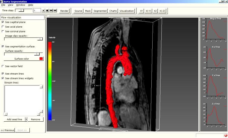
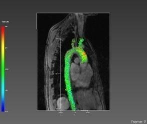
9 Aorta Velocities

10 Details Aorta Velocities, for step 4


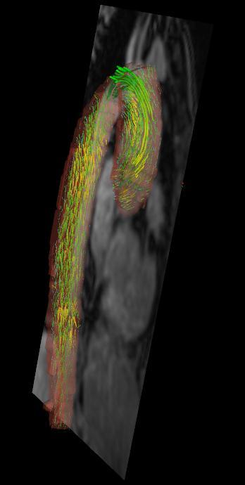
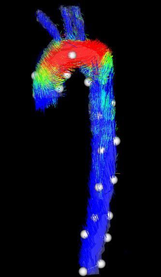
11 Details Aorta Velocities, all steps Aorta Coronal Plane Aorta Volume
12 WorkFlow DIPPO DCM Slicer ROI Filter 1 Filter N Filter 1 Filter N VTK Image Manager NoiseFilter Manager Segmentation Manager Numerical solver (CFD) Pre-Process VELOCITY GEOMETRY

, August 2007.")
13 Volume mesh Iso-Stuffing.(Direct volume meshing with quality warranty).(françois Labelle and Jonathan Richard Shewchuk, Isosurface Stuffing: Fast Tetrahedral Meshes with Good Dihedral Angles, ACM Transactions on Graphics 26(3), August Special issue on Proceedings of SIGGRAPH 2007)
14 CFD WSS CFD Velocities


15 Aorta dissection A multi-method approach towards understanding the pathophysiology of aortic dissections the complementary role of in-silico, in-vitro and in-vivo information Paula A. Rudenick, Maurizio Bordone, Bart H. Bijnens, Eduardo Soudah, David Garcia-Dorado, and Arturo Evangelista CIMNE & Vall d Hebron

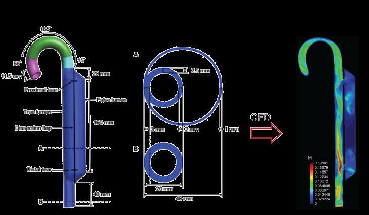
16 Aorta dissection CIMNE & Vall d Hebron
17 Conclusions - MRI 4D PC patient-specific data for Aorta studies. - More data are available for quantitative studies. - Combine MRI 4D with CFD Simulations (boundary conditions) - Improvement quantitative analysis - Determinate flow patterns and vortex in aorta. Future Studies - Velocity sensitive analysis - Compare 2D & 4D PC Image & CFD - Compare phantom model & 4D PC image CIMNE & Vall d Hebron
18 Thank you Eduardo Soudah CMBE 2011 Thursday, March 31 th
4D Magnetic Resonance Analysis. MR 4D Flow. Visualization and Quantification of Aortic Blood Flow
 4D Magnetic Resonance Analysis MR 4D Flow Visualization and Quantification of Aortic Blood Flow 4D Magnetic Resonance Analysis Complete assesment of your MR 4D Flow data Time-efficient and intuitive analysis
Medical-GiD: From Medical Images to Simulations, 4D MRI Flow Analysis
 Medical-GiD: From Medical Images to Simulations, 4D MRI Flow Analysis Eduardo Soudah, Julien Pennecot, Jorge S.Pérez, Maurizio Bordone and Eugenio Oñate. Abstract Medical imaging techniques, such as MRI
Blood Particle Trajectories in Phase-Contrast-MRI as Minimal Paths Computed with Anisotropic Fast Marching
 Blood Particle Trajectories in Phase-Contrast-MRI as Minimal Paths Computed with Anisotropic Fast Marching Michael Schwenke 1, Anja Hennemuth 1, Bernd Fischer 2, Ola Friman 1 1 Fraunhofer MEVIS, Institute
High dynamic range magnetic resonance flow imaging in the abdomen
 High dynamic range magnetic resonance flow imaging in the abdomen Christopher M. Sandino EE 367 Project Proposal 1 Motivation Time-resolved, volumetric phase-contrast magnetic resonance imaging (also known
Clinical Importance. Aortic Stenosis. Aortic Regurgitation. Ultrasound vs. MRI. Carotid Artery Stenosis
 Clinical Importance Rapid cardiovascular flow quantitation using sliceselective Fourier velocity encoding with spiral readouts Valve disease affects 10% of patients with heart disease in the U.S. Most
Life Sciences Applications: Modeling and Simulation for Biomedical Device Design SGC 2013
 Life Sciences Applications: Modeling and Simulation for Biomedical Device Design Kristian.Debus@cd-adapco.com SGC 2013 Modeling and Simulation for Biomedical Device Design Biomedical device design and
Isogeometric Analysis of Fluid-Structure Interaction
 Isogeometric Analysis of Fluid-Structure Interaction Y. Bazilevs, V.M. Calo, T.J.R. Hughes Institute for Computational Engineering and Sciences, The University of Texas at Austin, USA e-mail: {bazily,victor,hughes}@ices.utexas.edu
High dynamic range fusion of magnetic resonance flow imaging data
 High dynamic range fusion of magnetic resonance flow imaging data Christopher M. Sandino Stanford University EE 367 Final Project Report sandino@stanford.edu Abstract Time-resolved, volumetric phase-contrast
Adaptive Animations of Vortex Flow Extracted from Cardiac 4D PC-MRI Data
 Adaptive Animations of Vortex Flow Extracted from Cardiac 4D PCMRI Data Benjamin Köhler 1, Uta Preim 2, Matthias Grothoff 3, Matthias Gutberlet 3, Bernhard Preim 1 1 Dept. of Computer Graphics and Simulation,
3D3C & 2D3D Velocity Measurements Using Magnetic Resonance Velocimetry
 3D3C & 2D3D Velocity Measurements Using Magnetic Resonance Velocimetry Sven Grundmann Center of Smart Interfaces Technische Universität Darmstadt Flughafenstrasse 19 64347 Griesheim grundmann@csi-tu-darmstadt.de
DECISION SUPPORT SYSTEM FOR CARDIOVASCULAR PROBLEMS
 DECISION SUPPORT SYSTEM FOR CARDIOVASCULAR PROBLEMS E. Soudah R. López M. Bordone E.Oñate Monograph CIMNE DECISION SUPPORT SYSTEM FOR CARDIOVASCULAR PROBLEMS E.Soudah, R.López, M.Bordone and E.Oñate International
Application of level set based method for segmentation of blood vessels in angiography images
 Lodz University of Technology Faculty of Electrical, Electronic, Computer and Control Engineering Institute of Electronics PhD Thesis Application of level set based method for segmentation of blood vessels
GiD International Center for Numerical Methods in Engineering Gran Capitan s/n, Barcelona, Spain
 GiD 2010 5th Conference on Advances and Applications of GiD 1st Kratos Workshop M. Pasenau, E. Escolano, J. Suit, A. Coli, A. Melendo, A. Monros, F. Chico, P. Dadvand (Eds.) Monograph CIMNE N -118, May
Optical Flow Method for Blood Flow Velocimetry Based on Digital X-Ray Subtraction Angiography: A Brief Review
 Optical Flow Method for Blood Flow Velocimetry Based on Digital X-Ray Subtraction Angiography: A Brief Review Zifeng Yang* Department of Mechanical and Materials Engineering, Wright State University, Dayton,
Automated segmentation of blood-flow regions in large thoracic arteries using 3D-cine PC-MRI measurements
 Int J CARS (2012) 7:217 224 DOI 10.1007/s11548-011-0642-9 ORIGINAL ARTICLE Automated segmentation of blood-flow regions in large thoracic arteries using 3D-cine PC-MRI measurements Roy van Pelt Huy Nguyen
3D Vascular Segmentation using MRA Statistics and Velocity Field Information in PC-MRA
 3D Vascular Segmentation using MRA Statistics and Velocity Field Information in PC-MRA Albert C. S. Chung 1, J. Alison Noble 1, Paul Summers 2 and Michael Brady 1 1 Department of Engineering Science, Oxford
Skåne University Hospital Lund, Lund, Sweden 2 Deparment of Numerical Analysis, Centre for Mathematical Sciences, Lund University, Lund, Sweden
 Volume Tracking: A New Method for Visualization of Intracardiac Blood Flow from Three-Dimensional, Time-Resolved, Three-Component Magnetic Resonance Velocity Mapping Appendix: Theory and Numerical Implementation
SIMULATION D'IRM ANGIOGRAPHIQUE PAR EXTENSION DU LOGICIEL JEMRIS
 SIMULATION D'IRM ANGIOGRAPHIQUE PAR EXTENSION DU LOGICIEL JEMRIS Alexandre FORTIN Supervised by Emmanuel DURAND and Stéphanie SALMON Laboratoire de Mathématiques de Reims Model : clipart-fr.com VIVABRAIN
MRA Image Segmentation with Capillary Active Contour
 MRA Image Segmentation with Capillary Active Contour Pingkun Yan and Ashraf A. Kassim Department of Electrical & Computer Engineering, National University of Singapore {pingkun,ashraf}@nus.edu.sg Abstract.
CFD simulations of blood flow through abdominal part of aorta
 CFD simulations of blood flow through abdominal part of aorta Andrzej Polanczyk, Aleksandra Piechota Faculty of Process and Enviromental Engineering, Technical University of Lodz, Wolczanska 13 90-94 Lodz,
2D Vessel Segmentation Using Local Adaptive Contrast Enhancement
 2D Vessel Segmentation Using Local Adaptive Contrast Enhancement Dominik Schuldhaus 1,2, Martin Spiegel 1,2,3,4, Thomas Redel 3, Maria Polyanskaya 1,3, Tobias Struffert 2, Joachim Hornegger 1,4, Arnd Doerfler
A Workflow for Computational Fluid Dynamics Simulations using Patient-Specific Aortic Models
 2.8.8 A Workflow for Computational Fluid Dynamics Simulations using Patient-Specific Aortic Models D. Hazer 1,2, R. Unterhinninghofen 2, M. Kostrzewa 1, H.U. Kauczor 3, R. Dillmann 2, G.M. Richter 1 1)
Automatic Ascending Aorta Detection in CTA Datasets
 Automatic Ascending Aorta Detection in CTA Datasets Stefan C. Saur 1, Caroline Kühnel 2, Tobias Boskamp 2, Gábor Székely 1, Philippe Cattin 1,3 1 Computer Vision Laboratory, ETH Zurich, 8092 Zurich, Switzerland
Probabilistic Tracking and Model-based Segmentation of 3D Tubular Structures
 Probabilistic Tracking and Model-based Segmentation of 3D Tubular Structures Stefan Wörz, William J. Godinez, Karl Rohr University of Heidelberg, BIOQUANT, IPMB, and DKFZ Heidelberg, Dept. Bioinformatics
CPM Specifications Document Healthy Vertebral:
 CPM Specifications Document Healthy Vertebral: OSMSC 0078_0000, 0079_0000, 0166_000, 0167_0000 May 1, 2013 Version 1 Open Source Medical Software Corporation 2013 Open Source Medical Software Corporation.
Hemodynamics in the Thoracic Aorta using OpenFOAM: 4D PCMRI versus CFD. J. Casacuberta E. Soudah P. J. Gamez-Montero G. Raush R. Castilla J.S.
 Hemodynamics in the Thoracic Aorta using OpenFOAM: 4D PCMRI versus CFD J. Casacuberta E. Soudah P. J. Gamez-Montero G. Raush R. Castilla J.S. Perez Monograph CIMNE Nº-154, March 2015 Hemodynamics in the
Optimization of Flow Diverter Treatment for a Patient-specific Giant Aneurysm Using STAR-CCM+ László Daróczy, Philipp Berg, Gábor Janiga
 Optimization of Flow Diverter Treatment for a Patient-specific Giant Aneurysm Using STAR-CCM+ László Daróczy, Philipp Berg, Gábor Janiga Introduction Definition of aneurysm: permanent and locally limited
Phantom-based evaluation of a semi-automatic segmentation algorithm for cerebral vascular structures in 3D ultrasound angiography (3D USA)
") Phantom-based evaluation of a semi-automatic segmentation algorithm for cerebral vascular structures in 3D ultrasound angiography (3D USA) C. Chalopin¹, K. Krissian², A. Müns 3, F. Arlt 3, J. Meixensberger³,
Phantom-based evaluation of a semi-automatic segmentation algorithm for cerebral vascular structures in 3D ultrasound angiography (3D USA) C. Chalopin¹, K. Krissian², A. Müns 3, F. Arlt 3, J. Meixensberger³,
Multiscale Blood Vessel Segmentation in Retinal Fundus Images
 Multiscale Blood Vessel Segmentation in Retinal Fundus Images Attila Budai 1, Georg Michelson 2, Joachim Hornegger 1 1 Pattern Recognition Lab and Graduate School in Advanced Optical Technologies(SAOT),
Research Lines and RTD Project in Biomedical Engineering
 Research Lines and RTD Project in Biomedical Engineering CIMNE February 2009 Research Lines & RTD Project in Biomedical Engineering CIMNE February 2009 Centro Internacional de Métodos Numéricos en Ingeniería
Volume visualization. Volume visualization. Volume visualization methods. Sources of volume visualization. Sources of volume visualization
 Volume visualization Volume visualization Volumes are special cases of scalar data: regular 3D grids of scalars, typically interpreted as density values. Each data value is assumed to describe a cubic
Image Analysis, Geometrical Modelling and Image Synthesis for 3D Medical Imaging
 Image Analysis, Geometrical Modelling and Image Synthesis for 3D Medical Imaging J. SEQUEIRA Laboratoire d'informatique de Marseille - FRE CNRS 2246 Faculté des Sciences de Luminy, 163 avenue de Luminy,
From medical imaging to numerical simulations
 From medical imaging to numerical simulations Christophe Prud Homme, Vincent Chabannes, Marcela Szopos, Alexandre Ancel, Julien Jomier To cite this version: Christophe Prud Homme, Vincent Chabannes, Marcela
ANSYS AIM 16.0 Overview. AIM Program Management
 1 2015 ANSYS, Inc. September 27, 2015 ANSYS AIM 16.0 Overview AIM Program Management 2 2015 ANSYS, Inc. September 27, 2015 Today s Simulation Challenges Leveraging simulation across engineering organizations
Outline: Contrast-enhanced MRA
 Outline: Contrast-enhanced MRA Background Technique Clinical Indications Future Directions Disclosures: GE Health Care: Research support Consultant: Bracco, Bayer The Basics During rapid IV infusion, Gadolinium
11/18/ CPT Preauthorization Groupings Effective January 1, Computerized Tomography (CT) Abdomen 6. CPT Description SEGR CT01
 Abdomen 6. CPT Description SEGR CT01") Computerized Tomography (CT) 6 & 101 5 Upper Extremity 11 Lower Extremity 12 Head 3 Orbit 1 Sinus 2 Neck 4 7 Cervical Spine 8 Thoracic Spine 9 Lumbar Spine 10 Colon 13 CPT Description SEGR 74150 74160
Computerized Tomography (CT) 6 & 101 5 Upper Extremity 11 Lower Extremity 12 Head 3 Orbit 1 Sinus 2 Neck 4 7 Cervical Spine 8 Thoracic Spine 9 Lumbar Spine 10 Colon 13 CPT Description SEGR 74150 74160
Qualitative Comparison of Conventional and Oblique MRI for Detection of Herniated Spinal Discs
 Qualitative Comparison of Conventional and Oblique MRI for Detection of Herniated Spinal Discs Doug Dean Final Project Presentation ENGN 2500: Medical Image Analysis May 16, 2011 Outline Review of the
Single Breath-hold Abdominal T 1 Mapping using 3-D Cartesian Sampling and Spatiotemporally Constrained Reconstruction
 Single Breath-hold Abdominal T 1 Mapping using 3-D Cartesian Sampling and Spatiotemporally Constrained Reconstruction Felix Lugauer 1,3, Jens Wetzl 1, Christoph Forman 2, Manuel Schneider 1, Berthold Kiefer
SENSITIVITY MESH ANALYSIS OF BLOOD FLOW FOR STUDY HYPERELASTIC AORTA MODELS
 SENSITIVITY MESH ANALYSIS OF BLOOD FLOW FOR STUDY HYPERELASTIC AORTA MODELS Melendez, Noel Emmanuel (1), Vidal-Lesso A. (2) 1 [Mechanical Engineering, University of Guanajuato] [n.mahungmelendez@ugto.mx]
Numerical Validation of Hemodynamic Factors in Vascular Diseases.
 Numerical Validation of Hemodynamic Factors in Vascular Diseases. M. Bordone E.Oñate E. Soudah Monograph CIMNE Numerical Validation of Hemodynamic Factors in Vascular Diseases M. Bordone, E.Oñate, E. Soudah
Estimating Arterial Wall Shear Stress 1
 DEPARTMENT OF STATISTICS University of Wisconsin 1210 West Dayton St. Madison, WI 53706 TECHNICAL REPORT NO. 1088 December 12, 2003 Estimating Arterial Wall Shear Stress 1 John D. Carew 2 Departments of
Modeling and preoperative planning for kidney surgery
 Modeling and preoperative planning for kidney surgery Refael Vivanti Computer Aided Surgery and Medical Image Processing Lab Hebrew University of Jerusalem, Israel Advisor: Prof. Leo Joskowicz Clinical
48 4 Quantitative 3-D MRA: in vitro and in vivo results Endarterectomy Trial (NASCET) [216] and the European Carotid Surgery Trial (ECST) [92], both e
![48 4 Quantitative 3-D MRA: in vitro and in vivo results Endarterectomy Trial (NASCET) [216] and the European Carotid Surgery Trial (ECST) [92], both e](/thumbs/79/79296189.jpg "48 4 Quantitative 3-D MRA: in vitro and in vivo results Endarterectomy Trial (NASCET) [216] and the European Carotid Surgery Trial (ECST) [92], both e") True genius resides in the capacity for evaluation of uncertain, hazardous, and conflicting information. W. Churchill, 1874-1965 Chapter 4 Quantitative analysis of vascular morphology from 3-D MRA: in
True genius resides in the capacity for evaluation of uncertain, hazardous, and conflicting information. W. Churchill, 1874-1965 Chapter 4 Quantitative analysis of vascular morphology from 3-D MRA: in
AUTOMATED QUANTITATION OF NON-STEADY FLOW AND LUMEN AREA BASED ON TEMPORAL CORRELATION
 1 of 4 AUTOMATED QUATITATIO OF O-STEADY FOW AD UME AEA BASED O TEMPOA COEATIO Sang H. ee, oam Alperin Department of adiology, University of Illinois at Chicago, Chicago, Illinois USA Abstract A robust
QAngio XA 7.3. Quick Start Manual. July 31, v6.0
 QAngio XA 7.3 Quick Start Manual July 31, 2018 9.04.250.73.6 v6.0 Medis medical imaging systems bv Schuttersveld 9, 2316 XG Leiden, the Netherlands http://www.medis.nl Medis medical imaging systems bv
Modeling Mitral Valve Leaflets from Three-Dimensional Ultrasound
 Modeling Mitral Valve Leaflets from Three-Dimensional Ultrasound Robert J. Schneider 1, William C. Burke 1, Gerald R. Marx 3, Pedro J. del Nido 2, and Robert D. Howe 1 1 Harvard School of Engineering and
Applying vessel inlet/outlet conditions to patientspecific models embedded in Cartesian grids
 University of Iowa Iowa Research Online Theses and Dissertations Fall 2015 Applying vessel inlet/outlet conditions to patientspecific models embedded in Cartesian grids Aaron Matthew Goddard University
Visualization and Quantification of Helical Flow in the Aorta using 4D Flow MRI
 Linköpings universitet Institutionen för medicinsk teknik Master Thesis, 30 hp Biomedical Engineering Spring 2016 LiTH-IMT/BIT30-A-EX--16/537--SE Visualization and Quantification of Helical Flow in the
Vessel Explorer: a tool for quantitative measurements in CT and MR angiography
 Clinical applications Vessel Explorer: a tool for quantitative measurements in CT and MR angiography J. Oliván Bescós J. Sonnemans R. Habets J. Peters H. van den Bosch T. Leiner Healthcare Informatics/Patient
Magnetic Resonance Elastography (MRE) of Liver Disease
 of Liver Disease") Magnetic Resonance Elastography (MRE) of Liver Disease Authored by: Jennifer Dolan Fox, PhD VirtualScopics Inc. jennifer_fox@virtualscopics.com 1-585-249-6231 1. Overview of MRE Imaging MRE is a magnetic
Magnetic Resonance Elastography (MRE) of Liver Disease Authored by: Jennifer Dolan Fox, PhD VirtualScopics Inc. jennifer_fox@virtualscopics.com 1-585-249-6231 1. Overview of MRE Imaging MRE is a magnetic
Contours & Implicit Modelling 1
 Contouring & Implicit Modelling Visualisation Lecture 8 Institute for Perception, Action & Behaviour School of Informatics Contours & Implicit Modelling 1 Brief Recap Contouring Implicit Functions lecture
CFD VALIDATION FOR SURFACE COMBATANT 5415 STRAIGHT AHEAD AND STATIC DRIFT 20 DEGREE CONDITIONS USING STAR CCM+
 CFD VALIDATION FOR SURFACE COMBATANT 5415 STRAIGHT AHEAD AND STATIC DRIFT 20 DEGREE CONDITIONS USING STAR CCM+ by G. J. Grigoropoulos and I..S. Kefallinou 1. Introduction and setup 1. 1 Introduction The
Three-Dimensional Blood Vessel Modeling Method Considering IVUS Catheter Insertion
 Three-Dimensional Blood Vessel Modeling Method Considering IVUS Catheter Insertion Jinwon Son School of Mechanical Engineering, Chung-Ang University jinwon.son@cau.ac.kr Young Choi School of Mechanical
Extraction of quantitative measures of the aorta from four dimensional segmented MR data
 University of Iowa Iowa Research Online Theses and Dissertations 2008 Extraction of quantitative measures of the aorta from four dimensional segmented MR data Matthew T. Thomas University of Iowa Copyright
Quantitative IntraVascular UltraSound (QCU)
") Quantitative IntraVascular UltraSound (QCU) Authors: Jouke Dijkstra, Ph.D. and Johan H.C. Reiber, Ph.D., Leiden University Medical Center, Dept of Radiology, Leiden, The Netherlands Introduction: For decades,
Quantitative IntraVascular UltraSound (QCU) Authors: Jouke Dijkstra, Ph.D. and Johan H.C. Reiber, Ph.D., Leiden University Medical Center, Dept of Radiology, Leiden, The Netherlands Introduction: For decades,
Influence of principal component analysis acceleration factor on velocity measurement in 2D and 4D PC MRI
 https://doi.org/10.1007/s10334-018-0673-0 RESEARCH ARTICLE Influence of principal component analysis acceleration factor on velocity measurement in 2D and 4D PC MRI Gwenaël Pagé 1 Jérémie Bettoni 2 Anne
Tetrahedral Mesh Generation for Medical Imaging
 Tetrahedral Mesh Generation for Medical Imaging Andriy Fedorov 1,2,3, Nikos Chrisochoides 1,2,3, Ron Kikinis 2, and Simon Warfield 2,3 1 Department of Computer Science, College of William and Mary, Williamsburg,
BioIRC solutions. CFDVasc manual
 BioIRC solutions CFDVasc manual Main window of application is consisted from two parts: toolbar - which consist set of button for accessing variety of present functionalities image area area in which is
MITK-DI. A new Diffusion Imaging Component for MITK. Klaus Fritzsche, Hans-Peter Meinzer
 MITK-DI A new Diffusion Imaging Component for MITK Klaus Fritzsche, Hans-Peter Meinzer Division of Medical and Biological Informatics, DKFZ Heidelberg k.fritzsche@dkfz-heidelberg.de Abstract. Diffusion-MRI
Multiscale Blood Vessel Segmentation in Retinal Fundus Images
 Multiscale Blood Vessel Segmentation in Retinal Fundus Images Attila Budai 1, Georg Michelson 2, Joachim Hornegger 1 1 Pattern Recognition Lab and Graduate School in Advanced Optical Technologies(SAOT),
Flow Simulation How to Handle a Vortex Across a Pressure Boundary
 Flow Simulation How to Handle a Vortex Across a Pressure Boundary Overview This document describes why Flow Simulation will give a warning while solving stating that there is a vortex occurring across
Validation of two accelerated 4D flow MRI sequences at 3 T: a phantom study
 Ebel et al. European Radiology Experimental (2019) 3:10 https://doi.org/10.1186/s41747-019-0089-2 European Radiology Experimental ORIGINAL ARTICLE Validation of two accelerated 4D flow MRI sequences at
Building user interactive capabilities for imagebased modeling of patient-specific biological flows in single platform
 University of Iowa Iowa Research Online Theses and Dissertations Spring 2016 Building user interactive capabilities for imagebased modeling of patient-specific biological flows in single platform Liza
3D Volume Mesh Generation of Human Organs Using Surface Geometries Created from the Visible Human Data Set
 3D Volume Mesh Generation of Human Organs Using Surface Geometries Created from the Visible Human Data Set John M. Sullivan, Jr., Ziji Wu, and Anand Kulkarni Worcester Polytechnic Institute Worcester,
Lab Location: MRI, B2, Cardinal Carter Wing, St. Michael s Hospital, 30 Bond Street
 Lab Location: MRI, B2, Cardinal Carter Wing, St. Michael s Hospital, 30 Bond Street MRI is located in the sub basement of CC wing. From Queen or Victoria, follow the baby blue arrows and ride the CC south
Slide 1. Technical Aspects of Quality Control in Magnetic Resonance Imaging. Slide 2. Annual Compliance Testing. of MRI Systems.
 Slide 1 Technical Aspects of Quality Control in Magnetic Resonance Imaging Slide 2 Compliance Testing of MRI Systems, Ph.D. Department of Radiology Henry Ford Hospital, Detroit, MI Slide 3 Compliance Testing
Contrast Enhancement with Dual Energy CT for the Assessment of Atherosclerosis
 Contrast Enhancement with Dual Energy CT for the Assessment of Atherosclerosis Stefan C. Saur 1, Hatem Alkadhi 2, Luca Regazzoni 1, Simon Eugster 1, Gábor Székely 1, Philippe Cattin 1,3 1 Computer Vision
GE Healthcare CLINICAL GALLERY. Discovery * MR750w 3.0T. This brochure is intended for European healthcare professionals.
 GE Healthcare CLINICAL GALLERY Discovery * MR750w 3.0T This brochure is intended for European healthcare professionals. NEURO PROPELLER delivers high resolution, motion insensitive imaging in all planes.
Quantification of Vessel Wall Cyclic Strain Using Cine Phase Contrast Magnetic Resonance Imaging
 Annals of Biomedical Engineering, Vol. 30, pp. 1033 1045, 2002 Printed in the USA. All rights reserved. 0090-6964/2002/30 8 /1033/13/$15.00 Copyright 2002 Biomedical Engineering Society Quantification
7D Cardiac Flow MRI: Techniques & Automation of Reconstruction
 Cleveland State University EngagedScholarship@CSU ETD Archive 2012 7D Cardiac Flow MRI: Techniques & Automation of Reconstruction Michael G. Ambrosia Cleveland State University Follow this and additional
Dosimetric Analysis Report
 RT-safe 48, Artotinis str 116 33, Athens Greece +30 2107563691 info@rt-safe.com Dosimetric Analysis Report SAMPLE, for demonstration purposes only Date of report: ----------- Date of irradiation: -----------
Dimension Reduction for Big Data Analysis. Dan Shen. Department of Mathematics & Statistics University of South Florida.
 Dimension Reduction for Big Data Analysis Dan Shen Department of Mathematics & Statistics University of South Florida danshen@usf.edu October 24, 2014 1 Outline Multiscale weighted PCA for Image Analysis
Dynamic Contrast enhanced MRA
 Dynamic Contrast enhanced MRA Speaker: Yung-Chieh Chang Date : 106.07.22 Department of Radiology, Taichung Veterans General Hospital, Taichung, Taiwan 1 Outline Basic and advanced principles of Diffusion
Reconstruction of 3D Surface Meshes for Blood Flow Simulations of Intracranial Aneurysms
 Reconstruction of 3D Surface Meshes for Blood Flow Simulations of Intracranial Aneurysms Abstract: S. Glaßer¹, P. Berg², M. Neugebauer³, B. Preim¹ ¹Otto-von-Guericke University of Magdeburg, Department
A new approach to interoperability using HDF5
 A new approach to interoperability using HDF5 Second International Workshop on Software Solutions for Integrated Computational Materials Engineering ICME 2016 14 th April 2016, Barcelona, Spain Anshuman
DNV GL s 16th Technology Week
 OIL & GAS DNV GL s 16th Technology Week Advanced Simulation for Offshore Application: Application of CFD for Computing VIM of Floating Structures 1 SAFER, SMARTER, GREENER OUTLINE Introduction Elements
MITK-DI. A new Diffusion Imaging Component for MITK. Klaus Fritzsche, Hans-Peter Meinzer
 MITK-DI A new Diffusion Imaging Component for MITK Klaus Fritzsche, Hans-Peter Meinzer Division of Medical and Biological Informatics, DKFZ Heidelberg k.fritzsche@dkfz-heidelberg.de Abstract. Diffusion-MRI
Magnetic Resonance Imaging Velocity. Information. Joe Lee. April 4, 2000
 Locating Arteriovenous Malformations using Magnetic Resonance Imaging Velocity Information Joe Lee April 4, 2000 1 Introduction An arteriovenous malformation (AVM) is a congenital vascular defect where
Contours & Implicit Modelling 4
 Brief Recap Contouring & Implicit Modelling Contouring Implicit Functions Visualisation Lecture 8 lecture 6 Marching Cubes lecture 3 visualisation of a Quadric toby.breckon@ed.ac.uk Computer Vision Lab.
design as a constrained maximization problem. In principle, CODE seeks to maximize the b-value, defined as, where
 Optimal design of motion-compensated diffusion gradient waveforms Óscar Peña-Nogales 1, Rodrigo de Luis-Garcia 1, Santiago Aja-Fernández 1,Yuxin Zhang 2,3, James H. Holmes 2,Diego Hernando 2,3 1 Laboratorio
Dependence of Vessel Area Accuracy and Precision As a Function of MR Imaging Parameters and Boundary Detection Algorithm
 JOURNAL OF MAGNETIC RESONANCE IMAGING 25:1226 1234 (2007) Original Research Dependence of Vessel Area Accuracy and Precision As a Function of MR Imaging Parameters and Boundary Detection Algorithm Jing
Automatic Quantification of DTI Parameters along Fiber Bundles
 Automatic Quantification of DTI Parameters along Fiber Bundles Jan Klein 1, Simon Hermann 1, Olaf Konrad 1, Horst K. Hahn 1, and Heinz-Otto Peitgen 1 1 MeVis Research, 28359 Bremen Email: klein@mevis.de
Optimised Computational Functional Imaging for Arteries
 Optimised Computational Functional Imaging for Arteries Ramiro Moreno 1, Ming Chau 3, Shirod Jeetoo 1, Franck Nicoud 2, Frédéric Viart 3, and Hervé Rousseau 1 1 Institut de Médecine Moléculaire de Rangueil
Copyright 2017 Medical IP - Tutorial Medip v /2018, Revision
 Copyright 2017 Medical IP - Tutorial Medip v.1.0.0.9 01/2018, Revision 1.0.0.2 List of Contents 1. Introduction......................................................... 2 2. Overview..............................................................
RADIOMICS: potential role in the clinics and challenges
 27 giugno 2018 Dipartimento di Fisica Università degli Studi di Milano RADIOMICS: potential role in the clinics and challenges Dr. Francesca Botta Medical Physicist Istituto Europeo di Oncologia (Milano)
Quantitative Measurement of Kidney and Cyst Sizes in Patients with Autosomal Dominant Polycystic Kidney Disease (ADPKD)
") Quantitative Measurement of Kidney and Cyst Sizes in Patients with Autosomal Dominant Polycystic Kidney Disease (ADPKD) V Daum 1, H Helbig 1, R Janka 3,K-U Eckardt 2 and R Zeltner 2 1 Friedrich Alexander
Quantitative Measurement of Kidney and Cyst Sizes in Patients with Autosomal Dominant Polycystic Kidney Disease (ADPKD) V Daum 1, H Helbig 1, R Janka 3,K-U Eckardt 2 and R Zeltner 2 1 Friedrich Alexander
ACQUIRING AND PROCESSING SUSCEPTIBILITY WEIGHTED IMAGING (SWI) DATA ON GE 3.0T
 DATA ON GE 3.0T") ACQUIRING AND PROCESSING SUSCEPTIBILITY WEIGHTED IMAGING (SWI) DATA ON GE 3.0T Revision date: 12/13/2010 Overview Susceptibility Weighted Imaging (SWI) is a relatively new data acquisition and processing
ACQUIRING AND PROCESSING SUSCEPTIBILITY WEIGHTED IMAGING (SWI) DATA ON GE 3.0T Revision date: 12/13/2010 Overview Susceptibility Weighted Imaging (SWI) is a relatively new data acquisition and processing
11/1/13. Visualization. Scientific Visualization. Types of Data. Height Field. Contour Curves. Meshes
 CSCI 420 Computer Graphics Lecture 26 Visualization Height Fields and Contours Scalar Fields Volume Rendering Vector Fields [Angel Ch. 2.11] Jernej Barbic University of Southern California Scientific Visualization
Visualization. CSCI 420 Computer Graphics Lecture 26
 CSCI 420 Computer Graphics Lecture 26 Visualization Height Fields and Contours Scalar Fields Volume Rendering Vector Fields [Angel Ch. 11] Jernej Barbic University of Southern California 1 Scientific Visualization
MORPHOLOGY ANALYSIS OF HUMAN KNEE USING MR IMAGERY
 MORPHOLOGY ANALYSIS OF HUMAN KNEE USING MR IMAGERY D. Chetverikov 1,2, G. Renner 1 1 Computer and Automation Research Institute, Budapest, Hungary; 2 Eötvös Loránd University, Budapest, Hungary We present
Automatic Cerebral Aneurysm Detection in Multimodal Angiographic Images
 Automatic Cerebral Aneurysm Detection in Multimodal Angiographic Images Clemens M. Hentschke, Oliver Beuing, Rosa Nickl and Klaus D. Tönnies Abstract We propose a system to automatically detect cerebral
IEEE TRANSACTIONS ON MEDICAL IMAGING, VOL. 23, NO. 10, OCTOBER Jian Chen and Amir A. Amini*, Senior Member, IEEE
 IEEE TRANSACTIONS ON MEDICAL IMAGING, VOL. 23, NO. 10, OCTOBER 2004 1251 Quantifying 3-D Vascular Structures in MRA Images Using Hybrid PDE and Geometric Deformable Models Jian Chen and Amir A. Amini*,
MR Advance Techniques. Vascular Imaging. Class III
 MR Advance Techniques Vascular Imaging Class III 1 Vascular Imaging There are several methods that can be used to evaluate the cardiovascular systems with the use of MRI. MRI will aloud to evaluate morphology
COMPUTATIONAL FLUID DYNAMICS ANALYSIS OF ORIFICE PLATE METERING SITUATIONS UNDER ABNORMAL CONFIGURATIONS
 COMPUTATIONAL FLUID DYNAMICS ANALYSIS OF ORIFICE PLATE METERING SITUATIONS UNDER ABNORMAL CONFIGURATIONS Dr W. Malalasekera Version 3.0 August 2013 1 COMPUTATIONAL FLUID DYNAMICS ANALYSIS OF ORIFICE PLATE
Height Fields and Contours Scalar Fields Volume Rendering Vector Fields [Angel Ch. 12] April 23, 2002 Frank Pfenning Carnegie Mellon University
![Height Fields and Contours Scalar Fields Volume Rendering Vector Fields [Angel Ch. 12] April 23, 2002 Frank Pfenning Carnegie Mellon University](/thumbs/90/102611276.jpg "Height Fields and Contours Scalar Fields Volume Rendering Vector Fields [Angel Ch. 12] April 23, 2002 Frank Pfenning Carnegie Mellon University") 15-462 Computer Graphics I Lecture 21 Visualization Height Fields and Contours Scalar Fields Volume Rendering Vector Fields [Angel Ch. 12] April 23, 2002 Frank Pfenning Carnegie Mellon University http://www.cs.cmu.edu/~fp/courses/graphics/
15-462 Computer Graphics I Lecture 21 Visualization Height Fields and Contours Scalar Fields Volume Rendering Vector Fields [Angel Ch. 12] April 23, 2002 Frank Pfenning Carnegie Mellon University http://www.cs.cmu.edu/~fp/courses/graphics/
Brain Surface Conformal Spherical Mapping
 Brain Surface Conformal Spherical Mapping Min Zhang Department of Industrial Engineering, Arizona State University mzhang33@asu.edu Abstract It is well known and proved that any genus zero surface can
Image Processing Group: Research Activities in Medical Image Analysis
 Image Processing Group: Research Activities in Medical Image Analysis Sven Lončarić Faculty of Electrical Engineering and Computing University of Zagreb http://www.fer.unizg.hr/ipg Image Processing Group
Multi-Dimensional Flow-Preserving Compressed Sensing (MuFloCoS) for Time-Resolved Velocity-Encoded Phase Contrast MRI
 for Time-Resolved Velocity-Encoded Phase Contrast MRI") 400 IEEE TRANSACTIONS ON MEDICAL IMAGING, VOL. 34, NO. 2, FEBRUARY 2015 Multi-Dimensional Flow-Preserving Compressed Sensing (MuFloCoS) for Time-Resolved Velocity-Encoded Phase Contrast MRI Jana Hutter*,
400 IEEE TRANSACTIONS ON MEDICAL IMAGING, VOL. 34, NO. 2, FEBRUARY 2015 Multi-Dimensional Flow-Preserving Compressed Sensing (MuFloCoS) for Time-Resolved Velocity-Encoded Phase Contrast MRI Jana Hutter*,
ANALYSIS OF PULMONARY FIBROSIS IN MRI, USING AN ELASTIC REGISTRATION TECHNIQUE IN A MODEL OF FIBROSIS: Scleroderma
 ANALYSIS OF PULMONARY FIBROSIS IN MRI, USING AN ELASTIC REGISTRATION TECHNIQUE IN A MODEL OF FIBROSIS: Scleroderma ORAL DEFENSE 8 th of September 2017 Charlotte MARTIN Supervisor: Pr. MP REVEL M2 Bio Medical
Visualization Computer Graphics I Lecture 20
 15-462 Computer Graphics I Lecture 20 Visualization Height Fields and Contours Scalar Fields Volume Rendering Vector Fields [Angel Ch. 12] April 15, 2003 Frank Pfenning Carnegie Mellon University http://www.cs.cmu.edu/~fp/courses/graphics/