Outline: Contrast-enhanced MRA
|
|
|
- Conrad Foster
- 5 years ago
- Views:
Transcription

1 Outline: Contrast-enhanced MRA Background Technique Clinical Indications Future Directions Disclosures: GE Health Care: Research support Consultant: Bracco, Bayer
2 The Basics During rapid IV infusion, Gadolinium concentrated in arteries for 1 min. Gadolinium is a potent T1 relaxation agent in blood T1 blood 1200 ms <100 ms T1 blood Arterial MR signal enhancement is proportional to T1 shortening Off-label use of Gadolinium Contrast Agent
3 THE KEY TO MR-ANGIO SI T1 = 10 T1 = 50 ms T1 = 100 ms T1-shortening with paramagnetic contrast TR (ms)
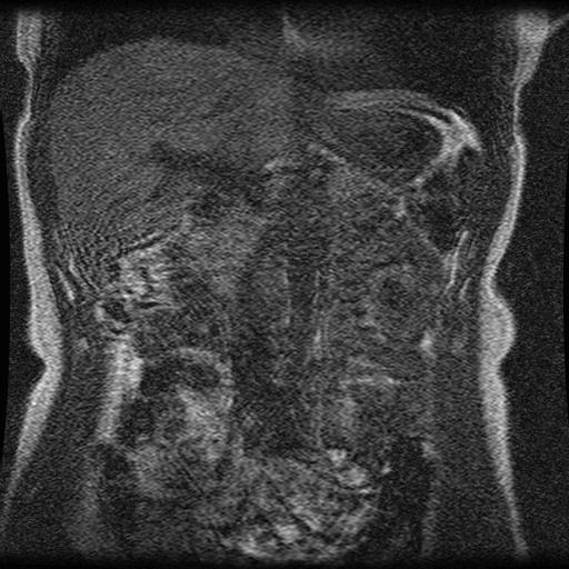
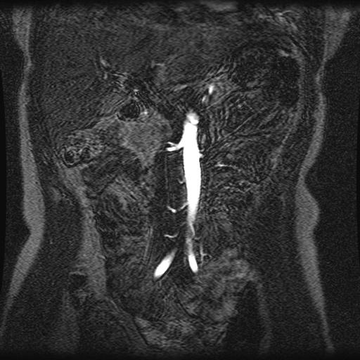
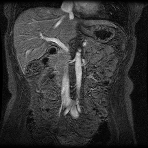
4 Contrast-enhanced MRA Pre During Post
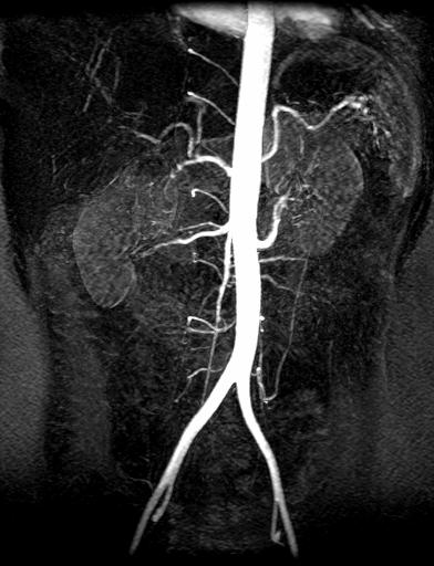
5 Contrast-enhanced MRA During Post


6 Contrast-enhanced MRA method Common features of technique: T1 weighted fast GRE 3D acquisition Tr < 5 ms Te < 1 ms Flip = 30 degrees Gadolinium Dose: 20 cc at 2 cc/s Nikola Tesla
 MRA is an off-label use of Gd flow rate = 3 ml/sec for renals")
7 HOW MUCH CONTRAST? Dose mmol/kg Aorta (20cc) Renal arteries/sma (30cc) Runoff 0.2 (40cc) MRA is an off-label use of Gd flow rate = 3 ml/sec for renals

8 Pitfalls: Timing Arterial Venous Tissue Time (s)

9 Timing Artifacts

10 Fourier Transform 45 echo

11 k-space Signal Image k y Detail k x Contrast


12 + = A = 1% A = 99% A = 100% + =
13 MRA Remains A Balancing Act Spatial Resolution SNR CNR Temporal Resolution k-space sampling and image reconstruction strategies help to achieve high spatial resolution time-resolved MR angiograms.
14 3D Time Resolved Imaging of Contrast Kinetics (TRICKS) aka: TREAT, DIRKS k z k z k y DC B A BCD ky Korosec et al., Magn. Reson. Med. 1996
15 3D TRICKS: Technique Contrast curve Artery Vein Time frame D A C A B A D A C A B A D A B(I) C(I) D(I) FFT Image at time frame 15
 Ave Korosec, et al.")
16 3D TRICKs Acquisition Time-Resolved Imaging of Contrast Kinetics k y k x k z C B A k-space 3D FFT image-space Scan Time = TR (PE Slice) Ave Korosec, et al., MRM 36:345-51;1996

/3 = 6.8 sec")
17 3D TRICKs Acquisition A B A C A B A C A B A B A C A ΔT = TR (PE Slice)/3 ΔT = 5 ms (128 32)/3 = 6.8 sec
 512 x 128 x16")

18 3D TRICKS TR = 10.8 (1996) 512 x 128 x16 Frame Time 5.6 s onstruction time 1996: 6 hours, one graduate stude
19 Outline: Time-resolved MRA Background Technique Clinical Indications Future Directions
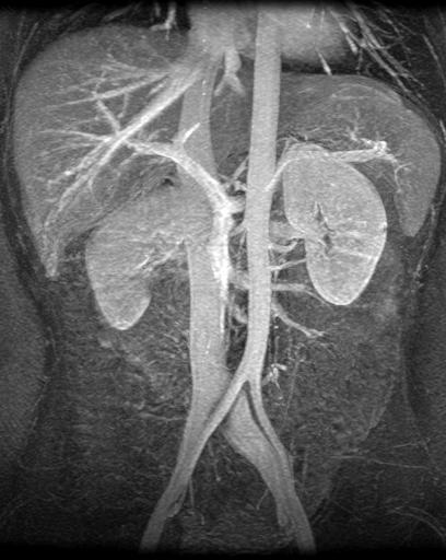
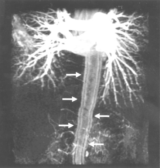
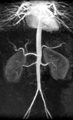
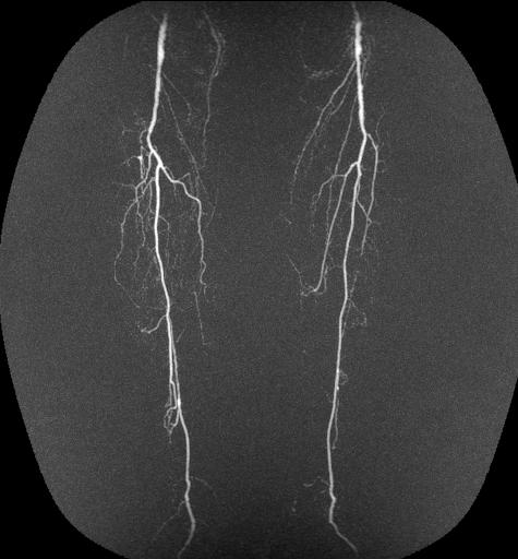
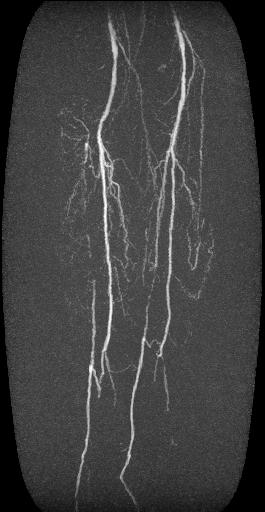
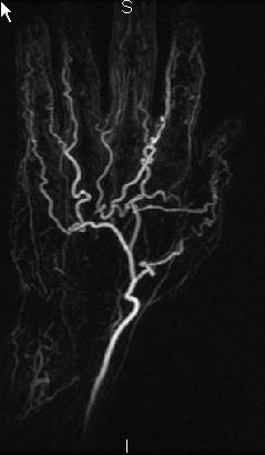
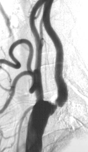
20 Clinical Indications Lower extremity runoff evaluation Asymmetric flow states Upper extremity MRA Mass evaluation and characterization Congenital heart disease Venous disease Aortic disease

21 Benefits of 3D CE MRA with subtraction 2D TOF 3D CEMRA BACKGROUND TISSUE SUPPRESSION


22 20 cc Gd Single Phase 3. Pelvis: Centric 3 2. Thighs: TRICKS 10 cc at 1 cc/sec 2 1. Distal Station Time-resolved MRA 10 cc Gd at 1 cc/sec 1


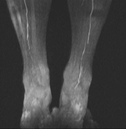
23 Improved Peripheral MRA Significantly more arteries diagnostic with TRICKS Significantly more venous contamination with moving SmartStep in lower station n=20, p < 0.05 Hany TF, et al Radiology 2001;221: Smartstep TRICKS

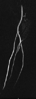
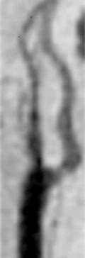
24 Benefits of time-resolved imaging protocol Left Popliteal Occlusion

25 Benefits of time-resolved imaging
26 Thromboangitis Obliterans


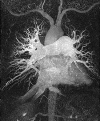
27 13 y/o with Tetrology of Fallot post-repair


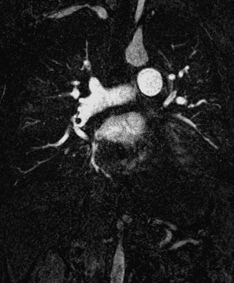
28 Right PA enlargement causes SVC obstruction Sagittal reformat


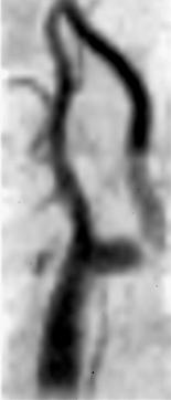
29 Vascular Access Evaluation Collapsed Time Frames Rotate MIP

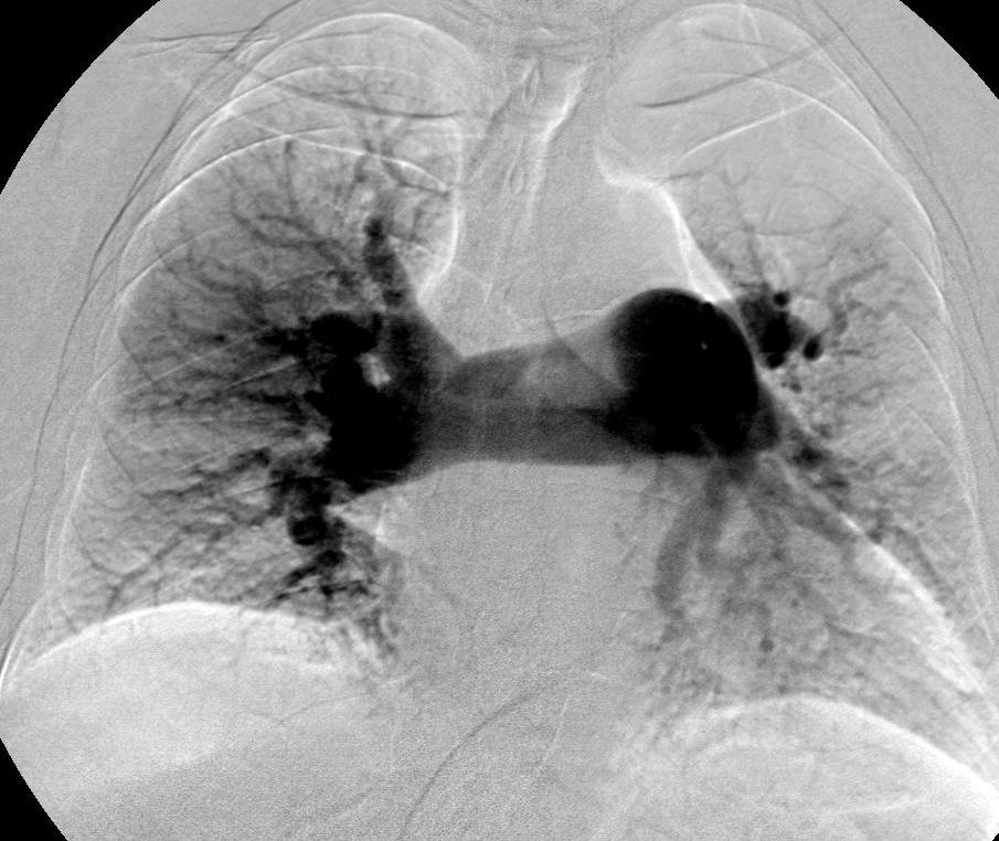
30 Secondary PAH due to chronic thromboembolic disease Courtesy of Stephan Schoenberg et al
31 Outline: Time-resolved MRA Background Technique Clinical Indications Future Directions Spatial Resolution SNR CNR Temporal Resolution
32 Traditional Cartesian sampling of k-k space 2D- FFT
 2D-")
33 Alternate Trajectories: Radial Sampling Sampling along radial spokes (2D-PR) 2D- FFT

34 Characteristics of Radial Sampling k x k y
35 HYPR Radial Acquisition Foot FOV = 300 mm 512 x 512 x 26 (ZIP 52) Voxel size 0.59 x 0.59 x 3.0 (-1.5) mm Frame Time = 2.0 Sec 16 proj/frame Speedup(Cartesian) = PE/16 proj 3D HYPR Speedup(radial) = proj/16 proj
36 Pitfalls: Resolution 3D TOF CE MRA DSA
37 Summary: Time-resolved MRA Eliminates need for accurate timing of contrast injection less need for radiologist supervision Allows high temporal and spatial resolution simultaneously Allows detection of non-uniform or asymmetric flow Automated process with essentially no post- processing Clinical indications expanding beyond arterial disease only Need for visualization with 4D processing to take full d t f ll th i f ti
Dynamic Contrast enhanced MRA
 Dynamic Contrast enhanced MRA Speaker: Yung-Chieh Chang Date : 106.07.22 Department of Radiology, Taichung Veterans General Hospital, Taichung, Taiwan 1 Outline Basic and advanced principles of Diffusion
Magnetic Resonance Angiography
 Magnetic Resonance Angiography Course: Advance MRI (BIOE 594) Instructors: Dr Xiaohong Joe Zhou Dr. Shadi Othman By, Nayan Pasad Phase Contrast Angiography By Moran 1982, Bryan et. Al. 1984 and Moran et.
Module 5: Dynamic Imaging and Phase Sharing. (true-fisp, TRICKS, CAPR, DISTAL, DISCO, HYPR) Review. Improving Temporal Resolution.
 Review. Improving Temporal Resolution.") MRES 7005 - Fast Imaging Techniques Module 5: Dynamic Imaging and Phase Sharing (true-fisp, TRICKS, CAPR, DISTAL, DISCO, HYPR) Review Improving Temporal Resolution True-FISP (I) True-FISP (II) Keyhole
MRES 7005 - Fast Imaging Techniques Module 5: Dynamic Imaging and Phase Sharing (true-fisp, TRICKS, CAPR, DISTAL, DISCO, HYPR) Review Improving Temporal Resolution True-FISP (I) True-FISP (II) Keyhole
MR Advance Techniques. Vascular Imaging. Class III
 MR Advance Techniques Vascular Imaging Class III 1 Vascular Imaging There are several methods that can be used to evaluate the cardiovascular systems with the use of MRI. MRI will aloud to evaluate morphology
GE Healthcare CLINICAL GALLERY. Discovery * MR750w 3.0T. This brochure is intended for European healthcare professionals.
 GE Healthcare CLINICAL GALLERY Discovery * MR750w 3.0T This brochure is intended for European healthcare professionals. NEURO PROPELLER delivers high resolution, motion insensitive imaging in all planes.
High-Resolution Time-Resolved Contrast-Enhanced MR Abdominal and Pulmonary Angiography Using a Spiral- TRICKS Sequence
 High-Resolution Time-Resolved Contrast-Enhanced MR Abdominal and Pulmonary Angiography Using a Spiral- TRICKS Sequence Jiang Du* and Mark Bydder Magnetic Resonance in Medicine 58:631 635 (2007) Both high
Fast Imaging Trajectories: Non-Cartesian Sampling (1)
") Fast Imaging Trajectories: Non-Cartesian Sampling (1) M229 Advanced Topics in MRI Holden H. Wu, Ph.D. 2018.05.03 Department of Radiological Sciences David Geffen School of Medicine at UCLA Class Business
Fast Imaging Trajectories: Non-Cartesian Sampling (1) M229 Advanced Topics in MRI Holden H. Wu, Ph.D. 2018.05.03 Department of Radiological Sciences David Geffen School of Medicine at UCLA Class Business
MRI. When to use What sequences. Outline 2012/09/19. Sequence: Definition. Basic Principles: Step 2. Basic Principles: Step 1. Govind Chavhan, MD
 MRI When to use What sequences Govind Chavhan, MD Assistant Professor and Staff Radiologist The Hospital For Sick Children, Toronto Planning Acquisition Post processing Interpretation Patient history and
Applications Guide for Interleaved
 Applications Guide for Interleaved rephase/dephase MRAV Authors: Yongquan Ye, Ph.D. Dongmei Wu, MS. Tested MAGNETOM Systems : 7TZ, TRIO a Tim System, Verio MR B15A (N4_VB15A_LATEST_20070519) MR B17A (N4_VB17A_LATEST_20090307_P8)
Initial Experience of Applying TWIST Dixon with Flexible View Sharing in Breast DCE-MRI
 Initial Experience of Applying TWIST Dixon with Flexible View Sharing in Breast DCE-MRI Yuan Le PhD 1, Hal D. Kipfer MD 1, Dominik M. Nickel PhD 2, Randall Kroeker PhD 2, Brian Dale PhD 2, Stephanie P.
Clinical Importance. Aortic Stenosis. Aortic Regurgitation. Ultrasound vs. MRI. Carotid Artery Stenosis
 Clinical Importance Rapid cardiovascular flow quantitation using sliceselective Fourier velocity encoding with spiral readouts Valve disease affects 10% of patients with heart disease in the U.S. Most
3D Radial Undersampling 7/19/2012. Artifact Removal SNR Restoration-- HYPR. Background: Time Resolved MR Angiography 4D DSA AND 4D FLUOROSCOPY:
 4D DSA AND 4D FLUOROSCOPY: Accelerated Applications using Undersampled Acquisition and Constrained Reconstruction Background: Time Resolved MR Angiography During the past 12 years we have been investigating
Compressed Sensing for Rapid MR Imaging
 Compressed Sensing for Rapid Imaging Michael Lustig1, Juan Santos1, David Donoho2 and John Pauly1 1 Electrical Engineering Department, Stanford University 2 Statistics Department, Stanford University rapid
Accelerated MRI Techniques: Basics of Parallel Imaging and Compressed Sensing
 Accelerated MRI Techniques: Basics of Parallel Imaging and Compressed Sensing Peng Hu, Ph.D. Associate Professor Department of Radiological Sciences PengHu@mednet.ucla.edu 310-267-6838 MRI... MRI has low
Lab Location: MRI, B2, Cardinal Carter Wing, St. Michael s Hospital, 30 Bond Street
 Lab Location: MRI, B2, Cardinal Carter Wing, St. Michael s Hospital, 30 Bond Street MRI is located in the sub basement of CC wing. From Queen or Victoria, follow the baby blue arrows and ride the CC south
(a Scrhon5 R2iwd b. P)jc%z 5. ivcr3. 1. I. ZOms Xn,s. 1E IDrAS boms. EE225E/BIOE265 Spring 2013 Principles of MRI. Assignment 8 Solutions
jc%z 5. ivcr3. 1. I. ZOms Xn,s. 1E IDrAS boms. EE225E/BIOE265 Spring 2013 Principles of MRI. Assignment 8 Solutions") EE225E/BIOE265 Spring 2013 Principles of MRI Miki Lustig Assignment 8 Solutions 1. Nishimura 7.1 P)jc%z 5 ivcr3. 1. I Due Wednesday April 10th, 2013 (a Scrhon5 R2iwd b 0 ZOms Xn,s r cx > qs 4-4 8ni6 4
EE225E/BIOE265 Spring 2013 Principles of MRI Miki Lustig Assignment 8 Solutions 1. Nishimura 7.1 P)jc%z 5 ivcr3. 1. I Due Wednesday April 10th, 2013 (a Scrhon5 R2iwd b 0 ZOms Xn,s r cx > qs 4-4 8ni6 4
Role of Parallel Imaging in High Field Functional MRI
 Role of Parallel Imaging in High Field Functional MRI Douglas C. Noll & Bradley P. Sutton Department of Biomedical Engineering, University of Michigan Supported by NIH Grant DA15410 & The Whitaker Foundation
SIEMENS MAGNETOM Verio syngo MR B15V
 \\USER\ZAHID_RESEARCH\MS\No Name\3D SWI TA: 6:39 PAT: 2 Voxel size: 1.0 0.5 2.0 mm Rel. SNR: 1.00 SIEMENS: gre Properties Prio Recon Before measurement After measurement Load to viewer Inline movie Auto
New Technology Allows Multiple Image Contrasts in a Single Scan
 These images were acquired with an investigational device. PD T2 T2 FLAIR T1 MAP T1 FLAIR PSIR T1 New Technology Allows Multiple Image Contrasts in a Single Scan MR exams can be time consuming. A typical
E. Mark Haacke, PhD. The MRI Institute for Biomedical Research Detroit, Michigan Wayne State University Detroit, Michigan 48201
 E. Mark Haacke, PhD The MRI Institute for Biomedical Research Detroit, Michigan 48202 Wayne State University Detroit, Michigan 48201 Acknowledgements The testing and establishment of these protocols has
MRI image formation 8/3/2016. Disclosure. Outlines. Chen Lin, PhD DABR 3. Indiana University School of Medicine and Indiana University Health
 MRI image formation Indiana University School of Medicine and Indiana University Health Disclosure No conflict of interest for this presentation 2 Outlines Data acquisition Spatial (Slice/Slab) selection
Sampling, Ordering, Interleaving
 Sampling, Ordering, Interleaving Sampling patterns and PSFs View ordering Modulation due to transients Temporal modulations Slice interleaving Sequential, Odd/even, bit-reversed Arbitrary Other considerations:
HST.583 Functional Magnetic Resonance Imaging: Data Acquisition and Analysis Fall 2008
 MIT OpenCourseWare http://ocw.mit.edu HST.583 Functional Magnetic Resonance Imaging: Data Acquisition and Analysis Fall 2008 For information about citing these materials or our Terms of Use, visit: http://ocw.mit.edu/terms.
CT Basics Principles of Spiral CT Dose. Always Thinking Ahead.
 1 CT Basics Principles of Spiral CT Dose 2 Who invented CT? 1963 - Alan Cormack developed a mathematical method of reconstructing images from x-ray projections Sir Godfrey Hounsfield worked for the Central
COBRE Scan Information
 COBRE Scan Information Below is more information on the directory structure for the COBRE imaging data. Also below are the imaging parameters for each series. Directory structure: var/www/html/dropbox/1139_anonymized/human:
Scan Acceleration with Rapid Gradient-Echo
 Scan Acceleration with Rapid Gradient-Echo Hsiao-Wen Chung ( 鍾孝文 ), Ph.D., Professor Dept. Electrical Engineering, National Taiwan Univ. Dept. Radiology, Tri-Service General Hospital 1 of 214 The Need
HST.583 Functional Magnetic Resonance Imaging: Data Acquisition and Analysis Fall 2008
 MIT OpenCourseWare http://ocw.mit.edu HST.583 Functional Magnetic Resonance Imaging: Data Acquisition and Analysis Fall 2008 For information about citing these materials or our Terms of Use, visit: http://ocw.mit.edu/terms.
Automatic Determination of Arterial Input Function for Dynamic Contrast Enhanced MRI in Tumor Assessment
 Automatic Determination of Arterial Input Function for Dynamic Contrast Enhanced MRI in Tumor Assessment Jeremy Chen, Jianhua Yao, and David Thomasson Diagnostic Radiology Department, Clinical Center,
Motion Artifacts and Suppression in MRI At a Glance
 Motion Artifacts and Suppression in MRI At a Glance Xiaodong Zhong, PhD MR R&D Collaborations Siemens Healthcare MRI Motion Artifacts and Suppression At a Glance Outline Background Physics Common Motion
7/31/2017. Historical Developments in Time-Resolved Angiography. Johns advice on how to streamline my teaching career
 Historical Developments in Time-Resolved Angiography Chuck MIstretta The University of Wisconsin-Madison 1971 Job Interview with John Cameron Chuck What did you do at for a high energy physics thesis at
Fast Imaging UCLA. Class Business. Class Business. Daniel B. Ennis, Ph.D. Magnetic Resonance Research Labs. Tuesday (3/7) from 6-9pm HW #1 HW #2
 from 6-9pm HW #1 HW #2") Fast Imaging Daniel B. Ennis, Ph.D. Magnetic Resonance Research Labs Class Business Tuesday (3/7) from 6-9pm 6:00-7:30pm Groups Avanto Sara Said, Yara Azar, April Pan Skyra Timothy Marcum, Diana Lopez,
Fast Imaging Daniel B. Ennis, Ph.D. Magnetic Resonance Research Labs Class Business Tuesday (3/7) from 6-9pm 6:00-7:30pm Groups Avanto Sara Said, Yara Azar, April Pan Skyra Timothy Marcum, Diana Lopez,
Slide 1. Technical Aspects of Quality Control in Magnetic Resonance Imaging. Slide 2. Annual Compliance Testing. of MRI Systems.
 Slide 1 Technical Aspects of Quality Control in Magnetic Resonance Imaging Slide 2 Compliance Testing of MRI Systems, Ph.D. Department of Radiology Henry Ford Hospital, Detroit, MI Slide 3 Compliance Testing
Abbie M. Diak, PhD Loyola University Medical Center Dept. of Radiation Oncology
 Abbie M. Diak, PhD Loyola University Medical Center Dept. of Radiation Oncology Outline High Spectral and Spatial Resolution MR Imaging (HiSS) What it is How to do it Ways to use it HiSS for Radiation
Sampling, Ordering, Interleaving
 Sampling, Ordering, Interleaving Sampling patterns and PSFs View ordering Modulation due to transients Temporal modulations Timing: cine, gating, triggering Slice interleaving Sequential, Odd/even, bit-reversed
MRI Physics II: Gradients, Imaging
 MRI Physics II: Gradients, Imaging Douglas C., Ph.D. Dept. of Biomedical Engineering University of Michigan, Ann Arbor Magnetic Fields in MRI B 0 The main magnetic field. Always on (0.5-7 T) Magnetizes
Functional MRI. Jerry Allison, Ph. D. Medical College of Georgia
 Functional MRI Jerry Allison, Ph. D. Medical College of Georgia BOLD Imaging Technique Blood Oxygen Level Dependent contrast can be used to map brain function Right Hand Motor Task Outline fmri BOLD Contrast
TOF-MRA Using Multi-Oblique-Stack Acquisition (MOSA)
") JOURNAL OF MAGNETIC RESONANCE IMAGING 26:432 436 (2007) Technical Note TOF-MRA Using Multi-Oblique-Stack Acquisition (MOSA) Ed X. Wu, PhD, 1,2 * Edward S. Hui, BEng, 1,2 and Jerry S. Cheung, BEng 1,2 Purpose:
JOURNAL OF MAGNETIC RESONANCE IMAGING 26:432 436 (2007) Technical Note TOF-MRA Using Multi-Oblique-Stack Acquisition (MOSA) Ed X. Wu, PhD, 1,2 * Edward S. Hui, BEng, 1,2 and Jerry S. Cheung, BEng 1,2 Purpose:
White Pixel Artifact. Caused by a noise spike during acquisition Spike in K-space <--> sinusoid in image space
 White Pixel Artifact Caused by a noise spike during acquisition Spike in K-space sinusoid in image space Susceptibility Artifacts Off-resonance artifacts caused by adjacent regions with different
11/18/ CPT Preauthorization Groupings Effective January 1, Computerized Tomography (CT) Abdomen 6. CPT Description SEGR CT01
 Abdomen 6. CPT Description SEGR CT01") Computerized Tomography (CT) 6 & 101 5 Upper Extremity 11 Lower Extremity 12 Head 3 Orbit 1 Sinus 2 Neck 4 7 Cervical Spine 8 Thoracic Spine 9 Lumbar Spine 10 Colon 13 CPT Description SEGR 74150 74160
Computerized Tomography (CT) 6 & 101 5 Upper Extremity 11 Lower Extremity 12 Head 3 Orbit 1 Sinus 2 Neck 4 7 Cervical Spine 8 Thoracic Spine 9 Lumbar Spine 10 Colon 13 CPT Description SEGR 74150 74160
k-space Interpretation of the Rose Model: Noise Limitation on the Detectable Resolution in MRI
 k-space Interpretation of the Rose Model: Noise Limitation on the Detectable Resolution in MRI Richard Watts and Yi Wang* Magnetic Resonance in Medicine 48:550 554 (2002) Noise limitation on the detected
SIEMENS MAGNETOM Avanto syngo MR B15
 \\USER\INVESTIGATORS\Ravi\ADNI-Subject\Localizer TA: 0:10 PAT: Voxel size: 1.9 1.5 8.0 mm Rel. SNR: 1.00 SIEMENS: gre Properties Prio Recon Before measurement After measurement Load to viewer Inline movie
M R I Physics Course
 M R I Physics Course Multichannel Technology & Parallel Imaging Nathan Yanasak, Ph.D. Jerry Allison Ph.D. Tom Lavin, B.S. Department of Radiology Medical College of Georgia References: 1) The Physics of
Image Acquisition Systems
 Image Acquisition Systems Goals and Terminology Conventional Radiography Axial Tomography Computer Axial Tomography (CAT) Magnetic Resonance Imaging (MRI) PET, SPECT Ultrasound Microscopy Imaging ITCS
Advanced Imaging Trajectories
 Advanced Imaging Trajectories Cartesian EPI Spiral Radial Projection 1 Radial and Projection Imaging Sample spokes Radial out : from k=0 to kmax Projection: from -kmax to kmax Trajectory design considerations
A Novel Iterative Thresholding Algorithm for Compressed Sensing Reconstruction of Quantitative MRI Parameters from Insufficient Data
 A Novel Iterative Thresholding Algorithm for Compressed Sensing Reconstruction of Quantitative MRI Parameters from Insufficient Data Alexey Samsonov, Julia Velikina Departments of Radiology and Medical
SIEMENS MAGNETOM Avanto syngo MR B15
 \\USER\INVESTIGATORS\Ravi\ADNI-phantom\QC Phantom-Localizer TA: 0:10 PAT: Voxel size: 1.9 1.5 8.0 mm Rel. SNR: 1.00 SIEMENS: gre Properties Prio Recon Before measurement After measurement Load to viewer
A preliminary study on adaptive field-of-view tracking in peripheral digital subtraction angiography
 Journal of X-Ray Science and Technology 11 (2003) 149 159 149 IOS Press A preliminary study on adaptive field-of-view tracking in peripheral digital subtraction angiography James R. Bennett a,b,er-weibai
Single Breath-hold Abdominal T 1 Mapping using 3-D Cartesian Sampling and Spatiotemporally Constrained Reconstruction
 Single Breath-hold Abdominal T 1 Mapping using 3-D Cartesian Sampling and Spatiotemporally Constrained Reconstruction Felix Lugauer 1,3, Jens Wetzl 1, Christoph Forman 2, Manuel Schneider 1, Berthold Kiefer
Constrained Reconstruction of Sparse Cardiac MR DTI Data
 Constrained Reconstruction of Sparse Cardiac MR DTI Data Ganesh Adluru 1,3, Edward Hsu, and Edward V.R. DiBella,3 1 Electrical and Computer Engineering department, 50 S. Central Campus Dr., MEB, University
A Virtual MR Scanner for Education
 A Virtual MR Scanner for Education Hackländer T, Schalla C, Trümper A, Mertens H, Hiltner J, Cramer BM Hospitals of the University Witten/Herdecke, Department of Radiology Wuppertal, Germany Purpose A
Advanced MRI Techniques (and Applications)
") Advanced MRI Techniques (and Applications) Jeffry R. Alger, PhD Department of Neurology Ahmanson-Lovelace Brain Mapping Center Brain Research Institute Jonsson Comprehensive Cancer Center University of
Advanced MRI Techniques (and Applications) Jeffry R. Alger, PhD Department of Neurology Ahmanson-Lovelace Brain Mapping Center Brain Research Institute Jonsson Comprehensive Cancer Center University of
ACQUIRING AND PROCESSING SUSCEPTIBILITY WEIGHTED IMAGING (SWI) DATA ON GE 3.0T
 DATA ON GE 3.0T") ACQUIRING AND PROCESSING SUSCEPTIBILITY WEIGHTED IMAGING (SWI) DATA ON GE 3.0T Revision date: 12/13/2010 Overview Susceptibility Weighted Imaging (SWI) is a relatively new data acquisition and processing
ACQUIRING AND PROCESSING SUSCEPTIBILITY WEIGHTED IMAGING (SWI) DATA ON GE 3.0T Revision date: 12/13/2010 Overview Susceptibility Weighted Imaging (SWI) is a relatively new data acquisition and processing
Patient Specific. Protocol Identification Number (PID) Must be on each individual. MRI Image
 Must be on each individual. MRI Image") MRI PROCEDURE GUIDELINES Patient Specific Protocol Identification Number (PID) Must be on each individual MRI Image [Protocol 11/04/13, Version 1.4] (Revised 09/03/2014) Page 1 of 72 Abbreviations: BH:
MRI PROCEDURE GUIDELINES Patient Specific Protocol Identification Number (PID) Must be on each individual MRI Image [Protocol 11/04/13, Version 1.4] (Revised 09/03/2014) Page 1 of 72 Abbreviations: BH:
HST.583 Functional Magnetic Resonance Imaging: Data Acquisition and Analysis Fall 2006
 MIT OpenCourseWare http://ocw.mit.edu HST.583 Functional Magnetic Resonance Imaging: Data Acquisition and Analysis Fall 2006 For information about citing these materials or our Terms of Use, visit: http://ocw.mit.edu/terms.
Fast Isotropic Volumetric Coronary MR Angiography Using Free-Breathing 3D Radial Balanced FFE Acquisition
 Fast Isotropic Volumetric Coronary MR Angiography Using Free-Breathing 3D Radial Balanced FFE Acquisition C. Stehning, 1 * P. Börnert, 2 K. Nehrke, 2 H. Eggers, 2 and O. Dössel 1 Magnetic Resonance in
Vessel Explorer: a tool for quantitative measurements in CT and MR angiography
 Clinical applications Vessel Explorer: a tool for quantitative measurements in CT and MR angiography J. Oliván Bescós J. Sonnemans R. Habets J. Peters H. van den Bosch T. Leiner Healthcare Informatics/Patient
Supplementary Figure 1
 Supplementary Figure 1 BOLD and CBV functional maps showing EPI versus line-scanning FLASH fmri. A. Colored BOLD and CBV functional maps are shown in the highlighted window (green frame) of the raw EPI
High dynamic range magnetic resonance flow imaging in the abdomen
 High dynamic range magnetic resonance flow imaging in the abdomen Christopher M. Sandino EE 367 Project Proposal 1 Motivation Time-resolved, volumetric phase-contrast magnetic resonance imaging (also known
Acknowledgments and financial disclosure
 AAPM 2012 Annual Meeting Digital breast tomosynthesis: basic understanding of physics principles James T. Dobbins III, Ph.D., FAAPM Director, Medical Physics Graduate Program Ravin Advanced Imaging Laboratories
Flip-angle-optimized fast dynamic T 1 mapping with a 3D gradient-echo sequence
 Flip-angle-optimized fast dynamic T 1 mapping with a 3D gradient-echo sequence Olaf Dietrich 1, Maximilian Freiermuth 1, Linus Willerding 2, Maximilian F. Reiser 1, Michael Peller 1 1 Josef Lissner Laboratory
Qualitative Comparison of Conventional and Oblique MRI for Detection of Herniated Spinal Discs
 Qualitative Comparison of Conventional and Oblique MRI for Detection of Herniated Spinal Discs Doug Dean Final Project Presentation ENGN 2500: Medical Image Analysis May 16, 2011 Outline Review of the
8/11/2009. Common Areas of Motion Problem. Motion Compensation Techniques and Applications. Type of Motion. What s your problem
 Common Areas of Motion Problem Motion Compensation Techniques and Applications Abdominal and cardiac imaging. Uncooperative patient, such as pediatric. Dynamic imaging and time series. Chen Lin, PhD Indiana
CP Generalize Concepts in Abstract Multi-dimensional Image Model Component Semantics. David Clunie.
 CP-1390 - Generalize Concepts in Abstract Multi-dimensional Image Model Semantics Page 1 STATUS Date of Last Update Person Assigned Submitter Name Submission Date Assigned 2014/06/09 David Clunie mailto:dclunie@dclunie.com
XI Signal-to-Noise (SNR)
") XI Signal-to-Noise (SNR) Lecture notes by Assaf Tal n(t) t. Noise. Characterizing Noise Noise is a random signal that gets added to all of our measurements. In D it looks like this: while in D
XI Signal-to-Noise (SNR) Lecture notes by Assaf Tal n(t) t. Noise. Characterizing Noise Noise is a random signal that gets added to all of our measurements. In D it looks like this: while in D
Module 4. K-Space Symmetry. Review. K-Space Review. K-Space Symmetry. Partial or Fractional Echo. Half or Partial Fourier HASTE
 MRES 7005 - Fast Imaging Techniques Module 4 K-Space Symmetry Review K-Space Review K-Space Symmetry Partial or Fractional Echo Half or Partial Fourier HASTE Conditions for successful reconstruction Interpolation
ADNI, ADNI_QH, SURVEY. Geometry. connection
 ADNI, ADNI_QH, SURVEY Geometry Coil selection = Head connection = d Multi coil Homogeneity correction ne FOV (mm) = 250.00 RFOV (%) = 100.00 Foldover suppression Matrix scan = 256 reconstruction = 256
Image Quality Assessment and Quality Assurance of Advanced Imaging Systems for IGRT. AAPM Penn-Ohio Chapter Sep 25, 2015 Soyoung Lee, PhD
 Image Quality Assessment and Quality Assurance of Advanced Imaging Systems for IGRT AAPM Penn-Ohio Chapter Sep 25, 2015 Soyoung Lee, PhD 1 Outline q Introduction q Imaging performances in 4D-CBCT Image
Breast MRI Accreditation Program Clinical Image Quality Guide
 Breast MRI Accreditation Program Clinical Image Quality Guide Introduction This document provides guidance on breast MRI clinical image quality and describes the criteria used by the ACR Breast MRI Accreditation
A novel noise removal using homomorphic normalization for multi-echo knee MRI
 A novel noise removal using homomorphic normalization for multi-echo knee MRI Xuenan Cui 1a),HakilKim 1b), Seongwook Hong 1c), and Kyu-Sung Kwack 2d) 1 School of Information and Communication Engineering,
SIEMENS MAGNETOM TrioTim syngo MR B17
 \\USER\KNARRGROUP\MultiBand\LavretskyMultiBand\trufi localizer 3-plane TA: 5.1 s PAT: Voxel size: 1.2 1.2 5. Rel. SNR: 1.00 SIEMENS: trufi Load to stamp Slice group 1 Slices 1 Dist. factor 20 % Phase enc.
MARP. Physicist s Role in ACR MRAP Accreditation May Carl R. Keener, Ph.D., DABMP, DABR Medical & Radiation Physics, Inc.
 Physicist s Role in ACR MRAP Accreditation May 2010 Carl R. Keener, Ph.D., DABMP, DABR keener@marpinc.com MARP Medical & Radiation Physics, Inc. disclosure ACR MRAP Physics subcommittee ACR MRAP phantom
XI Conference "Medical Informatics & Technologies" VALIDITY OF MRI BRAIN PERFUSION IMAGING METHOD
 XI Conference "Medical Informatics & Technologies" - 2006 medical imaging, MRI, brain perfusion Bartosz KARCZEWSKI 1, Jacek RUMIŃSKI 1 VALIDITY OF MRI BRAIN PERFUSION IMAGING METHOD Brain perfusion imaging
K-Space Trajectories and Spiral Scan
 K-Space and Spiral Scan Presented by: Novena Rangwala nrangw2@uic.edu 1 Outline K-space Gridding Reconstruction Features of Spiral Sampling Pulse Sequences Mathematical Basis of Spiral Scanning Variations
Computer-Tomography II: Image reconstruction and applications
 Computer-Tomography II: Image reconstruction and applications Prof. Dr. U. Oelfke DKFZ Heidelberg Department of Medical Physics (E040) Im Neuenheimer Feld 280 69120 Heidelberg, Germany u.oelfke@dkfz.de
CHAPTER 9: Magnetic Susceptibility Effects in High Field MRI
 Figure 1. In the brain, the gray matter has substantially more blood vessels and capillaries than white matter. The magnified image on the right displays the rich vasculature in gray matter forming porous,
3D3C & 2D3D Velocity Measurements Using Magnetic Resonance Velocimetry
 3D3C & 2D3D Velocity Measurements Using Magnetic Resonance Velocimetry Sven Grundmann Center of Smart Interfaces Technische Universität Darmstadt Flughafenstrasse 19 64347 Griesheim grundmann@csi-tu-darmstadt.de
Improved Spatial Localization in 3D MRSI with a Sequence Combining PSF-Choice, EPSI and a Resolution Enhancement Algorithm
 Improved Spatial Localization in 3D MRSI with a Sequence Combining PSF-Choice, EPSI and a Resolution Enhancement Algorithm L.P. Panych 1,3, B. Madore 1,3, W.S. Hoge 1,3, R.V. Mulkern 2,3 1 Brigham and
T 1 MAPPING FOR DCE-MRI
 T 1 MAPPING FOR DCE-MRI A dissertation submitted to the Faculty of Medicine, University of Malaya in partial fulfillment of the requirements for the degree of Master of Medical Physics By NURUN NAJWA BINTI
Error! Bookmark not defined. Error! Bookmark not defined.
 MRI-Neuro Protocols Brain Protocols... 2 WITH AND WITHOUT... 3 WITHOUT CONTRT... 4 HHT... 5 MS WITH AND WITHOUT CONTRT... 6 MS WITHOUT CONTRT... 7 PEDIATRIC... 8 PEDIATRIC WITH CONTRT... 9 PITUITARY NO
Spiral keyhole imaging for MR fingerprinting
 Spiral keyhole imaging for MR fingerprinting Guido Buonincontri 1, Laura Biagi 1,2, Pedro A Gómez 3,4, Rolf F Schulte 4, Michela Tosetti 1,2 1 IMAGO7 Research Center, Pisa, Italy 2 IRCCS Stella Maris,
SIEMENS MAGNETOM Skyra syngo MR D13
 Page 1 of 8 SIEMENS MAGNETOM Skyra syngo MR D13 \\USER\CIND\StudyProtocols\PTSA\*dm_ep2d_mono70_b0_p2_iso2.0 TA:1:05 PAT:2 Voxel size:2.0 2.0 2.0 mm Rel. SNR:1.00 :epse Properties Routine Prio Recon Load
GE Healthcare. AppsLinq* remote courses catalogue
 GE Healthcare AppsLinq* remote courses catalogue AppsLinq * remote training for MR Magnetic Resonance AppsLinq* a GE Training in Partnership (TiP) program, revolutionizes applications training with live,
Gradient-Echo. Spin-Echo. Echo planar. Assessment of Regional Function Assessment of Global. Parallel Imaging. Function. Steady State Imaging
 Gradient-Echo Spin-Echo James W. Goldfarb Ph.D. Department of Research and Education St. Francis Hospital Program in Biomedical Engineering SUNY Stony Brook Echo planar Assessment of Regional Function
8/1/2017. Current Technology: Energy Integrating Detectors. Principles, Pitfalls and Progress in Photon-Counting-Detector Technology.
 Photon Counting Detectors and Their Applications in Medical Imaging Principles, Pitfalls and Progress in Photon-Counting-Detector Technology Taly Gilat Schmidt, PhD Associate Professor Department of Biomedical
SPECIFICATIONS FOR A NEW STATE OF ART 16 SLICE ALL PURPOSE C. T. SCANNER
 SPECIFICATIONS FOR A NEW STATE OF ART 16 SLICE ALL PURPOSE C. T. SCANNER A) Scanner Design X-Ray generator and tube: 1. Scanner: Whole body spiral CT scanner (16 slices) of latest technology. 2. X-Ray
Background. Outline. Radiographic Tomosynthesis: Image Quality and Artifacts Reduction 1 / GE /
 Radiographic Tomosynthesis: Image Quality and Artifacts Reduction Baojun Li, Ph.D Department of Radiology Boston University Medical Center 2012 AAPM Annual Meeting Background Linear Trajectory Tomosynthesis
Sparse sampling in MRI: From basic theory to clinical application. R. Marc Lebel, PhD Department of Electrical Engineering Department of Radiology
 Sparse sampling in MRI: From basic theory to clinical application R. Marc Lebel, PhD Department of Electrical Engineering Department of Radiology Objective Provide an intuitive overview of compressed sensing
SPM8 for Basic and Clinical Investigators. Preprocessing. fmri Preprocessing
 SPM8 for Basic and Clinical Investigators Preprocessing fmri Preprocessing Slice timing correction Geometric distortion correction Head motion correction Temporal filtering Intensity normalization Spatial
Page 1 of 9. Protocol: adult_other_adni3basichumanprotocol25x_ _ _1. 3 Plane Localizer. 3 Plane Localizer PATIENT POSITION
 3 Localizer FOV 26.0 Slice Thickness 5.0 Slice Spacing 0.0 Freq 256 Phase 128 3-PLANE 3 Localizer Unswap Phase Correction Gradient Echo Imaging Options Seq, Fast Recon All Images 3 Localizer Pause / SCIC
Magnetic Resonance Imaging Velocity. Information. Joe Lee. April 4, 2000
 Locating Arteriovenous Malformations using Magnetic Resonance Imaging Velocity Information Joe Lee April 4, 2000 1 Introduction An arteriovenous malformation (AVM) is a congenital vascular defect where
CTA HEAD Perfusion AqONE without and with IV Contrast
 CTA HEAD Perfusion AqONE without and with IV Contrast Patient Position Adult Contrast Adult Injection Rate Supine IOML perpendicular to table top. IV: 100 ml with helical head CTA 50 ml without helical
Information about presenter
 Information about presenter 2013-now Engineer R&D ithera Medical GmbH 2011-2013 M.Sc. in Biomedical Computing (TU München) Thesis title: A General Reconstruction Framework for Constrained Optimisation
MRI Image Quality Assessment
 in partnership with MRI Image Quality Assessment David Collins CR-UK Cancer Imaging Centre, The Institute of Cancer Research Making the discoveries that defeat cancer Overview Current Practice Quality
Application of level set based method for segmentation of blood vessels in angiography images
 Lodz University of Technology Faculty of Electrical, Electronic, Computer and Control Engineering Institute of Electronics PhD Thesis Application of level set based method for segmentation of blood vessels
Use of MRI in Radiotherapy: Technical Consideration
 Use of MRI in Radiotherapy: Technical Consideration Yanle Hu, PhD Department of Radiation Oncology, Mayo Clinic Arizona 04/07/2018 2015 MFMER slide-1 Conflict of Interest: None 2015 MFMER slide-2 Objectives
VieW 3D. 3D Post-Processing WorKstation THE THIRD DIMENSION. Version 3.1
 VieW 3D 3D Post-Processing WorKstation THE THIRD DIMENSION Version 3.1 iq-view 3D THE FULLY-FEATURED 3D MEDICAL IMAGING SOLUTION FOR RADIOLOGISTS iq-view 3D contains all components of iq-view with the
US 1.
 US 1 Sample image: Normal pancreas seen on sonogram. Looking up from abdomen toward the head of the patient. The liver is in front of the pancreas. A vein draining the spleen is behind the pancreas http://www.radiologyinfo.org/photocat/photos.cfm?image=abdo-us-pancr.jpg&&subcategory=abdomen&&stop=9
Corso di laurea in Fisica A.A Fisica Medica 4 TC
 Corso di laurea in Fisica A.A. 2007-2008 Fisica Medica 4 TC Computed Tomography Principles 1. Projection measurement 2. Scanner systems 3. Scanning modes Basic Tomographic Principle The internal structure
Unaliasing by Fourier-Encoding the Overlaps Using the Temporal Dimension (UNFOLD), Applied to Cardiac Imaging and fmri
, Applied to Cardiac Imaging and fmri") 1999 ISMRM YOUNG INVESTIGATORS MOORE AWARD PAPERS Magnetic Resonance in Medicine 42:813 828 (1999) Unaliasing by Fourier-Encoding the Overlaps Using the Temporal Dimension (UNFOLD), Applied to Cardiac
1999 ISMRM YOUNG INVESTIGATORS MOORE AWARD PAPERS Magnetic Resonance in Medicine 42:813 828 (1999) Unaliasing by Fourier-Encoding the Overlaps Using the Temporal Dimension (UNFOLD), Applied to Cardiac
Steen Moeller Center for Magnetic Resonance research University of Minnesota
 Steen Moeller Center for Magnetic Resonance research University of Minnesota moeller@cmrr.umn.edu Lot of material is from a talk by Douglas C. Noll Department of Biomedical Engineering Functional MRI Laboratory